OPA3 Supports Pancreatic Tumor Growth Through Context-Dependent Metabolic and Hypoxic Adaptation
DOI:
https://doi.org/10.66505/cbtt.v1i4.72Keywords:
OPA3, pancreatic cancer, mitochondrial metabolism, tumor hypoxia, HIF-1α, KRAS, tumor microenvironmentAbstract
Background: Optic atrophy protein 3 (OPA3) regulates mitochondrial function, but its contribution to cancer progression and adaptation to hypoxic tumor microenvironments remains incompletely understood.
Methods: We examined OPA3 expression across 31 cancer types and investigated its function using CRISPR/Cas9 knockout in five cancer cell lines with different KRAS backgrounds. Proliferation, clonogenic growth, mitochondrial metabolism, and ATP production were evaluated in vitro. Pancreatic cancer xenografts, hypoxia experiments, and spatial transcriptomic analyses were used to assess tumor growth and HIF-1α-associated responses.
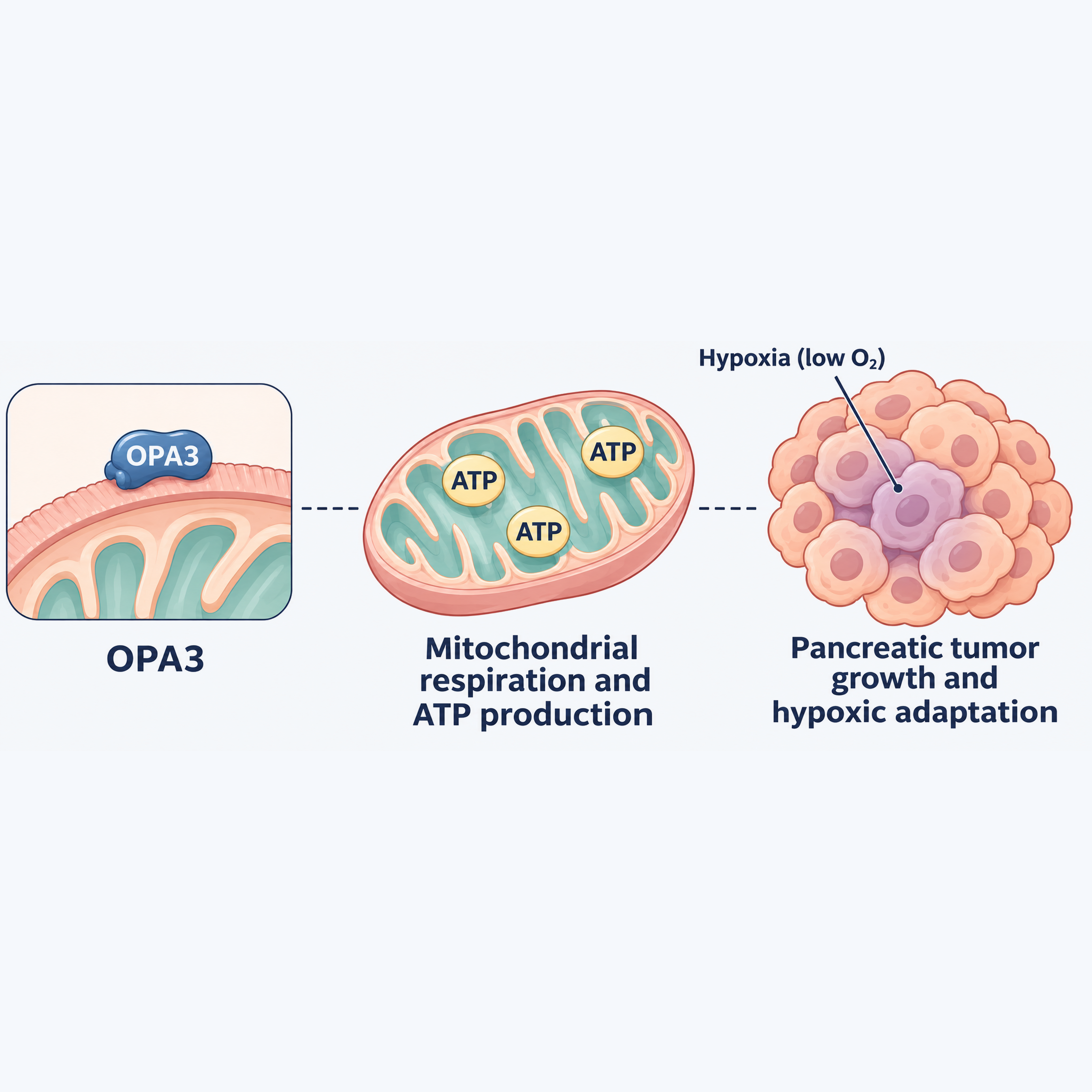
Results: OPA3 expression varied across cancer types. OPA3 knockout preferentially impaired proliferation and clonogenic growth in the KRAS-mutant cell lines examined, with reduced mitochondrial respiration and ATP production observed in PANC-1 cells. Despite limited effects on KRAS-wild-type BxPC-3 cells in vitro, OPA3 depletion suppressed both PANC-1 and BxPC-3 xenograft growth. Under prolonged hypoxia, OPA3-deficient cells exhibited reduced HIF-1α protein accumulation and impaired clonogenic growth. Spatial transcriptomic analyses identified a positive association between OPA3 and HIF1A expression in pancreatic tumor-associated tissues. However, these observations do not establish direct HIF-1α stabilization or a causal relationship in human tumors.
Conclusions: OPA3 supports pancreatic tumor growth through context-dependent processes associated with mitochondrial metabolism and hypoxic adaptation. Its effects extend beyond KRAS mutation status in xenograft models, although the underlying mechanisms require further validation. These findings identify OPA3 as a candidate regulator of tumor-microenvironment adaptation rather than an established therapeutic target.
References
1. Faubert B, Solmonson A, DeBerardinis RJ. Metabolic reprogramming and cancer progression. Science. 2020;368(6487):eaaw5473. doi:10.1126/science.aaw5473
2. Hanahan D. Hallmarks of cancer: new dimensions. Cancer Discov. 2022;12(1):31-46. doi:10.1158/2159-8290.CD-21-1059
3. Meng N, Glorieux C, Zhang Y, Liang L, Zeng P, Lu W, et al. Oncogenic K-ras induces mitochondrial OPA3 expression to promote energy metabolism in pancreatic cancer cells. Cancers (Basel). 2019;12(1):65. doi:10.3390/cancers12010065
4. Anikster Y, Kleta R, Shaag A, Gahl WA, Elpeleg O. Type III 3-methylglutaconic aciduria (optic atrophy plus syndrome, or Costeff optic atrophy syndrome): identification of the OPA3 gene and its founder mutation in Iraqi Jews. Am J Hum Genet. 2001;69(6):1218-1224. doi:10.1086/324651
5. Huizing M, Dorward H, Ly L, Klootwijk E, Kleta R, Skovby F, et al. OPA3, mutated in 3-methylglutaconic aciduria type III, encodes two transcripts targeted primarily to mitochondria. Mol Genet Metab. 2010;100(2):149-154. doi:10.1016/j.ymgme.2010.03.005
6. Ryu SW, Jeong HJ, Choi M, Karbowski M, Choi C. Optic atrophy 3 as a protein of the mitochondrial outer membrane induces mitochondrial fragmentation. Cell Mol Life Sci. 2010;67(16):2839-2850. doi:10.1007/s00018-010-0365-z
7. Tsai HW, Li CJ, Lin LT, Chiang AJ, Chen SN, Wen ZH, et al. Expression status and prognostic significance of mitochondrial dynamics OPA3 in human ovarian cancer. Aging (Albany NY). 2022;14(9):3874-3886. doi:10.18632/aging.204050
8. Yang Z, Zhang Y, Wang X, Huang J, Guo W, Wei P, et al. Putative biomarkers of malignant transformation of sinonasal inverted papilloma into squamous cell carcinoma. J Int Med Res. 2019;47(6):2371-2380. doi:10.1177/0300060519838385
9. Yin Y, Ma Z, Yuan S, Xu K, Wang X. OPA3 inhibits the cGAS-STING pathway mediated by mtDNA stress to promote colorectal cancer progression. In Vitro Cell Dev Biol Anim. 2025;61(2):165-177. doi:10.1007/s11626-024-01000-3
10. Wu H, Liu D. OPA3 overexpression modulates lipid droplet production and sensitizes colorectal cancer cells to bevacizumab treatment. Biocell. 2024;48(6):971-980. doi:10.32604/biocell.2024.049466
11. Kang YJ, Pan L, Liu Y, Rong Z, Liu J, Liu F. GEPIA3: Enhanced drug sensitivity and interaction network analysis for cancer research. Nucleic Acids Res. 2025;53(W1):W283-W290. doi:10.1093/nar/gkaf423
12. Nusinow DP, Szpyt J, Ghandi M, Rose CM, McDonald ER 3rd, Kalocsay M, et al. Quantitative proteomics of the cancer cell line encyclopedia. Cell. 2020;180(2):387-402.e16. doi:10.1016/j.cell.2019.12.023
13. Wolf FA, Angerer P, Theis FJ. SCANPY: large-scale single-cell gene expression data analysis. Genome Biol. 2018;19(1):15. doi:10.1186/s13059-017-1382-0
14. van Dijk D, Sharma R, Nainys J, Yim K, Kathail P, Carr AJ, et al. Recovering gene interactions from single-cell data using data diffusion. Cell. 2018;174(3):716-729.e27. doi:10.1016/j.cell.2018.05.061
15. Bardeesy N, DePinho RA. Pancreatic cancer biology and genetics. Nat Rev Cancer. 2002;2(12):897-909. doi:10.1038/nrc949
16. Cancer Genome Atlas Network. Comprehensive molecular characterization of human colon and rectal cancer. Nature. 2012;487(7407):330-337. doi:10.1038/nature11252
17. Herbst RS, Heymach JV, Lippman SM. Lung cancer. N Engl J Med. 2008;359(13):1367-1380. doi:10.1056/NEJMra0802714
18. Suvac A, Ashton J, Bristow RG. Tumour hypoxia in driving genomic instability and tumour evolution. Nat Rev Cancer. 2025;25(3):167-188. doi:10.1038/s41568-024-00781-9
19. Jing X, Yang F, Shao C, Wei K, Xie M, Shen H, et al. Role of hypoxia in cancer therapy by regulating the tumor microenvironment. Mol Cancer. 2019;18(1):157. doi:10.1186/s12943-019-1089-9
20. Wang Y, Ying X, Wang Y, Zou Z, Yuan A, Xiao Z, et al. Hydrogen sulfide alleviates mitochondrial damage and ferroptosis by regulating OPA3-NFS1 axis in doxorubicin-induced cardiotoxicity. Cell Signal. 2023;107:110655. doi:10.1016/j.cellsig.2023.110655
21. Schito L, Semenza GL. Hypoxia-inducible factors: master regulators of cancer progression. Trends Cancer. 2016;2(12):758-770. doi:10.1016/j.trecan.2016.10.016
22. Semenza GL. Hypoxia-inducible factors in physiology and medicine. Cell. 2012;148(3):399-408. doi:10.1016/j.cell.2012.01.021
23. Gilkes DM, Semenza GL, Wirtz D. Hypoxia and the extracellular matrix: drivers of tumour metastasis. Nat Rev Cancer. 2014;14(6):430-439. doi:10.1038/nrc3726
24. Binnewies M, Roberts EW, Kersten K, Chan V, Fearon DF, Merad M, et al. Understanding the tumor immune microenvironment (TIME) for effective therapy. Nat Med. 2018;24(5):541-550. doi:10.1038/s41591-018-0014-x
25. Xu M, Zhang T, Xia R, Wei Y, Wei X. Targeting the tumor stroma for cancer therapy. Mol Cancer. 2022;21(1):208. doi:10.1186/s12943-022-01670-1
26. Sun L, Zhang H, Gao P. Metabolic reprogramming and epigenetic modifications on the path to cancer. Protein Cell. 2022;13(12):877-919. doi:10.1007/s13238-021-00846-7
Published
How to Cite
Issue
Section
License
Copyright (c) 2026 Zichen Qiu, Ning Meng, Wenwen Ding, Rūta Steponaitienė, Christophe Glorieux

This work is licensed under a Creative Commons Attribution 4.0 International License.
Copyright
Authors publishing in Cancer Biome and Targeted Therapy retain full copyright of their work. By submitting a manuscript, authors grant the publisher (GCINC Press) a non-exclusive license to publish, distribute, and archive the article, and to identify itself as the original publisher.
License
All articles are published open access under the terms of the Creative Commons Attribution 4.0 International License (CC BY 4.0).
https://creativecommons.org/licenses/by/4.0/. This license permits unrestricted use, distribution, reproduction, and adaptation in any medium, including for commercial purposes, provided that:
- Proper attribution is given to the original author(s) and source,
- A link to the license is provided, and
- Any changes made are clearly indicated.
Author Rights
Authors retain the right to:
- Use their article in future works (e.g., books, theses, lectures)
- Share and archive the final published version on institutional repositories or personal websites
- Adapt or translate their work, or authorize others to do so, with proper citation
Reuse by Third Parties
Content is licensed under the Creative Commons Attribution 4.0 International License (CC BY 4.0). Third parties may copy, redistribute, remix, transform, and build upon the material for any purpose, including commercial use, provided that appropriate credit is given to the original author(s).
Archiving and Preservation
All articles are made freely available immediately upon publication, without embargo. Cancer Biome and Targeted Therapy is hosted on the Open Journal Systems (OJS) platform, developed by the Public Knowledge Project (PKP). The journal participates in long-term digital preservation through the PKP Preservation Network (PKP PN) using the LOCKSS system. Authors are encouraged to self-archive in institutional repositories, disciplinary archives, and preprint servers in accordance with the license terms.




Funding data
-
Lietuvos Mokslo Taryba
Grant numbers S-A-UEI-23-7