Targeting Human Telomerase in Cancer: Therapeutic Limitations and New Opportunities
1 State Key Laboratory of Mechanism and Quality of Chinese Medicine, Institute of Chinese Medical Sciences, University of Macau, Macao SAR, China
2 Department of Pharmacy and Pharmaceutical Sciences, Faculty of Medicine, University of Macau, Macao SAR, China
3 Department of Biological Sciences, Faculty of Science, University of Macau, Macao SAR, China
Correspondence: He Song (hesong@um.edu.mo)
Received: June 18, 2026
Accepted: September 14, 2026
Published: November 15, 2026
© 2026 The Author(s). Published by GCINC Press, Spokane, Washington, United States. Open Access licensed under a Creative Commons Attribution 4.0 International License (CC BY 4.0), which permits unrestricted use, distribution, and reproduction in any medium, provided the original author(s) and source are credited. To view a copy of this license, visit: Creative Commons Attribution 4.0 International License (CC BY 4.0)
Abstract
Telomerase reactivation sustains telomere maintenance in most human cancers, making the enzyme an attractive therapeutic target. However, the clinical development of telomerase inhibitors has been constrained by delayed responses, toxicity in normal proliferative tissues, and incomplete understanding of inhibitor binding to human telomerase. Structural studies of insect TERT-like proteins, ciliate and yeast holoenzymes, and human telomerase have clarified the organization of the catalytic machinery and the conformational changes required for telomeric repeat synthesis. A recent series of nine cryo-electron microscopy structures of human telomerase, with and without BIBR1532, identified an inhibitor-binding pocket between the TERT finger and palm regions. These structures revealed how BIBR1532 interferes with template-DNA duplex repositioning, providing a mechanistic explanation for its inhibition of repeat-addition processivity. Importantly, the human binding site differs substantially from the pocket previously identified in an insect TERT-like protein, illustrating the limitations of extrapolating inhibitor recognition across species. This Review integrates advances in telomerase structural biology with the therapeutic development of RNA-template antagonists, nucleotide analogs, and direct TERT inhibitors. We critically examine the translational limitations of these approaches, including the gap between biochemical inhibition and antitumor efficacy. Finally, we discuss how the newly characterized human TERT pocket could inform medicinal chemistry and computational drug design, while identifying the experimental and clinical evidence needed to establish its therapeutic potential.
Keywords
Telomerase; telomere maintenance; telomerase reverse transcriptase (TERT); telomerase inhibitors; BIBR1532; structure-guided drug design.
1. Introduction
The inability of conventional DNA polymerases to fully replicate the ends of linear chromosomes was recognized in the 1970s and led to the prediction that a dedicated activity must compensate for terminal sequence loss (1). Telomerase activity was subsequently identified in Tetrahymena in 1985, establishing an RNA-templated mechanism for adding telomeric repeats to chromosome ends (2). Human telomeric DNA consists of tandem TTAGGG repeats ending in a G-rich 3′ overhang and is protected by shelterin and associated factors that suppress inappropriate DNA-damage signaling, nucleolytic degradation, and chromosome fusion (3, 4). Direct measurements established that telomeres shorten during serial proliferation of human cells, and subsequent work defined distinct replication and end-processing challenges at leading- and lagging-strand telomeres (4–7). Critically short or uncapped telomeres activate a persistent DNA-damage response and senescence, whereas checkpoint bypass permits dicentric chromosome formation and crisis-associated genome rearrangement (8, 9). Telomerase counteracts this process by copying an internal RNA template to extend chromosome 3′ ends (2, 4). The minimal catalytic machinery comprises telomerase reverse transcriptase (TERT) and telomerase RNA (TER; hTR or TERC in humans), whereas accessory proteins regulate RNA maturation, ribonucleoprotein assembly, trafficking, telomere recruitment, and processive repeat synthesis (4, 10, 11).
An important distinction throughout this Review is that biochemical inhibition of telomerase does not necessarily translate into rapid tumor-cell killing. The consequences of enzyme inhibition depend on baseline telomere length, proliferative rate, checkpoint integrity, and the extent to which a tumor relies on telomerase rather than alternative telomere-maintenance mechanisms. Moreover, normal hematopoietic and other proliferative compartments retain telomerase activity, limiting the assumption that TERT-directed agents will be intrinsically tumor selective (10, 50). These considerations provide the clinical context in which structural advances must be evaluated.
Telomerase regulation is extensively rewired during tumorigenesis (10, 12, 13). TERT expression is low or undetectable in most differentiated somatic cells, whereas telomerase is reactivated in most human malignancies and provides the telomere-maintenance capacity required to escape senescence and crisis (10, 12, 14). Reactivation can arise through TERT promoter mutations, promoter rearrangements, copy-number gain, altered DNA methylation and chromatin organization, enhancer activation, or oncogenic transcriptional programs (12, 13). The recurrent −124C > T and −146C > T promoter substitutions create de novo ETS-family motifs, and GABP selectively recognizes and activates the mutant promoter (15–18). Telomerase activation is therefore not merely a marker of transformation: stabilization of short telomeres permits sustained clonal expansion while preserving sufficient chromosome-end function for continued tumor evolution (4, 10, 19). A smaller subset of cancers, particularly several sarcoma and glioma subtypes, maintains telomeres through alternative lengthening of telomeres (ALT), underscoring that telomere maintenance is nearly universal in advanced malignancy even when the responsible mechanism differs (20).
Structural studies have progressively defined telomerase RNA architecture, catalytic organization, and assembly requirements (21). Early solution structures identified a conserved triple-helix arrangement in the human telomerase RNA pseudoknot and established the importance of its tertiary interactions for enzymatic activity (22). Subsequent analyses clarified the organization of human telomerase RNA and the contribution of its conserved domains to ribonucleoprotein assembly (23). Functional studies of the TERT N-terminal domain identified its contribution to the catalytic reaction (24), while structural characterization of telomerase RNA processing elements revealed distinct requirements for RNA maturation and Cajal body localization (25). Single-molecule mechanical studies characterized folding and unfolding pathways of an isolated human telomerase RNA pseudoknot (26), while mutational analyses supported the functional importance of the pseudoknot structure (27). Despite these advances, the low abundance, compositional heterogeneity, and conformational flexibility of intact telomerase initially limited high-resolution structural analysis. Crystal structures of the Tribolium castaneum TERT-like protein established the architecture of the catalytic ring and its interactions with RNA-DNA substrates (28–31).
Comparative structural analysis subsequently identified a conserved organization of the human and Tetrahymena catalytic cores, including the telomerase-specific TEN-TRAP module (32). Electron microscopy of Tetrahymena telomerase first established the overall organization of its multisubunit holoenzyme (33, 34). Subsequent structures defined the trajectory of telomeric DNA through the enzyme and revealed conformational changes associated with successive steps of repeat synthesis (35, 36). Structural characterization of human telomerase initially identified an active dimeric population, although its physiological significance remained uncertain (37). Cryo-EM analysis subsequently resolved the overall architecture of a substrate-bound human holoenzyme, providing a foundation for higher-resolution studies (38). Atomic structures then defined the organization of the human catalytic core, including the template-DNA path, RNA-protein interfaces, and conformational features associated with telomeric repeat synthesis (39, 40). Structures incorporating the shelterin proteins TPP1 and POT1 subsequently clarified how telomerase recruitment and DNA retention contribute to repeat-addition processivity (41, 42).
More recent work resolved the human telomerase H/ACA ribonucleoprotein at near-atomic resolution, providing insight into RNA recognition and the structural consequences of disease-associated mutations (43). A subsequent study identified an H/ACA-mediated dimeric assembly and linked disruption of dimer formation to defects in telomerase assembly, activity, and telomere maintenance (44). The budding yeast telomerase structure revealed a distinct arrangement of RNA and accessory proteins surrounding a conserved TERT-centered catalytic core, illustrating the evolutionary diversity of holoenzyme organization (45). Recent cryo-EM structures of nucleotide-bound human telomerase have resolved distinct catalytic intermediates during telomeric DNA repeat addition, providing structural insight into nucleotide incorporation and associated conformational changes (46). Complementary biochemical and kinetic analyses previously identified a rate-limiting nucleotide-incorporation step during the human telomerase repeat-addition cycle (47). Together, these findings connect structural intermediates with experimentally characterized catalytic kinetics.
Together, these studies establish the structural foundation for investigating human telomerase inhibition. However, the differences in RNA architecture, accessory proteins, conformational states, and local binding-pocket geometry among species limit the direct extrapolation of inhibitor-binding mechanisms from nonhuman systems to human TERT. Direct pharmacological strategies target three components of the telomerase reaction: the hTR template, the TERT protein, and nucleotide utilization (48, 49). Approval of the hTR-template antagonist imetelstat established clinical proof that telomerase can be inhibited therapeutically, although its indication, intravenous administration, and hematological toxicity leave a need for complementary approaches (48, 50). Nucleotide analogs exploit telomerase-mediated DNA synthesis, whereas BIBR1532 remains the best-characterized non-nucleosidic inhibitor of TERT (48, 49).
Telomerase-related therapies also include suppression of TERT transcription or epigenetic reactivation, stabilization of telomeric G-quadruplexes, and TERT-directed vaccination; these modalities do not directly engage the catalytic RNP and are not discussed in detail here (13, 51, 52). Accordingly, this Review first examines how telomerase reactivation creates a cancer dependency that provides the biological rationale for therapeutic targeting. We then consider comparative telomerase structures to distinguish conserved catalytic principles from lineage-specific features and to define the architecture relevant to human drug development. On this structural foundation, we discuss the major classes of direct telomerase inhibition and their translational limitations, before focusing on the recently defined human telomerase-BIBR1532 structures as a case study of how mechanistic structural information can enable structure-guided development of next-generation TERT inhibitors.
2. Telomerase Reactivation Drives Replicative Immortality in Cancer
In human cells, telomere length reflects the balance between sequence loss and compensatory extension rather than a simple count of cell divisions (4–7). Replication-associated shortening is compounded by end-processing reactions that generate the terminal 3′ overhang, oxidative damage within G-rich repeats, and occasional replication-fork failure (3–7). As telomeres shorten, shelterin-dependent end protection becomes increasingly vulnerable, and critically short or uncapped telomeres activate ATM- or ATR-dependent signaling and stable replicative senescence (3, 8). If checkpoint control is lost, chromosome ends undergo fusion, producing dicentric chromosomes that break during mitosis and initiate fusion–breakage cycles (3, 9). Telomere crisis is therefore highly deleterious but also mutagenic, generating complex rearrangements that can accelerate malignant evolution (9). Telomerase changes this trajectory by restoring telomeric repeats and chromosome-end protection, thereby coupling telomere stabilization to long-term proliferative competence (4, 10).
Selection for telomerase reactivation commonly converges on TERT, which is rate-limiting for enzymatic activity in many cellular settings (10). Recurrent TERT promoter mutations create de novo ETS-family motifs, whereas structural rearrangements can reposition strong enhancers near TERT (12, 13, 15–18). Additional tumors use focal amplification, altered promoter methylation and chromatin accessibility, or transcriptional inputs from MYC, MAPK, Wnt/β-catenin, and NF-κB pathways (12, 13). These mechanisms need not restore long telomeres; cancer cells frequently maintain short but functional telomeres, indicating that the selected advantage is avoidance of lethal telomere dysfunction rather than reversal of previous attrition (4, 10). The timing of reactivation is also important: transient telomere instability can promote genome diversification early in tumorigenesis, whereas subsequent telomerase activation stabilizes clones that have acquired advantageous oncogenic alterations (9, 19). In tumors that depend on telomerase, its reactivation can stabilize critically short telomeres and permit continued clonal expansion after checkpoint escape. However, the timing and selective consequences of reactivation differ among tumor lineages, and ALT-positive cancers demonstrate that replicative immortality does not universally require TERT activation (19).
The distribution of telomerase activation reflects tumor lineage and the route of transformation (12, 20, 53). A pan-cancer analysis of 6,835 tumors detected TERT expression in 73% of evaluable cancers and linked it to promoter mutation, structural rearrangement, amplification, transcript fusion, and promoter methylation (12). This estimate should not be interpreted as the prevalence of functionally active telomerase. TERT transcript abundance, telomerase enzymatic activity, and dependence on telomerase-mediated telomere maintenance are related but distinct measurements. Their concordance may vary with tumor purity, cellular composition, transcript abundance, and the presence of alternative telomere-maintenance pathways. Therapeutic stratification will therefore require functional or appropriately validated surrogate biomarkers rather than TERT expression alone. TERT promoter mutations are particularly frequent in cutaneous melanoma, glioblastoma and other diffuse gliomas, urothelial carcinoma, hepatocellular carcinoma, and subsets of thyroid carcinoma (15–17, 53).
In many epithelial cancers and hematological malignancies, however, telomerase activation occurs without recurrent promoter mutation, indicating reliance on alternative transcriptional or epigenetic mechanisms (12–14). By contrast, ALT is enriched in specific mesenchymal and neuroendocrine contexts, including subsets of sarcomas, astrocytic tumors, and pancreatic neuroendocrine tumors (20). These patterns suggest that therapeutic sensitivity will depend on the operative telomere-maintenance mechanism, baseline telomere reserve, proliferative history, and the integrity of senescence and cell-death pathways (12, 20).
Telomerase is an attractive therapeutic target in many cancers, but its inhibition differs from the acute blockade of oncogenic signaling pathways. Agents that act primarily through progressive telomere shortening may require multiple cell divisions before producing sustained antiproliferative effects, whereas agents that induce telomere dysfunction can act more rapidly (48). Baseline telomere length, proliferative rate, and checkpoint integrity may influence treatment responses, while alternative lengthening of telomeres (ALT) provides a distinct telomere-maintenance mechanism that could limit the applicability of telomerase-directed therapies (12, 20). Normal hematopoietic and other proliferative compartments also require consideration because sustained telomere dysfunction may compromise tissue renewal (7).
Telomerase offers several intervention points beyond the polymerase active site. Its activity depends on hTR folding and maturation, ribonucleoprotein assembly, intracellular trafficking, and recruitment to chromosome ends (10, 11). Early structural studies of the isolated insect TERT-like protein established the organization of the catalytic ring and its interaction with RNA–DNA substrates (28, 29). Comparative analysis subsequently identified the conserved TEN-TRAP architecture of human and Tetrahymena TERT (32). Studies of the Tetrahymena holoenzyme clarified the arrangement of accessory proteins and the conformational changes accompanying repeat synthesis (34–36). In human telomerase, the initial substrate-bound holoenzyme reconstruction established an architectural framework (38), while later atomic structures defined the catalytic core and its interactions with hTR and telomeric DNA (39, 40). Structures incorporating TPP1 and POT1 further clarified telomere recruitment and DNA retention during processive synthesis (41, 42). More recent nucleotide-bound structures revealed catalytic intermediates that complement earlier biochemical characterization of the rate-limiting nucleotide-incorporation step (46, 47).
These structural advances provide a foundation for therapeutic development, but conserved catalytic architecture does not guarantee conserved inhibitor-binding sites. Differences in RNA organization, accessory proteins, and local amino acid environments can limit the direct extrapolation of inhibitor-binding mechanisms from nonhuman systems to human telomerase (21, 32). The distinct BIBR1532-binding sites identified in insect and human TERT illustrate this limitation and emphasize the importance of studying inhibitors in the authentic human enzyme (30, 49).
3. Structural Diversity of Telomerase Holoenzymes
High-resolution structural analysis of the TERT core began with the putative TERT-like protein from the red flour beetle Tribolium castaneum (tcTERT), which can be produced as a stable recombinant protein (28). The 2008 apo structure showed that the telomerase RNA-binding domain (TRBD), reverse transcriptase domain (RT), and C-terminal extension (CTE) form a ring-like architecture adapted to surround a template-primer substrate (28). A subsequent tcTERT structure with an engineered RNA-DNA duplex showed how a template–primer duplex can be accommodated and stabilized within the TERT catalytic ring, although the RNA component was not derived from a physiological telomerase RNA template (29, 30). tcTERT later yielded the first BIBR1532-bound TERT structure, and additional RNA–DNA-bound structures revealed a central cavity and substrate flexibility relevant to template translocation (30, 31). These studies provided an important protein-level framework for TERT catalysis (Fig. 1A) (28–31). However, tcTERT lacks both the telomerase-essential N-terminal (TEN) domain and the telomerase repeat-addition-processivity (RAP) motif (TRAP), two coevolved telomerase-specific elements present in confirmed human and Tetrahymena TERTs (32). An endogenous beetle TER has also not been identified (31, 32). Thus, tcTERT is a useful reductionist model for the TRBD-RT-CTE ring but does not reproduce the complete domain organization or RNP environment of a physiological telomerase holoenzyme (28–32).
Tetrahymena thermophila provided the transition from an isolated catalytic protein to a multisubunit telomerase holoenzyme (33–36). Negative-stain electron microscopy initially resolved the overall organization of the Tetrahymena telomerase holoenzyme and localized six of the seven protein subunits known at the time (33). The first cryo-EM holoenzyme structure subsequently revealed three interconnected subcomplexes and previously unrecognized RPA- and CST-related components (34). Complementary p65–RNA structures clarified RNA recognition and assembly interactions (54, 55). A higher-resolution structure subsequently traced TER through the TERT ring, defined the single-stranded DNA exit path, and showed how p50 and the Teb1-Teb2-Teb3 complex connect the catalytic core to telomeric DNA (35). Structures at successive nucleotide-addition states then visualized movement of the template–DNA duplex and surrounding TERT elements during repeat synthesis (36). More recently, cryo-EM structures of Tetrahymena telomerase-bound CST, with and without polymerase α-primase (PolαPrim), established the p75-p45-p19 complex as Ctc1-Stn1-Ten1 (TtCST) and revealed how CST associates with p50 and recruits PolαPrim to coordinate telomeric G-strand extension with C-strand synthesis (56). The resulting architecture places the TERT-TER-p65 catalytic core beside a p50-centered interaction hub and the Teb RNP, explaining how RNA folding, telomere recruitment, DNA handling, and repeat-addition processivity are coordinated in the ciliate holoenzyme (Fig. 1B) (33–36).
Human telomerase structures extended these principles to the enzyme directly implicated in cancer and telomere biology disorders. Early NMR studies characterized the conserved tertiary structure and dynamics of the human telomerase RNA pseudoknot (22, 57), while complementary structural analyses defined essential elements of its CR4/5 activation domain (58). Subsequent work established the broader architecture of human telomerase RNA, providing a foundation for structural analysis of the assembled enzyme (23, 59). A negative-stain reconstruction subsequently identified an active dimeric population, although the physiological relevance of this assembly remains uncertain (37).
In 2018, cryo-electron microscopy resolved the overall architecture of a substrate-bound human telomerase holoenzyme, establishing a framework for higher-resolution structural studies (38). Comparative analysis subsequently identified a conserved TEN-TRAP organization in human and Tetrahymena TERT, clarifying the arrangement of telomerase-specific domains within the catalytic core (32). Atomic structures reported in 2021 then defined the template–DNA path, RNA- and DNA-binding interfaces, an H2A-H2B dimer associated with CR4/5, and the bilobal organization of the catalytic core and H/ACA ribonucleoprotein (39, 40). Structures incorporating TPP1 and the TPP1-POT1 complex further characterized the interfaces involved in telomerase recruitment, DNA retention, and repeat-addition processivity (41, 42).
More recent structural studies resolved the human H/ACA ribonucleoprotein at 2.7 Å, providing additional insight into RNA-protein recognition and the effects of disease-associated mutations (43). A subsequent study characterized an H/ACA-mediated telomerase dimer and provided cellular evidence supporting its functional importance, although its physiological abundance and precise role remain incompletely resolved (44). Together, these structures define human telomerase as a modular ribonucleoprotein in which the catalytic core, RNA-processing machinery, and telomere-recruitment factors perform distinct but coordinated functions (Fig. 1C).
The cryo-EM structure of Saccharomyces cerevisiae telomerase completed a major comparative gap (45). The unusually large TLC1 RNA organizes Est2, the TERT orthologue, together with Est1, Est3, and the Pop1-Pop6-Pop7 complex (45). The structure revealed a stable core assembled around these factors and identified a zinc-finger element in Est2 that contributes to telomerase function (Fig. 1D) (45). This architecture differs markedly from the ciliate p50-Teb organization and the vertebrate H/ACA RNP, yet it preserves a conserved TERT-centered catalytic core. However, the TLC1 RNA template and its interactions with the DNA primer were not resolved in the cryo-EM structure (45). Across eukaryotic lineages, the catalytic chemistry and core geometry of RNA-templated DNA synthesis are conserved, whereas the RNA scaffolds and accessory proteins governing telomerase maturation, recruitment, substrate retention, and regulation have diversified (32). Comparative structures therefore provide complementary insights into conserved catalytic mechanisms and lineage-specific holoenzyme organization. However, the relevance of model systems to structure-guided drug development depends on whether individual binding pockets and associated conformational networks are conserved in human telomerase (38, 49).
The Tribolium TERT-like protein established the conserved TRBD-RT-CTE ring and provided an important structural framework for understanding telomerase polymerase architecture (28, 29). Subsequent comparative analyses further clarified the organization of its catalytic core and its relationship to telomerase proteins from other eukaryotic lineages (30, 31). Nevertheless, the absence of TEN and TRAP domains, the lack of an identified endogenous TER, and differences in the local sequence environment limit this model's utility for predicting human ligand-binding sites (32). In contrast, Tetrahymena telomerase provides a more complete model for investigating TEN–TRAP organization and the contributions of accessory proteins to holoenzyme function (33, 34). Structural and mechanistic studies have also advanced understanding of telomeric DNA handling, repeat-addition processivity, and the conformational transitions associated with repeat synthesis (35, 36). Despite these conserved features, its accessory proteins and local binding environments remain lineage-specific. The lack of BIBR1532 inhibition of Tetrahymena telomerase further demonstrates that conservation of the overall catalytic architecture does not necessarily confer equivalent inhibitor sensitivity (49).
The budding yeast holoenzyme illustrates a different aspect of telomerase evolution: conserved TERT-centered catalytic machinery can function within a substantially divergent RNA-protein assembly. Its structure is therefore particularly informative for understanding the evolutionary flexibility of holoenzyme organization rather than for predicting human drug-binding pockets (45). Human telomerase structures, by comparison, establish the domain organization and RNA-protein environment of the therapeutic target itself (38, 39). Additional structural studies have characterized the organization of the human catalytic core, the trajectory of its DNA substrate, and conformational features associated with telomere repeat synthesis (40–42). Analyses of distinct structural and functional states have further refined the mechanistic framework for understanding human telomerase activity and regulation (43, 44).
Together with complementary studies of human telomerase structure and function (46, 47), these findings establish the molecular context required to evaluate target-specific binding pockets and develop structure-guided inhibitors. They also provide the foundation for examining human telomerase catalysis and the repeat-addition cycle in the following section.
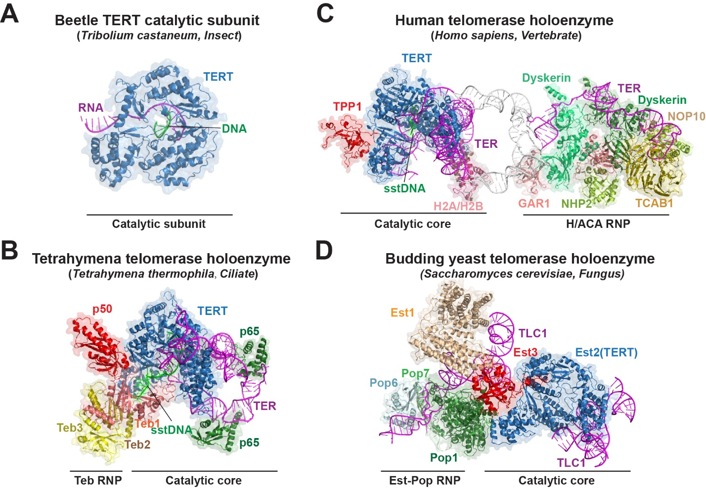
A recurring limitation of comparative telomerase structural studies is that conservation of the overall catalytic fold does not necessarily ensure conservation of ligand recognition or inhibitor sensitivity (30, 49). Species-specific amino acid substitutions, differences in RNA architecture and accessory-protein interactions, and distinct conformational equilibria can alter the geometry, accessibility, and chemical environment of potential drug-binding pockets. Consequently, compounds developed using isolated or nonhuman TERT constructs require renewed biochemical and structural validation in the assembled human enzyme. This distinction is particularly important for BIBR1532. The binding pocket originally identified in the Tribolium TERT-like protein differs substantially from the site subsequently observed in human telomerase, illustrating the limitations of extrapolating inhibitor-binding mechanisms across species. These findings underscore the importance of human telomerase structures for establishing target engagement, interpreting species-dependent inhibitor responses, and guiding the development of compounds directed against therapeutically relevant binding sites.
4. Structural Basis of Human Telomerase Repeat Synthesis
At the center of the human telomerase holoenzyme, hTERT, hTR, and telomeric DNA assemble into a compact catalytic core (39). hTERT comprises four major domains, TEN, TRBD, RT, and CTE, with the telomerase-specific IFD-TRAP insertion located within the RT region (Fig. 2A) (32). The TRBD, RT, and CTE domains form a ring around the short template–DNA duplex, while TEN and IFD-TRAP cap one face of the ring and contribute to the DNA-binding channel (Fig. 2A, B) (40). hTR is not merely a passive template: its template-pseudoknot domain traverses the catalytic core and helps define the template boundary, whereas CR4/5 contacts distinct surfaces of TRBD and CTE and is stabilized by H2A-H2B (39). These RNA-protein interactions organize the catalytic core and help position the DNA 3′ end and incoming nucleotide for DNA synthesis. Within this architecture, TRBD anchors conserved hTR elements and helps position the RNA template, whereas the RT palm contains the canonical catalytic motifs and acidic residues that coordinate divalent metal ions (32). The finger and motif-B elements contribute to nucleotide entry and template interactions, while CTE constrains the template-DNA duplex and its trajectory (40). TEN and TRAP form a telomerase-specific module involved in DNA handling and repeat-addition processivity. Structural studies of active human telomerase have also defined the interaction of this region with the shelterin protein TPP1 and its role in telomerase recruitment (41, 42). Together, these domains couple RNA recognition and nucleotide addition to DNA retention and template repositioning, enabling successive rounds of telomere-repeat synthesis.
Human telomerase initiates synthesis by aligning the telomeric DNA 3′ end with its internal RNA template and extending the primer through successive nucleotide-addition steps. Upon completion of a repeat, the short template–DNA duplex dissociates, the RNA template repositions, and the DNA 3′ end realigns with the beginning of the template. The DNA product remains associated with the enzyme, permitting another round of synthesis and thereby supporting repeat-addition processivity (Fig. 2C) (46, 47). Nucleotide-bound structures show a four-base-pair primer–template duplex, with the incoming nucleotide forming an additional base pair (46). A separate structural study identified a five-base-pair alignment intermediate that requires one-nucleotide repositioning before the first incoming nucleotide can occupy the active site (49). Unlike many conventional DNA polymerases and HIV-1 reverse transcriptase, human TERT undergoes little finger movement upon dNTP binding, with only limited rearrangement of its active-site geometry (49). Telomerase catalysis therefore relies heavily on precise duplex positioning and translocation rather than a large finger-closing transition.
Biochemical analysis identified the first nucleotide-addition step as intrinsically slow, suggesting that initiation represents a kinetically distinct stage of the telomerase catalytic cycle (47). Subsequent cryo-EM structures representing initiation, elongation, and pre-termination states revealed a largely preformed active-site architecture in which surrounding RNA and TERT elements accommodate progression through the repeat (46). Together, these findings indicate that efficient repeat synthesis requires coordinated duplex positioning, DNA anchoring, RNA flexibility, and domain movements, rather than nucleotide incorporation alone. In particular, maintaining the nascent DNA within the enzyme while repositioning the RNA template is essential for successive rounds of repeat synthesis. These mechanistic features reveal therapeutic opportunities beyond the nucleotide-binding site. Imetelstat blocks productive template–primer pairing through sequence-specific antagonism of hTR (60, 61), whereas BIBR1532 perturbs the conformational network involved in duplex repositioning (49). Human telomerase structures thus provide a mechanistic framework for developing inhibitors that disrupt substrate engagement, nucleotide utilization, product retention, or duplex translocation. Targeting these distinct stages may enable therapeutic strategies that exploit the specialized catalytic cycle of telomerase rather than the conserved polymerase active site alone.
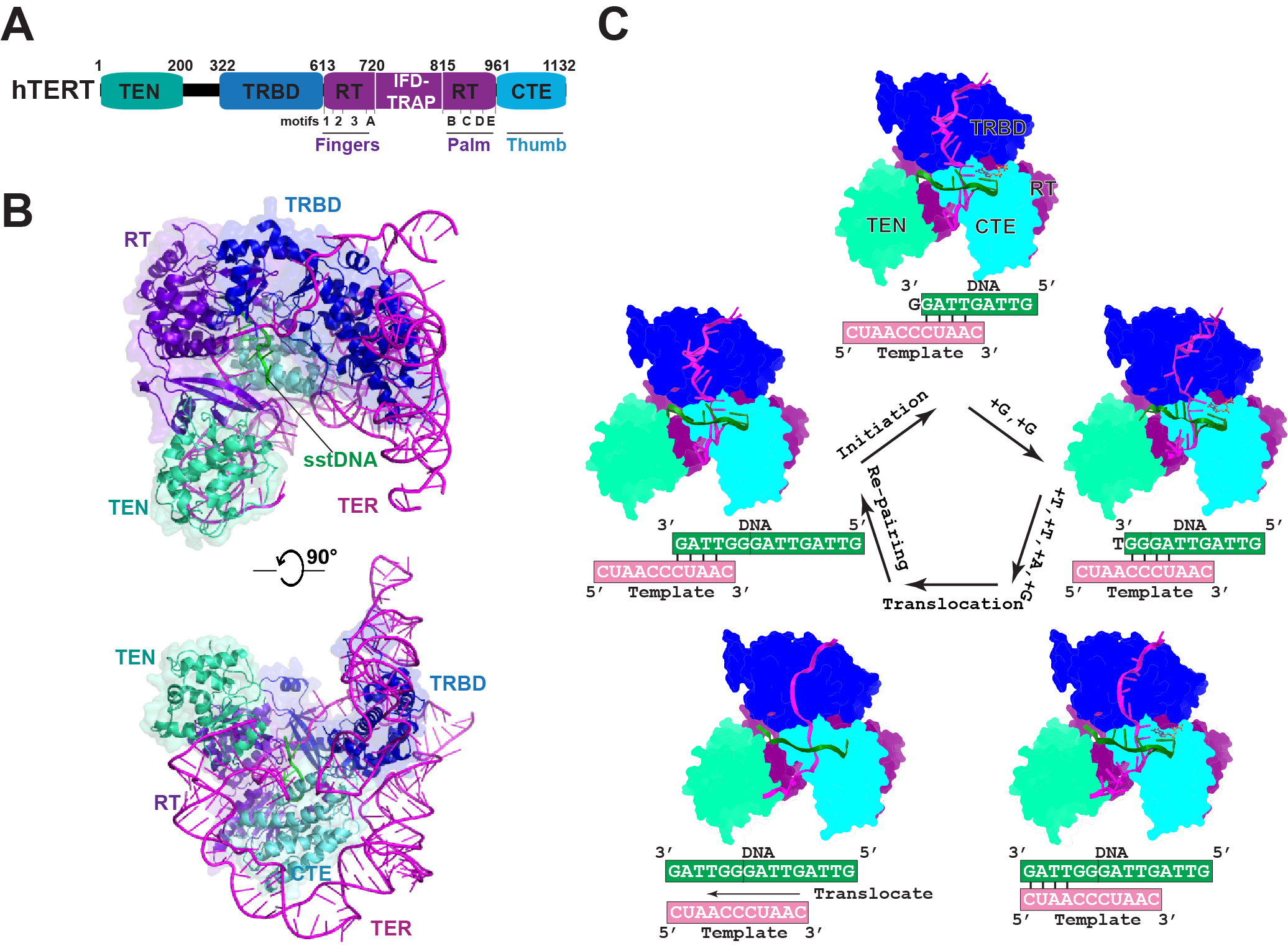
Although cryo-EM has resolved multiple catalytic intermediates, these structures represent experimentally stabilized populations rather than continuous observations of individual enzyme molecules undergoing catalysis. Their mechanistic interpretation therefore requires complementary kinetic measurements and targeted perturbation experiments to establish the sequence and functional significance of the observed states. In particular, the relative occupancy of individual states under structural conditions may not accurately reflect their distribution during telomere extension in living cells, where shelterin interactions, substrate availability, and additional telomere-associated factors can influence enzyme recruitment, catalytic dynamics, and repeat-addition processivity.
5. Direct Strategies for Human Telomerase Inhibition
Telomerase reactivation in the majority of human cancers provides a strong rationale for targeting telomere maintenance as an anticancer strategy (4, 10). Therapeutic approaches can be broadly divided into direct inhibition of the telomerase catalytic machinery and indirect strategies that suppress telomerase expression or disrupt telomere maintenance (48). Despite substantial preclinical progress, clinical translation remains challenging. Inhibitors that act primarily through progressive telomere shortening may require prolonged treatment before producing antitumor effects, while telomerase inhibition can also affect normal hematopoietic progenitors (48, 63). Additional challenges include variation in tumor telomere length, the potential for alternative lengthening of telomeres (ALT), and delivery constraints associated with oligonucleotide therapeutics (20, 61). Table 1 summarizes representative strategies, their mechanisms, development status, advantages, and limitations. Among direct approaches, the hTR-template antagonist imetelstat has progressed to phase III clinical evaluation and regulatory approval for a defined population of patients with lower-risk myelodysplastic syndromes (50, 63). The following discussion focuses on the major classes of direct telomerase inhibition.
Table 1. Therapeutic strategies targeting telomerase and telomere maintenance in cancer.
| Strategy | Agents | Molecular mechanism | Development status | Potential advantage | Limitations |
|---|---|---|---|---|---|
| hTR-template antagonism | Imetelstat. | Binds the hTR/TERC template and blocks productive primer pairing and telomere repeat synthesis. | FDA approved for transfusion-dependent anemia in eligible adults with lower-risk myelodysplastic syndromes. | Sequence-specific telomerase inhibition with proven reduction in transfusion dependence in eligible patients with MDS (50, 60, 63). | Intravenous dosing; frequent thrombocytopenia and neutropenia; benefit established in a restricted hematologic indication (50, 63). |
| Nucleoside analogs | AZT; 6-thio-dG. | After intracellular activation, AZT-TP inhibits telomerase-mediated DNA extension, whereas 6-thio-dGTP incorporation induces telomere dysfunction and can stall repeat synthesis (64–66). | 6-thio-dG (ateganosine/THIO), sequenced with cemiplimab, is being evaluated in phase II and III NSCLC trials (NCT05208944; NCT06908304). | 6-thio-dG exploits telomerase-mediated incorporation to induce telomere dysfunction without requiring progressive telomere shortening (65, 66). | AZT has limited telomerase selectivity. Both agents require intracellular activation; potential off-target effects and effects on normal cells require consideration (48, 64–66). |
| Direct hTERT inhibition | BIBR1532 . | Binds the hTERT RT finger–palm pocket and restricts conformational changes required for template–DNA duplex repositioning. | Preclinical / chemical probe | A structurally characterized human TERT pocket provides a basis for developing non-nucleotide-site inhibitors (49). | Limited cellular potency and uncertain pharmacokinetics; derivatives require validation of target engagement, selectivity, and in vivo efficacy (49, 67–69). |
| Telomeric G-quadruplex stabilization | Telomestatin and related G4 ligands. | Stabilizes telomeric G-quadruplex DNA, limiting productive telomerase access and perturbing telomere function | Preclinical | Targets telomeric DNA without direct engagement of the telomerase catalytic complex (51). | Selectivity among cellular G-quadruplexes is challenging, with potential effects on replication and transcription (48, 51). |
| TERT-directed immunotherapy | VX-001 and related TERT vaccines. | Can elicit TERT-specific T-cell responses without directly inhibiting telomerase catalysis (52). | Clinical trials | Targets a tumor-associated antigen without requiring access to the telomerase catalytic pocket (52). | Responses depend on HLA type and immune context; VX-001 did not demonstrate an overall-survival benefit in the randomized phase II trial (52). |
Abbreviations: ALT, alternative lengthening of telomeres; AZT, zidovudine (3′-azido-3′-deoxythymidine); AZT-TP, zidovudine triphosphate; FDA, US Food and Drug Administration; G4, G-quadruplex; HLA, human leukocyte antigen; hTERT, human telomerase reverse transcriptase; hTR, human telomerase RNA; RT, reverse transcriptase; TERC, telomerase RNA component; TERT, telomerase reverse transcriptase; 6-thio-dG, 6-thio-2′-deoxyguanosine; 6-thio-dGTP, 6-thio-2′-deoxyguanosine triphosphate.
Direct approaches to telomerase inhibition include hTR-template antagonism, interference with nucleotide utilization, and small-molecule inhibition of TERT (Fig. 3) (48). Oligonucleotide antagonists bind the hTR template and prevent productive primer pairing (60, 61); nucleoside metabolites interfere with telomerase-mediated DNA synthesis or induce telomere dysfunction following incorporation (64–66); and BIBR1532 binds a distinct TERT pocket and impairs template–DNA duplex repositioning and repeat-addition processivity (49, 67). These mechanisms differ from indirect strategies that suppress TERT transcription, alter telomeric chromatin, stabilize telomeric G-quadruplexes, or elicit TERT-directed immunity (13, 51, 52). The central question for direct inhibition is whether a defined component or catalytic state of telomerase can be engaged with sufficient selectivity, potency, and pharmacological control to produce a therapeutic window (48).
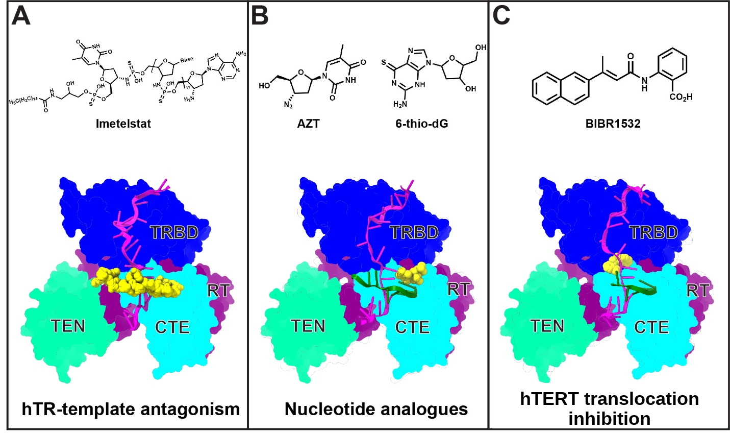
Imetelstat (GRN163L) is a lipid-conjugated, 13-nucleotide oligonucleotide complementary to the template region of human telomerase RNA (hTR). By competitively blocking access of telomeric DNA to the RNA template, it directly inhibits telomerase activity rather than binding to the TERT catalytic pocket (Fig. 3) (60, 61). Its clinical development culminated in the randomized, double-blind phase III IMerge trial in patients with transfusion-dependent lower-risk myelodysplastic syndromes (MDS) who were refractory to, had relapsed after, or were ineligible for erythropoiesis-stimulating agents. Red blood cell transfusion independence lasting at least eight weeks was achieved in 40% of imetelstat-treated patients compared with 15% of placebo-treated patients; the corresponding rates for transfusion independence lasting at least 24 weeks were 28% and 3%, respectively (63). On June 6, 2024, the US Food and Drug Administration approved imetelstat for adults with low- to intermediate-1-risk MDS with transfusion-dependent anemia who meet the specified transfusion-burden and prior-treatment criteria (50). However, grade 3-4 neutropenia and thrombocytopenia were frequent in the phase III trial, underscoring the importance of hematologic toxicity monitoring (63). Although imetelstat establishes the clinical feasibility of directly targeting telomerase, its demonstrated benefit in this setting is reduced transfusion dependence; these findings do not support efficacy against solid tumors or a survival benefit.
Nucleoside analogs can interfere with telomerase-mediated DNA synthesis following intracellular conversion to their nucleotide metabolites (Fig. 3B) (48, 64–66). AZT is converted to AZT triphosphate, which lacks the 3′-hydroxyl group required for continued DNA elongation. Early studies in immortalized human cell lines linked reverse-transcriptase inhibitor exposure to altered telomerase activity and telomere shortening (64). However, limited selectivity among cellular polymerases constrains its potential application as a telomerase-directed anticancer agent (48). A mechanistically distinct approach uses 6-thio-2′-deoxyguanosine (6-thio-dG), which is metabolized to 6-thio-dGTP and incorporated by telomerase into newly synthesized telomeric DNA, inducing telomere dysfunction in telomerase-positive cancer models (65). Subsequent work showed that incorporation can inhibit further repeat synthesis after template translocation, producing a nonproductive telomerase complex rather than immediate enzyme dissociation (66). Unlike inhibitors that act primarily through progressive telomere shortening, 6-thio-dG can induce telomeric damage without prolonged telomere attrition. Nevertheless, intracellular activation, competition with endogenous nucleotide pools, potential off-target effects, and the therapeutic window remain important considerations (48, 65, 66).
The distinction between gradual telomere shortening and acute telomere dysfunction has important therapeutic implications. Inhibition of telomerase can require sustained treatment before progressive telomere attrition limits proliferation (48, 60, 61), whereas 6-thio-dG incorporation can induce telomeric dysfunction without prolonged telomere shortening (65, 66). However, rapid telomere dysfunction does not necessarily confer tumor selectivity. The extent of off-target nucleotide incorporation, dependence on intracellular metabolic activation, effects on normal progenitor cells, and durability of tumor responses require direct comparison in clinically relevant models. These considerations make it premature to infer comparative therapeutic efficacy from biochemical differences alone.
BIBR1532 was identified as a selective, non-nucleosidic telomerase inhibitor associated with progressive telomere shortening and delayed proliferative arrest in telomerase-positive cancer-cell models (62). Subsequent kinetic analysis characterized mixed-type noncompetitive inhibition and reduced repeat-addition processivity, consistent with a binding site distinct from the DNA-primer and dNTP sites (67). BIBR1532 therefore became the principal chemical probe for TERT-directed inhibition (Fig. 3C) (62, 67). However, limited cellular potency, incompletely characterized pharmacokinetic properties, and the historical lack of a validated human binding site have constrained its clinical development (48, 49). Previous medicinal-chemistry studies explored BIBR1532-derived analogs and related scaffolds, using ligand-based design and preclinical activity assays to investigate their telomerase-inhibitory potential (68, 69). However, these compounds were developed before the human BIBR1532-binding pocket was structurally characterized. The identification of this pocket now provides an experimentally defined framework for future structure-guided optimization (49).
6. Structural Basis of BIBR1532-Mediated Human TERT Inhibition
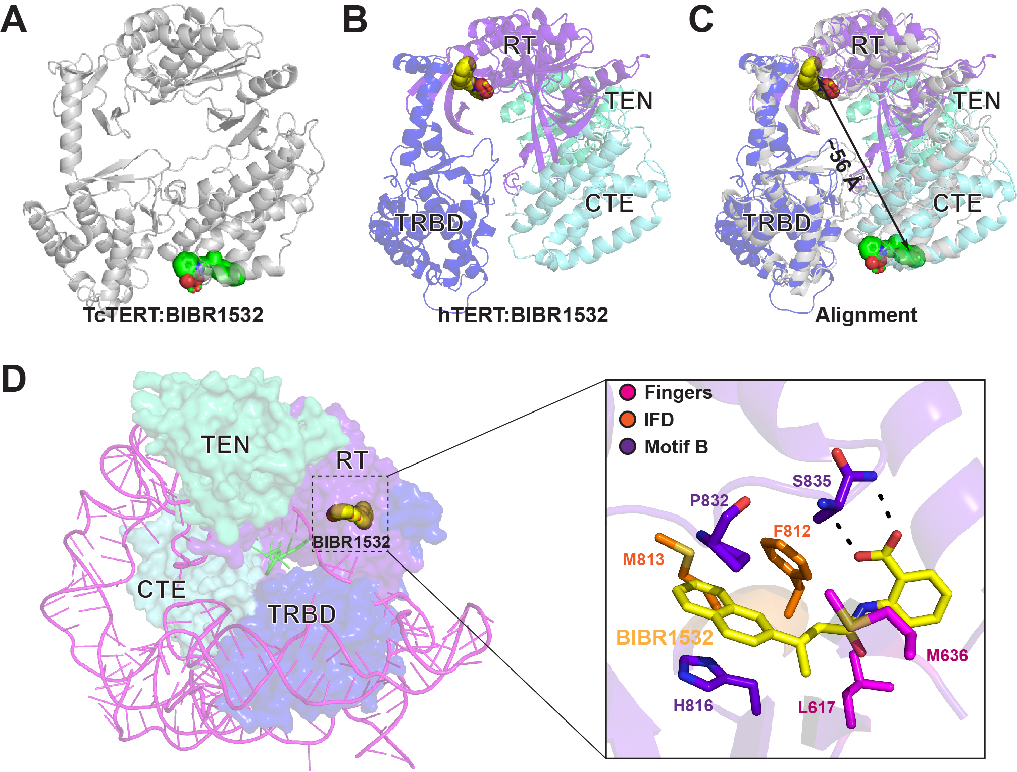
Early biochemical studies established that BIBR1532 inhibits human telomerase without directly competing with nucleotide substrates, although its binding site initially remained unknown (67). The first structural evidence for BIBR1532 binding came from a crystal structure of the inhibitor bound to the isolated Tribolium castaneum TERT-like protein (tcTERT). In this beetle model, BIBR1532 occupied a hydrophobic pocket formed by the FVYL region of the C-terminal extension (CTE), or thumb domain, outside the catalytic center (Fig. 4A) (30). However, a subsequent RNA-DNA-bound tcTERT structure revealed an alternative conformation in which residues T487–N492 form an α-helix, positioning L490 and F494 within the proposed pocket and preventing inhibitor occupancy (31). These findings indicated that the beetle CTE pocket is conformationally variable. Sequence and structural differences, including replacement of beetle F494 with asparagine in human TERT, further limited direct extrapolation to the human enzyme (30, 32). The absence of an identified endogenous Tribolium telomerase RNA and the incomplete domain architecture of the isolated beetle protein introduced additional limitations (31, 32). Although subsequent medicinal-chemistry studies investigated BIBR1532-related scaffolds, these efforts preceded experimental identification of its binding site in human telomerase (68, 69).
More recently, nine cryo-EM structures of human telomerase, encompassing inhibitor-bound and inhibitor-free catalytic states, identified a distinct BIBR1532-binding pocket within the human TERT reverse transcriptase domain, between the finger and palm regions (Fig. 4B, C) (49). Unlike the beetle CTE pocket, the human site lies near the template–DNA duplex but remains separate from the catalytic center, consistent with the previously observed noncompetitive inhibition (49, 67). The ligand is surrounded by residues from the finger, motif 3, motif B, the motif-B loop, and the IFD-TRAP connecting region (49). Within this pocket, S835 forms a hydrogen bond with the BIBR1532 carboxylate, H816 stacks against its naphthalene group, and L617 rearranges to accommodate the ligand (Fig. 4D) (49). Mutational analysis linked these interactions to inhibitor sensitivity, providing experimental support for the human binding-site assignment (49). Thus, the beetle and human structures identify spatially and mechanistically distinct binding sites; the human structures, rather than the earlier beetle model, establish the molecular basis for interpreting BIBR1532 inhibition of human telomerase.
Comparison of inhibitor-bound and inhibitor-free states supports a mechanism in which BIBR1532 impairs template–DNA duplex repositioning rather than directly occupying the nucleotide-binding site (49). Before adding the first nucleotide, the initially aligned template–DNA duplex undergoes an additional one-nucleotide repositioning step to reach a catalytically competent register. BIBR1532-bound particles populated distorted and partially translocated intermediates, indicating that ligand binding slows this transition. The pocket lies within a network formed by the finger β-sheet, motif-B loop, and template-interacting elements; pocket occupation restrains the coordinated protein and RNA-template dynamics required for the template to follow the movement of the DNA primer (49).
BIBR1532 preferentially inhibits the intrinsically slow first nucleotide-addition step (47, 49). The authors proposed that pyrophosphate release helps drive subsequent duplex translocation, contributing to the smaller inhibitory effects at later steps (49). Together, the structural, biochemical, and mutational results support a model in which BIBR1532 inhibits human telomerase by restricting the conformational changes required for template–DNA duplex repositioning rather than competing directly with incoming nucleotides (49, 67). The preferential effect on the slow first nucleotide-addition step is consistent with this mechanism, although further time-resolved and cellular studies are needed to define how pocket occupancy influences the complete catalytic cycle under physiological conditions.
An important unresolved question is whether the conformational states captured in purified telomerase accurately represent inhibitor action at chromosome ends in cancer cells. The structural experiments establish the location of BIBR1532 and its effects on the catalytic cycle, but they do not directly measure the duration of intracellular target occupancy or the influence of shelterin-associated factors, local substrate availability, and telomere-associated chromatin. These variables could alter inhibitor sensitivity or the consequences of impaired repeat synthesis. Complementary studies in cancer cells should therefore connect pocket-specific target engagement with telomerase activity, telomere dysfunction, and sustained proliferative responses. Such experiments would also help distinguish effects caused by direct telomerase inhibition from unrelated activities of candidate compounds.
Discussion
The human telomerase–BIBR1532 structures establish an experimentally validated small-molecule-binding pocket in hTERT that is linked to an essential catalytic transition (49). By locating BIBR1532 between the finger and palm regions of the reverse transcriptase domain, these structures demonstrate that telomerase can be inhibited through interference with template–DNA duplex repositioning without direct occupation of the nucleotide-binding site (49). This finding provides a human-specific structural foundation for inhibitor development, particularly because the human pocket differs from the binding site previously identified in the beetle TERT-like protein (30, 49). Nevertheless, BIBR1532 remains a structural and biochemical starting point rather than an established anticancer drug candidate. Earlier medicinal-chemistry studies of BIBR1532-related compounds support the feasibility of scaffold modification, but their optimization preceded identification of the human binding pocket and should not be interpreted as human-structure-guided drug development (68, 69).
The next challenge is to translate this structural information into compounds with improved potency, selectivity, physicochemical properties, and cellular exposure. The resolved human pocket provides opportunities to preserve the S835 hydrogen bond and interactions involving H816 and L617 while exploring adjacent regions near the motif-B loop, motif 3N entrance, and IFD-TRAP connecting region (49). Because pocket conformation and ligand accessibility may vary across catalytic states, lead optimization should consider both inhibitor-bound and inhibitor-free structures. Candidate compounds should first be assessed through quantitative binding measurements, telomerase activity and repeat-addition processivity assays, inhibitor-bound structural studies, and mutational analysis of pocket residues. Compounds with convincing biochemical activity should then undergo cellular target-engagement studies, evaluation of telomere dysfunction and antiproliferative effects, selectivity testing, and pharmacokinetic–pharmacodynamic assessment. These experiments are essential because binding to purified telomerase does not establish adequate intracellular exposure, sustained target occupancy, or an acceptable therapeutic window.
Clinical translation will also require identifying tumors in which telomerase inhibition is likely to produce a meaningful therapeutic effect. Telomerase activation, baseline telomere length, proliferative capacity, and the presence of alternative lengthening of telomeres (ALT) may influence treatment response (12, 20). Highly proliferative, telomerase-dependent tumors with relatively short telomeres and no detectable ALT activity could be investigated in initial proof-of-concept studies, although these selection criteria remain hypothetical and require prospective validation. TERT expression alone is unlikely to provide a sufficient predictive biomarker; functional telomerase activity, telomere-maintenance mechanism, checkpoint status, and intratumoral heterogeneity should also be considered. Resistance to direct TERT inhibition also requires systematic investigation. Mutations that alter the newly characterized binding pocket could potentially reduce inhibitor affinity while preserving sufficient catalytic activity, although clinically relevant resistance substitutions have not been established. Tumors may also differ in their capacity to tolerate telomere dysfunction or adapt through alternative telomere-maintenance mechanisms (20, 48, 62, 65). Longitudinal studies using molecularly characterized cancer models, including models with distinct checkpoint backgrounds and telomere lengths, could help distinguish primary resistance from adaptation during treatment. Serial measurements of telomerase activity, telomere length, telomere-associated DNA damage, and ALT-associated phenotypes would be particularly informative. These studies should precede claims that human-pocket-directed TERT inhibitors can overcome resistance associated with existing telomerase-directed treatments.
Combination approaches may help address delayed responses or adaptive resistance. For example, telomerase-dependent incorporation of 6-thio-2′-deoxyguanosine offers a mechanistically distinct means of inducing telomere dysfunction (65). Combinations with agents that increase replication stress or impair DNA-damage tolerance also merit investigation, but their development should depend on experimentally demonstrated benefit and acceptable toxicity. Hematologic safety is particularly important, as the clinical experience with imetelstat has demonstrated substantial risks of neutropenia and thrombocytopenia (63).
DiffSBDD uses protein-pocket-conditioned equivariant diffusion to generate three-dimensional ligands and supports molecular redesign and computational property optimization (70). CMD-GEN combines pocket-conditioned pharmacophore generation with molecular design and conformation alignment, with experimental validation involving PARP1/2 inhibitors (71). PocketXMol provides an atom-level framework for docking, de novo design, fragment growing, fragment linking, and molecular optimization (72). None of these methods has yet been established as effective for developing human TERT inhibitors. Nevertheless, the human telomerase–BIBR1532 structures provide a defined ligand pose, experimentally characterized contact residues, and multiple catalytic conformations that could support scaffold optimization and exploration of alternative chemotypes (49). Computational approaches should account for pocket flexibility, the neighboring RNA–DNA duplex, and relevant solvent interactions rather than relying on a single static structure. Their principal contribution would be to prioritize chemically diverse candidates for the staged experimental program outlined above, potentially expanding the range of human TERT inhibitors beyond BIBR1532-related scaffolds.
References
1. Olovnikov AM. A theory of marginotomy. The incomplete copying of template margin in enzymic synthesis of polynucleotides and biological significance of the phenomenon. J Theor Biol. 1973;41(1):181–190. https://doi.org/10.1016/0022-5193(73)90198-7
2. Greider CW, Blackburn EH. Identification of a specific telomere terminal transferase activity in Tetrahymena extracts. Cell. 1985;43(2 Pt 1):405–413. https://doi.org/10.1016/0092-8674(85)90170-9
3. de Lange T. Shelterin-mediated telomere protection. Annu Rev Genet. 2018;52:223–247. https://doi.org/10.1146/annurev-genet-032918-021921
4. Shay JW, Wright WE. Telomeres and telomerase: three decades of progress. Nat Rev Genet. 2019;20(5):299–309. https://doi.org/10.1038/s41576-019-0099-1
5. Harley CB, Futcher AB, Greider CW. Telomeres shorten during ageing of human fibroblasts. Nature. 1990;345(6274):458–460. https://doi.org/10.1038/345458a0
6. Takai H, Aria V, Borges P, Yeeles JTP, de Lange T. CST-polymerase α-primase solves a second telomere end-replication problem. Nature. 2024;627(8004):664–670. https://doi.org/10.1038/s41586-024-07137-1
7. Chakravarti D, LaBella KA, DePinho RA. Telomeres: history, health, and hallmarks of aging. Cell. 2021;184(2):306–322. https://doi.org/10.1016/j.cell.2020.12.028
8. d'Adda di Fagagna F, Reaper PM, Clay-Farrace L, Fiegler H, Carr P, Von Zglinicki T, et al. A DNA damage checkpoint response in telomere-initiated senescence. Nature. 2003;426(6963):194–198. https://doi.org/10.1038/nature02118
9. Maciejowski J, Li Y, Bosco N, Campbell PJ, de Lange T. Chromothripsis and kataegis induced by telomere crisis. Cell. 2015;163(7):1641–1654. https://doi.org/10.1016/j.cell.2015.11.054
10. Roake CM, Artandi SE. Regulation of human telomerase in homeostasis and disease. Nat Rev Mol Cell Biol. 2020;21(7):384–397. https://doi.org/10.1038/s41580-020-0234-z
11. Schmidt JC, Cech TR. Human telomerase: biogenesis, trafficking, recruitment, and activation. Genes Dev. 2015;29(11):1095–1105. https://doi.org/10.1101/gad.263863.115
12. Barthel FP, Wei W, Tang M, Martinez-Ledesma E, Hu X, Amin SB, et al. Systematic analysis of telomere length and somatic alterations in 31 cancer types. Nat Genet. 2017;49(3):349–357. https://doi.org/10.1038/ng.3781
13. Sharma S, Chowdhury S. Emerging mechanisms of telomerase reactivation in cancer. Trends Cancer. 2022;8(8):632–641. https://doi.org/10.1016/j.trecan.2022.03.005
14. Kim NW, Piatyszek MA, Prowse KR, Harley CB, West MD, Ho PL, et al. Specific association of human telomerase activity with immortal cells and cancer. Science. 1994;266(5193):2011–2015. https://doi.org/10.1126/science.7605428
15. Horn S, Figl A, Rachakonda PS, Fischer C, Sucker A, Gast A, et al. TERT promoter mutations in familial and sporadic melanoma. Science. 2013;339(6122):959–961. https://doi.org/10.1126/science.1230062
16. Huang FW, Hodis E, Xu MJ, Kryukov GV, Chin L, Garraway LA. Highly recurrent TERT promoter mutations in human melanoma. Science. 2013;339(6122):957–959. https://doi.org/10.1126/science.1229259
17. Killela PJ, Reitman ZJ, Jiao Y, Bettegowda C, Agrawal N, Diaz LA Jr, et al. TERT promoter mutations occur frequently in gliomas and a subset of tumors derived from cells with low rates of self-renewal. Proc Natl Acad Sci U S A. 2013;110(15):6021–6026. https://doi.org/10.1073/pnas.1303607110
18. Bell RJ, Rube HT, Kreig A, Mancini A, Fouse SD, Nagarajan RP, et al. Cancer. The transcription factor GABP selectively binds and activates the mutant TERT promoter in cancer. Science. 2015;348(6238):1036–1039. https://doi.org/10.1126/science.aab0015
19. Hanahan D. Hallmarks of cancer: new dimensions. Cancer Discov. 2022;12(1):31–46. https://doi.org/10.1158/2159-8290.Cd-21-1059
20. Heaphy CM, Subhawong AP, Hong SM, Goggins MG, Montgomery EA, Gabrielson E, et al. Prevalence of the alternative lengthening of telomeres telomere maintenance mechanism in human cancer subtypes. Am J Pathol. 2011;179(4):1608–1615. https://doi.org/10.1016/j.ajpath.2011.06.018
21. He Y, Feigon J. Telomerase structural biology comes of age. Curr Opin Struct Biol. 2022;76:102446. https://doi.org/10.1016/j.sbi.2022.102446
22. Theimer CA, Blois CA, Feigon J. Structure of the human telomerase RNA pseudoknot reveals conserved tertiary interactions essential for function. Mol Cell. 2005;17(5):671–682. https://doi.org/10.1016/j.molcel.2005.01.017
23. Zhang Q, Kim NK, Feigon J. Architecture of human telomerase RNA. Proc Natl Acad Sci U S A. 2011;108(51):20325–20332. https://doi.org/10.1073/pnas.1100279108
24. Jurczyluk J, Nouwens AS, Holien JK, Adams TE, Lovrecz GO, Parker MW, et al. Direct involvement of the TEN domain at the active site of human telomerase. Nucleic Acids Res. 2011;39(5):1774–1788. https://doi.org/10.1093/nar/gkq1083
25. Theimer CA, Jády BE, Chim N, Richard P, Breece KE, Kiss T, et al. Structural and functional characterization of human telomerase RNA processing and Cajal body localization signals. Mol Cell. 2007;27(6):869–881. https://doi.org/10.1016/j.molcel.2007.07.017
26. Chen G, Wen JD, Tinoco I Jr. Single-molecule mechanical unfolding and folding of a pseudoknot in human telomerase RNA. RNA. 2007;13(12):2175–2188. https://doi.org/10.1261/rna.676707
27. Chen JL, Greider CW. Functional analysis of the pseudoknot structure in human telomerase RNA. Proc Natl Acad Sci U S A. 2005;102(23):8080–8085. https://doi.org/10.1073/pnas.0502259102
28. Gillis AJ, Schuller AP, Skordalakes E. Structure of the Tribolium castaneum telomerase catalytic subunit TERT. Nature. 2008;455(7213):633–637. https://doi.org/10.1038/nature07283
29. Mitchell M, Gillis A, Futahashi M, Fujiwara H, Skordalakes E. Structural basis for telomerase catalytic subunit TERT binding to RNA template and telomeric DNA. Nat Struct Mol Biol. 2010;17(4):513–518. https://doi.org/10.1038/nsmb.1777
30. Bryan C, Rice C, Hoffman H, Harkisheimer M, Sweeney M, Skordalakes E. Structural basis of telomerase inhibition by the highly specific BIBR1532. Structure. 2015;23(10):1934–1942. https://doi.org/10.1016/j.str.2015.08.006
31. Choi WS, Weng PJ, Yang W. Flexibility of telomerase in binding the RNA template and DNA telomeric repeat. Proc Natl Acad Sci U S A. 2022;119(1):e2116159118. https://doi.org/10.1073/pnas.2116159118
32. Wang Y, Gallagher-Jones M, Sušac L, Song H, Feigon J. A structurally conserved human and Tetrahymena telomerase catalytic core. Proc Natl Acad Sci U S A. 2020;117(49):31078–31087. https://doi.org/10.1073/pnas.2011684117
33. Jiang J, Miracco EJ, Hong K, Eckert B, Chan H, Cash DD, et al. The architecture of Tetrahymena telomerase holoenzyme. Nature. 2013;496(7444):187–192. https://doi.org/10.1038/nature12062
34. Jiang J, Chan H, Cash DD, Miracco EJ, Ogorzalek Loo RR, Upton HE, et al. Structure of Tetrahymena telomerase reveals previously unknown subunits, functions, and interactions. Science. 2015;350(6260):aab4070. https://doi.org/10.1126/science.aab4070
35. Jiang J, Wang Y, Sušac L, Chan H, Basu R, Zhou ZH, et al. Structure of telomerase with telomeric DNA. Cell. 2018;173(5):1179–1190.e13. https://doi.org/10.1016/j.cell.2018.04.038
36. He Y, Wang Y, Liu B, Helmling C, Sušac L, Cheng R, et al. Structures of telomerase at several steps of telomere repeat synthesis. Nature. 2021;593(7859):454–459. https://doi.org/10.1038/s41586-021-03529-9
37. Sauerwald A, Sandin S, Cristofari G, Scheres SHW, Lingner J, Rhodes D. Structure of active dimeric human telomerase. Nat Struct Mol Biol. 2013;20(4):454–460. https://doi.org/10.1038/nsmb.2530
38. Nguyen THD, Tam J, Wu RA, Greber BJ, Toso D, Nogales E, et al. Cryo-EM structure of substrate-bound human telomerase holoenzyme. Nature. 2018;557(7704):190–195. https://doi.org/10.1038/s41586-018-0062-x
39. Ghanim GE, Fountain AJ, van Roon AM, Rangan R, Das R, Collins K, et al. Structure of human telomerase holoenzyme with bound telomeric DNA. Nature. 2021;593(7859):449–453. https://doi.org/10.1038/s41586-021-03415-4
40. Wan F, Ding Y, Zhang Y, Wu Z, Li S, Yang L, et al. Zipper head mechanism of telomere synthesis by human telomerase. Cell Res. 2021;31(12):1275–1290. https://doi.org/10.1038/s41422-021-00586-7
41. Liu B, He Y, Wang Y, Song H, Zhou ZH, Feigon J. Structure of active human telomerase with telomere shelterin protein TPP1. Nature. 2022;604(7906):578–583. https://doi.org/10.1038/s41586-022-04582-8
42. Sekne Z, Ghanim GE, van Roon AM, Nguyen THD. Structural basis of human telomerase recruitment by TPP1-POT1. Science. 2022;375(6585):1173–1176. https://doi.org/10.1126/science.abn6840
43. Ghanim GE, Sekne Z, Balch S, van Roon AM, Nguyen THD. 2.7 Å cryo-EM structure of human telomerase H/ACA ribonucleoprotein. Nat Commun. 2024;15(1):746. https://doi.org/10.1038/s41467-024-45002-x
44. Balch S, Sekne Z, Franco-Echevarría E, Ludzia P, Kretsch RC, Sun W, et al. Cryo-EM structure of human telomerase dimer reveals H/ACA RNP-mediated dimerization. Science. 2025;389(6756):eadr5817. https://doi.org/10.1126/science.adr5817
45. Hu H, Neumann H, Teplitz GM, Franco-Echevarría E, Chartrand P, Wellinger RJ, et al. Cryo-electron microscopy structure of the budding yeast telomerase holoenzyme. Science. 2026;391(6792):eadz5344. https://doi.org/10.1126/science.adz5344
46. Balch S, Franco-Echevarría E, Ghanim GE, Kretsch RC, Das R, Nguyen THD. Structures of nucleotide-bound human telomerase at several steps of its telomeric DNA repeat addition cycle. Nat Commun. 2026;17(1):1847. https://doi.org/10.1038/s41467-026-68560-8
47. Chen Y, Podlevsky JD, Logeswaran D, Chen JJ. A single nucleotide incorporation step limits human telomerase repeat addition activity. EMBO J. 2018;37(6):e97953. https://doi.org/10.15252/embj.201797953
48. Siteni S, Grichuk A, Shay JW. Telomerase in cancer therapeutics. Cold Spring Harb Perspect Biol. 2024;16(12):a041703. https://doi.org/10.1101/cshperspect.a041703
49. Wang Y, Liu B, He Y, Feigon J. Structures of human telomerase with BIBR1532 reveal novel mechanism of inhibition. Nat Chem Biol. 2026;22(11):1854–1862. https://doi.org/10.1038/s41589-026-02238-6
50. Kim N, Pulte ED, Ehrlich LA, Ionan AC, Haupert S, Vallejo J, et al. US Food and Drug Administration approval summary: imetelstat for selected patients with low- to intermediate-1 risk myelodysplastic syndromes with transfusion-dependent anemia. J Clin Oncol. 2025;43(35):3760–3768. https://doi.org/10.1200/JCO-25-01369
51. Kim MY, Vankayalapati H, Shin-Ya K, Wierzba K, Hurley LH. Telomestatin, a potent telomerase inhibitor that interacts quite specifically with the human telomeric intramolecular G-quadruplex. J Am Chem Soc. 2002;124(10):2098–2099. https://doi.org/10.1021/ja017308q
52. Gridelli C, Ciuleanu T, Domine M, Szczesna A, Bover I, Cobo M, et al. Clinical activity of a hTERT (VX-001) cancer vaccine as post-chemotherapy maintenance immunotherapy in patients with stage IV non-small cell lung cancer: final results of a randomised phase 2 clinical trial. Br J Cancer. 2020;122(10):1461–1466. https://doi.org/10.1038/s41416-020-0785-y
53. Vinagre J, Almeida A, Pópulo H, Batista R, Lyra J, Pinto V, et al. Frequency of TERT promoter mutations in human cancers. Nat Commun. 2013;4:2185. https://doi.org/10.1038/ncomms3185
54. Singh M, Wang Z, Koo BK, Patel A, Cascio D, Collins K, et al. Structural basis for telomerase RNA recognition and RNP assembly by the holoenzyme La family protein p65. Mol Cell. 2012;47(1):16–26. https://doi.org/10.1016/j.molcel.2012.05.018
55. Wang Y, He Y, Wang Y, Yang Y, Singh M, Eichhorn CD, et al. Structure of LARP7 protein p65-telomerase RNA complex in telomerase revealed by cryo-EM and NMR. J Mol Biol. 2023;435(11):168044. https://doi.org/10.1016/j.jmb.2023.168044
56. He Y, Song H, Chan H, Liu B, Wang Y, Sušac L, et al. Structure of Tetrahymena telomerase-bound CST with polymerase α-primase. Nature. 2022;608(7924):813–818. https://doi.org/10.1038/s41586-022-04931-7
57. Kim NK, Zhang Q, Zhou J, Theimer CA, Peterson RD, Feigon J. Solution structure and dynamics of the wild-type pseudoknot of human telomerase RNA. J Mol Biol. 2008;384(5):1249–1261. https://doi.org/10.1016/j.jmb.2008.10.005
58. Leeper TC, Varani G. The structure of an enzyme-activating fragment of human telomerase RNA. RNA. 2005;11(4):394–403. https://doi.org/10.1261/rna.7222505
59. Wang Y, Yesselman JD, Zhang Q, Kang M, Feigon J. Structural conservation in the template/pseudoknot domain of vertebrate telomerase RNA from teleost fish to human. Proc Natl Acad Sci U S A. 2016;113(35):E5125–E5134. https://doi.org/10.1073/pnas.1607411113
60. Herbert BS, Pongracz K, Shay JW, Gryaznov SM. Oligonucleotide N3′→P5′ phosphoramidates as efficient telomerase inhibitors. Oncogene. 2002;21(4):638–642. https://doi.org/10.1038/sj.onc.1205064
61. Herbert BS, Gellert GC, Hochreiter A, Pongracz K, Wright WE, Zielinska D, et al. Lipid modification of GRN163, an N3′ → P5′ thio-phosphoramidate oligonucleotide, enhances the potency of telomerase inhibition. Oncogene. 2005;24(33):5262–5268. https://doi.org/10.1038/sj.onc.1208760
62. Damm K, Hemmann U, Garin-Chesa P, Hauel N, Kauffmann I, Priepke H, et al. A highly selective telomerase inhibitor limiting human cancer cell proliferation. EMBO J. 2001;20(24):6958–6968. https://doi.org/10.1093/emboj/20.24.6958
63. Platzbecker U, Santini V, Fenaux P, Sekeres MA, Savona MR, Madanat YF, et al. Imetelstat in patients with lower-risk myelodysplastic syndromes who have relapsed or are refractory to erythropoiesis-stimulating agents (IMerge): a multinational, randomised, double-blind, placebo-controlled, phase 3 trial. Lancet. 2024;403(10423):249–260. https://doi.org/10.1016/s0140-6736(23)01724-5
64. Strahl C, Blackburn EH. Effects of reverse transcriptase inhibitors on telomere length and telomerase activity in two immortalized human cell lines. Mol Cell Biol. 1996;16(1):53–65. https://doi.org/10.1128/mcb.16.1.53
65. Mender I, Gryaznov S, Dikmen ZG, Wright WE, Shay JW. Induction of telomere dysfunction mediated by the telomerase substrate precursor 6-thio-2'-deoxyguanosine. Cancer Discov. 2015;5(1):82–95. https://doi.org/10.1158/2159-8290.Cd-14-0609
66. Sanford SL, Badstübner M, Gerber M, Mannherz W, Lampl N, Dannenberg R, et al. Therapeutic 6-thio-deoxyguanosine inhibits telomere elongation in cancer cells by inducing a non-productive stalled telomerase complex. Nat Commun. 2026;17(1):6. https://doi.org/10.1038/s41467-025-66534-w
67. Pascolo E, Wenz C, Lingner J, Hauel N, Priepke H, Kauffmann I, et al. Mechanism of human telomerase inhibition by BIBR1532, a synthetic, non-nucleosidic drug candidate. J Biol Chem. 2002;277(18):15566–15572. https://doi.org/10.1074/jbc.M201266200
68. Al-Karmalawy AA, Nafie MS, Shaldam MA, Elmaaty AA, Antar SA, El-Hamaky AA, et al. Ligand-based design on the dog-bone-shaped BIBR1532 pharmacophoric features and synthesis of novel analogues as promising telomerase inhibitors with in vitro and in vivo evaluations. J Med Chem. 2023;66(1):777–792. https://doi.org/10.1021/acs.jmedchem.2c01668
69. Al-Karmalawy AA, Mousa MHA, Sharaky M, Mourad MAE, El-Dessouki AM, Hamouda AO, et al. Lead optimization of BIBR1591 to improve its telomerase inhibitory activity: design and synthesis of novel four chemical series with in silico, in vitro, and in vivo preclinical assessments. J Med Chem. 2024;67(1):492–512. https://doi.org/10.1021/acs.jmedchem.3c01708
70. Schneuing A, Harris C, Du Y, Didi K, Jamasb A, Igashov I, et al. Structure-based drug design with equivariant diffusion models. Nat Comput Sci. 2024;4(12):899–909. https://doi.org/10.1038/s43588-024-00737-x
71. Zou Y, Guo T, Fu Z, Guo Z, Bo W, Yan D, et al. A structure-based framework for selective inhibitor design and optimization. Commun Biol. 2025;8(1):422. https://doi.org/10.1038/s42003-025-07840-3
72. Peng X, Guo R, Guo F, Wang Z, Sun J, Guan J, et al. Unified modeling of 3D molecular generation via atomic interactions with PocketXMol. Cell. 2026;189(7):1904–1922.e28. https://doi.org/10.1016/j.cell.2026.01.003
Declarations
Funding Statement
This work was supported by the Macau Science and Technology Development Fund (0143/2025/ITP2), University of Macau (MYRG-GRG2024-00283-ICMS-UMDF), and the State Key Laboratory of Mechanism and Quality of Chinese Medicine (0002/2025/NRP).
Competing Interest
The authors declare no financial or personal relationships with other individuals or organizations that could inappropriately influence or bias the content of this work. All authors have read the final version of the manuscript and confirm that there are no competing interests.
Consent for Publication
Consent for publication: All authors have approved the final version of the manuscript.
Use of Artificial Intelligence Disclosure
This article was written by human contributors. Artificial intelligence-based tools were used to improve grammar, language, and readability without affecting the article's scientific content, data interpretation, or conclusions. The authors reviewed and verified all content to ensure its accuracy and integrity.
Data Availability Statement
No datasets were generated or analyzed in the current study.
Ethics approval and consent to participate
Not applicable. This review did not involve new studies with human participants or animals.
Authors’ affiliations
1. State Key Laboratory of Mechanism and Quality of Chinese Medicine, Institute of Chinese Medical Sciences, University of Macau, Macao SAR, China
2. Department of Pharmacy and Pharmaceutical Sciences, Faculty of Medicine, University of Macau, Macao SAR, China
3. Department of Biological Sciences, Faculty of Science, University of Macau, Macao SAR, China
CRediT authorship contribution statement
Yongyi Wu: Visualization, Writing – review & editing. He Song: Conceptualization, Writing – review & editing. All authors approved the final manuscript.
ORCID ID
Yongyi Wu: https://orcid.org/0009-0009-3698-9299