Targeting Mitosis to Reprogram the Tumor Microenvironment: Combination Strategies for Cancer Immunotherapy
1 Department of Digestive Disease, The First Affiliated Hospital of USTC, Division of Life Sciences and Medicine, University of Science and Technology of China, Hefei, 230027, China.
2 State Key Laboratory of Immune Response and Immunotherapy, Division of Life Sciences and Medicine, University of Science and Technology of China, Hefei, 230027, China.
Correspondence: Fazhi Yu (fzy1988@ustc.edu.cn); Zhenye Yang (zhenye@ustc.edu.cn)
Received: May 13, 2026
Accepted: September 15, 2026
Published: November 15, 2026
© 2026 The Author(s). Published by GCINC Press, Spokane, Washington, United States. Open Access licensed under a Creative Commons Attribution 4.0 International License (CC BY 4.0), which permits unrestricted use, distribution, and reproduction in any medium, provided the original author(s) and source are credited. To view a copy of this license, visit: Creative Commons Attribution 4.0 International License (CC BY 4.0)
Abstract
Dysregulated mitosis is a therapeutic vulnerability in cancer, but the consequences of mitotic targeting extend beyond tumor-cell death. Chromosome missegregation, micronucleus formation, and chromatin-bridge exposure can activate cytosolic DNA sensing and alter inflammatory signaling, immune recognition, and stromal interactions within the tumor microenvironment (TME). These responses are not uniformly antitumor. Acute mitotic perturbation may enhance innate immune activation and tumor-cell susceptibility to immune elimination, whereas persistent chromosomal instability can promote inflammatory adaptation, myeloid immunosuppression, and immune escape. This distinction has important implications for combining antimitotic agents with immunotherapy. Here, we examine the molecular mechanisms linking mitotic disruption to TME remodeling, with particular emphasis on signaling through cyclic GMP–AMP synthase (cGAS) and stimulator of interferon genes (STING), immune checkpoint regulation, myeloid-cell responses, and stromal reorganization. We evaluate combination strategies involving immune checkpoint blockade, innate immune modulation, myeloid targeting, radiotherapy, and cell-death-enhancing interventions. Although preclinical studies provide mechanistic support for several approaches, clinical evidence remains uneven, and the immune consequences of individual antimitotic agents are difficult to predict across tumor types. We propose a testable framework integrating baseline chromosomal instability, p53 status, innate immune competence, and early treatment-induced biomarkers to guide therapeutic selection and scheduling. Translating this framework into clinical practice will require prospective validation, clearer separation of drug-specific effects from general mitotic stress responses, and evidence that immune remodeling improves patient outcomes.
Keywords
Mitotic stress; chromosomal instability; tumor microenvironment; cGAS–STING signaling; cancer immunotherapy; immune resistance; combination therapy; predictive biomarkers.
1. Introduction
Chromosome imbalance and defective chromosome segregation can contribute to genomic instability and tumor development (1, 2). This understanding provided part of the rationale for developing microtubule-targeting agents, including taxanes and vinca alkaloids, as anticancer therapies (3, 4). Historically, the therapeutic focus has been largely tumor cell-intrinsic, emphasizing the dependence of proliferating cancer cells on coordinated spindle assembly, chromosome segregation, and cytokinesis (5, 6). Disrupting these processes can trigger mitotic arrest, chromosome missegregation, or cell death, although the outcome depends on the extent of mitotic damage and tumor cells' capacity to adapt (7–10). More recently, research on chromosomal instability (CIN) has established that mitotic errors can generate micronuclei and chromatin bridges, providing potential sources of cytosolic DNA (11, 12). Recognition of this DNA by innate immune sensing pathways, particularly cGAS–STING, can initiate inflammatory signaling that extends beyond the affected tumor cell (13, 14). However, these responses do not uniformly promote antitumor immunity. Depending on the persistence of genomic instability and the surrounding immune context, mitotic stress may contribute to either immune activation or tumor-promoting inflammatory adaptation (15, 16). Thus, mitotic disruption should be considered not only a mechanism of tumor-cell injury but also a potential regulator of tumor microenvironment (TME) remodeling.
Tumor mitosis often occurs under replication stress, centrosome amplification, spindle defects, altered checkpoint regulation, and pre-existing genomic instability (1, 17, 18). Genomic instability is an important feature of cancer, and patterns of chromosomal instability vary across tumors (19, 20). These observations motivate investigation of context-specific therapeutic vulnerabilities but do not establish dependence on a particular mitotic target. Chromosomal instability can drive gene amplification through chromothripsis (21), while whole-genome doubling can create distinct dependencies on mitotic machinery (22). Selected aneuploid cancer cells are vulnerable to mitotic checkpoint inhibition (23), and whole-genome duplication can also generate genetic instability during the subsequent S phase (24). These are distinct mechanisms; sensitivity to checkpoint inhibition cannot be inferred from weakened checkpoint control alone. However, these vulnerabilities are not universal, as their therapeutic significance depends on tumor lineage, the extent of pre-existing mitotic dysfunction, and the capacity of tumor cells to adapt. This vulnerability has motivated therapies targeting microtubules, Aurora kinases, PLK1, survivin, and other components of spindle assembly, chromosome segregation, and cytokinesis (3, 25–27). However, the clinical activity of many next-generation antimitotic drugs has been limited by toxicity in proliferative normal tissues, incomplete target exposure, mitotic slippage, adaptive stress responses, and intratumoral heterogeneity (25, 28–32).
Importantly, mitotic perturbation can have consequences beyond immediate tumor-cell death. Mitotic checkpoint dysregulation and abnormal mitotic progression can generate chromosome abnormalities or polyploid states, and some affected cells remain viable (2, 33, 34). These abnormalities may alter cytosolic DNA sensing and downstream inflammatory signaling (11, 12). However, the immunological consequences are context-dependent. Acute mitotic injury may promote antitumor immune activation, whereas persistent chromosomal instability can favor chronic inflammation, myeloid immunosuppression, and immune escape (13–15, 35).
In this Review, we use these observations to propose a unifying framework in which mitosis-targeting therapy is considered not only a cytotoxic intervention but also a potential inducer of transient tumor-state and microenvironmental change. We emphasize that 'state conversion' is an organizing concept rather than a clinically validated therapeutic category. A central, testable hypothesis is that the quality, duration, and resolution of mitotic injury determine whether the induced state becomes immune-activating, immune-suppressive, or tumor-adaptive, and therefore which combination therapy, if any, is most likely to be effective.
This framework has important translational implications because the therapeutic consequences of mitotic perturbation depend on the immune and stromal context of the tumor microenvironment. Related evidence from G1-directed CDK4/6 inhibition shows that cell-cycle interventions can enhance antigen presentation and T-cell activation (36, 37). These findings provide a basis for comparison with mitotic targeting rather than direct evidence of its effects. CDK4/6-related signaling can regulate PD-L1 stability (38), while abemaciclib enhanced immune activation and anti-PD-L1 efficacy in preclinical models (39). Post-translational regulation is an additional layer of checkpoint control (40). YM155 has also been investigated with docetaxel in preclinical melanoma models, providing a separate example of combining a survivin-directed approach with a microtubule-targeting agent (41). Innate immune modulation, myeloid targeting, radiotherapy, and cell-death-enhancing interventions offer additional candidate partners, as discussed below. However, these approaches differ substantially in mechanistic rationale and clinical validation, and their benefits cannot be assumed across tumor types. The central question is therefore not simply whether an antimitotic drug disrupts cell division, but whether the resulting tumor and TME changes can be reliably measured, linked to therapeutic vulnerabilities, and exploited within an appropriate treatment window before adaptive resistance emerges.
On the basis of this perspective, this Review is organized into four major sections. First, we summarize the molecular control of tumor cell division and the principal therapeutic vulnerabilities embedded within this machinery. Second, we discuss how mitosis-targeting therapies remodel the TME through CIN, cytosolic DNA sensing, inflammatory rewiring, NK cell recognition, and immune checkpoint regulation, thereby shaping antitumor immunity. Third, we outline rational combination strategies built upon these mechanisms, including integration with immune checkpoint blockade, innate immune agonism, myeloid reprogramming, and radiotherapy. Finally, we consider the key translational challenges that must be addressed, including efficacy, biomarker development, treatment scheduling, and toxicity management, if cell-division-targeting strategies are to be fully integrated into next-generation cancer immunotherapy.
2. Tumor cell division control and therapeutic targeting of mitotic vulnerabilities
The mitotic program as a therapeutic target
Faithful mitosis requires coordinated mitotic entry, centrosome maturation, bipolar spindle assembly, chromosome congression, spindle-assembly checkpoint satisfaction, chromosome segregation, mitotic exit, and cytokinesis (5–8). In tumor cells, this machinery often operates under replication stress, supernumerary centrosomes, abnormal kinetochore-microtubule attachments, altered checkpoint regulation, and pre-existing genome instability (7, 18, 42, 43). Recent reviews discuss how mitotic dysregulation may create therapeutic opportunities, while emphasizing differences among targets, agents, and tumor contexts (44–46).
This stress-dependent reliance on the mitotic machinery creates a therapeutically exploitable vulnerability. Tumor cells depend on continued cell division to sustain growth, yet many operate under structural, mechanical, and genomic constraints that reduce their tolerance to further mitotic disruption (47–49). This principle helps explain why mitotic regulators, including Aurora kinases, PLK1, spindle-assembly checkpoint components, kinesins, microtubules, and cytokinetic machinery, have long been considered attractive anticancer targets (47, 50–53). These components are not merely executors of mitosis; they also help stabilize a division program that is already functioning under substantial cellular stress (Figure 1).
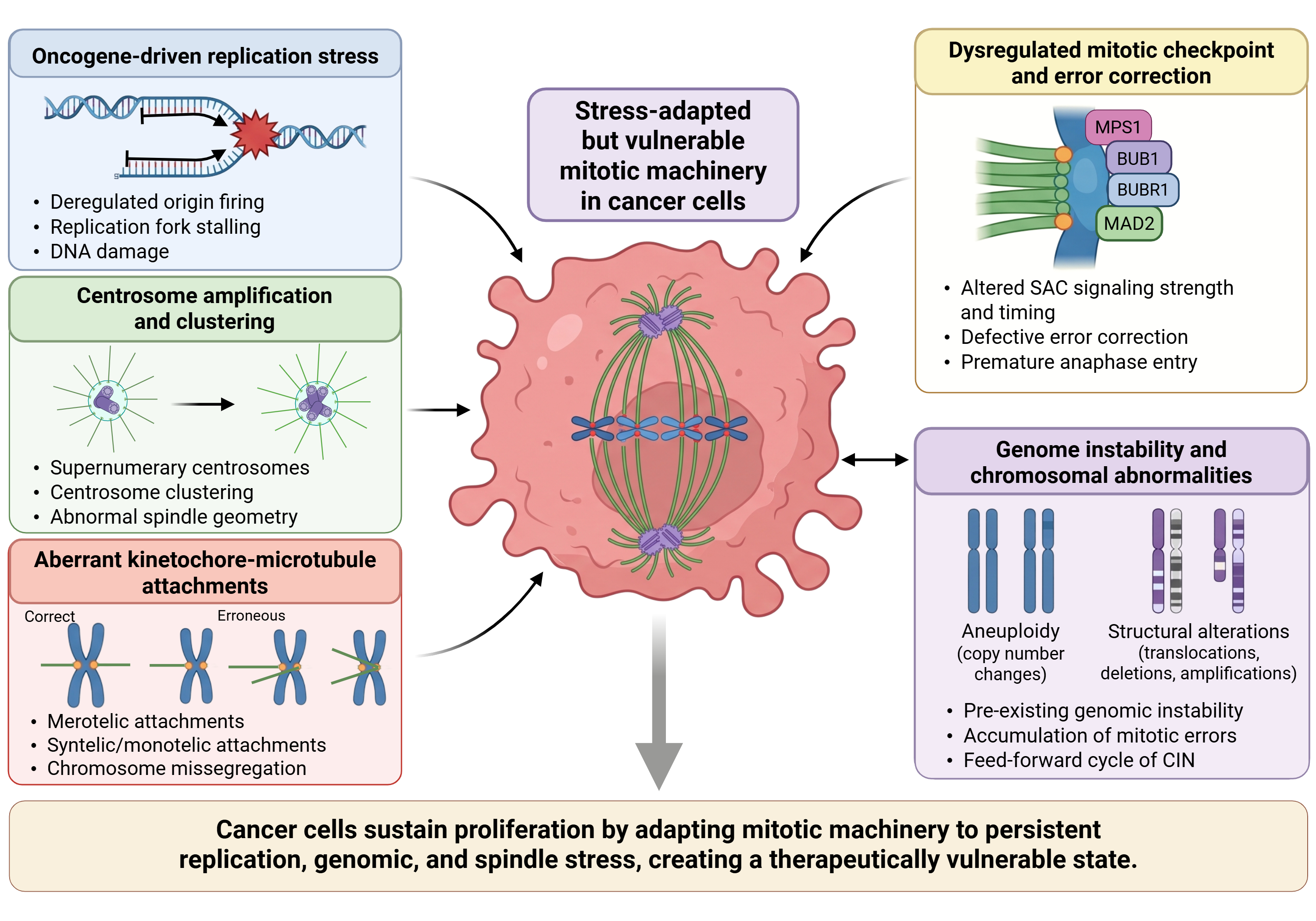
However, the therapeutic significance of mitotic vulnerability extends beyond direct tumor cell killing. When mitotic control is disrupted, tumor cells may undergo apoptosis or enter mitotic catastrophe, which can culminate in cell death or senescence. Other cells may survive with chromosome missegregation, micronuclei, chromatin bridges, polyploidy, or persistent chromosomal instability (9, 33, 34, 54). Surviving cells can exhibit abnormal post-mitotic states (9). Reviews of genome instability and immunity describe context-dependent inflammatory and immune consequences of such abnormalities, rather than a uniform immune response (55–57). Thus, the same vulnerabilities that make mitosis an attractive cytotoxic target also create a route through which cell-division perturbation can remodel the tumor microenvironment.
Dependence on mitotic regulatory kinases
Mitotic kinases such as Aurora A, Aurora B, and PLK1 are central components of the regulatory network that coordinates mitotic progression and enables cells to cope with mitotic stress (5, 47, 50–52). Their dysregulation can support malignant proliferation while simultaneously creating therapeutic vulnerabilities. Importantly, however, tumor dependence on these kinases is determined not simply by expression level, but also by lineage, cellular state, and the extent of pre-existing mitotic dysfunction (44–46, 58).
Aurora kinases occupy a central position in the mitotic regulatory network. Aurora A primarily controls centrosome maturation, spindle assembly, and mitotic entry, whereas Aurora B, as the catalytic core of the chromosomal passenger complex (CPC), governs chromosome alignment, correction of kinetochore-microtubule attachment errors, and cytokinesis (50–52). In many cancers, Aurora kinases are amplified, overexpressed, or aberrantly activated, thereby supporting proliferative fitness while simultaneously promoting chromosome segregation defects and genome instability (47, 50, 51). They are therefore both drivers of malignant growth and archetypal nodes for therapeutic mitotic targeting (47, 51).
PLK1 likewise exerts broad influence over mitotic progression. It coordinates mitotic entry, centrosome dynamics, spindle assembly, chromosome segregation, and cytokinesis, while also participating in stress responses that extend beyond mitosis itself (53, 59, 60). Across multiple tumor types, high PLK1 expression is associated with aggressive disease, rapid growth, and poor prognosis, and PLK1 inhibition can impose substantial proliferative stress (47, 53, 59). Studies and reviews of antimitotic therapies describe heterogeneous cellular responses, including death during mitotic arrest and survival following abnormal mitotic progression (28, 49). The mechanisms underlying these outcomes require agent-specific investigation and should not be assumed to apply equally to every PLK1 inhibitor. These observations indicate that tumor dependence on mitotic kinases is not simply a matter of expression level, but a context-dependent liability shaped by lineage, cell state, and the degree of pre-existing mitotic dysfunction (48, 49). From this perspective, tumor dependence on the cell-cycle kinase network is not merely dependence on proliferative “switches”, but on an integrated support system that enables cells to complete replication and division despite chronic stress (44–46).
These kinases act not only as executors of cell division, but also as stabilizers of a stressed mitotic state (5, 47, 48). In some tumors, elevated kinase activity mainly sustains proliferative throughput. In others, it buffers chromosome segregation defects, replication stress, or centrosome abnormalities (7, 48, 53). This creates a form of cell-cycle kinase dependence that is highly context-dependent and not fully captured by expression analysis alone (28, 48, 49).
Dependence on the cytoskeleton and cell cortex during mitotic progression
The cytoskeleton provides the structural basis for mitosis: microtubules organize spindle assembly and chromosome segregation, whereas actin, myosin, and the cell cortex control mitotic rounding, cleavage-furrow formation, and cytokinesis (8, 61–63). Tumor cells frequently execute these processes under abnormal mechanical and genomic conditions, creating additional structural vulnerabilities. Microtubule-targeting agents (MTAs) remain the most clinically successful class of mitosis-related drugs. Taxanes stabilize microtubules, whereas vinca alkaloids and related agents destabilize them, but both classes ultimately disrupt spindle dynamics and impair chromosome segregation (3, 9, 10).
Historically, their activity was often attributed to prolonged mitotic arrest. Later work, however, showed that under exposure conditions more relevant to clinical treatment, MTAs can also induce multipolar divisions, chromosome missegregation, and micronucleation. This distinction is important because it means that MTAs do not simply determine whether tumor cells live or die; they also shape the form of genome stress produced and, consequently, the downstream biological consequences of that stress (4, 64).
The importance of cortical and cytokinetic machinery is also increasingly evident. Defects in mitotic progression or cytokinesis can produce abnormal cellular outcomes, including polyploidization, cell death, or senescence (34). Whether a particular cortical defect subsequently produces persistent genomic instability or inflammatory signaling requires direct investigation. Dependence on mitotic kinases and dependence on mitotic structures should therefore not be viewed as separate vulnerabilities. Rather, they are functionally intertwined and together determine whether mitosis can still be completed despite accumulated stress. Once this structural support system is disrupted, tumor cells may either undergo rapid death or enter a more unstable, but potentially more adaptive, abnormal state (34).
Immunogenic cell death provides a general framework for understanding communication between injured tumor cells and the immune system (16). Separately, original studies show that selected MTAs can induce chromosome missegregation on multipolar spindles (64). Whether this mitotic mechanism produces a particular immune outcome requires additional evidence. These immune-relevant effects are context-dependent and should be distinguished from their established cytotoxic activity.
Tumor-intrinsic consequences of mitotic targeting
Targeting mitosis can induce a wide spectrum of tumor-intrinsic outcomes, including mitotic arrest, metabolic dysregulation, apoptosis, senescence, polyploidization and aneuploidization. Mitotic catastrophe describes a response to defective mitosis that can lead to cell death or senescence, rather than a separate terminal death modality (34). These processes and outcomes are neither mutually exclusive nor fixed in sequence. A single tumor cell may arrest in mitosis, undergo slippage, transiently survive in an abnormal ploidy state, and later progress towards delayed cell death, senescence, or continued proliferation with increased genome instability. Such variability can reflect differences in apoptotic competence, cellular context, and the nature or duration of mitotic perturbation (28, 34).
Non-lethal outcomes of mitotic perturbation, including polyploidization and persistent chromosomal instability (CIN), should not be regarded solely as evidence of incomplete drug efficacy. These abnormal cellular states can have biological consequences beyond immediate cytotoxicity (34). In selected experimental models, drug-induced hyperploidy enhances natural killer (NK) cell recognition, whereas aneuploid senescent cells can activate NF-κB-dependent programs that promote NK-cell-mediated clearance (65, 66). Thus, failure to eliminate tumor cells immediately does not necessarily imply biological irrelevance. Instead, surviving cells may enter altered states that influence subsequent tumor evolution and immune recognition. Whether these changes ultimately favor immune-mediated elimination or tumor adaptation remains an important context-dependent question (Table 1). Immune effects are agent- and model-dependent and should not be assumed for every compound listed. Survivin vaccines are included for comparison and do not directly inhibit the mitotic machinery.
Table 1. Representative drug classes targeting cell division and mitotic machinery, and their potential immune-microenvironmental consequences.
| Drug class | Representative agents | Primary target / lesion | Tumor-cell consequence | Immune or TME-relevant implication |
|---|---|---|---|---|
| Microtubule-stabilizing agents | Paclitaxel, docetaxel, nab-paclitaxel, cabazitaxel | Microtubule dynamics and spindle function | Mitotic arrest, multipolar division, chromosome missegregation, micronuclei, apoptosis | Agent- and context-dependent ICD and inflammatory effects (10, 16, 67); selected taxane-containing ICB regimens have clinical evidence (68–70) |
| Microtubule-destabilizing agents | Vincristine, vinblastine, vinorelbine, eribulin | Microtubule polymerization and spindle assembly | Spindle defects, mitotic arrest or slippage, apoptosis; mitotic catastrophe may precede death or senescence | Agent-dependent vascular and immune effects; eribulin has preclinical vascular-remodeling evidence (71) |
| Aurora A-directed inhibitors | Alisertib (MLN8237), LY3295668, LSN3321213; ENMD-2076 (multikinase inhibitor). | Aurora A; ENMD-2076 also inhibits angiogenic kinases including VEGFRs and FGFRs (72). | Abnormal spindle formation, polyploidy, micronuclei, apoptosis or senescence. | Preclinical PD-L1, interferon, and myeloid effects (73–76); early clinical alisertib–ICB evidence is limited (77). |
| Aurora B / pan-Aurora inhibitors | Barasertib (AZD1152), danusertib, tozasertib. | Aurora B-dependent chromosome segregation and cytokinesis; multiple Aurora kinases for pan-inhibitors. | Failed cytokinesis, polyploidization, chromosome segregation errors. | Potential effects on DNA sensing and CIN; immune consequences require agent-specific validation (50, 51, 78). |
| PLK1 inhibitors | Volasertib, onvansertib, BI 2536. | PLK1-dependent mitotic entry, spindle assembly, and cytokinesis. | G2/M accumulation, spindle defects, apoptosis; sensitivity depends on tumor context. | Clinical combinations with chemotherapy have been studied (27, 29); immune-state-based combinations remain investigational (44). |
| MPS1/TTK inhibitors | BOS172722, BAY 1217389, CFI-402257, OSU13. | Spindle assembly checkpoint. | Premature mitotic progression, chromosome missegregation, aneuploidy, and cell death. | DNA-sensing and immunotherapy effects have been reported with decitabine followed by pulse MPS1 inhibition (79), and with OSU13 in selected preclinical models (80). Equivalent effects are not established for every listed agent. |
| Kinesin/spindle motor inhibitors | Filanesib (ARRY-520), ispinesib, litronesib. | Mitotic kinesins, particularly Eg5/KIF11. | Monopolar spindle formation, prolonged mitotic arrest, apoptosis or slippage. | Immune effects remain incompletely characterized; clinical development has encountered efficacy and tolerability limitations (48, 49). |
| Survivin-related therapies | YM155; survivin antisense oligonucleotides; survivin-targeted vaccines. | Modality-dependent: survivin depletion; survivin antigen targeting by vaccines, which do not directly inhibit mitosis; or YM155-associated SLC35F2-dependent genotoxic activity (81). | Survivin depletion: apoptosis sensitization and CPC-related mitotic defects; vaccines: no direct mitotic lesion. | YM155–docetaxel combinations have preclinical antitumor evidence (41); this study does not establish immune remodeling or cGAS–PD-L1 regulation. |
Abbreviations: CIN, chromosomal instability; CPC, chromosomal passenger complex; FGFR, fibroblast growth factor receptor; ICD, immunogenic cell death; ICB, immune checkpoint blockade; KIF11, kinesin family member 11; MPS1, monopolar spindle 1 kinase (TTK); PD-L1, programmed death-ligand 1; PLK1, polo-like kinase 1; SLC35F2, solute carrier family 35 member F2; TME, tumor microenvironment; VEGFR, vascular endothelial growth factor receptor.
Why mitosis-targeting therapy often underperforms
The limited efficacy of mitosis-targeting therapy, particularly as monotherapy, reflects both pharmacological and biological constraints. First, toxicity and pharmacological limitations can restrict effective treatment of tumor cells while sparing normal proliferative tissues (49). Second, inducing chromosome abnormalities does not necessarily eliminate affected cells; experimental checkpoint dysregulation can generate abnormalities compatible with tumor development (2). Third, tumor cells may survive prolonged arrest or abnormal mitotic progression (28, 34). Fourth, intratumoral heterogeneity can produce differences in susceptibility to mitotic disruption, while chromosome segregation errors may generate additional genomic diversity among surviving cells.
These limitations help explain why disrupting mitosis does not consistently translate into durable therapeutic responses. Importantly, the in vivo consequences of mitosis-targeting agents are not limited to suppression of tumor-cell proliferation. Increasing evidence indicates that selected agents can modulate immune-related molecules on tumor cells. For example, Aurora A inhibition can alter PD-L1 expression, while nuclear Aurora A can regulate PD-L1 transcription (75, 82). These agent-specific findings should be distinguished from the general mechanisms of checkpoint-protein turnover discussed in reference 40. Persistent CIN can promote inflammatory adaptation and suppressive microenvironmental changes in selected tumor models (13, 15). Whether a particular antimitotic treatment generates the same persistent state requires direct assessment. Poor efficacy therefore does not always mean tumor-cell killing is simply insufficient; it may also reflect complex adaptive responses, some of which partially counteract the intended antitumor effect.
The clinical experience with alisertib provides a representative example of the challenges associated with translating mitotic targeting into therapeutic benefit. Despite strong preclinical rationale, the TBCRC041 phase II trial in endocrine-resistant metastatic breast cancer did not show a clear advantage of alisertib plus fulvestrant over alisertib alone (83). Although this trial did not establish a benefit from adding fulvestrant to alisertib in the studied population, its findings cannot determine whether immune-directed combinations would be effective in other therapeutic settings. The result instead highlights the need to distinguish inadequate target engagement, tumor-intrinsic resistance, and treatment-induced microenvironmental adaptation before selecting combination partners. These considerations motivate testing antimitotic agents as potential modifiers of tumor and microenvironmental states, in addition to their established cytotoxic effects. Such remodeling may provide additional therapeutic value in rationally selected combination strategies, but its contribution to clinical benefit remains to be established through prospective studies incorporating mechanistic and immune-response biomarkers.
3. Tumor microenvironment remodeling induced by mitosis-targeting therapies
The effects of mitosis-targeting therapies extend beyond suppression of tumor-cell proliferation, as mitotic injury can alter communication between malignant cells and the surrounding tumor microenvironment (TME). Tumor cells exposed to mitotic stress may release inflammatory mediators, damage-associated signals, and chemokines that influence immune-cell recruitment, activation, and local inflammatory responses (13, 15). However, the biological consequences of these signals depend on the broader immune context, including the abundance, polarization, and functional states of macrophage populations (84–86). These responses can affect the composition and functional state of tumor-associated macrophages, neutrophils, and other myeloid populations, potentially shifting the balance between antitumor immunity and immunosuppression (87–89).
Importantly, changes in immune-cell abundance do not necessarily indicate improved antitumor function, as recruited populations may acquire distinct inflammatory or suppressive phenotypes depending on local signaling conditions. Myeloid-derived suppressor cells (MDSCs) and cancer-associated fibroblasts (CAFs) also regulate immune suppression and tissue organization, contributing to complex interactions among inflammatory signaling, stromal remodeling, and immune-cell accessibility (90–92). Selected drug formulations and microtubule-targeting agents can also alter stromal or vascular compartments, potentially modifying tissue architecture and immune-cell trafficking, although these effects need not depend on mitosis in the affected non-malignant cells (71, 93, 94).
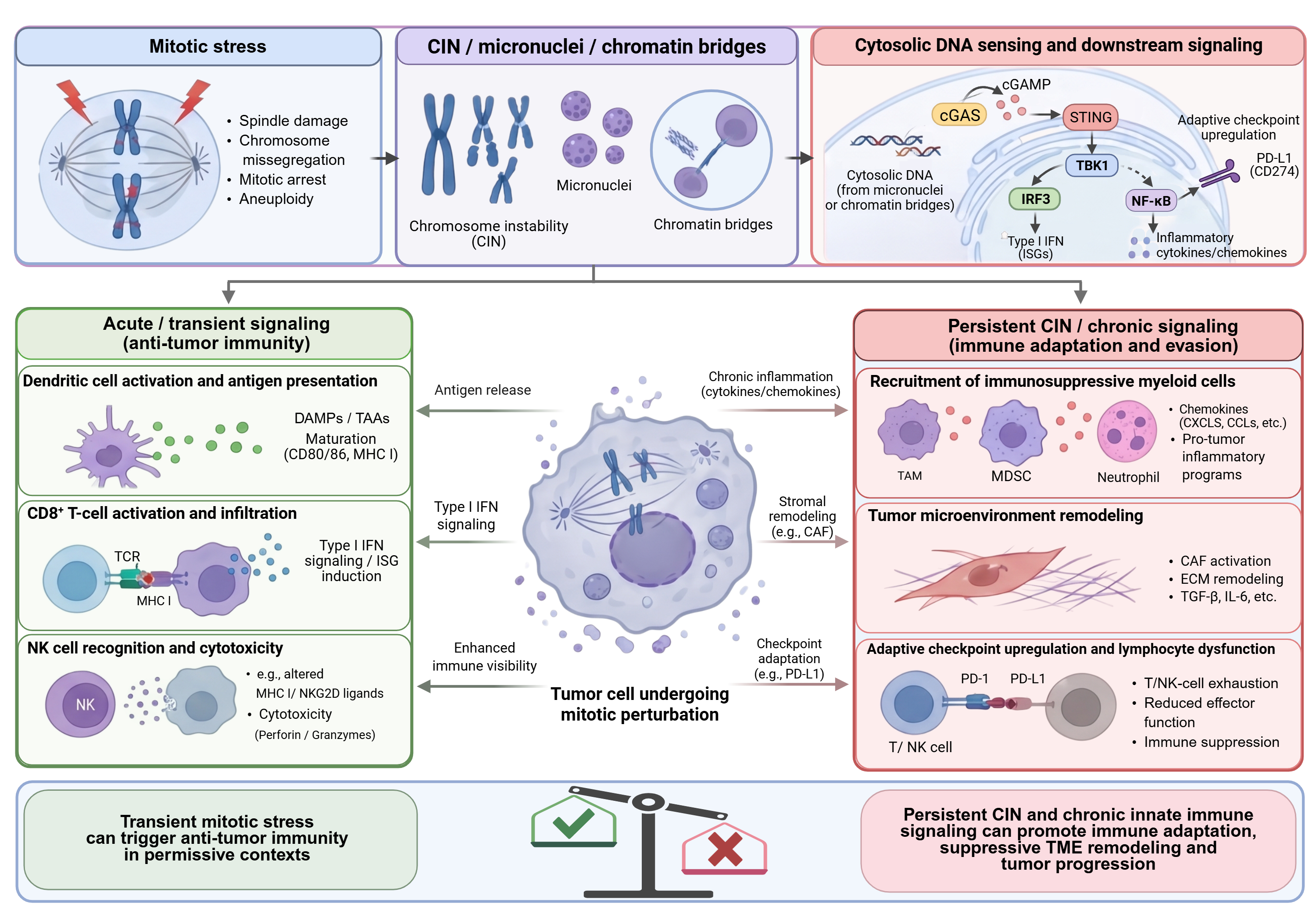
However, these effects vary with the therapeutic agent, drug exposure, tumor type, and pre-existing microenvironmental conditions. The timing and persistence of treatment-induced signaling may further influence whether these changes support effective immune responses or create conditions that favor adaptive resistance. Consequently, mitotic perturbation should not be assumed to produce uniformly beneficial immune remodeling, and distinguishing tumor-cell-mediated effects from direct actions on non-malignant cells remains an important challenge for combination therapy development. This distinction is particularly relevant when interpreting preclinical studies in which cytotoxicity, inflammatory activation, and stromal alterations occur simultaneously. Overall, these effects may favor immune activation, adaptive inflammation, or immunosuppression depending on the type, intensity, and persistence of the perturbation (Figure 2).
Direct action on tumor cells to remodel the tumor microenvironment
CIN as a driver of non-cell-autonomous signaling
Chromosomal instability (CIN) is increasingly recognized not only as a source of genetic diversity but also as a driver of inflammatory signaling and tumor microenvironment (TME) remodeling (13, 15). Persistent chromosome missegregation generates aneuploidy, lagging chromosomes, chromatin bridges, and micronuclei, some of which expose genomic DNA to cytosolic sensing pathways (17, 18). The cGAS–STING pathway provides a potential connection between genome abnormalities and inflammatory signaling, although its consequences depend on cellular context and pathway competence (95). However, the consequences of this signaling are not uniformly antitumor. Depending on the cellular context and duration of pathway activation, cGAS–STING signaling may promote immune surveillance or contribute to chronic inflammation, immune suppression, and tumor progression (96). These opposing outcomes have important therapeutic implications, as the effectiveness of STING-directed interventions may depend on pathway competence, cellular localization, and the pre-existing immune microenvironment (97). Recent evidence in esophageal adenocarcinoma further demonstrates that CIN can shape the TME through a cGAS–chemokine–myeloid signaling axis, providing a direct link between chromosome segregation defects and immune-cell remodeling (98). Nevertheless, whether these mechanisms operate consistently across tumor types or can be therapeutically exploited following mitosis-targeting treatment remains uncertain. CIN therefore has two separable consequences: it generates genetic diversity that can accelerate tumor evolution, and it can produce DNA structures and inflammatory cues that are sensed by immune and stromal compartments (13, 15). The net outcome is context-dependent; increased immune visibility and tumor-promoting adaptation are both documented consequences of mitotic instability.
Micronuclei, chromatin bridges, and the cGAS–STING pathway
Early studies established that cGAS can accumulate on chromatin exposed by micronuclear-envelope rupture, linking genome instability to innate immune signaling (11, 12). However, cGAS recruitment is not equivalent to enzymatic activation or productive downstream STING signaling. The relative contributions of micronuclei and chromatin bridges vary with the type of mitotic or genotoxic stress and require functional pathway assessment (99, 100). These observations extend the significance of mitotic errors beyond tumor-cell death by linking chromosome segregation defects to innate immune recognition. Recent evidence in esophageal adenocarcinoma demonstrates that CIN can reshape the tumor microenvironment through a cGAS–chemokine–myeloid signaling axis, illustrating how genomic instability can influence immune-cell composition (98). More broadly, the cGAS–cGAMP–STING pathway connects cytosolic DNA sensing to inflammatory signaling, senescence, and cancer-associated immune responses (78). Recognition of cytosolic chromatin fragments by cGAS can also promote cellular senescence, indicating that DNA sensing may influence cell fate independently of immediate immune-mediated tumor clearance (101).
These mechanisms have stimulated interest in therapeutic modulation of STING, although pathway heterogeneity, inflammatory toxicity, and context-dependent signaling remain important translational challenges (102). Importantly, chromosome missegregation can also activate RNA-sensing pathways that enhance antitumor immunity, indicating that immune responses to mitotic errors are not restricted to cytosolic DNA sensing (103). A colorectal-cancer-focused review further illustrates the context-dependent roles of cGAS–STING signaling in cancer immunity and treatment response, but does not establish a general benefit from mitosis-targeting combinations (104). Accordingly, STING-directed strategies offer potential opportunities to enhance antitumor immunity, but their effectiveness depends on the cellular source, intensity, and duration of pathway activation (105). Together, these findings support investigating mitosis-targeting therapies as potential modulators of tumor immune recognition rather than assuming that mitotic damage necessarily produces productive antitumor immunity.
Subsequent studies have revealed that cGAS–STING activation does not invariably promote antitumor immunity. In chromosomally unstable cancers, persistent activation of this pathway can support tumor-cell survival through IL-6-dependent inflammatory signaling, illustrating how CIN-associated innate immune responses may be redirected toward tumor-promoting adaptation (106). These findings highlight the importance of signaling duration, tumor-cell context, and downstream inflammatory programs in determining the biological consequences of mitotic instability. Thus, therapeutic strategies designed to enhance cGAS–STING signaling must account for its potential to promote tumor survival rather than assuming that increased pathway activation will improve antitumor immunity.
An important limitation is that micronucleus formation, cGAS recruitment, STING activation, and productive antitumor immunity represent distinct biological events. Evidence for one cannot establish the others. Moreover, tumor-cell-intrinsic STING signaling may produce outcomes different from those arising in dendritic cells or other host immune populations. Studies that measure micronuclei or inflammatory transcripts without confirming pathway activation, cellular source, and immune function may therefore overestimate the immunotherapeutic significance of mitotic injury.
Not all micronuclei are equally immunogenic
Not all micronuclei have the same capacity to support innate immune signaling. Micronuclear-envelope rupture can permit cGAS access to chromatin (11, 12), but nucleosome binding can inhibit cGAS enzymatic activity (107). Functional studies further show that cGAS recruitment to micronuclei need not predict cGAMP production, and chromatin bridges can be predominant triggers under particular experimental conditions (99, 100). Micronuclei can also undergo DNA breakage and chromosome pulverization (108), emphasizing that genome remodeling and immune activation are distinct consequences. Thus, micronucleus abundance should be evaluated alongside functional DNA-sensing and immune readouts rather than used as a surrogate for pathway activation (Table 2).
Table 2. Candidate response patterns after mitotic and related cell-cycle perturbations, and potential immunotherapeutic opportunities.
| Observed or candidate response pattern | Representative inducing agents | Potential favorable immune outcome | Potential unfavorable immune outcome | Proposed combination strategy |
|---|---|---|---|---|
| Acute mitotic arrest followed by apoptosis | Taxanes, vinca alkaloids, Aurora kinase inhibitors, PLK1 inhibitors. | Potential ICD-related activation (16); related ploidy and HMGB1 evidence does not establish that apoptosis is immunogenic (109, 110). | Myelosuppression, lymphocyte depletion, and impaired immune responses with excessive drug exposure. | Potential immune recognition or clearance (65, 66), apoptosis-enhancing combination activity (115), and immune elimination of arrested cells (116); selective targeting of post-mitotic survivors remains unestablished. |
| Micronuclei- and cytosolic DNA-associated stress | Selected microtubule-targeting agents and MPS1/TTK inhibitors; Aurora B inhibitors and radiotherapy combinations, require agent- and regimen-specific validation. | Context-dependent cGAS–STING activation; micronuclei or cGAS recruitment alone are insufficient evidence (11, 12, 99, 100). | Persistent inflammatory signaling, IL-6-dependent tumor-cell survival, and immunosuppressive adaptation in selected models (13, 15, 106). | Investigate checkpoint blockade or STING-directed interventions according to functional pathway activity and tumor immune context; proposed pairings require direct combination evidence. |
| Checkpoint-adapted inflammatory state | Aurora A inhibitors; related G1-directed CDK4/6 and DNA-damage-response interventions for comparison. | Context-dependent interferon signaling, antigen presentation, and T-cell recruitment. | Context-dependent PD-L1 upregulation, inflammatory adaptation, or impaired immune function (38, 75, 111, 112). | Investigate PD-1/PD-L1 blockade in biomarker-selected settings; assess PD-L1, interferon-response signatures, and immune infiltration. |
| Myeloid-dominant inflammatory remodeling | Selected Aurora A inhibitors, mitotic kinase inhibitors, microtubule-targeting agents, and CIN-associated tumor states. | Activation of antigen-presenting myeloid populations in selected settings. | Suppressive macrophage, neutrophil, or MDSC states can emerge or persist; some Aurora A interventions instead reduce myeloid suppression (15, 73, 113). | Evaluate CXCR2 targeting where supported (114). CSF1R-, CCR2-, and PI3Kγ-directed combinations remain context-specific hypotheses requiring direct combination evidence. |
| Abnormal post-mitotic survivor state | Sublethal mitotic kinase inhibition; mitotic slippage following microtubule-targeting treatment. | Potential susceptibility to immune recognition and elimination in selected models (65, 66, 116). | Persistence of polyploid or aneuploid cells, genomic diversification, treatment resistance, and immune escape. | Investigate strategies targeting survival dependencies or immune vulnerabilities of residual cells; determine optimal sequencing experimentally. |
Response patterns may coexist. CDK4/6 and DNA-damage-response inhibitors are included as related interventions, and the proposed combinations have differing levels of evidence and require context-specific validation.
Abbreviations: CCR2, C-C chemokine receptor 2; CDK4/6, cyclin-dependent kinases 4 and 6; CIN, chromosomal instability; CSF1R, colony-stimulating factor 1 receptor; CXCR2, C-X-C chemokine receptor 2; cGAS, cyclic GMP–AMP synthase; DC, dendritic cell; ICB, immune checkpoint blockade; ICD, immunogenic cell death; IL-6, interleukin-6; MDSCs, myeloid-derived suppressor cells; MPS1/TTK, monopolar spindle 1 kinase; NK, natural killer; PD-1, programmed cell death protein 1; PD-L1, programmed death-ligand 1; PI3Kγ, phosphoinositide 3-kinase gamma; PLK1, polo-like kinase 1; STING, stimulator of interferon genes.
Inflammatory rewiring and immune checkpoint adaptation
Mitotic perturbation can alter tumor-cell transcriptional and secretory programs, influencing communication with surrounding immune and stromal cells. Aurora A inhibition illustrates the context-dependent immunological consequences of these changes. In experimental breast cancer models, Aurora A inhibition can reduce myeloid-mediated immunosuppression and enhance responses to PD-L1 blockade (73). In colorectal cancer models, Aurora A inhibition increased tumor-cell IL-10 expression and promoted CD8+ T-cell infiltration through IL-10 receptor signaling (74). However, these effects are not uniformly immunostimulatory. Aurora A inhibition can increase tumor-cell PD-L1 expression, potentially limiting its antitumor efficacy and providing a rationale for combination therapy (75). In that study, reduced cGAS phosphorylation accompanied STING–NF-κB signaling without detectable TBK1–IRF3 activation, distinguishing this effect from a simple mitotic-error model. In small-cell lung cancer models, Aurora A inhibition has also been associated with restoration of interferon signaling and improved responses to PD-L1 blockade (76). Conversely, nuclear Aurora A can promote PD-L1-mediated immune suppression through MYC-dependent transcriptional regulation in triple-negative breast cancer (82). Together, these findings indicate that Aurora A-dependent immune regulation varies across tumor contexts and that the therapeutic consequences of Aurora A inhibition depend on the balance between immunostimulatory and immune-evasive responses.
Microtubule-targeting agents as immunologically active therapies
The immunological effects of microtubule-targeting agents (MTAs) have likely been underestimated for many years. Reviews and selected experimental studies describe immune-related effects of chemotherapy, including paclitaxel-associated inflammatory responses (16, 67, 117). The TONIC trial provides separate clinical context for induction treatment before PD-1 blockade, but did not test a taxane induction arm (118). The immunological consequences of taxane-containing regimens extend beyond direct cytotoxicity, although their contribution to clinical efficacy remains difficult to isolate. Chemotherapy-induced immunogenic cell death can promote the release of tumor antigens and immunostimulatory signals, potentially enabling cooperation with immunotherapy (117).
More broadly, the immunogenicity of treatment-induced cell death depends on the nature of the cellular damage and the capacity of the surrounding immune microenvironment to respond (119). A commentary proposes that interferon-associated macrophage reprogramming may contribute to paclitaxel-based chemo-immunotherapy; this interpretation should be distinguished from direct experimental evidence (120). Nevertheless, the clinical benefit of taxane-containing immunotherapy regimens cannot be attributed specifically to mitotic stress-induced immune remodeling. Establishing such a contribution will require treatment-specific pharmacodynamic studies and comparative clinical evidence. The immunological consequences of MTAs may also vary with drug selection, dose, and treatment schedule, emphasizing the need to define conditions that favor productive antitumor immunity.
Effects on myeloid, lymphoid, and stromal compartments
Myeloid cells as major responders to mitotic perturbation
Myeloid cells are important mediators of inflammatory and immunosuppressive responses within the tumor microenvironment. Tumor-associated macrophages can respond to damage-associated signals and inflammatory mediators, influencing antigen presentation, cytokine production, and T-cell activity (86, 87). Myeloid-derived suppressor cells (MDSCs) can further restrict antitumor immunity through multiple suppressive mechanisms, although their abundance and functional states vary across tumor contexts (89, 90). Consequently, inflammatory signals generated by mitotic injury may influence myeloid-cell recruitment and function, potentially promoting antitumor immunity or immunosuppressive remodeling. These responses can arise from tumor-derived signals or direct drug effects on myeloid cells, and the latter need not involve mitosis. For example, macrophage-expressed JAG1 activates NOTCH2 on ovarian cancer cells and promotes paclitaxel resistance in experimental models (113). In breast cancer models, Aurora A inhibition has been shown to reduce myeloid-mediated immunosuppression and enhance the efficacy of anti-PD-L1 therapy (73).
Overall, the phenotypic and functional plasticity of tumor-associated myeloid cells suggests that changes in myeloid-cell states, rather than lymphocyte abundance alone, may contribute to the microenvironmental consequences of mitotic perturbation (86). Neutrophils deserve special attention in this regard. It is now well established that neutrophils are highly heterogeneous and context-dependent, with functions ranging from direct tumoricidal activity to immunosuppression, angiogenesis, and promotion of metastasis (121–123). This functional diversity makes neutrophils potential downstream mediators of treatment-associated TME remodeling, but their specific contribution after individual mitosis-targeting interventions requires direct validation (121–124). This may be especially important in tumors characterized by strong inflammatory rewiring but weak adaptive immune engagement.
Lymphoid consequences and reversal of the “immune-cold” state
The ultimate therapeutic significance of TME remodeling lies in its effects on antitumor immunity. Mitosis-targeting therapies may enhance lymphocyte engagement and antitumor immune recognition in selected tumor contexts. Aurora A inhibition has been shown to promote CD8+ T-cell infiltration through changes in tumor-cell IL-10 production (74), while combined targeting of Aurora A and Bcl-xL has been reported to remodel the immune microenvironment in experimental breast cancer models (115). In KRAS–LKB1-mutant lung cancer models, decitabine followed by pulse MPS1 inhibition restored T-cell infiltration and enhanced anti-PD-1 efficacy, illustrating a combination-dependent route to increased tumor immunogenicity (79).
However, these findings are largely preclinical, and whether comparable immune conversion can be achieved consistently across tumor types remains uncertain. Nevertheless, increased CD8+ T-cell infiltration does not necessarily indicate restored antitumor immunity. Infiltrating lymphocytes may remain dysfunctional, spatially excluded from malignant cells, or restrained by suppressive myeloid populations. Consequently, claims of immune-cold-to-immune-hot conversion should be supported by functional T-cell measurements and tumor-control outcomes, not infiltration alone. Although these findings support the potential of mitosis-targeting strategies to enhance antitumor immune responses, the extent to which changes in lymphoid infiltration predict therapeutic benefit remains uncertain. Established immunotherapy biomarkers, including PD-L1 expression and tumor mutational burden, have context-dependent predictive value, highlighting the limitations of relying on individual immune measurements (125).
This point is especially important in the era of cancer immunotherapy. Impaired immune entry, priming, and effector function can coexist in tumors resistant to immune checkpoint blockade. Whether mitosis-targeting treatment can overcome these barriers requires tumor- and regimen-specific evaluation. If mitosis-targeting therapies can alter this state, then an additional therapeutic value may lie in establishing a microenvironmental context in which other therapies become effective (76, 79).
Stromal remodeling: CAFs, endothelial cells and extracellular matrix
In addition to immune cells, cancer-associated fibroblasts (CAFs) are increasingly recognized as central regulators of the TME. They are no longer viewed merely as passive structural components, but rather as highly heterogeneous and functionally active stromal cells that shape extracellular matrix architecture, produce immunoregulatory cytokines, restrict T-cell infiltration, and help establish myeloid-suppressive niches (126, 127). As such, CAFs are likely to play an important role in determining how mitosis-targeting therapies influence the broader tissue environment.
A clinical study of nab-paclitaxel plus gemcitabine, with complementary preclinical analyses, reported stromal alterations and reduced tumor stiffness in pancreatic cancer (93). The contribution of nab-paclitaxel cannot be isolated from the clinical combination alone. However, these stromal effects appear to be context-dependent, as patient-derived xenograft studies have not uniformly observed stromal depletion or increased vascular perfusion after nab-paclitaxel treatment (128). Conversely, CAF-derived matrix signals can reduce sensitivity to microtubule-directed chemotherapy, underscoring a bidirectional interaction between MTAs and the stroma (129). Thus, rather than uniformly depleting CAFs, different MTAs may remodel CAF states and matrix organization in an agent-, dose-, and context-dependent manner.
Such stromal remodeling may have important consequences for both immune-cell trafficking and drug accessibility. The density and orientation of collagen- and fibronectin-rich stromal networks can directly constrain T-cell migration and prevent lymphocytes from entering tumor-cell nests (130). Moreover, experimental normalization of myofibroblastic CAFs can overcome CAF-mediated CD8+ T-cell exclusion and restore responsiveness to immunotherapy (131). Stromal targeting can also influence therapeutic delivery: selective accumulation of a docetaxel nanoparticle in αSMA-positive tumor-associated fibroblasts reduced stromal density and was associated with a greater than tenfold increase in tumor perfusion in pancreatic cancer models (94).
Together, these studies provide a mechanistic basis for the idea that MTA-induced changes in CAF contractility, focal-adhesion signaling and extracellular-matrix architecture could lower physical barriers to immune-cell entry and drug penetration. Nevertheless, direct causal evidence demonstrating that MTA-induced CAF cytoskeletal remodeling itself drives improved antitumor immune infiltration remains limited. Endothelial cells provide an additional stromal route through which microtubule-directed therapy can reshape the TME. Eribulin, for example, can remodel abnormal tumor vasculature, increase perfusion, and enhance the intratumoral accumulation of liposomal therapeutics; these vascular changes were also accompanied by increased infiltration of CD11b+ immune cells, while NK-cell depletion partially reduced the antitumor activity of eribulin (71). Thus, changes in vascular organization, tissue stiffness and extracellular-matrix architecture may act in parallel to influence oxygenation, drug delivery, inflammatory gradients and immune-cell accessibility. Future studies should therefore move beyond a tumor cell–immune cell binary and incorporate a broader tissue-level framework that includes tumor cells, immune cells, fibroblasts, vasculature and extracellular matrix.
Overall, mitotic injury can influence the TME through tumor-derived DNA and inflammatory signals, adaptive checkpoint programs, myeloid and lymphoid responses, and stromal remodeling. For MTAs in particular, emerging evidence indicates that their effects extend beyond mitotic arrest in tumor cells to include effects on fibroblast and stromal organization, extracellular-matrix architecture and tumor vasculature. The relative contribution of each layer varies across tumor types, agents, and treatment schedules, providing the mechanistic basis for the combination strategies discussed below.
4. Rational combination strategies enabled by mitotic targeting
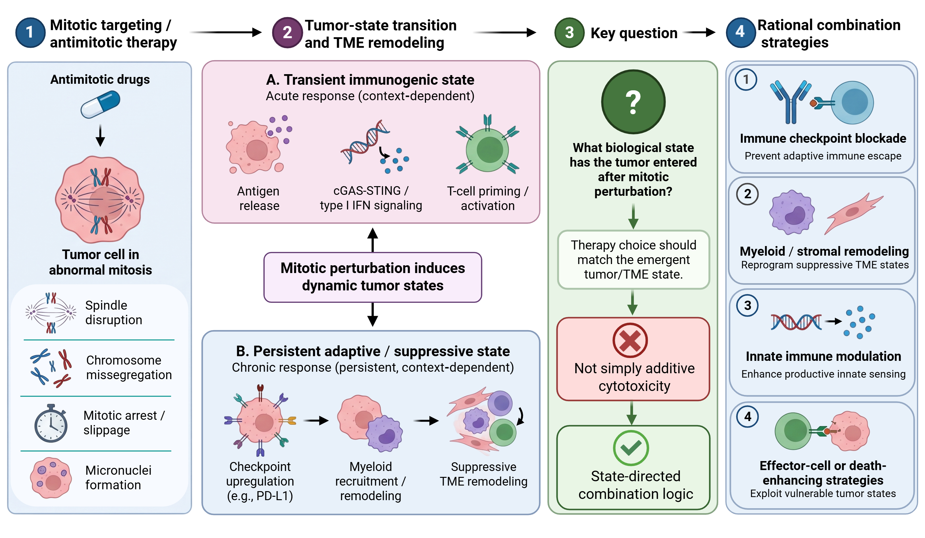
Mitotic perturbation can produce diverse cellular outcomes, including prolonged mitotic arrest, mitotic slippage, and cell death, depending on checkpoint activity and the capacity of tumor cells to adapt to mitotic stress (132). However, the clinical limitations of several mitosis-targeting agents show that disrupting cell division does not necessarily translate into durable therapeutic benefit, challenging the assumption that mitotic inhibition alone is sufficient for effective cancer treatment (133). Advances in mitotic targeting have therefore emphasized alternative vulnerabilities and therapeutic strategies that account for the complexity of mitotic regulation and tumor-cell responses (134). Mechanistically, the dynamic regulation of spindle assembly checkpoint signaling helps determine whether cells maintain mitotic arrest or progress despite chromosome segregation defects (135). Building on these observations and the immune and stromal consequences discussed above, we propose tumor-state conversion as an organizing framework rather than an established clinical treatment category. In this framework, mitotic injury may transiently produce immune-activating, checkpoint-adapted, myeloid-suppressive, or residual-survival response patterns that may coexist. Identifying the predominant response and its duration may help guide the selection and timing of combination therapies.
This framework leads to a testable combination principle: partner therapy should be selected according to the induced state and its timing rather than added empirically. The following strategies are therefore presented with different levels of evidence, from clinically established chemotherapy-ICB combinations to preclinical or hypothesis-generating approaches, and should not be interpreted as equally validated (Figure 3).
Combination with immune checkpoint blockade
Once mitotic targeting is viewed as a TME-remodeling intervention, combination with immune checkpoint blockade (ICB) becomes not merely an empirical option but a mechanistically supported strategy in selected preclinical settings. Selected mitotic abnormalities can enhance tumor immune recognition, as illustrated by ploidy-associated immunosurveillance (109). Immunogenic cell death provides an additional route to immune priming, but its effectiveness depends on the relevant danger signals and host sensing pathways (110). Interferon-associated responses and changes in T-cell infiltration have also been reported in specific Aurora A- or MPS1-targeted experimental models (74, 76, 79). If these changes can be sustained and amplified in a timely manner, PD-1/PD-L1 blockade may convert transient immune activation into a more durable antitumor response (136, 137). When mitotic perturbation induces PD-L1 expression, checkpoint blockade may counteract one component of adaptive immune resistance. However, PD-L1 induction alone does not establish dependence on the PD-1/PD-L1 axis or predict clinical benefit from its inhibition. Functional immune assessment and tumor-specific validation remain necessary (111, 112, 138).
Aurora A inhibition provides a representative example of this logic. Alisertib has shown myeloid effects and PD-L1-associated adaptive responses in selected preclinical models (73, 75). In SCLC models, the Aurora A inhibitor LSN3321213 induced mitotic accumulation and restored interferon-associated expression, supporting combination with PD-L1 blockade (76). Thus, combining Aurora A inhibition with PD-L1 blockade has a mechanistic rationale because it may counteract an adaptive immune-regulatory response elicited by Aurora A inhibition. This response can involve direct cGAS regulation as well as changes in cell-cycle state, and the contribution of mitotic disruption itself requires validation (75). Interferon signaling can regulate checkpoint expression and resistance programs (111, 112), but extension of this combination rationale to other mitosis-targeting agents requires agent-specific evidence.
Clinical evidence supports selected chemotherapy–ICB combinations in defined patient populations, but benefit is not uniform across regimens. IMpassion130 demonstrated improved progression-free survival with atezolizumab plus nab-paclitaxel in advanced triple-negative breast cancer (68), whereas IMpassion131 did not demonstrate a progression-free-survival benefit from adding atezolizumab to paclitaxel (69). KEYNOTE-355 showed an overall-survival benefit from pembrolizumab plus chemotherapy in patients with a PD-L1 combined positive score of at least 10; its chemotherapy options included taxanes or gemcitabine–carboplatin (70). More directly related to mitotic kinase targeting, an early-phase trial of alisertib plus pembrolizumab in refractory, Rb-deficient head and neck squamous cell carcinoma reported prolonged stable disease in some patients but no objective responses (77). These studies support context-specific investigation of combinations, without establishing mitotic stress-induced immune remodeling as the mechanism of clinical benefit or a general indication for all antimitotic–ICB regimens.
Combination with myeloid-targeted strategies
The immunological consequences of mitotic injury may depend partly on how myeloid cells respond to tumor-derived inflammatory signals. Tumor-associated macrophages regulate inflammatory signaling and T-cell responses (86), whereas MDSCs can suppress antitumor immunity through multiple mechanisms (89). Neutrophils exhibit substantial functional plasticity, potentially supporting either antitumor immunity or tumor-promoting inflammation depending on the surrounding microenvironment (122). These observations provide a rationale for combining mitosis-targeting therapies with interventions that modulate suppressive myeloid populations. However, whether such combinations consistently enhance therapeutic efficacy remains to be established.
This logic is particularly relevant in tumors that, following mitosis-targeting therapy, display marked chemokine rewiring, enhanced myeloid recruitment, or dominance of suppressive myeloid programs. In these settings, the failure of therapy to generate durable benefit may not reflect insufficient inflammation, but rather the fact that inflammation has been organized into an immunosuppressive tissue response. Under such circumstances, blocking selected tumor–myeloid communication pathways provides a testable combination rationale. For example, disrupting CXCR2-mediated MDSC trafficking enhanced anti-PD-1 efficacy in experimental models (114). Broader approaches to macrophage reprogramming or selective modulation of suppressive myeloid populations have additional mechanistic rationales (86, 88–90), but their benefit with specific antimitotic agents remains to be demonstrated.
Myeloid-targeted strategies may improve the therapeutic consequences of mitotic injury by limiting immunosuppressive responses rather than simply increasing cytotoxic pressure. In tumors characterized by suppressive macrophage activity or MDSC accumulation, interventions that modify myeloid-cell recruitment and function may help restore antitumor immune responses (89, 114). However, combining mitosis-targeting agents with immunomodulatory therapies also requires careful consideration of treatment-related toxicity, with risks evaluated for the particular drugs and schedule. Reports of immune-related adverse events across kinase-targeted cancer therapies provide general safety context rather than combination-specific evidence (139). Thus, the therapeutic potential of these combinations must be evaluated alongside their safety, and whether myeloid-directed interventions consistently enhance responses to mitosis-targeting therapies remains to be established.
Combination with innate immune pathway modulation
The cGAS–STING pathway provides a mechanistic link between mitotic abnormalities and innate immune signaling. Chromosome missegregation, micronucleus formation, and exposure of genomic DNA to the cytosol can activate cGAS-dependent inflammatory responses, connecting mitotic injury to changes in the tumor immune microenvironment (11–13). Upon recognition of cytosolic DNA, cGAS generates cyclic GMP–AMP (cGAMP), which activates STING and downstream signaling pathways involved in type I interferon production and inflammatory gene expression (78, 140). Host STING-dependent DNA sensing supports antitumor immune priming in experimental models (141). Separately, preserving extracellular cGAMP through ENPP1 inhibition enhanced radiation-associated tumor control in mice (142). Whether these mechanisms can improve responses to mitosis-targeting agents requires direct validation.
However, the therapeutic consequences of cGAS–STING activation are context-dependent. cGAS-mediated sensing of cytosolic chromatin can influence senescence-associated responses (101), while broader reviews describe context-dependent inflammatory and immune consequences of cGAS signaling (140). These findings do not establish that signaling duration alone determines therapeutic benefit. Extracellular cGAMP availability provides another regulatory layer: ENPP1-mediated degradation can limit its immune-stimulatory effects (142). Consequently, the efficacy of these combinations likely depends on pathway competence in both malignant cells and host immune compartments, the intensity and duration of innate immune activation, and the timing of treatment. Their therapeutic benefit requires direct experimental and clinical validation.
Accordingly, combining mitosis-targeting therapies with innate immune modulators should aim to amplify productive DNA-sensing responses without sustaining tumor-promoting inflammation. The consequences of cGAS–STING activation depend on the cellular context and duration of signaling, making temporal assessment of pathway activation particularly important (13, 78). Persistent inflammatory signaling may also promote tumor-cell survival and adaptive resistance rather than effective immune clearance (106). A further translational concern is that pharmacological STING agonism may not reproduce the spatial and temporal patterns of endogenous DNA sensing following mitotic injury. Therapeutic outcomes may differ depending on whether signaling occurs predominantly in malignant cells, antigen-presenting cells, or stromal compartments. Combination studies should therefore assess pathway competence, cell-specific activation, and signaling duration before interpreting increased inflammatory activity as evidence of therapeutic synergy. Equally important, the biological effects of microtubule-targeting agents in patients cannot be assumed to arise predominantly from mitotic arrest, as their antitumor activity may involve additional mechanisms beyond direct disruption of mitosis (143). Consequently, future combination studies should verify mitotic target engagement and downstream DNA-sensing responses independently rather than assuming that exposure to a microtubule-targeting agent necessarily produces therapeutically relevant cGAS–STING activation.
Combination with cell therapy
Combining mitosis-targeting therapies with cell-based immunotherapy represents an emerging strategy based on the possibility that mitotic abnormalities can increase tumor-cell susceptibility to immune recognition. Mitotic perturbation may generate polyploid or aneuploid cells that exhibit altered stress responses and immune-recognition signals. Experimental studies have shown that drug-induced hyperploidy can stimulate NK-cell responses through NKG2D- and DNAM-1-dependent mechanisms (65). Similarly, aneuploid senescent cells can activate NF-κB-associated signaling that promotes their clearance by NK cells (66). Beyond these mechanisms, centrosome amplification can trigger PIDD1-dependent inflammatory signaling and immunosurveillance, providing another link between abnormal cell division and immune recognition (144).
More broadly, immunosurveillance contributes to tissue homeostasis by identifying and eliminating cells with potentially harmful abnormalities (145). Consistent with this principle, experimental chromosome missegregation has been shown to generate cells with complex karyotypes that undergo cell-cycle arrest and subsequent immune-mediated elimination (116). Together, these findings suggest that selected mitotic abnormalities may create opportunities for immune-cell-mediated clearance. However, whether these responses can be reliably exploited by adoptive T-cell, NK-cell, or engineered cell therapies remains uncertain, particularly because surviving tumor cells may undergo further adaptation and selection.
These observations support investigating whether mitotic perturbation creates a transient period of increased susceptibility to NK-cell, CAR-NK-cell, or T-cell-based interventions. Its duration and therapeutic accessibility have not been established. Tumor heterogeneity and clonal evolution can generate populations with different capacities for survival and immune evasion (146), while treatment-associated selection may favor resistant subclones (147). Prospective studies should therefore define the kinetics of stress-ligand expression, immune recognition, and residual-cell survival, while also assessing drug effects on the infused effector cells. This would permit experimental evaluation of whether cell therapy can eliminate post-mitotic survivors and which treatment sequence preserves immune-cell function.
Combination with apoptosis-enhancing and ferroptosis-enhancing strategies
A major limitation of mitosis-targeting therapies is that disruption of mitotic progression does not invariably result in tumor-cell death. Depending on checkpoint activity and cellular stress responses, tumor cells may undergo prolonged mitotic arrest, mitotic slippage, or survival following abnormal division (132). Cells that escape mitotic death may persist with altered ploidy or chromosomal instability, potentially contributing to subsequent therapeutic resistance (33). These outcomes provide a rationale for combining mitosis-targeting agents with apoptosis-enhancing therapies to increase the likelihood of irreversible tumor-cell elimination before surviving populations undergo further adaptation.
The combination of alisertib and the BH3 mimetic navitoclax (ABT263) illustrates an experimental strategy targeting Aurora A and anti-apoptotic survival signaling. In experimental breast cancer models, this combination has demonstrated synergistic antitumor activity accompanied by changes in the tumor immune microenvironment (115). These findings do not by themselves establish that mitotic disruption mediates the immune effects. Mechanistically, Aurora A inhibition disrupts mitotic regulation and can increase cellular vulnerability to additional therapies, supporting combination approaches designed to overcome the limitations of single-agent treatment (148). Bcl-xL inhibition may further promote apoptosis in tumor cells that would otherwise survive mitotic stress, although the extent of this effect depends on cellular context and apoptotic dependence. More broadly, researchers are investigating Aurora A-targeted combination strategies to improve therapeutic efficacy and limit adaptive resistance (149). Whether accelerated elimination of mitotically damaged cells also reduces persistent inflammatory signaling or improves subsequent antitumor immune responses remains to be established.
A related example from broader cell-cycle and radiotherapy research illustrates how checkpoint rewiring can engage multiple death programs. In irradiated breast cancer cells, FBXW7-targeted DNAzyme-mediated stabilization of phosphorylated p53 enforced persistent G2 arrest, impaired DNA repair, and enhanced apoptosis, while simultaneously suppressing the SLC7A11–GSH–GPX4 antioxidant axis. Combined with Fe²⁺-mediated Fenton chemistry and radiation-induced ROS, this checkpoint rewiring amplified ferroptotic damage, coupling prolonged cell-cycle arrest with both apoptotic and ferroptotic cell death (150). This genotoxic-stress model illustrates coordinated apoptosis and ferroptosis during prolonged G2 arrest; it does not by itself establish a mechanism involving mitotic arrest.
An emerging body of evidence further indicates that the relationship between mitotic or cell-cycle perturbation and ferroptosis is highly context-dependent rather than uniformly sensitizing. Ferroptosis is driven by iron-dependent peroxidation of polyunsaturated fatty-acid-containing phospholipids and is tightly controlled by cellular lipid, iron, and redox metabolism (151). A recent breast-cancer study centered on Tongguanteng injection (TGT) provides evidence connecting AURKA-related cell-cycle control to ferroptosis. TGT reduced AURKA expression and was associated with G2/M accumulation, iron accumulation, GSH depletion, and increased lipid peroxidation. AURKA knockdown or alisertib treatment also induced G2/M accumulation and ferroptotic responses. TGT disrupted the AURKA–KEAP1 interaction and promoted NRF2 nuclear translocation and HO-1-associated ferroptosis; AURKA overexpression counteracted TGT effects, while TGT combined with docetaxel enhanced ferroptosis sensitivity (152). These findings suggest that, in specific molecular contexts, targeting a mitotic regulator can simultaneously impair cell-cycle progression and create a ferroptosis-permissive state.
However, cell-cycle arrest can also establish a ferroptosis-protected state. Cell-cycle arrest induced at different phases, including G2/M accumulation following nocodazole treatment or CDK1 deficiency, has been shown to suppress lipid peroxidation and ferroptosis (153). Mechanistically, arrested cells increase DGAT-dependent triacylglycerol synthesis and lipid-droplet formation, thereby sequestering excess PUFAs away from peroxidation-prone membrane phospholipids. Pharmacological or genetic inhibition of DGAT redirects PUFAs from TAG storage toward phospholipids and re-sensitizes arrested cells to ferroptosis (153). Notably, this mechanism also applies to slow-cycling, therapy-resistant tumor populations that accumulate lipid droplets and display increased ferroptosis resistance; combined DGAT inhibition and ferroptosis induction can overcome this protective state (153).
A complementary ferroptosis-protective program has been identified following FOXO3 activation in experimental models including hTERT-RPE-1 cells. FOXO3-mediated p27-dependent G1 arrest contributes to resistance to ferroptosis, but the protection extends beyond cell-cycle arrest itself. FOXO3 activation lowers cellular H₂O₂, reduces intracellular iron partly by regulating TFR1, and decreases the expression of ACSL4 and PEX5, collectively limiting hydroxyl-radical generation, lipid peroxidation, and ferroptotic susceptibility (154). These observations further support the concept that cell-cycle state and ferroptosis competence are functionally coupled through metabolic and stress-response programs rather than through cell-cycle arrest alone.
Taken together, the ferroptotic consequence of mitotic or cell-cycle perturbation appears to depend not on arrest per se, but on accompanying remodeling of PUFA partitioning, iron availability, ROS production, and antioxidant defenses. This context dependence has important implications for combination therapy. Direct ferroptosis induction may be particularly effective when mitotic perturbation increases oxidative stress or labile iron, or suppresses antioxidant defenses, whereas co-targeting adaptive lipid-storage or antioxidant programs may be necessary when arrested cells instead enter a ferroptosis-resistant state. For example, DGAT inhibition may expose PUFA pools that have been diverted into lipid droplets, thereby restoring ferroptosis sensitivity in slow-cycling or treatment-resistant populations (153).
Future studies should therefore define the metabolic state created by individual mitotic interventions using lipid-peroxidation measurements, ferroptosis-rescue experiments, and biomarkers of PUFA-phospholipid remodeling, iron handling, and antioxidant capacity, rather than assuming that mitotic stress intrinsically sensitizes tumors to ferroptosis. The opposing effects reported across experimental systems also raise a mechanistic attribution problem. G2/M accumulation alone does not distinguish G2 arrest from mitotic arrest. Kinase inhibition, oxidative stress, and altered lipid metabolism often occur together, making it difficult to determine which event drives ferroptotic susceptibility.
Genetic rescue experiments, orthogonal pharmacological inhibitors, and lipid-peroxidation measurements are needed to separate mitotic arrest from kinase-specific and metabolic effects. Until such evidence is available, ferroptosis-enhancing combinations should be considered context-dependent experimental strategies rather than broadly applicable extensions of antimitotic therapy. Increased ferroptotic tumor-cell death does not by itself establish improved antitumor immunity; combination studies should also assess immune-cell function and immunotherapy outcomes.
Combination with radiotherapy and DNA-damaging therapies
Radiotherapy and other DNA-damaging treatments intersect with mitosis-targeting therapies through their effects on genomic integrity, chromosome segregation, and innate immune signaling. DNA damage that persists into mitosis can contribute to chromosome segregation abnormalities, micronucleus formation, and subsequent exposure of genomic DNA to cytosolic sensing mechanisms (11–13). Recognition of cytosolic DNA by cGAS can activate STING-dependent inflammatory signaling, providing a mechanistic connection between genomic injury and immune responses (78). These observations support investigating whether combining DNA-damaging treatments with selected mitosis-targeting agents can increase tumor immunogenicity by coordinating DNA damage and its downstream consequences. However, greater genomic damage does not necessarily produce stronger antitumor immunity, because persistent chromosomal instability and inflammatory signaling may also promote immune suppression or tumor adaptation (13). Consequently, the therapeutic value of these combinations likely depends on treatment sequence, damage intensity, and the duration of innate immune activation rather than simply the cumulative extent of cellular injury. Agent-specific mechanisms can further alter the immune consequences of genomic damage. For example, HO-1 can redistribute cGAS and STING and blunt radiotherapy-associated immune signaling (155). Such findings reinforce the need to measure functional DNA sensing rather than infer immune activation from damage burden alone.
A significant translational challenge in combining mitosis-targeting therapies with radiotherapy or other DNA-damaging treatments is determining how treatment intensity, sequence, and scheduling influence the balance between tumor-cell elimination and immune modulation. Experimental mitotic checkpoint dysregulation can generate chromosome abnormalities and promote tumorigenesis (2), reinforcing the need to distinguish genomic injury from effective tumor-cell elimination. Moreover, conventional chemotherapy and targeted anticancer agents can exert immunological effects that extend beyond direct cytotoxicity, with outcomes depending on the nature of treatment-induced cellular stress and its consequences for the tumor microenvironment (156).
Evidence from chemo-immunotherapy research further emphasizes that the immunological consequences of treatment must be considered when designing combinations intended to enhance antitumor immunity (157). Conversely, tumor heterogeneity, immunoediting, and immune-evasion mechanisms may limit the durability of initially favorable immune responses (158). Together, these observations support investigating treatment schedules that generate sufficient genomic stress to promote tumor-cell elimination and productive immune activation while limiting prolonged inflammatory signaling and selection of resistant populations. Rather than pursuing maximal DNA damage, future studies should test whether a controlled, temporally restricted period of genomic stress can improve therapeutic outcomes. The optimal dose, sequence, and duration of such combinations remain to be established.
Delivery systems and biomaterial-enabled strategies
Precision delivery systems may help address a longstanding limitation of mitosis-targeting therapies: systemic toxicity resulting from drug exposure in normal proliferative tissues. Nanoparticle formulations offer opportunities to modify drug distribution and increase intratumoral exposure. In patients with solid tumors, a direct comparison of nanoparticle-entrapped docetaxel with conventional docetaxel demonstrated differences in intratumoral drug exposure, providing clinical evidence that formulation can influence drug delivery to tumor tissue (159). Similarly, a phase III trial comparing albumin-bound paclitaxel with conventional solvent-based paclitaxel in breast cancer showed that formulation changes can affect clinical efficacy and tolerability, although these benefits do not necessarily establish tumor-selective delivery (160).
Beyond pharmacokinetic optimization, delivery platforms may also preserve or enhance the immunological activity of microtubule-targeting agents. In experimental tumor models, a paclitaxel-loaded nanomicelle maintained treatment-associated immune activation and improved responsiveness to PD-1 blockade, supporting the potential integration of drug delivery with immunotherapy (161). Local biomaterial-based approaches provide another strategy for coordinating cytotoxic treatment and immune stimulation. In murine hepatocellular carcinoma and breast cancer models, peritumoral administration of an alginate hydrogel containing nab-paclitaxel and the immunostimulant R837 enhanced ICD-associated and antitumor immune responses (162). In addition, researchers have developed cascade-responsive nanosystems to control drug exposure at multiple sites and improve experimental chemo-immunotherapy outcomes (163). Collectively, these findings suggest that delivery platforms may contribute to combination efficacy by influencing drug distribution, local exposure, and treatment-associated immune responses. However, further validation is needed to confirm their ability to improve the therapeutic index of mitosis-targeting immunotherapy combinations.
More importantly, in the current therapeutic framework, delivery technology should no longer be viewed simply as a formulation-level optimization issue, but as part of the therapeutic mechanism itself. Drug delivery can influence cellular exposure, exposure kinetics, and local treatment effects. Whether these changes coordinate mitotic injury with productive immune remodeling requires formulation-specific pharmacodynamic evidence. If an additional value of mitosis-targeting therapy lies in its ability to induce local immunogenic conversion, then restricting that process as much as possible to the tumor site and coupling it spatiotemporally with immune stimulation carries clear mechanistic significance (159, 161–163). For strategies intended to be combined with immunotherapy, biomaterial platforms may also serve as co-delivery systems that enable local, coordinated release of mitotic inhibitors, innate immune agonists, checkpoint-blocking agents, or myeloid-modulating drugs. Delivery systems may therefore support coordinated combination treatment, while the proposed restoration of immunosurveillance remains a broader biological rationale (164).
Biomarkers and patient stratification
For clinical translation, biomarker development should prioritize a limited set of mechanistically informative variables rather than an unrestricted panel of candidate markers. As a testable baseline framework, we propose three complementary measurements: chromosomal instability (CIN) burden, p53 status, and cGAS–STING pathway competence. CIN refers to ongoing numerical or structural chromosome changes. Candidate assays should distinguish direct measurements of segregation errors or evolving single-cell heterogeneity from static proxies such as aneuploidy, copy-number burden, or whole-genome doubling. The selected CIN-related measure reflects pre-existing chromosome-segregation abnormalities or genomic heterogeneity and has emerged as a potential variable for therapeutic stratification, although its predictive value remains dependent on tumor context and treatment modality (165). p53 status provides information about cellular responses to mitotic stress, including checkpoint regulation, cell-cycle arrest, and survival following abnormal division. Experimental evidence that paclitaxel can induce p53- and p21-associated cell-cycle responses rather than mitotic arrest in a cell-type-dependent manner illustrates the importance of this context (166). Nevertheless, low-dose paclitaxel can also induce a p53-dependent arrest in the subsequent cell cycle (167).
These studies motivate considering p53 pathway status, but do not validate it as a predictor in the proposed clinical framework. The third variable, cGAS–STING competence in malignant cells and relevant host immune compartments, may be particularly relevant when mitosis-targeting therapies are intended to enhance antitumor immunity. In KRAS–LKB1-mutant lung cancer models, decitabine-mediated STING derepression followed by pulse MPS1 inhibition restored immune infiltration and enhanced anti-PD-1 efficacy (79). This result supports assessing both pathway competence and treatment-induced functional signaling. However, the presence of micronuclei or chromosomal abnormalities does not necessarily establish productive cGAS–STING signaling. Functional pathway assessment may therefore be needed to distinguish tumors capable of mounting an innate immune response from those with impaired downstream signaling. Together, these three variables provide a hypothesis-driven framework for patient stratification that requires prospective validation before clinical implementation.
Early on-treatment assessment could focus on four complementary features: micronucleus and chromatin-bridge frequency, a compact interferon-stimulated-gene (ISG) score, changes in PD-L1 expression, and shifts in myeloid versus CD8+ T-cell populations. Micronucleus formation and chromatin-bridge accumulation provide morphological indicators of chromosome-segregation abnormalities following mitotic perturbation (11, 12). However, these abnormalities should not be interpreted as direct evidence of functional cGAS activation. Experimental studies have demonstrated that chromatin bridges, rather than micronuclei, can be the predominant triggers of cGAS activation following drug-induced mitotic errors (99). More recent measurements further indicate that cGAS recruitment to micronuclei does not necessarily predict downstream cGAMP production, emphasizing the need for functional pathway assessment (100).
A compact ISG score could therefore provide a complementary transcriptional readout of inflammatory pathway activation. Paclitaxel-induced micronucleation has been associated with cGAS–STING-dependent inflammatory signaling (168), while TTK inhibition has been shown to activate tumor STING and enhance experimental immunotherapy responses (80). An IFN-γ-related mRNA signature has shown associations with response to PD-1 blockade (169), supporting a role for transcriptional immune monitoring. However, this type II interferon-related signature should not be treated as a validated type I interferon or cGAS–STING-specific assay. In parallel, increased PD-L1 expression may indicate an adaptive checkpoint response following mitotic perturbation, as observed in experimental studies of Aurora A inhibition (75). Finally, changes in myeloid and CD8+ T-cell populations may help distinguish potentially productive immune activation from chronic chromosomal instability-associated immunosuppressive remodeling (15). Together, these measurements provide a hypothesis-driven approach to evaluating whether mitosis-targeting treatment induces an immune-activating, checkpoint-adapted, or myeloid-suppressive tumor state.
This two-stage biomarker panel is proposed as a testable framework, not a validated clinical assay. Baseline markers could inform patient stratification, whereas early on-treatment measurements could help determine whether mitosis-targeting therapy induces an immune-activating, checkpoint-adapted, or myeloid-suppressive tumor state. Prospective studies will be required to establish the predictive value of these measurements and their utility for guiding treatment selection and timing. Additional exploratory measurements, including direct cGAMP or pathway-activation readouts, can be layered onto this core panel in mechanistic studies, but should not obscure the variables most directly linked to treatment selection and timing. The proposed biomarker panel also faces substantial analytical and biological limitations. Chromosomal instability varies spatially within tumors, p53 mutation status does not fully capture pathway function, and cGAS–STING expression does not necessarily indicate signaling competence. Similarly, interferon signatures and PD-L1 expression can arise through mechanisms unrelated to mitotic perturbation. Prospective studies should establish assay reproducibility, tumor-specific thresholds, sampling intervals, and the incremental predictive value of combined markers before clinical implementation.
The clinical value of this framework will ultimately depend on prospective validation of predefined assay thresholds and sampling windows, particularly because both CIN and treatment-induced immune responses are dynamic and context-dependent (165, 170). When repeated tumor biopsies are impractical, longitudinal blood-based biomarkers could complement tissue-based measurements to capture systemic treatment responses, while recognizing that liquid biopsy does not fully reproduce the spatial information provided by tumor tissue (170, 171).
5. Challenges and future perspectives
Defining immune-activating versus immune-suppressive mitotic stress
A central challenge in this field is that mitotic stress is neither uniformly tumor suppressive nor consistently immunostimulatory. Chromosome segregation errors can generate micronuclei that expose genomic DNA to cytosolic sensing mechanisms (11, 12). Under appropriate cellular conditions, activation of the cGAS–STING pathway can induce type I interferon signaling and potentially enhance antitumor immune responses (78). In contrast, persistent chromosomal instability can promote chronic inflammatory signaling and, in certain tumor contexts, contribute to metastatic progression and immune evasion (13, 18). However, the biological consequences of aneuploidy are heterogeneous and depend on cellular context, the extent of genomic imbalance, and the capacity of tumor cells to adapt to chromosomal abnormalities (172). Therefore, the therapeutic objective should not be to maximize mitotic damage indiscriminately, but to induce sufficient tumor-cell vulnerability while limiting the emergence of persistent genomic instability and adaptive survival programs.
This distinction requires assessment of the biological consequences of mitotic errors rather than their frequency alone. Micronuclei and chromatin bridges can differ in their capacity to expose genomic DNA to cytosolic sensing mechanisms (11, 12). Aneuploidy and polyploidy may produce additional consequences involving altered gene dosage, cellular stress responses, and changes in proliferative capacity (173). Moreover, cells that survive chromosome missegregation can undergo cell-cycle arrest or elimination, illustrating that similar mitotic abnormalities do not necessarily produce equivalent cellular outcomes (116). The immunological consequences of these alterations are likely to depend on DNA accessibility, cGAS–STING competence, inflammatory signaling dynamics, cell-death responses, and interactions with the surrounding microenvironment. Thus, the central question is not simply whether mitotic damage has occurred, but which abnormalities arise, how long they persist, and whether their downstream consequences favor antitumor immunity or tumor adaptation. A useful conceptual framework is to distinguish mitotic stress according to its downstream immune consequences. Cytoplasmic chromatin can engage inflammatory signaling in senescence and cancer (14), and the cGAS–STING pathway provides a broader connection between DNA sensing and cancer-associated immune responses (78).
These observations support investigating, rather than assuming, an immunogenic consequence of mitotic stress. Evidence of micronucleus formation or inflammatory gene expression should therefore be interpreted alongside functional measurements of DNA sensing, immune-cell activity, and tumor control. In contrast, persistent chromosomal instability may sustain inflammatory signaling and promote tumor adaptation through NF-κB-associated pathways and immunosuppressive microenvironmental changes (13, 18, 43). These immune-activating and immune-suppressive response patterns should be regarded as context-dependent, potentially overlapping outcomes rather than mutually exclusive biological categories. Their assessment should also distinguish signaling within malignant cells from responses in immune and stromal compartments, because a similar molecular readout may have different implications across these populations. Longitudinal sampling could help determine whether an initially favorable response persists, resolves, or becomes accompanied by adaptive resistance. Such measurements would provide a basis for testing whether particular combinations or treatment sequences improve immune-mediated tumor control, rather than selecting partners solely from the presence of inflammatory markers. Establishing measurable criteria to distinguish these responses will be important for developing rational combinations of mitosis-targeting therapies and cancer immunotherapy (Figure 4).
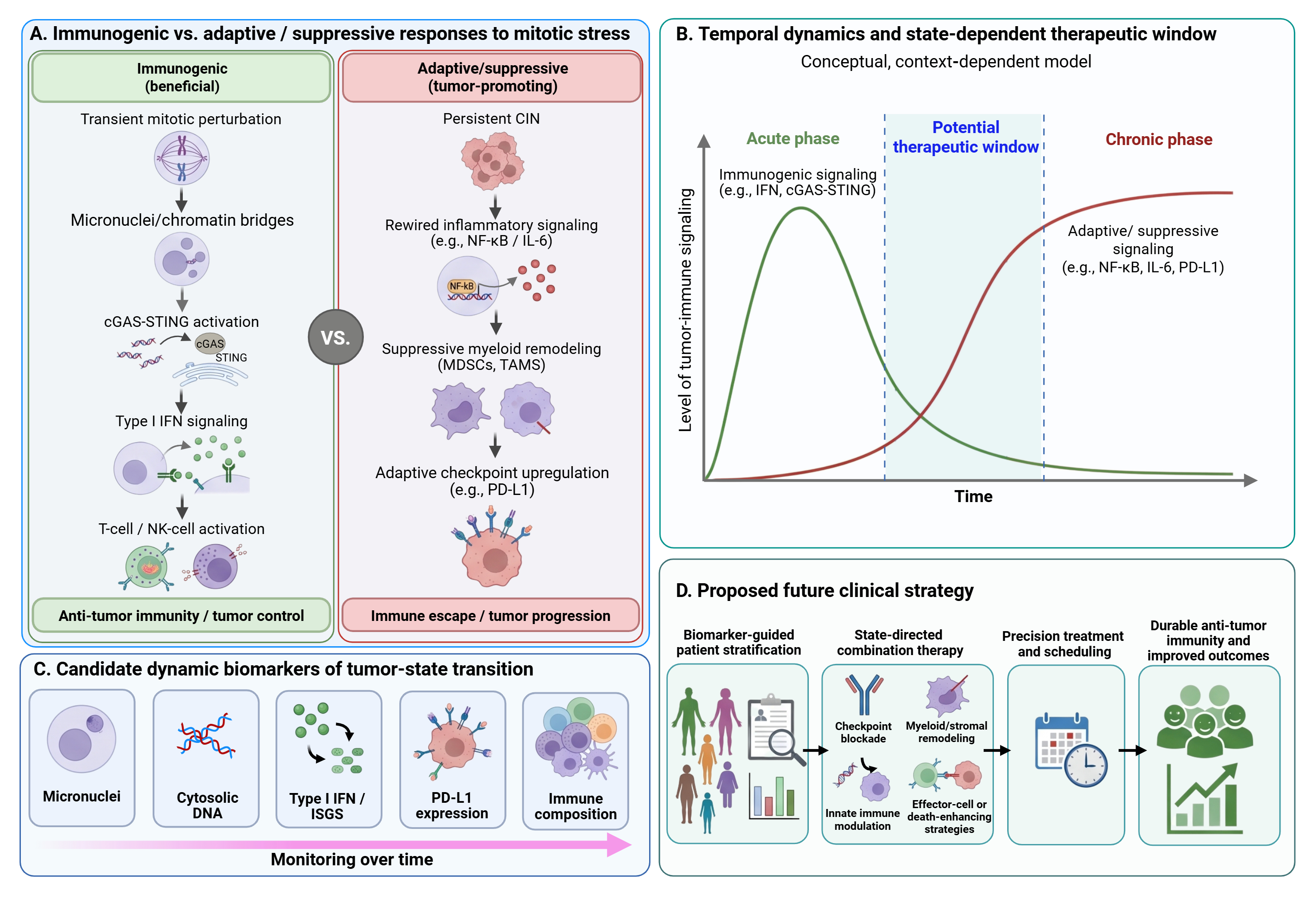
Acute versus chronic mitotic dysregulation
The distinction between acute and chronic mitotic dysregulation may influence therapeutic outcome, but timing alone does not determine whether a response is antitumor or immune-suppressive. Acute chromosome-segregation errors can generate micronuclei and expose genomic DNA to cytosolic sensing mechanisms (11, 12). When downstream cGAS–STING signaling remains functional, these events may promote inflammatory responses that increase tumor immunogenicity and potentially facilitate immune-mediated clearance (78). However, the consequences of mitotic injury can change when damaged tumor cells survive and develop persistent chromosomal instability. Chronic instability may contribute to inflammatory adaptation, tumor progression, and immune evasion in certain cellular contexts (13, 18). Moreover, aneuploidy can affect tumor-cell fitness and survival in heterogeneous ways, suggesting that the consequences of sustained chromosomal abnormalities depend on individual tumors' capacity to tolerate genomic imbalance (172). This distinction has direct implications for treatment scheduling. Continuous or excessive inhibition of mitotic regulators may not always be optimal. Experimental links between checkpoint dysregulation and tumorigenesis (2), uncertainties about the mechanisms of microtubule-targeting agents in patient tumors (143), and the immunological effects of anticancer treatment (156) motivate investigation of alternative schedules. Whether intermittent dosing reduces persistent CIN-bearing residual populations remains unproven. In other words, the goal may not be persistent suppression of cell division, but creating a controlled, time-limited stress state that can be rapidly converted into cell death or productive immunity.
This view challenges the traditional maximum tolerated dose paradigm. For mitosis-targeting therapy, dose and schedule are not merely pharmacological variables; they are biological determinants of immune outcome. A regimen that maximizes cytotoxicity may not be the same as one that maximizes immunogenic conversion. Future studies should therefore consider concepts such as immune-effective dose, state-converting dose, or combination-priming schedule, in addition to conventional toxicity and response metrics. These are investigational concepts rather than established dosing recommendations. They may help identify regimens that induce immune susceptibility while limiting persistent immune-suppressive or tumor-adaptive signaling.
Context dependence and tumor heterogeneity
Tumors differ substantially in their dependence on mitotic regulators, tolerance of chromosome missegregation, apoptotic competence, p53 status, innate immune signaling, and baseline tumor microenvironment composition. The biological consequences of aneuploidy depend on cellular context and the capacity of tumor cells to tolerate genomic imbalance (172). Furthermore, intratumoral heterogeneity and adaptive resistance mechanisms can contribute to divergent therapeutic responses, even among tumors of the same histological type (147). These differences support investigating why similar mitosis-targeting interventions produce distinct cellular and immunological outcomes across tumor models.
Intratumoral heterogeneity further complicates therapeutic responses to mitosis-targeting agents. Within a single tumor, genetically and phenotypically distinct subclones may differ in their susceptibility to treatment-induced stress, apoptotic responses, and capacity to develop resistance (147). Mitotic perturbation may introduce additional chromosome segregation abnormalities, potentially increasing genomic diversity among surviving tumor cells (13). However, the consequences of these abnormalities depend on cellular context, because aneuploidy can impair cellular fitness while also providing opportunities for adaptation and clonal selection (172). Consequently, treatment may eliminate susceptible tumor populations while favoring the persistence or expansion of subclones that tolerate genomic instability. Whether these surviving populations also acquire enhanced immune-evasive properties requires further investigation.
This context dependence indicates that mitosis-targeting therapies cannot be optimized solely based on target expression. Aurora A, Aurora B, and PLK1 are central regulators of mitotic progression, and their dysregulation can contribute to malignant proliferation and therapeutic vulnerability (47, 50). However, expression of these regulators does not necessarily establish functional dependence or predict sensitivity to their inhibition. The biological consequences of mitotic targeting depend on checkpoint integrity, apoptotic competence, and the capacity of tumor cells to survive abnormal division (134). Furthermore, the therapeutic potential of targeting cell-cycle regulators depends on tumor-specific molecular vulnerabilities and the broader regulatory context (174). For combinations intended to enhance antitumor immunity, patient stratification should therefore incorporate both tumor-intrinsic characteristics and immune-related variables, including CIN burden, cGAS–STING competence, inflammatory responses, and tumor microenvironment composition. The predictive value of such an integrated approach remains to be established prospectively.
Dynamic biomarkers for tumor-state conversion
A pragmatic longitudinal strategy could build on the baseline and early on-treatment panel in Section 4.8, without treating individual markers as validated predictors. Sampling should distinguish ongoing chromosome missegregation from static aneuploidy-related states (172, 173), evaluate p53 pathway function as appropriate, and resolve DNA-sensing responses in malignant and host immune cells. The immediate priorities are assay reproducibility, informative sampling intervals, and whether longitudinal changes predict functional immune responses or clinical benefit beyond baseline measurements.
Serial tumor biopsies provide spatial and cell-resolved information on mitotic damage and tumor-microenvironmental remodeling, but their invasiveness and susceptibility to sampling heterogeneity limit repeated assessment (175). Blood-based approaches offer complementary opportunities for longitudinal monitoring of tumor and immune responses (171). Circulating tumor DNA (ctDNA) can be sampled serially to assess changes in tumor burden and tumor-derived molecular alterations, potentially providing insights into clonal dynamics during treatment (176). Peripheral immune profiling, including cellular and molecular measurements, may additionally reveal changes in circulating immune populations and treatment-associated immune responses (177). However, blood-based measurements cannot fully reproduce the spatial organization and cellular interactions within the tumor microenvironment (175). Integrating strategically timed tumor biopsies with more frequent ctDNA and peripheral immune assessments may therefore provide a practical framework for investigating the magnitude, kinetics, and durability of therapy-induced tumor-state conversion.
Clinical trial design for state-converting therapies
If mitosis-targeting agents are used to prime tumors for immunotherapy, early-phase trials could incorporate mechanism-informed pharmacodynamic endpoints alongside conventional safety and efficacy measures. Clinical evidence from a defined chemotherapy–checkpoint inhibitor regimen (68), along with broader evidence on the immunological effects of anticancer agents (156), supports investigating mechanism-informed combinations. Neither source establishes optimal biomarkers for mitotic kinase inhibitor regimens. The immediate objective would be to determine whether treatment induces the intended tumor and immune changes, rather than assuming that these changes necessarily predict clinical benefit. Candidate endpoints could include the baseline and early on-treatment biomarkers described above, supplemented by measures of stromal accessibility or NK-cell activation when supported by the proposed mechanism. Paired tumor biopsies and longitudinal blood sampling could then be evaluated for their ability to capture these changes reproducibly and feasibly in prospective trials. Treatment timing and sequence should be evaluated as biological variables rather than determined solely by conventional dosing schedules. For example, treatment-induced PD-L1 expression could provide a rationale for investigating the timing of checkpoint blockade (75, 76), whereas increased recruitment of suppressive myeloid populations could motivate evaluation of complementary myeloid-targeted interventions (114). These approaches represent hypotheses for prospective testing rather than established scheduling recommendations. Patient selection based solely on mitotic-target expression is unlikely to capture the complexity of treatment-induced tumor and immune responses. Future stratification strategies could therefore integrate a limited set of baseline biomarkers with early pharmacodynamic measurements, including changes in mitotic damage, innate immune signaling, and tumor microenvironment composition. Such panels should remain sufficiently focused to permit analytical validation and prospective evaluation of their predictive value.
An essential design requirement is to distinguish pharmacodynamic evidence of tumor-state alteration from evidence of therapeutic benefit. Early-phase trials should incorporate predefined biological endpoints but should not substitute these measurements for objective response, progression-free survival, or other clinically meaningful outcomes. Randomized comparisons, where feasible, will be necessary to establish whether biomarker-guided sequencing improves efficacy beyond conventional combination treatment. Negative trials should also evaluate whether failure reflects inadequate drug exposure, absent pathway competence, inappropriate treatment timing, or biological resistance.
Toxicity and therapeutic window
Toxicity remains a major barrier for therapies targeting cell division. Normal proliferative tissues, including bone marrow, gastrointestinal epithelium and hair follicles, depend on many of the same cell-cycle and mitotic programs as tumor cells. This shared dependence limits the therapeutic window of antimitotic drugs and has contributed to the clinical challenges faced by several next-generation mitotic inhibitors (3, 49). Combination therapy can further intensify this problem. Adding immune checkpoint blockade, innate immune agonists, radiotherapy, myeloid-targeted therapy, or apoptosis-enhancing agents may improve efficacy, but may also increase hematologic toxicity, inflammatory toxicity, tissue injury, or immune-related adverse events (49, 139, 178). Therefore, the goal should not be simply to add more therapies, but to design combinations in which each component has a defined mechanistic role and can be delivered at a biologically effective, tolerable dose.
Several strategies warrant investigation to improve the therapeutic window of mitosis-targeting therapies. Pulse or intermittent dosing may reduce cumulative toxicity and allow recovery of normal proliferative tissues, although the optimal schedule depends on the agent and its pharmacological properties (49, 179). Dose and sequence optimization may also influence treatment-induced antitumor immunity, as demonstrated in studies of immunogenic chemotherapy, but these findings require validation for specific mitosis-targeting regimens (180, 181). Tumor-directed drug delivery and localized release systems offer additional approaches for modifying intratumoral drug exposure and potentially reducing systemic toxicity (159, 162). Early pharmacodynamic and safety measurements could support evaluation of candidate doses and schedules (182). Patient-specific dose adaptation would require separate prospective validation. Importantly, toxicity management should be integrated with therapeutic objectives rather than considered solely a constraint on dose escalation. In selected settings, a biologically effective dose that produces the intended tumor or immune response without unacceptable toxicity may be preferable to pursuing the maximum tolerated dose (182). Whether this approach improves clinical outcomes in mitosis-targeting immunotherapy combinations remains to be established.
Translational scope of the proposed framework
The proposed state-conversion framework seeks to connect established mitotic biology with measurable immune and stromal consequences. It does not imply that all antimitotic agents function as immunotherapies or that a given degree of chromosomal instability produces equivalent immune outcomes across tumor types. Its translational utility should therefore be evaluated prospectively by examining three linked variables: the type of mitotic injury induced, the resulting tumor and microenvironmental responses, and whether a mechanistically matched combination therapy improves clinical outcomes within an acceptable therapeutic window. Across the available literature, a major limitation is the predominance of mechanistic studies conducted in selected cell lines, engineered systems, and immunocompetent mouse models. These systems provide valuable causal evidence but do not fully capture human tumor heterogeneity, prior treatment exposure, immune dysfunction, and clinical dosing constraints. Furthermore, studies often differ in drug concentration, exposure duration, tumor lineage, and methods for assessing immune activation, limiting direct comparison. Resolving these inconsistencies will require coordinated studies in patient-derived systems and prospective clinical cohorts, with standardized measurements linking mitotic injury to functional immune responses and treatment outcomes.
Discussion
Mitotic vulnerability remains a fundamental feature of cancer, but targeting cell division extends beyond immediate cytotoxicity. Mitotic perturbation can trigger DNA-sensing, inflammatory, checkpoint, and stromal responses that may promote immune-mediated tumor clearance or facilitate adaptive survival, depending on the magnitude, duration, and cellular context of mitotic stress. These effects can also be agent-specific. For example, selected microtubule-targeting drugs can remodel tumor vasculature through pericyte phenotype switching, potentially improving tumor perfusion, T-cell infiltration, and responsiveness to immunotherapy (183). A major translational priority is therefore to distinguish immune-activating from immune-suppressive responses to mitotic stress using focused baseline and early on-treatment biomarkers and to prospectively determine the optimal timing and sequence of combination therapies.
Resistance represents another important challenge for mitosis-targeting immunotherapy combinations. Under sustained therapeutic and immune pressure, immunoediting may favor the survival of tumor clones with reduced antigenicity or impaired antigen presentation (158). Defective antigen processing and presentation represent established mechanisms of tumor immune escape (184). Moreover, acquired resistance to PD-1 blockade has been associated with alterations affecting antigen presentation and interferon signaling in melanoma, illustrating how tumor-intrinsic changes can undermine initially effective antitumor immunity (185). Persistent chromosomal instability can also promote inflammatory adaptation and immune-evasive phenotypes (13). Notably, cGAS–STING signaling can support IL-6-dependent survival in chromosomally unstable cancer cells, showing that activating innate DNA-sensing pathways does not necessarily produce effective antitumor immunity (106).
These adaptive responses may coexist with changes in the tumor microenvironment that favor suppressive myeloid populations (15). Because resistance mechanisms may evolve during treatment, longitudinal assessment of tumor and immune states will be important for determining whether an initially favorable response is sustained or replaced by adaptive resistance. The immunological importance of stromal barriers is further supported by evidence that TGF-β-associated stromal signaling can restrict T-cell infiltration and attenuate responses to PD-L1 blockade (186). Although this finding was not established specifically in the context of mitosis-targeting therapy, it highlights a potential mechanism through which stromal remodeling could influence the efficacy of immunotherapy combinations.
The central hypothesis of this Review is therefore testable: the therapeutic value of mitotic targeting in immunotherapy combinations should depend not simply on the extent of mitotic disruption, but on whether a defined mitotic lesion generates a measurable, temporally constrained and therapeutically actionable immune state. Establishing this relationship in mechanistically informed models and prospective clinical studies will be essential for translating mitotic-immune interactions into rational patient selection, treatment scheduling and resistance-management strategies.
References
1. Holland AJ, Cleveland DW. Boveri revisited: chromosomal instability, aneuploidy and tumorigenesis. Nat Rev Mol Cell Biol. 2009;10(7):478–487. https://doi.org/10.1038/nrm2718
2. Sotillo R, Hernando E, Díaz-Rodríguez E, Teruya-Feldstein J, Cordón-Cardo C, Lowe SW, et al. Mad2 overexpression promotes aneuploidy and tumorigenesis in mice. Cancer Cell. 2007;11(1):9–23. https://doi.org/10.1016/j.ccr.2006.10.019
3. Jordan MA, Wilson L. Microtubules as a target for anticancer drugs. Nat Rev Cancer. 2004;4(4):253–265. https://doi.org/10.1038/nrc1317
4. Zasadil LM, Andersen KA, Yeum D, Rocque GB, Wilke LG, Tevaarwerk AJ, et al. Cytotoxicity of paclitaxel in breast cancer is due to chromosome missegregation on multipolar spindles. Sci Transl Med. 2014;6(229):229ra43. https://doi.org/10.1126/scitranslmed.3007965
5. Nigg EA. Mitotic kinases as regulators of cell division and its checkpoints. Nat Rev Mol Cell Biol. 2001;2(1):21–32. https://doi.org/10.1038/35048096
6. Musacchio A, Salmon ED. The spindle-assembly checkpoint in space and time. Nat Rev Mol Cell Biol. 2007;8(5):379–393. https://doi.org/10.1038/nrm2163
7. Holland AJ, Cleveland DW. Losing balance: the origin and impact of aneuploidy in cancer. EMBO Rep. 2012;13(6):501–514. https://doi.org/10.1038/embor.2012.55
8. Prosser SL, Pelletier L. Mitotic spindle assembly in animal cells: a fine balancing act. Nat Rev Mol Cell Biol. 2017;18(3):187–201. https://doi.org/10.1038/nrm.2016.162
9. Weaver BA. How Taxol/paclitaxel kills cancer cells. Mol Biol Cell. 2014;25(18):2677–2681. https://doi.org/10.1091/mbc.E14-04-0916
10. Serpico AF, Visconti R, Grieco D. Exploiting immune-dependent effects of microtubule-targeting agents to improve efficacy and tolerability of cancer treatment. Cell Death Dis. 2020;11(5):361. https://doi.org/10.1038/s41419-020-2567-0
11. Mackenzie KJ, Carroll P, Martin CA, Murina O, Fluteau A, Simpson DJ, et al. cGAS surveillance of micronuclei links genome instability to innate immunity. Nature. 2017;548(7668):461–465. https://doi.org/10.1038/nature23449
12. Harding SM, Benci JL, Irianto J, Discher DE, Minn AJ, Greenberg RA. Mitotic progression following DNA damage enables pattern recognition within micronuclei. Nature. 2017;548(7668):466–470. https://doi.org/10.1038/nature23470
13. Bakhoum SF, Ngo B, Laughney AM, Cavallo JA, Murphy CJ, Ly P, et al. Chromosomal instability drives metastasis through a cytosolic DNA response. Nature. 2018;553(7689):467–472. https://doi.org/10.1038/nature25432
14. Dou Z, Ghosh K, Vizioli MG, Zhu J, Sen P, Wangensteen KJ, et al. Cytoplasmic chromatin triggers inflammation in senescence and cancer. Nature. 2017;550(7676):402–406. https://doi.org/10.1038/nature24050
15. Li J, Hubisz MJ, Earlie EM, Duran MA, Hong C, Varela AA, et al. Non-cell-autonomous cancer progression from chromosomal instability. Nature. 2023;620(7976):1080–1088. https://doi.org/10.1038/s41586-023-06464-z
16. Galluzzi L, Buqué A, Kepp O, Zitvogel L, Kroemer G. Immunogenic cell death in cancer and infectious disease. Nat Rev Immunol. 2017;17(2):97–111. https://doi.org/10.1038/nri.2016.107
17. Hanahan D. Hallmarks of cancer—then and now, and beyond. Cell. 2026;189(8):2254–2277. https://doi.org/10.1016/j.cell.2025.12.049
18. Bakhoum SF, Cantley LC. The multifaceted role of chromosomal instability in cancer and its microenvironment. Cell. 2018;174(6):1347–1360. https://doi.org/10.1016/j.cell.2018.08.027
19. Hanahan D. Hallmarks of cancer: new dimensions. Cancer Discov. 2022;12(1):31–46. https://doi.org/10.1158/2159-8290.CD-21-1059
20. Drews RM, Hernando B, Tarabichi M, Haase K, Lesluyes T, Smith PS, et al. A pan-cancer compendium of chromosomal instability. Nature. 2022;606(7916):976–983. https://doi.org/10.1038/s41586-022-04789-9
21. Shoshani O, Brunner SF, Yaeger R, Ly P, Nechemia-Arbely Y, Kim DH, et al. Chromothripsis drives the evolution of gene amplification in cancer. Nature. 2021;591(7848):137–141. https://doi.org/10.1038/s41586-020-03064-z
22. Quinton RJ, DiDomizio A, Vittoria MA, Kotýnková K, Ticas CJ, Patel S, et al. Whole-genome doubling confers unique genetic vulnerabilities on tumour cells. Nature. 2021;590(7846):492–497. https://doi.org/10.1038/s41586-020-03133-3
23. Cohen-Sharir Y, McFarland JM, Abdusamad M, Marquis C, Bernhard SV, Kazachkova M, et al. Aneuploidy renders cancer cells vulnerable to mitotic checkpoint inhibition. Nature. 2021;590(7846):486–491. https://doi.org/10.1038/s41586-020-03114-6
24. Gemble S, Wardenaar R, Keuper K, Srivastava N, Nano M, Macé AS, et al. Genetic instability from a single S phase after whole-genome duplication. Nature. 2022;604(7904):146–151. https://doi.org/10.1038/s41586-022-04578-4
25. Tolcher AW, Quinn DI, Ferrari A, Ahmann F, Giaccone G, Drake T, et al. A phase II study of YM155, a novel small-molecule suppressor of survivin, in castration-resistant taxane-pretreated prostate cancer. Ann Oncol. 2012;23(4):968–973. https://doi.org/10.1093/annonc/mdr353
26. Melichar B, Adenis A, Lockhart AC, Bennouna J, Dees EC, Kayaleh O, et al. Safety and activity of alisertib, an investigational aurora kinase A inhibitor, in patients with breast cancer, small-cell lung cancer, non-small-cell lung cancer, head and neck squamous-cell carcinoma, and gastro-oesophageal adenocarcinoma: a five-arm phase 2 study. Lancet Oncol. 2015;16(4):395–405. https://doi.org/10.1016/S1470-2045(15)70051-3
27. Ahn DH, Ridinger M, Cannon TL, Mendelsohn L, Starr JS, Hubbard JM, et al. Onvansertib in combination with chemotherapy and bevacizumab in second-line treatment of KRAS-mutant metastatic colorectal cancer: a single-arm, phase II trial. J Clin Oncol. 2025;43(7):840–851. https://doi.org/10.1200/JCO-24-01266
28. Gascoigne KE, Taylor SS. Cancer cells display profound intra- and interline variation following prolonged exposure to antimitotic drugs. Cancer Cell. 2008;14(2):111–122. https://doi.org/10.1016/j.ccr.2008.07.002
29. Döhner H, Symeonidis A, Deeren D, Demeter J, Sanz MA, Anagnostopoulos A, et al. Adjunctive volasertib in patients with acute myeloid leukemia not eligible for standard induction therapy: a randomized, phase 3 trial. Hemasphere. 2021;5(8):e617. https://doi.org/10.1097/hs9.0000000000000617
30. Di Bona M, Bakhoum SF. Micronuclei and cancer. Cancer Discov. 2024;14(2):214–226. https://doi.org/10.1158/2159-8290.CD-23-1073
31. Yang R, Kwan W, Du Y, Yan R, Zang L, Li C, et al. Drug-induced senescence by aurora kinase inhibitors attenuates innate immune response of macrophages on gastric cancer organoids. Cancer Lett. 2024;598:217106. https://doi.org/10.1016/j.canlet.2024.217106
32. Ogawa Y, Fisher L, Matsumoto T. The impact of polyploid giant cancer cells: the root of stress resilience. Cancer Sci. 2025;116(11):2949–2958. https://doi.org/10.1111/cas.70191
33. Liu P, Wang L, Yu H. Polyploid giant cancer cells: origin, possible pathways of formation, characteristics, and mechanisms of regulation. Front Cell Dev Biol. 2024;12:1410637. https://doi.org/10.3389/fcell.2024.1410637
34. Vitale I, Galluzzi L, Castedo M, Kroemer G. Mitotic catastrophe: a mechanism for avoiding genomic instability. Nat Rev Mol Cell Biol. 2011;12(6):385–392. https://doi.org/10.1038/nrm3115
35. Sistigu A, Yamazaki T, Vacchelli E, Chaba K, Enot DP, Adam J, et al. Cancer cell-autonomous contribution of type I interferon signaling to the efficacy of chemotherapy. Nat Med. 2014;20(11):1301–1309. https://doi.org/10.1038/nm.3708
36. Goel S, DeCristo MJ, Watt AC, BrinJones H, Sceneay J, Li BB, et al. CDK4/6 inhibition triggers anti-tumour immunity. Nature. 2017;548(7668):471–475. https://doi.org/10.1038/nature23465
37. Deng J, Wang ES, Jenkins RW, Li S, Dries R, Yates K, et al. CDK4/6 inhibition augments antitumor immunity by enhancing T-cell activation. Cancer Discov. 2018;8(2):216–233. https://doi.org/10.1158/2159-8290.CD-17-0915
38. Zhang J, Bu X, Wang H, Zhu Y, Geng Y, Nihira NT, et al. Cyclin D-CDK4 kinase destabilizes PD-L1 via cullin 3-SPOP to control cancer immune surveillance. Nature. 2018;553(7686):91–95. https://doi.org/10.1038/nature25015
39. Schaer DA, Beckmann RP, Dempsey JA, Huber L, Forest A, Amaladas N, et al. The CDK4/6 inhibitor abemaciclib induces a T cell inflamed tumor microenvironment and enhances the efficacy of PD-L1 checkpoint blockade. Cell Rep. 2018;22(11):2978–2994. https://doi.org/10.1016/j.celrep.2018.02.053
40. Hu X, Wang J, Chu M, Liu Y, Wang ZW, Zhu X. Emerging role of ubiquitination in the regulation of PD-1/PD-L1 in cancer immunotherapy. Mol Ther. 2021;29(3):908–919. https://doi.org/10.1016/j.ymthe.2020.12.032
41. Yamanaka K, Nakahara T, Yamauchi T, Kita A, Takeuchi M, Kiyonaga F, et al. Antitumor activity of YM155, a selective small-molecule survivin suppressant, alone and in combination with docetaxel in human malignant melanoma models. Clin Cancer Res. 2011;17(16):5423–5431. https://doi.org/10.1158/1078-0432.CCR-10-3410
42. Ganem NJ, Godinho SA, Pellman D. A mechanism linking extra centrosomes to chromosomal instability. Nature. 2009;460(7252):278–282. https://doi.org/10.1038/nature08136
43. Chen X, Agustinus AS, Li J, DiBona M, Bakhoum SF. Chromosomal instability as a driver of cancer progression. Nat Rev Genet. 2025;26(1):31–46. https://doi.org/10.1038/s41576-024-00761-7
44. Wang W, Zhao R, Wang Y, Pan L, Luan F, Fu G. PLK1 in cancer therapy: a comprehensive review of immunomodulatory mechanisms and therapeutic opportunities. Front Immunol. 2025;16:1602752. https://doi.org/10.3389/fimmu.2025.1602752
45. Salinas Y, Chauhan SC, Bandyopadhyay D. Small-molecule mitotic inhibitors as anticancer agents: discovery, classification, mechanisms of action, and clinical trials. Int J Mol Sci. 2025;26(7):3279. https://doi.org/10.3390/ijms26073279
46. Pinto B, Silva JPN, Silva PMA, Sarmento B, Carvalho-Tavares J, Bousbaa H. Mitotic machinery dysregulation in lung cancer: biological roles, therapeutic targeting, and combination strategies. Pharmaceutics. 2026;18(4):402. https://doi.org/10.3390/pharmaceutics18040402
47. Lens SMA, Voest EE, Medema RH. Shared and separate functions of polo-like kinases and aurora kinases in cancer. Nat Rev Cancer. 2010;10(12):825–841. https://doi.org/10.1038/nrc2964
48. Doménech E, Malumbres M. Mitosis-targeting therapies: a troubleshooting guide. Curr Opin Pharmacol. 2013;13(4):519–528. https://doi.org/10.1016/j.coph.2013.03.011
49. Jackson JR, Patrick DR, Dar MM, Huang PS. Targeted anti-mitotic therapies: can we improve on tubulin agents? Nat Rev Cancer. 2007;7(2):107–117. https://doi.org/10.1038/nrc2049
50. Carmena M, Ruchaud S, Earnshaw WC. Making the Auroras glow: regulation of Aurora A and B kinase function by interacting proteins. Curr Opin Cell Biol. 2009;21(6):796–805. https://doi.org/10.1016/j.ceb.2009.09.008
51. Krenn V, Musacchio A. The Aurora B kinase in chromosome bi-orientation and spindle checkpoint signaling. Front Oncol. 2015;5:225. https://doi.org/10.3389/fonc.2015.00225
52. Vader G, Lens SMA. The Aurora kinase family in cell division and cancer. Biochim Biophys Acta. 2008;1786(1):60–72. https://doi.org/10.1016/j.bbcan.2008.07.003
53. Strebhardt K. Multifaceted polo-like kinases: drug targets and antitargets for cancer therapy. Nat Rev Drug Discov. 2010;9(8):643–660. https://doi.org/10.1038/nrd3184
54. Compton DA. Mechanisms of aneuploidy. Curr Opin Cell Biol. 2011;23(1):109–113. https://doi.org/10.1016/j.ceb.2010.08.007
55. Kuang X, Li J. Chromosome instability and aneuploidy as context-dependent activators or inhibitors of antitumor immunity. Front Immunol. 2022;13:895961. https://doi.org/10.3389/fimmu.2022.895961
56. Requesens M, Foijer F, Nijman HW, de Bruyn M. Genomic instability as a driver and suppressor of anti-tumor immunity. Front Immunol. 2024;15:1462496. https://doi.org/10.3389/fimmu.2024.1462496
57. Chabanon RM, Danlos FX, Ouali K, Postel-Vinay S. Genome instability and crosstalk with the immune response. Genome Med. 2025;17(1):139. https://doi.org/10.1186/s13073-025-01509-6
58. Lee CH, Minh Nguyen T, Lee Y, Choi SG, Nguyen PN, Park JH, et al. Overcoming therapeutic resistance in triple-negative breast cancer: targeting the undrugged kinome. Int J Mol Sci. 2026;27(1):450. https://doi.org/10.3390/ijms27010450
59. Joukov V, De Nicolo A. Aurora-PLK1 cascades as key signaling modules in the regulation of mitosis. Sci Signal. 2018;11(543):eaar4195. https://doi.org/10.1126/scisignal.aar4195
60. Zitouni S, Nabais C, Jana SC, Guerrero A, Bettencourt-Dias M. Polo-like kinases: structural variations lead to multiple functions. Nat Rev Mol Cell Biol. 2014;15(7):433–452. https://doi.org/10.1038/nrm3819
61. McIntosh JR. Mitosis. Cold Spring Harb Perspect Biol. 2016;8(9):a023218. https://doi.org/10.1101/cshperspect.a023218
62. Lancaster OM, Baum B. Shaping up to divide: coordinating actin and microtubule cytoskeletal remodelling during mitosis. Semin Cell Dev Biol. 2014;34:109–115. https://doi.org/10.1016/j.semcdb.2014.02.015
63. Stewart MP, Helenius J, Toyoda Y, Ramanathan SP, Muller DJ, Hyman AA. Hydrostatic pressure and the actomyosin cortex drive mitotic cell rounding. Nature. 2011;469(7329):226–230. https://doi.org/10.1038/nature09642
64. Zhou AS, Tucker JB, Scribano CM, Lynch AR, Carlsen CL, Pop-Vicas ST, et al. Diverse microtubule-targeted anticancer agents kill cells by inducing chromosome missegregation on multipolar spindles. PLoS Biol. 2023;21(10):e3002339. https://doi.org/10.1371/journal.pbio.3002339
65. Acebes-Huerta A, Lorenzo-Herrero S, Folgueras AR, Huergo-Zapico L, Lopez-Larrea C, Lopez-Soto A, et al. Drug-induced hyperploidy stimulates an antitumor NK cell response mediated by NKG2D and DNAM-1 receptors. Oncoimmunology. 2016;5(2):e1074378. https://doi.org/10.1080/2162402X.2015.1074378
66. Wang RW, Viganò S, Ben-David U, Amon A, Santaguida S. Aneuploid senescent cells activate NF-κB to promote their immune clearance by NK cells. EMBO Rep. 2021;22(8):e52032. https://doi.org/10.15252/embr.202052032
67. Serpico AF, Pisauro C, Grieco D. cGAS-dependent proinflammatory and immune homeostatic effects of the microtubule-targeting agent paclitaxel. Front Immunol. 2023;14:1127623. https://doi.org/10.3389/fimmu.2023.1127623
68. Schmid P, Adams S, Rugo HS, Schneeweiss A, Barrios CH, Iwata H, et al. Atezolizumab and nab-paclitaxel in advanced triple-negative breast cancer. N Engl J Med. 2018;379(22):2108–2121. https://doi.org/10.1056/NEJMoa1809615
69. Miles D, Gligorov J, André F, Cameron D, Schneeweiss A, Barrios C, et al.; IMpassion131 investigators. Primary results from IMpassion131, a double-blind, placebo-controlled, randomised phase III trial of first-line paclitaxel with or without atezolizumab for unresectable locally advanced/metastatic triple-negative breast cancer. Ann Oncol. 2021;32(8):994–1004. https://doi.org/10.1016/j.annonc.2021.05.801
70. Cortes J, Rugo HS, Cescon DW, Im SA, Yusof MM, Gallardo C, et al.; KEYNOTE-355 Investigators. Pembrolizumab plus chemotherapy in advanced triple-negative breast cancer. N Engl J Med. 2022;387(3):217–226. https://doi.org/10.1056/NEJMoa2202809
71. Ito K, Hamamichi S, Abe T, Akagi T, Shirota H, Kawano S, et al. Antitumor effects of eribulin depend on modulation of the tumor microenvironment by vascular remodeling in mouse models. Cancer Sci. 2017;108(11):2273–2280. https://doi.org/10.1111/cas.13392
72. Fletcher GC, Brokx RD, Denny TA, Hembrough TA, Plum SM, Fogler WE, et al. ENMD-2076 is an orally active kinase inhibitor with antiangiogenic and antiproliferative mechanisms of action. Mol Cancer Ther. 2011;10(1):126–137. https://doi.org/10.1158/1535-7163.MCT-10-0574
73. Yin T, Zhao ZB, Guo J, Wang T, Yang JB, Wang C, et al. Aurora A inhibition eliminates myeloid cell-mediated immunosuppression and enhances the efficacy of anti-PD-L1 therapy in breast cancer. Cancer Res. 2019;79(13):3431–3444. https://doi.org/10.1158/0008-5472.CAN-18-3397
74. Han J, Jiang Z, Wang C, Chen X, Li R, Sun N, et al. Inhibition of Aurora-A promotes CD8+ T-cell infiltration by mediating IL10 production in cancer cells. Mol Cancer Res. 2020;18(10):1589–1602. https://doi.org/10.1158/1541-7786.MCR-19-1226
75. Wang X, Huang J, Liu F, Yu Q, Wang R, Wang J, et al. Aurora A kinase inhibition compromises its antitumor efficacy by elevating PD-L1 expression. J Clin Invest. 2023;133(9):e161929. https://doi.org/10.1172/JCI161929
76. Li Y, Mahadevan NR, Duplaquet L, Hong D, Durmaz YT, Jones KL, et al. Aurora A kinase inhibition induces accumulation of SCLC tumor cells in mitosis with restored interferon signaling to increase response to PD-L1. Cell Rep Med. 2023;4(11):101282. https://doi.org/10.1016/j.xcrm.2023.101282
77. Johnson FM, O'Hara MP, Yapindi L, Jiang P, Tran HT, Reuben A, et al. Phase I/II study of the Aurora kinase A inhibitor alisertib and pembrolizumab in refractory, Rb-deficient head and neck squamous cell carcinomas. Clin Cancer Res. 2025;31(3):479–490. https://doi.org/10.1158/1078-0432.CCR-24-2290
78. Li T, Chen ZJ. The cGAS-cGAMP-STING pathway connects DNA damage to inflammation, senescence, and cancer. J Exp Med. 2018;215(5):1287–1299. https://doi.org/10.1084/jem.20180139
79. Kitajima S, Tani T, Springer BF, Campisi M, Osaki T, Haratani K, et al. MPS1 inhibition primes immunogenicity of KRAS-LKB1 mutant lung cancer. Cancer Cell. 2022;40(10):1128–1144.e8. https://doi.org/10.1016/j.ccell.2022.08.015
80. Bharti V, Kumar A, Wang Y, Roychowdhury N, de Lima Bellan D, Kassaye BB, et al. TTK inhibitor OSU13 promotes immunotherapy responses by activating tumor STING. JCI Insight. 2024;9(15):e177523. https://doi.org/10.1172/jci.insight.177523
81. Winter GE, Radic B, Mayor-Ruiz C, Blomen VA, Trefzer C, Kandasamy RK, et al. The solute carrier SLC35F2 enables YM155-mediated DNA damage toxicity. Nat Chem Biol. 2014;10(9):768–773. https://doi.org/10.1038/nchembio.1590
82. Sun S, Zhou W, Li X, Peng F, Yan M, Zhan Y, et al. Nuclear Aurora kinase A triggers programmed death-ligand 1-mediated immune suppression by activating MYC transcription in triple-negative breast cancer. Cancer Commun (Lond). 2021;41(9):851–866. https://doi.org/10.1002/cac2.12190
83. Haddad TC, Suman VJ, D'Assoro AB, Carter JM, Giridhar KV, McMenomy BP, et al. Evaluation of alisertib alone or combined with fulvestrant in patients with endocrine-resistant advanced breast cancer: the phase 2 TBCRC041 randomized clinical trial. JAMA Oncol. 2023;9(6):815–824. https://doi.org/10.1001/jamaoncol.2022.7949
84. Binnewies M, Roberts EW, Kersten K, Chan V, Fearon DF, Merad M, et al. Understanding the tumor immune microenvironment (TIME) for effective therapy. Nat Med. 2018;24(5):541–550. https://doi.org/10.1038/s41591-018-0014-x
85. Pitt JM, Marabelle A, Eggermont A, Soria JC, Kroemer G, Zitvogel L. Targeting the tumor microenvironment: removing obstruction to anticancer immune responses and immunotherapy. Ann Oncol. 2016;27(8):1482–1492. https://doi.org/10.1093/annonc/mdw168
86. DeNardo DG, Ruffell B. Macrophages as regulators of tumour immunity and immunotherapy. Nat Rev Immunol. 2019;19(6):369–382. https://doi.org/10.1038/s41577-019-0127-6
87. Chen S, Saeed AFUH, Liu Q, Jiang Q, Xu H, Xiao GG, et al. Macrophages in immunoregulation and therapeutics. Signal Transduct Target Ther. 2023;8(1):207. https://doi.org/10.1038/s41392-023-01452-1
88. Mantovani A, Marchesi F, Malesci A, Laghi L, Allavena P. Tumour-associated macrophages as treatment targets in oncology. Nat Rev Clin Oncol. 2017;14(7):399–416. https://doi.org/10.1038/nrclinonc.2016.217
89. Li K, Shi H, Zhang B, Ou X, Ma Q, Chen Y, et al. Myeloid-derived suppressor cells as immunosuppressive regulators and therapeutic targets in cancer. Signal Transduct Target Ther. 2021;6(1):362. https://doi.org/10.1038/s41392-021-00670-9
90. Veglia F, Sanseviero E, Gabrilovich DI. Myeloid-derived suppressor cells in the era of increasing myeloid cell diversity. Nat Rev Immunol. 2021;21(8):485–498. https://doi.org/10.1038/s41577-020-00490-y
91. Monteran L, Erez N. The dark side of fibroblasts: cancer-associated fibroblasts as mediators of immunosuppression in the tumor microenvironment. Front Immunol. 2019;10:1835. https://doi.org/10.3389/fimmu.2019.01835
92. Mao X, Xu J, Wang W, Liang C, Hua J, Liu J, et al. Crosstalk between cancer-associated fibroblasts and immune cells in the tumor microenvironment: new findings and future perspectives. Mol Cancer. 2021;20(1):131. https://doi.org/10.1186/s12943-021-01428-1
93. Alvarez R, Musteanu M, Garcia-Garcia E, Lopez-Casas PP, Megias D, Guerra C, et al. Stromal disrupting effects of nab-paclitaxel in pancreatic cancer. Br J Cancer. 2013;109(4):926–933. https://doi.org/10.1038/bjc.2013.415
94. Ernsting MJ, Hoang B, Lohse I, Undzys E, Cao P, Do T, et al. Targeting of metastasis-promoting tumor-associated fibroblasts and modulation of pancreatic tumor-associated stroma with a carboxymethylcellulose-docetaxel nanoparticle. J Control Release. 2015;206:122–130. https://doi.org/10.1016/j.jconrel.2015.03.023
95. Shen M, Jiang X, Peng Q, Oyang L, Ren Z, Wang J, et al. The cGAS‒STING pathway in cancer immunity: mechanisms, challenges, and therapeutic implications. J Hematol Oncol. 2025;18(1):40. https://doi.org/10.1186/s13045-025-01691-5
96. Yue B, Gao W, Lovell JF, Jin H, Huang J. The cGAS–STING pathway in cancer immunity: dual roles, therapeutic strategies, and clinical challenges. Essays Biochem. 2025;69(2):63–75. https://doi.org/10.1042/EBC20253006
97. Shi J, Zhang Y, Zhao N, Seki E, Ma L, Kocic G, et al. Precision targeting of STING: challenges, innovations, and clinical outlook for cancer therapy. Innovation (Camb). 2026;7(1):101074. https://doi.org/10.1016/j.xinn.2025.101074
98. Beernaert B, Jady-Clark RL, Shah P, Ramon-Gil E, Lawson NM, Brodtman ZD, et al. Chromosomal instability shapes the tumor microenvironment of esophageal adenocarcinoma via a cGAS–chemokine–myeloid axis. Sci Adv. 2026;12(11):eaeb1611. https://doi.org/10.1126/sciadv.aeb1611
99. Flynn PJ, Koch PD, Mitchison TJ. Chromatin bridges, not micronuclei, activate cGAS after drug-induced mitotic errors in human cells. Proc Natl Acad Sci U S A. 2021;118(48):e2103585118. https://doi.org/10.1073/pnas.2103585118
100. Lebrec V, Kanellou A, Davies LR, Afshar N, Kujirai T, Pickering RJ, et al. A versatile cGAMP reporter reveals principles of cGAS activation by DNA damage and chromosome instability. Nat Cell Biol. 2026;28(9):1957–1971. https://doi.org/10.1038/s41556-026-02037-0
101. Glück S, Guey B, Gulen MF, Wolter K, Kang TW, Schmacke NA, et al. Innate immune sensing of cytosolic chromatin fragments through cGAS promotes senescence. Nat Cell Biol. 2017;19(9):1061–1070. https://doi.org/10.1038/ncb3586
102. Lu C, Wang W, Fu YX. Opportunities and challenges of targeting cGAS–STING in cancer. Nat Rev Cancer. 2026;26(3):200–216. https://doi.org/10.1038/s41568-025-00894-9
103. Sasaki N, Homme M, Murayama T, Osaki T, Tenma T, An T, et al. RNA sensing induced by chromosome missegregation augments anti-tumor immunity. Mol Cell. 2025;85(4):770–786.e7. https://doi.org/10.1016/j.molcel.2024.11.025
104. Li WJ, Dong GH, Bi Y, Han YY, Sun LL, Wang T, et al. The cGAS‒STING pathway in colorectal cancer: bridging innate immunity and therapeutic strategies. J Exp Clin Cancer Res. 2025;44(1):286. https://doi.org/10.1186/s13046-025-03544-y
105. Fahey CG, Cordova AF, Gedeon PC, Barbie DA. Targeting STING to generate therapeutic anti-tumor immunity. Cancer Cell. 2026;44(2):260–280. https://doi.org/10.1016/j.ccell.2025.12.002
106. Hong C, Schubert M, Tijhuis AE, Requesens M, Roorda M, van den Brink A, et al. cGAS–STING drives the IL-6-dependent survival of chromosomally instable cancers. Nature. 2022;607(7918):366–373. https://doi.org/10.1038/s41586-022-04847-2
107. Kujirai T, Zierhut C, Takizawa Y, Kim R, Negishi L, Uruma N, et al. Structural basis for the inhibition of cGAS by nucleosomes. Science. 2020;370(6515):455–458. https://doi.org/10.1126/science.abd0237
108. Crasta K, Ganem NJ, Dagher R, Lantermann AB, Ivanova EV, Pan Y, et al. DNA breaks and chromosome pulverization from errors in mitosis. Nature. 2012;482(7383):53–58. https://doi.org/10.1038/nature10802
109. Senovilla L, Vitale I, Martins I, Tailler M, Pailleret C, Michaud M, et al. An immunosurveillance mechanism controls cancer cell ploidy. Science. 2012;337(6102):1678–1684. https://doi.org/10.1126/science.1224922
110. Yamazaki T, Hannani D, Poirier-Colame V, Ladoire S, Locher C, Sistigu A, et al. Defective immunogenic cell death of HMGB1-deficient tumors: compensatory therapy with TLR4 agonists. Cell Death Differ. 2014;21(1):69–78. https://doi.org/10.1038/cdd.2013.72
111. Benci JL, Xu B, Qiu Y, Wu TJ, Dada H, Twyman-Saint Victor C, et al. Tumor interferon signaling regulates a multigenic resistance program to immune checkpoint blockade. Cell. 2016;167(6):1540–1554.e12. https://doi.org/10.1016/j.cell.2016.11.022
112. Garcia-Diaz A, Shin DS, Moreno BH, Saco J, Escuin-Ordinas H, Rodriguez GA, et al. Interferon receptor signaling pathways regulating PD-L1 and PD-L2 expression. Cell Rep. 2017;19(6):1189–1201. https://doi.org/10.1016/j.celrep.2017.04.031
113. Yu F, Zhou Q, Yu W, Zhou T, Cao C, Xie Y, et al. Tumor-associated macrophages promote chemoresistance to paclitaxel via activating NOTCH2-JAG1 juxtacrine signaling. Mol Cancer. 2026;25(1):135. https://doi.org/10.1186/s12943-025-02546-w
114. Highfill SL, Cui Y, Giles AJ, Smith JP, Zhang H, Morse E, et al. Disruption of CXCR2-mediated MDSC tumor trafficking enhances anti-PD1 efficacy. Sci Transl Med. 2014;6(237):237ra67. https://doi.org/10.1126/scitranslmed.3007974
115. Liu M, Guo J, Liu W, Yang Z, Yu F. Dual targeting of Aurora-A and Bcl-xL synergistically reshapes the immune microenvironment and induces apoptosis in breast cancer. Cancer Sci. 2025;116(7):1823–1835. https://doi.org/10.1111/cas.70072
116. Santaguida S, Richardson A, Iyer DR, M'Saad O, Zasadil L, Knouse KA, et al. Chromosome mis-segregation generates cell-cycle-arrested cells with complex karyotypes that are eliminated by the immune system. Dev Cell. 2017;41(6):638–651.e5. https://doi.org/10.1016/j.devcel.2017.05.022
117. Zhai J, Gu X, Liu Y, Hu Y, Jiang Y, Zhang Z. Chemotherapeutic and targeted drugs-induced immunogenic cell death in cancer models and antitumor therapy: an update review. Front Pharmacol. 2023;14:1152934. https://doi.org/10.3389/fphar.2023.1152934
118. Voorwerk L, Slagter M, Horlings HM, Sikorska K, van de Vijver KK, de Maaker M, et al. Immune induction strategies in metastatic triple-negative breast cancer to enhance the sensitivity to PD-1 blockade: the TONIC trial. Nat Med. 2019;25(6):920–928. https://doi.org/10.1038/s41591-019-0432-4
119. Catanzaro E, Beltrán-Visiedo M, Galluzzi L, Krysko DV. Immunogenicity of cell death and cancer immunotherapy with immune checkpoint inhibitors. Cell Mol Immunol. 2025;22(1):24–39. https://doi.org/10.1038/s41423-024-01245-8
120. Kalinski P, Kokolus KM, Gandhi S. Paclitaxel, interferons and functional reprogramming of tumor-associated macrophages in optimized chemo-immunotherapy. J Immunother Cancer. 2025;13(5):e010960. https://doi.org/10.1136/jitc-2024-010960
121. Coffelt SB, Wellenstein MD, de Visser KE. Neutrophils in cancer: neutral no more. Nat Rev Cancer. 2016;16(7):431–446. https://doi.org/10.1038/nrc.2016.52
122. Jaillon S, Ponzetta A, Di Mitri D, Santoni A, Bonecchi R, Mantovani A. Neutrophil diversity and plasticity in tumour progression and therapy. Nat Rev Cancer. 2020;20(9):485–503. https://doi.org/10.1038/s41568-020-0281-y
123. Xiong S, Dong L, Cheng L. Neutrophils in cancer carcinogenesis and metastasis. J Hematol Oncol. 2021;14(1):173. https://doi.org/10.1186/s13045-021-01187-y
124. Feng Y, Liu G, Li H, Cheng L. Target neutrophil heterogeneity and plasticity in cancer. J Hematol Oncol. 2025;18(1):79. https://doi.org/10.1186/s13045-025-01731-0
125. Wang SL, Chan TA. Navigating established and emerging biomarkers for immune checkpoint inhibitor therapy. Cancer Cell. 2025;43(4):641–664. https://doi.org/10.1016/j.ccell.2025.03.006
126. Barrett RL, Puré E. Cancer-associated fibroblasts and their influence on tumor immunity and immunotherapy. ELife. 2020;9:e57243. https://doi.org/10.7554/eLife.57243
127. Desbois M, Wang Y. Cancer-associated fibroblasts: key players in shaping the tumor immune microenvironment. Immunol Rev. 2021;302(1):241–258. https://doi.org/10.1111/imr.12982
128. Kim H, Samuel S, Lopez-Casas P, Grizzle W, Hidalgo M, Kovar J, et al. SPARC-independent delivery of nab-paclitaxel without depleting tumor stroma in patient-derived pancreatic cancer xenografts. Mol Cancer Ther. 2016;15(4):680–688. https://doi.org/10.1158/1535-7163.MCT-15-0764
129. Li X, Li Q, Yu X, Li H, Huang G. Reverse of microtubule-directed chemotherapeutic drugs resistance induced by cancer-associated fibroblasts in breast cancer. Onco Targets Ther. 2019;12:7963–7973. https://doi.org/10.2147/OTT.S211043
130. Salmon H, Franciszkiewicz K, Damotte D, Dieu-Nosjean MC, Validire P, Trautmann A, et al. Matrix architecture defines the preferential localization and migration of T cells into the stroma of human lung tumors. J Clin Invest. 2012;122(3):899–910. https://doi.org/10.1172/JCI45817
131. Ford K, Hanley CJ, Mellone M, Szyndralewiez C, Heitz F, Wiesel P, et al. NOX4 inhibition potentiates immunotherapy by overcoming cancer-associated fibroblast-mediated CD8 T-cell exclusion from tumors. Cancer Res. 2020;80(9):1846–1860. https://doi.org/10.1158/0008-5472.CAN-19-3158
132. Weaver BAA, Cleveland DW. Decoding the links between mitosis, cancer, and chemotherapy: the mitotic checkpoint, adaptation, and cell death. Cancer Cell. 2005;8(1):7–12. https://doi.org/10.1016/j.ccr.2005.06.011
133. Komlodi-Pasztor E, Sackett DL, Fojo AT. Inhibitors targeting mitosis: tales of how great drugs against a promising target were brought down by a flawed rationale. Clin Cancer Res. 2012;18(1):51–63. https://doi.org/10.1158/1078-0432.CCR-11-0999
134. Dominguez-Brauer C, Thu KL, Mason JM, Blaser H, Bray MR, Mak TW. Targeting mitosis in cancer: emerging strategies. Mol Cell. 2015;60(4):524–536. https://doi.org/10.1016/j.molcel.2015.11.006
135. Musacchio A. The molecular biology of spindle assembly checkpoint signaling dynamics. Curr Biol. 2015;25(20):R1002–R1018. https://doi.org/10.1016/j.cub.2015.08.051
136. Iwai Y, Ishida M, Tanaka Y, Okazaki T, Honjo T, Minato N. Involvement of PD-L1 on tumor cells in the escape from host immune system and tumor immunotherapy by PD-L1 blockade. Proc Natl Acad Sci U S A. 2002;99(19):12293–12297. https://doi.org/10.1073/pnas.192461099
137. Sharma P, Allison JP. The future of immune checkpoint therapy. Science. 2015;348(6230):56–61. https://doi.org/10.1126/science.aaa8172
138. Topalian SL, Drake CG, Pardoll DM. Immune checkpoint blockade: a common denominator approach to cancer therapy. Cancer Cell. 2015;27(4):450–461. https://doi.org/10.1016/j.ccell.2015.03.001
139. Ramos-Casals M, Flores-Chávez A, Brito-Zerón P, Lambotte O, Mariette X. Immune-related adverse events of cancer immunotherapies targeting kinases. Pharmacol Ther. 2022;237:108250. https://doi.org/10.1016/j.pharmthera.2022.108250
140. Ablasser A, Chen ZJ. cGAS in action: expanding roles in immunity and inflammation. Science. 2019;363(6431):eaat8657. https://doi.org/10.1126/science.aat8657
141. Woo SR, Fuertes MB, Corrales L, Spranger S, Furdyna MJ, Leung MYK, et al. STING-dependent cytosolic DNA sensing mediates innate immune recognition of immunogenic tumors. Immunity. 2014;41(5):830–842. https://doi.org/10.1016/j.immuni.2014.10.017
142. Carozza JA, Böhnert V, Nguyen KC, Skariah G, Shaw KE, Brown JA, et al. Extracellular cGAMP is a cancer-cell-produced immunotransmitter involved in radiation-induced anticancer immunity. Nat Cancer. 2020;1(2):184–196. https://doi.org/10.1038/s43018-020-0028-4
143. Komlodi-Pasztor E, Sackett D, Wilkerson J, Fojo T. Mitosis is not a key target of microtubule agents in patient tumors. Nat Rev Clin Oncol. 2011;8(4):244–250. https://doi.org/10.1038/nrclinonc.2010.228
144. Garcia-Carpio I, Braun VZ, Weiler ES, Leone M, Niñerola S, Barco A, et al. Extra centrosomes induce PIDD1-mediated inflammation and immunosurveillance. EMBO J. 2023;42(20):e113510. https://doi.org/10.15252/embj.2023113510
145. Senovilla L, Galluzzi L, Zitvogel L, Kroemer G. Immunosurveillance as a regulator of tissue homeostasis. Trends Immunol. 2013;34(10):471–481. https://doi.org/10.1016/j.it.2013.06.005
146. McGranahan N, Swanton C. Clonal heterogeneity and tumor evolution: past, present, and the future. Cell. 2017;168(4):613–628. https://doi.org/10.1016/j.cell.2017.01.018
147. Dagogo-Jack I, Shaw AT. Tumour heterogeneity and resistance to cancer therapies. Nat Rev Clin Oncol. 2018;15(2):81–94. https://doi.org/10.1038/nrclinonc.2017.166
148. Mou PK, Yang EJ, Shi C, Ren G, Tao S, Shim JS. Aurora kinase A, a synthetic lethal target for precision cancer medicine. Exp Mol Med. 2021;53(5):835–847. https://doi.org/10.1038/s12276-021-00635-6
149. Du R, Huang C, Liu K, Li X, Dong Z. Targeting AURKA in cancer: molecular mechanisms and opportunities for cancer therapy. Mol Cancer. 2021;20(1):15. https://doi.org/10.1186/s12943-020-01305-3
150. Yu J, Zhang Y, Li L, Xiang Y, Yao X, Zhao Y, et al. Coordination-driven FBXW7 DNAzyme-Fe nanoassembly enables a binary switch of breast cancer cell cycle checkpoint responses for enhanced ferroptosis-radiotherapy. Acta Biomater. 2023;169:434–450. https://doi.org/10.1016/j.actbio.2023.07.042
151. Stockwell BR, Friedmann Angeli JP, Bayir H, Bush AI, Conrad M, Dixon SJ, et al. Ferroptosis: a regulated cell death nexus linking metabolism, redox biology, and disease. Cell. 2017;171(2):273–285. https://doi.org/10.1016/j.cell.2017.09.021
152. Liang H, Ding Q, Chen J, Zhao S, Zhou Y, Hu Y, et al. Tongguanteng injection induces cell cycle arrest and drives ferroptosis through AURKA/KEAP1/NRF2 axis in breast cancer. Phytomedicine. 2026;156:158250. https://doi.org/10.1016/j.phymed.2026.158250
153. Lee H, Horbath A, Kondiparthi L, Meena JK, Lei G, Dasgupta S, et al. Cell cycle arrest induces lipid droplet formation and confers ferroptosis resistance. Nat Commun. 2024;15(1):79. https://doi.org/10.1038/s41467-023-44412-7
154. Huang H, van Sligtenhorst M, Smits AMM, Gulersonmez C, Stigter E, Dansen TB, et al. Activation of a FOXO3-induced cell cycle arrest regulates ferroptosis. Cell Death Discov. 2025;11(1):465. https://doi.org/10.1038/s41420-025-02760-x
155. Zhang C, Deng Z, Wu J, Ding C, Li Z, Xu Z, et al. HO-1 impairs the efficacy of radiotherapy by redistributing cGAS and STING in tumors. J Clin Invest. 2024;134(23):e181044. https://doi.org/10.1172/JCI181044
156. Galluzzi L, Buqué A, Kepp O, Zitvogel L, Kroemer G. Immunological effects of conventional chemotherapy and targeted anticancer agents. Cancer Cell. 2015;28(6):690–714. https://doi.org/10.1016/j.ccell.2015.10.012
157. Galluzzi L, Humeau J, Buqué A, Zitvogel L, Kroemer G. Immunostimulation with chemotherapy in the era of immune checkpoint inhibitors. Nat Rev Clin Oncol. 2020;17(12):725–741. https://doi.org/10.1038/s41571-020-0413-z
158. Roerden M, Spranger S. Cancer immune evasion, immunoediting and intratumour heterogeneity. Nat Rev Immunol. 2025;25(5):353–369. https://doi.org/10.1038/s41577-024-01111-8
159. Atrafi F, van Eerden RAG, van Hylckama Vlieg MAM, Oomen-de Hoop E, de Bruijn P, Lolkema MP, et al. Intratumoral comparison of nanoparticle entrapped docetaxel (CPC634) with conventional docetaxel in patients with solid tumors. Clin Cancer Res. 2020;26(14):3537–3545. https://doi.org/10.1158/1078-0432.CCR-20-0008
160. Gradishar WJ, Tjulandin S, Davidson N, Shaw H, Desai N, Bhar P, et al. Phase III trial of nanoparticle albumin-bound paclitaxel compared with polyethylated castor oil–based paclitaxel in women with breast cancer. J Clin Oncol. 2005;23(31):7794–7803. https://doi.org/10.1200/JCO.2005.04.937
161. Yang Q, Shi G, Chen X, Lin Y, Cheng L, Jiang Q, et al. Nanomicelle protects the immune activation effects of paclitaxel and sensitizes tumors to anti-PD-1 immunotherapy. Theranostics. 2020;10(18):8382–8399. https://doi.org/10.7150/thno.45391
162. Liu Q, Xu R, Shen J, Tao Y, Shao J, Ke Y, et al. In situ chemoimmunotherapy hydrogel elicits immunogenic cell death and evokes efficient antitumor immune response. J Transl Med. 2024;22(1):341. https://doi.org/10.1186/s12967-024-05102-0
163. Zhang J, Zhang Y, Zhao B, Lv M, Chen E, Zhao C, et al. Cascade-responsive hierarchical nanosystems for multisite specific drug exposure and boosted chemoimmunotherapy. ACS Appl Mater Interfaces. 2021;13(49):58319–58328. https://doi.org/10.1021/acsami.1c16636
164. Zitvogel L, Galluzzi L, Smyth MJ, Kroemer G. Mechanism of action of conventional and targeted anticancer therapies: reinstating immunosurveillance. Immunity. 2013;39(1):74–88. https://doi.org/10.1016/j.immuni.2013.06.014
165. Al-Rawi DH, Lettera E, Li J, DiBona M, Bakhoum SF. Targeting chromosomal instability in patients with cancer. Nat Rev Clin Oncol. 2024;21(9):645–659. https://doi.org/10.1038/s41571-024-00923-w
166. Giannakakou P, Robey R, Fojo T, Blagosklonny MV. Low concentrations of paclitaxel induce cell type-dependent p53, p21 and G1/G2 arrest instead of mitotic arrest: molecular determinants of paclitaxel-induced cytotoxicity. Oncogene. 2001;20(29):3806–3813. https://doi.org/10.1038/sj.onc.1204487
167. Demidenko ZN, Kalurupalle S, Hanko C, Lim CU, Broude E, Blagosklonny MV. Mechanism of G1-like arrest by low concentrations of paclitaxel: next cell cycle p53-dependent arrest with sub G1 DNA content mediated by prolonged mitosis. Oncogene. 2008;27(32):4402–4410. https://doi.org/10.1038/onc.2008.82
168. Hu Y, Manasrah BK, McGregor SM, Lera RF, Norman RX, Tucker JB, et al. Paclitaxel induces micronucleation and activates pro-inflammatory cGAS–STING signaling in triple-negative breast cancer. Mol Cancer Ther. 2021;20(12):2553–2567. https://doi.org/10.1158/1535-7163.Mct-21-0195
169. Ayers M, Lunceford J, Nebozhyn M, Murphy E, Loboda A, Kaufman DR, et al. IFN-γ-related mRNA profile predicts clinical response to PD-1 blockade. J Clin Invest. 2017;127(8):2930–2940. https://doi.org/10.1172/JCI91190
170. Holder AM, Dedeilia A, Sierra-Davidson K, Cohen S, Liu D, Parikh A, et al. Defining clinically useful biomarkers of immune checkpoint inhibitors in solid tumours. Nat Rev Cancer. 2024;24(7):498–512. https://doi.org/10.1038/s41568-024-00705-7
171. Pantel K, Masmoudi D, Alix-Panabieres C. Towards liquid biopsy-based analysis of antitumour immunity. Nat Rev Clin Oncol. 2026;23(10):766–791. https://doi.org/10.1038/s41571-026-01181-8
172. Ben-David U, Amon A. Context is everything: aneuploidy in cancer. Nat Rev Genet. 2020;21(1):44–62. https://doi.org/10.1038/s41576-019-0171-x
173. Santaguida S, Amon A. Short- and long-term effects of chromosome mis-segregation and aneuploidy. Nat Rev Mol Cell Biol. 2015;16(8):473–485. https://doi.org/10.1038/nrm4025
174. Otto T, Sicinski P. Cell cycle proteins as promising targets in cancer therapy. Nat Rev Cancer. 2017;17(2):93–115. https://doi.org/10.1038/nrc.2016.138
175. Hegde PS, Karanikas V, Evers S. The where, the when, and the how of immune monitoring for cancer immunotherapies in the era of checkpoint inhibition. Clin Cancer Res. 2016;22(8):1865–1874. https://doi.org/10.1158/1078-0432.CCR-15-1507
176. Siravegna G, Marsoni S, Siena S, Bardelli A. Integrating liquid biopsies into the management of cancer. Nat Rev Clin Oncol. 2017;14(9):531–548. https://doi.org/10.1038/nrclinonc.2017.14
177. Murray JC, Sivapalan L, Hummelink K, Balan A, White JR, Niknafs N, et al. Elucidating the heterogeneity of immunotherapy response and immune-related toxicities by longitudinal ctDNA and immune cell compartment tracking in lung cancer. Clin Cancer Res. 2024;30(2):389–403. https://doi.org/10.1158/1078-0432.CCR-23-1469
178. Das S, Johnson DB. Immune-related adverse events and anti-tumor efficacy of immune checkpoint inhibitors. J Immunother Cancer. 2019;7(1):306. https://doi.org/10.1186/s40425-019-0805-8
179. Kollareddy M, Zheleva D, Dzubak P, Brahmkshatriya PS, Lepsik M, Hajduch M. Aurora kinase inhibitors: progress towards the clinic. Invest New Drugs. 2012;30(6):2411–2432. https://doi.org/10.1007/s10637-012-9798-6
180. Wu J, Waxman DJ. Immunogenic chemotherapy: dose and schedule dependence and combination with immunotherapy. Cancer Lett. 2018;419:210–221. https://doi.org/10.1016/j.canlet.2018.01.050
181. Chang CL, Hsu YT, Wu CC, Lai YZ, Wang C, Yang YC, et al. Dose-dense chemotherapy improves mechanisms of antitumor immune response. Cancer Res. 2013;73(1):119–127. https://doi.org/10.1158/0008-5472.CAN-12-2225
182. Gao W, Liu J, Shtylla B, Venkatakrishnan K, Yin D, Shah M, et al. Realizing the promise of Project Optimus: challenges and emerging opportunities for dose optimization in oncology drug development. CPT Pharmacometrics Syst Pharmacol. 2024;13(5):691–709. https://doi.org/10.1002/psp4.13079
183. He B, Wood KH, Li ZJ, Ermer JA, Li J, Bastow ER, et al. Selective tubulin-binding drugs induce pericyte phenotype switching and anti-cancer immunity. EMBO Mol Med. 2025;17(5):1071–1100. https://doi.org/10.1038/s44321-025-00222-6
184. Yang K, Halima A, Chan TA. Antigen presentation in cancer - mechanisms and clinical implications for immunotherapy. Nat Rev Clin Oncol. 2023;20(9):604–623. https://doi.org/10.1038/s41571-023-00789-4
185. Zaretsky JM, Garcia-Diaz A, Shin DS, Escuin-Ordinas H, Hugo W, Hu-Lieskovan S, et al. Mutations associated with acquired resistance to PD-1 blockade in melanoma. N Engl J Med. 2016;375(9):819–829. https://doi.org/10.1056/NEJMoa1604958
186. Mariathasan S, Turley SJ, Nickles D, Castiglioni A, Yuen K, Wang Y, et al. TGFβ attenuates tumour response to PD-L1 blockade by contributing to exclusion of T cells. Nature. 2018;554(7693):544–548. https://doi.org/10.1038/nature25501
Declarations
Funding Statement
This work was supported by the Noncommunicable Chronic Diseases-National Science and Technology Major Project, Grant No. 2023ZD0507500; the Research Funds of the Center for Advanced Interdisciplinary Science and Biomedicine of IHM of USTC, Grant No. QYPY20220017 to Z.Y.; and the Anhui Provincial Natural Science Foundation, Grant No. 2508085MH187 to F.Y.
Competing Interest
The authors declare no financial or personal relationships with other individuals or organizations that could inappropriately influence or bias the content of this work. All authors have read the final version of the manuscript and confirm that there are no competing interests.
Consent for Publication
Consent for publication: All authors have approved the final version of the manuscript.
Use of Artificial Intelligence Disclosure
This article was written by human contributors. Artificial intelligence (AI)-based tools were used to improve grammar, language, and readability without altering the scientific content, data interpretation, or conclusions. The authors reviewed and verified the final manuscript, including all AI-assisted content, and take full responsibility for its accuracy and integrity.
Data Availability Statement
“No datasets were generated or analyzed in the current study.”
Ethics approval and consent to participate
Not applicable. This review reports no new studies involving human participants or animals.
Authors’ affiliations
1. Department of Digestive Disease, The First Affiliated Hospital of USTC, Division of Life Sciences and Medicine, University of Science and Technology of China, Hefei, 230027, China.
2. State Key Laboratory of Immune Response and Immunotherapy, Division of Life Sciences and Medicine, University of Science and Technology of China, Hefei, 230027, China.
CRediT authorship contribution statement
Fazhi Yu: Conceptualization, manuscript framework design, writing-original draft. Shuaishuai Ji: Visualization, preliminary figure design. Jing Guo: Conceptualization, manuscript framework design, writing-review and editing. Zhenye Yang: Conceptualization, manuscript framework design, writing-review and editing, supervision.
ORCID ID
Fazhi Yu: https://orcid.org/0000-0001-8586-0998
Shuaishuai Ji: https://orcid.org/0009-0005-0752-7934
Jing Guo: https://orcid.org/0000-0002-0470-5417
Zhenye Yang: https://orcid.org/0000-0001-7797-5891