Chromatin Remodeling in Cancer Stemness, Plasticity, and Therapeutic Resistance: Mechanisms and Targeted Strategies
1 School of Basic Medicine, Changzhi Medical College, Changzhi, Shanxi, China.
2 Department of Human Anatomy, School of Basic Medicine, Tongji Medical College, Huazhong University of Science and Technology, Wuhan, China.
3 Institute of Precision Cancer Medicine and Pathology, School of Medicine, MOE Key Laboratory of Tumor Molecular Biology, State Key Laboratory of Bioactive Molecules and Druggability Assessment, Jinan University, Guangzhou, China.
4 Department of Radiotherapy, The Third Affiliated Hospital of Kunming Medical University, Yunnan Cancer Hospital, Peking University Cancer Hospital Yunnan; Xishan District, Kunming, Yunnan, China.
5 School of Basic Medicine, Changzhi Medical College, Changzhi, Shanxi, China; MOE Key Laboratory of Tumor Molecular Biology, State Key Laboratory of Bioactive Molecules and Druggability Assessment, Institute of Precision Cancer Medicine and Pathology, School of Medicine, Jinan University, Guangzhou, China.
Correspondence: Yiqiang Zhang (yiqiangzhang@czmc.edu.cn); Wei Xiong (panda_wei_wei@163.com)
* Equal contributors to this work.
Received: May 19, 2026
Accepted: August 21, 2026
Published: November 15, 2026
© 2026 The Author(s). Published by GCINC Press, Spokane, Washington, United States. Open Access licensed under a Creative Commons Attribution 4.0 International License (CC BY 4.0), which permits unrestricted use, distribution, and reproduction in any medium, provided the original author(s) and source are credited. To view a copy of this license, visit: Creative Commons Attribution 4.0 International License (CC BY 4.0)
Abstract
Cancer stem cells (CSCs) comprise phenotypically plastic tumor-cell states associated with tumor propagation, recurrence, and resistance to therapy. Their self-renewal, differentiation potential, and phenotypic plasticity are shaped by genetic, transcriptional, metabolic, and epigenetic mechanisms, including ATP-dependent chromatin remodeling. This article reviews recent research on how chromatin remodeling regulates CSC characteristics. It first summarizes current research on how chromatin remodeling profoundly affects core stemness-related transcriptional networks and key signaling pathways through mechanisms such as changes in chromatin accessibility, nucleosome positioning, and histone variant exchange. Furthermore, this article explores how chromatin remodeling endows CSCs with plasticity, enabling them to adapt to microenvironmental stress and leading to therapy resistance. Finally, it analyzes emerging therapeutic strategies targeting specific chromatin remodeling complexes or factors to investigate CSC-associated vulnerabilities and potential approaches to treatment resistance, aiming to provide new theoretical foundations and potential intervention targets for developing more effective precision cancer therapies. Although these regulatory dependencies identify potential therapeutic vulnerabilities across several tumor types, most evidence remains preclinical, and their therapeutic relevance likely depends on tumor lineage, remodeling-complex composition, genomic context, and the extent to which CSC-selective dependencies can be separated from chromatin-remodeling requirements in normal tissues.
Keywords
Chromatin remodeling; CSCs; epigenetics; plasticity; stemness regulation; targeted therapy; ATP-dependent chromatin remodelers; SWI/SNF complex.
1. Introduction
Cancer stem cells (CSCs) are functionally defined by self-renewal and the capacity to sustain tumor growth. Lineage-tracing experiments in mouse intestinal adenomas demonstrated that Lgr5-positive cells generated additional stem-like cells and differentiated progeny while supporting continued adenoma growth (1). CSC-associated states have also been implicated in metastatic competence, recurrence, and treatment resistance, although these properties vary across tumors and may change through cell-state transitions (2). Genetic and epigenetic mechanisms jointly influence stemness-associated states. In breast cancer models, a poised chromatin configuration at the ZEB1 promoter facilitated transitions from non-CSC to CSC states, providing direct evidence linking epigenetic regulation to malignant cell plasticity (3). ATP-dependent chromatin remodelers, classified into the SWI/SNF, ISWI, CHD, and INO80 families, contribute to transcriptional regulation by altering nucleosome organization and access to genomic DNA (4).
Chromatin remodeling determines gene transcriptional activity by altering chromatin structure and accessibility, thereby playing a switch-like role in cell fate determination. Chromatin-remodeling dependencies vary across malignant contexts. Glioblastoma studies link chromatin regulation to stem-like behavior and treatment resistance (5), whereas experiments in acute leukemia demonstrated that BRG1 supports leukemia maintenance and an oncogenic transcriptional program involving distal MYC enhancers (6). For example, in head and neck squamous cell carcinoma, zinc finger and SCAN domain‑containing 4 (ZSCAN4) promotes hyperacetylation of histone H3, remodeling the chromatin state at the promoters of stemness genes such as octamer‑binding transcription factor 3/4 (OCT3/4, encoded by POU5F1) and nanog homeobox (NANOG), thereby maintaining the CSC phenotype (7). Similarly, in glioblastoma, chromatin-remodeling programs link stem-like behavior to therapy resistance, thereby shaping the epigenetic identity of CSCs (5).
Integrated transcription-factor and chromatin analyses in glioblastoma identified POU3F2, SOX2, SALL2, and OLIG2 as a core regulatory network that can reprogram differentiated tumor cells toward a tumor-propagating state (8). Separately, ATAC-seq analysis of patient-derived glioblastoma stem-cell cultures identified functionally distinct chromatin-accessibility subgroups associated with patient survival (9). For instance, in castration-resistant prostate cancer and triple-negative breast cancer (TNBC), the oncoprotein mucin 1 C‑terminal subunit (MUC1-C) drives alterations in global chromatin structure, inducing regions of differential accessibility across the genome that correlate with differentially expressed genes (10). In these experimental models, MUC1-C promoted chromatin-accessibility changes and JUN/BAF occupancy at enhancer-like regulatory regions (10). In prostate cancer models, MUC1-C induced PBAF-component expression and cooperated with NRF2 and PBRM1 at SLC7A11, linking chromatin accessibility and antioxidant regulation with CSC-associated properties (11). Understanding how chromatin remodeling shapes the epigenetic identity of CSCs not only helps elucidate the underlying mechanisms of tumorigenesis and progression but also opens avenues for developing novel therapies targeting this resilient cell population (12).
This review examines how chromatin-remodeling systems intersect with CSC-associated transcriptional programs, plasticity, and therapeutic resistance, distinguishes mechanistic evidence from associations, and highlights the barriers that currently limit clinical translation.
2. Classification of Chromatin Remodeling Complexes in Tumor Stem Cells
Core Families of ATP-Dependent Chromatin Remodeling Complexes
ATP-dependent chromatin remodeling complexes primarily regulate chromatin structure and gene expression. They harness energy from ATP hydrolysis to alter chromatin accessibility by sliding, evicting, assembling, or editing nucleosomes (4). ATP-dependent chromatin remodelers are classified into four core families: SWI/SNF, ISWI, CHD, and INO80 (4). Direct evidence linking remodeling activity to cell identity includes experiments showing that CHD1 maintains open chromatin and pluripotency in mouse embryonic stem cells (13). These families play indispensable roles in maintaining genomic stability and determining cell fate, and their dysfunction is closely associated with various diseases, including cancer (14). Understanding these remodeling complexes is essential for elucidating disease regulatory networks and developing targeted therapies. However, cancer stemness is inconsistently defined, with evidence ranging from stem-cell markers and tumorsphere formation to lineage tracing and functional tumor re-initiation. These endpoints are not interchangeable, and changes in chromatin accessibility or remodeler expression alone do not establish a causal role in CSC maintenance.
SWI/SNF Complex
The SWI/SNF complex is a key ATP-dependent chromatin remodeling complex. Its core function is to use energy from ATP hydrolysis to increase chromatin accessibility by sliding or evicting nucleosomes, thereby promoting gene transcription (15). In cancer biology, mutations in the SWI/SNF complex are closely associated with the occurrence and progression of various tumors (Figure 1).
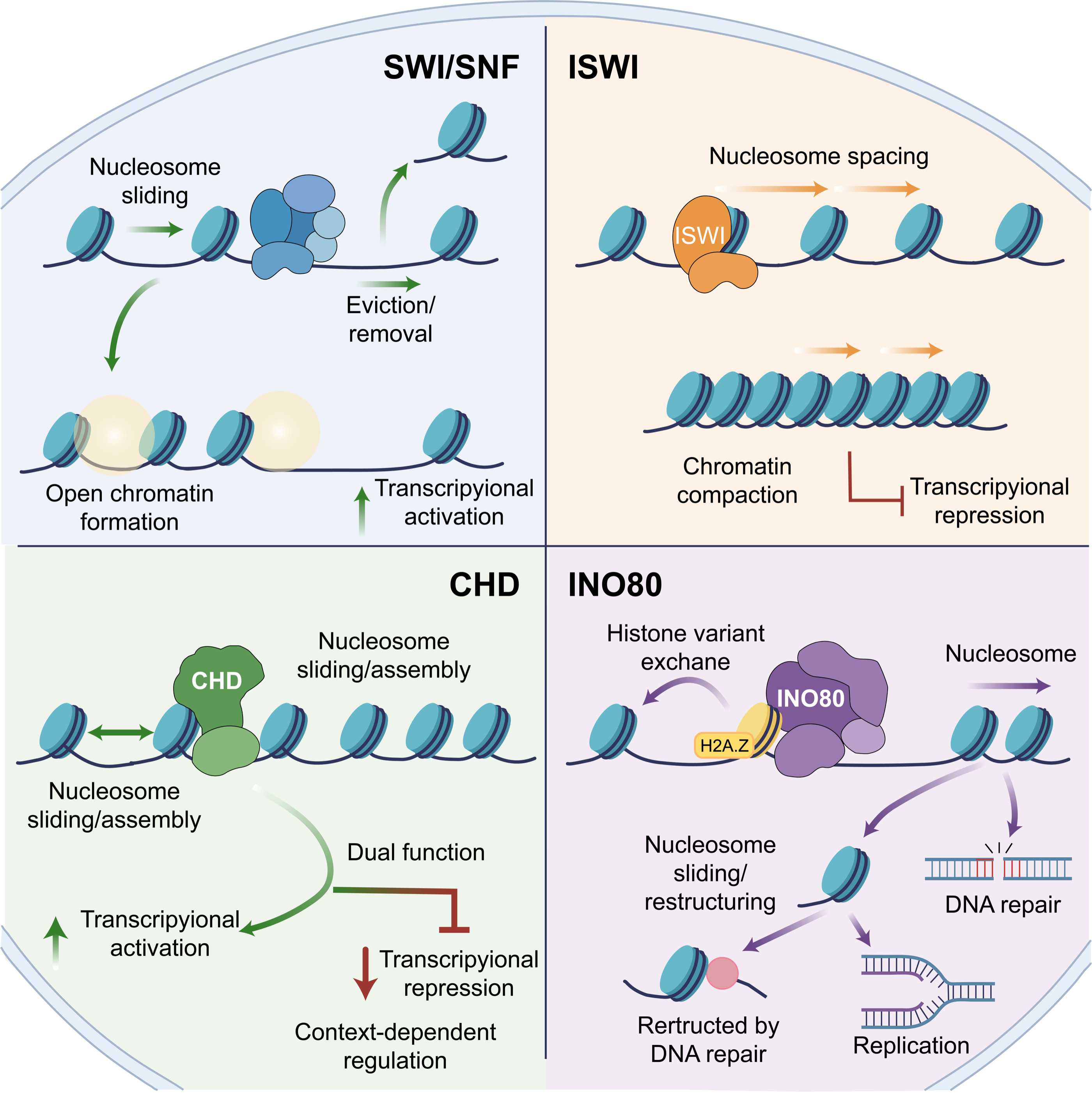
Its functional abnormalities can lead to widespread dysregulation of chromatin structure, affecting cancer cell fate decisions (14). In particular, the SWI/SNF complex plays a key role in regulating CSCs. In MUC1-C-dependent experimental models, MUC1-C engages BAF and PBAF complexes and is associated with genome-wide changes in chromatin accessibility and stemness-related transcriptional programs (11).
Long noncoding RNAs can influence CSC-associated properties through interactions with chromatin-remodeling machinery. In liver CSCs, lncBRM associated with BRM and promoted a BRG1/BRM switch within BAF complexes, activating YAP1 signaling and supporting self-renewal and tumor initiation (16). In embryonic stem cells, specific BAF complexes (esBAF) and their subunits (such as BRG1) are crucial for maintaining an open chromatin state at the promoters of pluripotency genes (such as OCT4 and SOX2). These observations provide a mechanistic framework for understanding related dependencies in CSCs, although evidence from embryonic stem cells should not be interpreted as direct evidence of the same regulatory architecture in malignant stem-like populations (17). The SWI/SNF complex is not limited to transcriptional activation; it also counteracts Polycomb Repressive Complex (PRC)-mediated gene silencing, creating a dynamic balance that supports cell-fate reprogramming and CSC stemness maintenance (11). Therefore, targeting the SWI/SNF complex or its regulatory network offers new therapeutic strategies to disrupt CSC stemness and overcome therapy resistance.
Importantly, SWI/SNF activity cannot be classified uniformly as pro-stemness or tumor-promoting. Loss-of-function alterations in ARID1A, SMARCB1, PBRM1, or SMARCA4 can themselves promote tumorigenesis, whereas other tumor contexts retain or acquire dependence on residual SWI/SNF activity. The therapeutic consequence is therefore genotype- and lineage-dependent: inhibiting a remodeler required by one CSC state could be ineffective, or potentially counterproductive, in tumors in which loss of that activity contributes to malignant progression. Therapeutic strategies should consequently be framed around context-specific dependencies rather than global inhibition of SWI/SNF function.
ISWI Complex
The ISWI complex is another important class of ATP-dependent chromatin remodeling complexes. Its main functions include regular nucleosome spacing and chromatin assembly, playing a core role in maintaining higher-order chromatin structure and mediating gene silencing (4, 18). ISWI complexes regulate nucleosome spacing and chromatin organization, with transcriptional effects that depend on complex composition, genomic location, and cellular context (18). Their developmental functions provide a rationale for investigating roles in cell-state regulation, but do not establish a universal mechanism of CSC quiescence (Figure 1). Quiescent or slowly cycling states occur in subsets of tumor cells and may contribute to treatment persistence; however, proliferative behavior varies across tumor types and cellular states (19). In glioma models, CD133-positive tumor cells exhibited preferential activation of DNA damage checkpoints and enhanced repair following irradiation, contributing to radioresistance (20). ISWI-family complexes have been implicated in cancer-associated transcriptional and genome-maintenance processes, but their contributions to CSC quiescence and treatment persistence require model-specific functional evidence (21). In glioblastoma, chromatin regulation has been implicated in stem-like behavior and treatment resistance (5), while altered chromatin insulation provides a separate framework for understanding dysregulated gene expression (22). These observations do not directly establish that ISWI complexes maintain glioma stem-cell quiescence or confer resistance to temozolomide and radiation. More direct evidence connects an ISWI-family ATPase to glioblastoma-associated stemness. In experimental glioblastoma models, miR-146a targeted POU3F2 and SMARCA5, reducing stemness-associated properties and increasing sensitivity to temozolomide (23). These findings implicate SMARCA5-associated regulation in treatment response without establishing that its effects are mediated through cellular quiescence. Whether ISWI dependence constitutes a therapeutically exploitable vulnerability remains less well established than for selected SWI/SNF dependencies, and direct functional studies in patient-derived CSC models will be required to distinguish causal roles from associations with quiescent chromatin states.
CHD Complex
The CHD complex family is characterized by members containing chromodomain and helicase/ATPase domains, playing a unique role in the regulation of chromatin dynamics (4, 24). These complexes participate in processes such as nucleosome sliding, histone variant incorporation, and transcriptional regulation (Figure 1). In mouse embryonic stem cells, CHD1 depletion caused heterochromatin accumulation and impaired pluripotency maintenance, directly linking this remodeler to chromatin organization and cellular identity (13). Whether comparable dependencies operate in malignant stem-like populations requires tumor-specific functional evidence. CHD4, a member of the CHD family and a core ATP-dependent chromatin-remodeling subunit of the nucleosome remodeling and deacetylase (NuRD) complex, regulates transcription, DNA-damage responses, and chromatin organization in cancer cells (25). CHD4 also contributes to genome maintenance and has been implicated in treatment resistance through context-dependent effects on DNA repair, cell-cycle regulation, and other stress-response pathways (25).
In TNBC models, MUC1-C/MYC signaling induced expression of the NuRD components MTA1, MBD3, and CHD4 and contributed to repression of luminal differentiation programs, linking MUC1-C to chromatin-associated dedifferentiation (26). In TNBC, low FBXW7 expression correlates with poor prognosis; FBXW7 mediates ubiquitin-proteasomal degradation of CHD4 to restrain CHD4-induced β-catenin nuclear translocation and Wnt/β-catenin pathway activation, thereby suppressing cancer stemness, malignant cellular phenotypes, and in vivo tumorigenicity of TNBC cells (27). Chromatin-remodeling abnormalities can contribute to transcriptional states associated with recurrence and therapy resistance, although their relative importance varies according to tumor lineage, genotype, and treatment context (28). CHD complexes influence CSC fate decisions by regulating the chromatin state of key stemness genes (such as SOX2, OCT4) and differentiation-related genes. Their dysfunction may lead to global changes in chromatin accessibility, enabling CSCs to adapt dynamically to therapeutic pressure and survive (29, 30). Therefore, targeting CHD complexes or the chromatin-remodeling pathways they mediate holds promise for interfering with the stemness-maintenance network of CSCs, offering new strategies to overcome cancer recurrence.
CHD-family proteins also differ substantially in complex membership, genomic localization, and biological function; they should therefore not be treated as a single therapeutic class. In particular, evidence linking individual CHD proteins to CSC maintenance remains uneven across tumor types, and several proposed mechanisms derive from conventional cancer-cell models rather than prospectively defined CSC populations.
INO80 Complex
INO80-family complexes regulate nucleosome organization and histone-variant dynamics. H2A.Z deposition is principally associated with SWR1/SRCAP/p400-related complexes, whereas canonical INO80 regulates nucleosome positioning and context-dependent H2A.Z exchange or occupancy (4, 31). These activities can influence transcriptional regulation during cell-state transitions (Figure 1). During embryonic development and cell differentiation, precise switching of gene expression programs requires large-scale chromatin remodeling. The INO80 complex is an ATP-dependent chromatin remodeler involved in nucleosome organization and histone-variant regulation (4). Its effects on H2A.Z occupancy are context-dependent. In pluripotent stem cells, INO80 promotes H2A.Z occupancy at bivalent promoters and facilitates the transition from naïve to primed pluripotency, demonstrating a role in chromatin regulation during cell-fate transitions (31). These findings provide a mechanistic basis for investigating whether INO80-dependent H2A.Z regulation also contributes to CSC plasticity. Whether the developmental mechanisms described above also regulate malignant stem-like states requires direct investigation in the relevant tumor models. CSCs need to maintain high epigenetic plasticity to switch between self-renewal and differentiation and to adapt to the tumor microenvironment and therapeutic pressure (30, 32).
The dynamic exchange of H2A.Z mediated by the INO80 complex may be a key regulator of this plasticity. For example, enrichment of H2A.Z at gene promoters is closely associated with transcriptional potential, and its abnormal distribution is linked to the occurrence and development of various cancers (31). In hepatocellular carcinoma, the long noncoding RNA HAND2-AS1 recruits the INO80 chromatin-remodeling complex to the promoter of bone morphogenetic protein receptor type 1A (BMPR1A), promoting its transcription and activating BMP signaling to sustain liver cancer stem cell (CSC) self-renewal. Experimental depletion of INO80 impairs liver CSC self-renewal and suppresses tumor growth, providing direct evidence for its contribution to liver cancer stemness (33). In glioblastoma, chromatin-remodeling programs are likewise implicated in glioblastoma stem cell (GSC) maintenance and therapeutic resistance, although the specific contribution of INO80 requires further investigation (5). Furthermore, ATP-dependent chromatin remodelers, including INO80 and SWI/SNF, regulate nucleosome organization and transcriptional accessibility. Their activities may influence stress-response and stemness-associated transcriptional programs, although direct functional cooperation between these complexes in CSCs remains to be established (34).
Therefore, targeting the INO80 complex or the H2A.Z exchange process it catalyzes could interfere with transcriptional programs that support selected CSC states. However, the therapeutic window for INO80 inhibition remains uncertain because INO80-family complexes also participate in DNA replication, DNA repair, and genome maintenance in normal cells.
Somatic Mutations and Expression Dysregulation of Chromatin Remodeling Complex Genes
Chromatin remodeling complexes, particularly the SWI/SNF (BAF/PBAF) family, play a central role in maintaining genomic stability and regulating gene expression. Large-scale cancer genome sequencing studies have revealed that mutations in genes encoding SWI/SNF subunits occur in over 20% of cancers, making them among the most frequently mutated epigenetic regulators in human cancers (14). These mutations are widespread across solid tumors and hematological malignancies. For instance, in hepatocellular carcinoma, AT-rich interaction domain (ARID) genes such as AT‑rich interaction domain 1A/B (ARID1A/B) and AT‑rich interaction domain 2 (ARID2) frequently undergo loss-of-function mutations (35).
In melanoma, exome sequencing also commonly identifies loss-of-function mutations in SWI/SNF subunit-encoding genes (36). A study of gastric cancer in the Mizo population of India further confirmed that ARID1A is a significantly mutated gene, with its mutations closely associated with abnormalities in the chromatin remodeling pathway (37). These mutations are not isolated events; they lead to fundamental changes in global chromatin accessibility by disrupting the integrity of the SWI/SNF complex. As ATP-driven molecular motors, SWI/SNF complexes normally reconfigure nucleosome structure and control genomic DNA accessibility. Different SWI/SNF subunit alterations can change complex assembly, recruitment, or residual activity, with consequences that depend on the affected subunit and tumor context. Mutation frequency alone, however, does not establish a CSC-specific mechanism. SWI/SNF alterations can influence proliferation, differentiation, DNA repair, immune signaling, and lineage identity independently of stemness. Studies that infer CSC regulation from bulk-tumor genomic associations should therefore be distinguished from experiments that directly test self-renewal, tumor initiation, or reversible stem-like state transitions.
Chromatin-regulatory abnormalities can alter enhancer activity and differentiation-associated transcription. Distinct mechanisms include loss of tumor-suppressive functions and oncogenic recruitment of otherwise functional remodeling machinery. For example, in fusion-negative rhabdomyosarcoma (FN-RMS), the transcription factor twist family bHLH transcription factor 2 (TWIST2) interacts with chromatin-remodeling factors SMARCA4 and CHD3 to regulate histone modifications at distal enhancers, thereby activating growth-related genes and suppressing myogenic differentiation genes, maintaining the undifferentiated state and tumorigenicity of cells (38). This illustrates oncogenic recruitment of chromatin regulators rather than a demonstrated consequence of SWI/SNF loss-of-function mutations. The SWI/SNF complex also contributes to genome maintenance. Experimental depletion of its ATPase subunit BRG1 increases R-loop accumulation, transcription–replication conflicts, and R-loop-dependent DNA damage, demonstrating a role for SWI/SNF-mediated chromatin remodeling in preserving genomic integrity (39).
Beyond high-frequency somatic mutations, chromatin-remodeling complex subunits often show aberrant expression, and this dysregulation often synergizes with specific oncogenic signals. For example, the well-known oncoprotein MYC can upregulate certain chromatin-remodeling factors, thereby jointly driving transcriptional programs that promote stemness. This dysregulation may also profoundly affect the tumor microenvironment. Studies indicate that in cancers with mutations in SWI/SNF complex subunits (such as PBRM1, SMARCA4, ARID2), the tumor immune microenvironment exhibits distinct activation states. In particular, in gastric cancer with low SMARCA4 expression, T cell effector (such as CD8B), central memory T cell markers, and mature B cell markers (such as CD20, CD38) are upregulated, whereas cancer-related genes like MYC and aurora kinase B (AURKB) are downregulated, and features of tertiary lymphoid structures are observed (40).
This suggests that abnormalities in chromatin-remodeling complexes not only directly alter the epigenetic state and stemness of tumor cells but may also indirectly affect cancer progression and response to therapies (such as immune checkpoint blockade) by reshaping the tumor immune microenvironment. In summary, somatic mutations and expression dysregulation of chromatin-remodeling complex genes regulate CSC characteristics across multiple dimensions and levels—by altering global chromatin accessibility, disrupting normal gene regulatory networks, inducing genomic instability, and synergizing with oncogenic signals, serving as a key bridge between epigenetic abnormalities and tumorigenesis and development.
Characteristics of CSCs-Specific Chromatin Accessibility Landscapes
CSC-associated regulatory programs can involve differences in chromatin organization, enhancer activity, and gene expression, although the evidence depends on the experimental system. Comparative epigenomic profiling of glioma stem cells and neural stem/progenitor cells identified distinct DNA-modification patterns and enhancer landscapes associated with differential gene regulation (41). In glioblastoma stem cells, a super-enhancer-associated program promoted polyunsaturated fatty-acid synthesis and supported EGFR signaling, providing direct evidence that enhancer regulation can sustain a tumor-associated metabolic dependency (42). Comparative chromatin-state analysis in glioblastoma CSCs identified activation of developmental transcription-factor programs, including ASCL1-dependent regulation of Wnt signaling. Functional experiments established a requirement for ASCL1 in CSC maintenance and tumorigenicity (43). These findings illustrate distinct regulatory mechanisms rather than a shared pattern of increased accessibility at SOX2, NANOG, and ALDH1A1 loci across cancers.
Histone-defined regulatory landscapes and transcription-factor networks provide complementary evidence of stemness-associated gene regulation. Profiling of primary gliomas identified subtype-associated enhancer states and bivalent chromatin domains enriched for developmental regulators, revealing similarities to glioma stem-cell regulatory programs (44). In a separate study, a SOX2-associated network involving OLIG2 and ZEB1 supported glioma-initiating properties; co-expression of these factors generated glioma-initiating cells from tumor-suppressor-deficient astrocytes in experimental models (45). Together, these findings connect regulatory chromatin states with functional transcriptional programs, without establishing that all CSCs exhibit globally increased accessibility.
A methodological caveat is that differential accessibility is not equivalent to functional enhancer activity or remodeler dependence. ATAC-seq identifies accessible DNA but does not establish which remodeling complex created or maintains that state. Moreover, CSC enrichment based on surface markers can capture heterogeneous populations whose chromatin profiles partly reflect lineage, cell-cycle state, or environmental exposure rather than stemness itself. Integrating accessibility profiling with perturbation experiments, transcription-factor occupancy, and functional CSC assays is therefore necessary to assign causality.
Histone modifications, transcription-factor activity, and chromatin accessibility represent related but distinct layers of gene regulation. In glioma stem-cell models, profiling of DNA modifications and enhancer-associated histone marks revealed regulatory landscapes that differed from those of neural stem/progenitor cells (41). In TNBC models, DOT1L-dependent H3K79 methylation supported stemness-associated transcriptional and metabolic programs (46). Single-cell chromatin-accessibility profiling of primary glioblastomas, combined with analysis of patient-derived glioblastoma stem-cell models, identified distinct regulatory states associated with different transcription-factor dependencies and invasive properties (47). These findings demonstrate heterogeneity within stem-like tumor populations rather than a uniform increase in accessibility at all stemness-associated genes. However, gene expression and marker enrichment alone do not establish increased chromatin accessibility or transcription-factor occupancy at the corresponding regulatory regions. Together, these studies illustrate different mechanisms associated with stemness rather than a single experimentally demonstrated chromatin-accessibility feedback circuit (Figure 2).
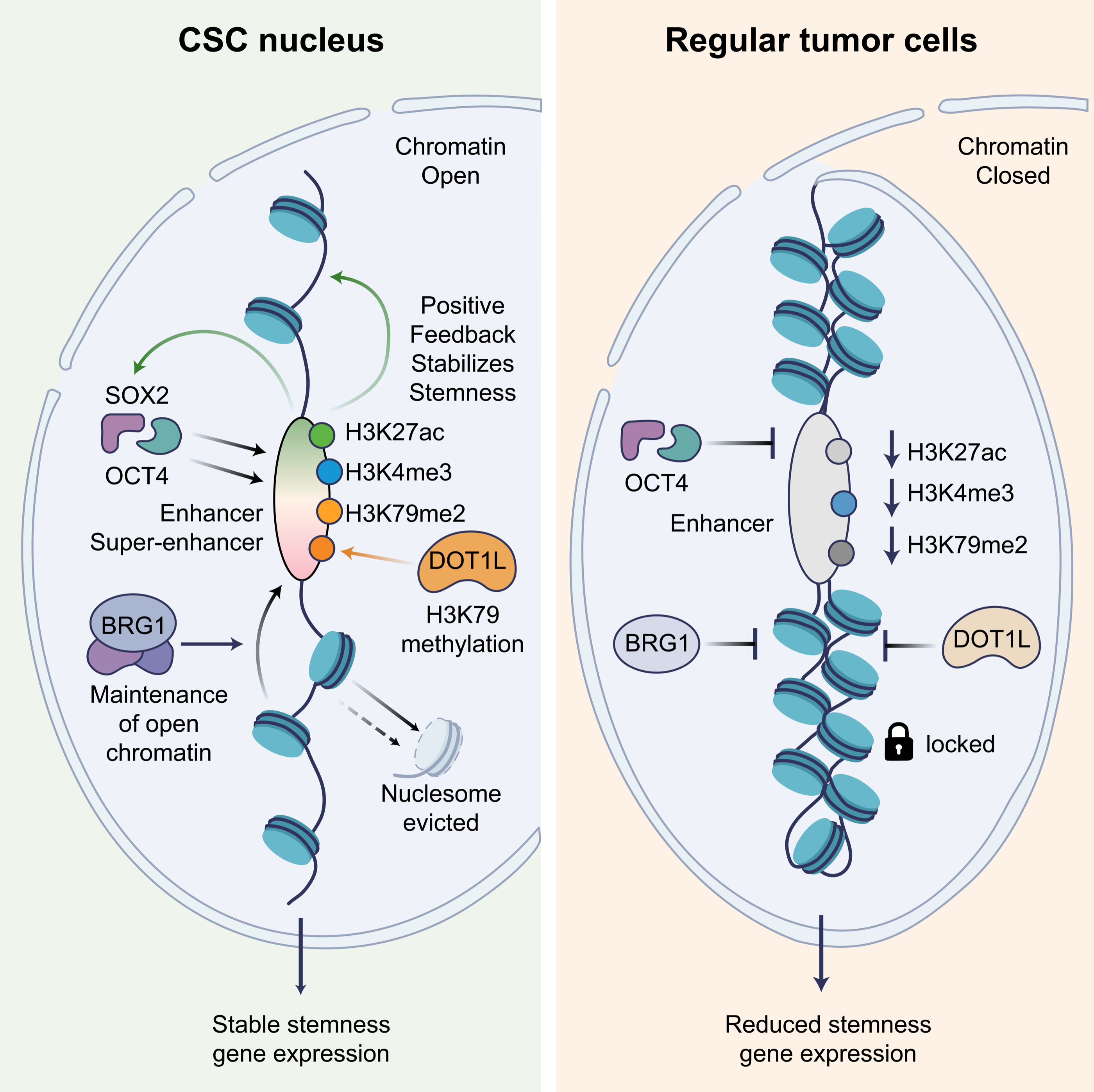
The establishment and maintenance of open chromatin states are not passive; they depend on the precise recruitment of chromatin remodeling complexes to key regions. These complexes dynamically regulate chromatin accessibility by consuming ATP to slide, evict, or reorganize nucleosomes. In CSCs, a series of chromatin-remodeling factors has been shown to be essential for maintaining stemness. For example, the core catalytic subunit BRG1 of the SWI/SNF chromatin remodeling complex plays a key role in colorectal CSCs. Specific knockout of Brg1 in cells expressing Dclk1 (a marker of intestinal tumor stem cells) significantly inhibits tumor growth and reduces stem cell self-renewal capacity through increased apoptosis and downregulation of stemness-related gene expression (48). These results support a functional requirement for Brg1 in the intestinal tumor stem-cell population studied. However, the knockout phenotype alone does not identify the genomic sites where Brg1 acts or demonstrate that altered accessibility at those sites explains the observed effects. Similarly, the histone methyltransferase DOT1L is highly expressed in TNBC CSCs. It regulates stemness-related gene expression programs, including oxidative phosphorylation, MYC target genes, and the Wnt pathway, by catalyzing H3K79 methylation, thereby maintaining CSC self-renewal capacity (46) (Figure 2). Glioma stem-cell profiling further demonstrated heterogeneous epigenomic and transcriptional responses to differentiation cues, highlighting the context-dependent relationship between chromatin state and cellular identity (41). These observations do not identify which ATP-dependent remodeling complexes causally establish the observed epigenomic states. These chromatin remodeling complexes and modifying enzymes act like architects and switches of the genome that can be recruited by CSC-specific signaling pathways or transcription factors. This consolidates the open chromatin landscape that supports stemness by remodeling local nucleosome structures, ultimately determining the functional state of CSCs and their resistance to therapy.
3. Molecular Mechanisms of Chromatin Remodeling Regulating Tumor Stem Cells
Synergy Between Remodeling Complexes and Pluripotency Factors OCT4/SOX2/NANOG
Chromatin-remodeling complexes and stemness-associated transcription factors contribute to context-dependent regulatory programs. Mammalian SWI/SNF complexes use ATP hydrolysis to reposition or evict nucleosomes, and their composition and recruitment influence transcription and cell identity (49, 50). Their regulatory effects depend on the genomic regions engaged and the transcriptional machinery available within a particular cellular state. In glioblastoma, POU3F2, SOX2, SALL2, and OLIG2 were identified as a core network that can reprogram differentiated tumor cells toward a tumor-propagating state (8). MUC1-C-associated studies provide a separate framework linking BAF/PBAF activity to stemness-related transcriptional programs (11). These examples illustrate distinct routes through which transcriptional regulators and chromatin-associated processes can support malignant cell identity. Biochemical and structural studies explain how remodelers engage nucleosomes, but do not themselves demonstrate a universal OCT4/SOX2/NANOG–SWI/SNF recruitment circuit in CSCs (34, 51).
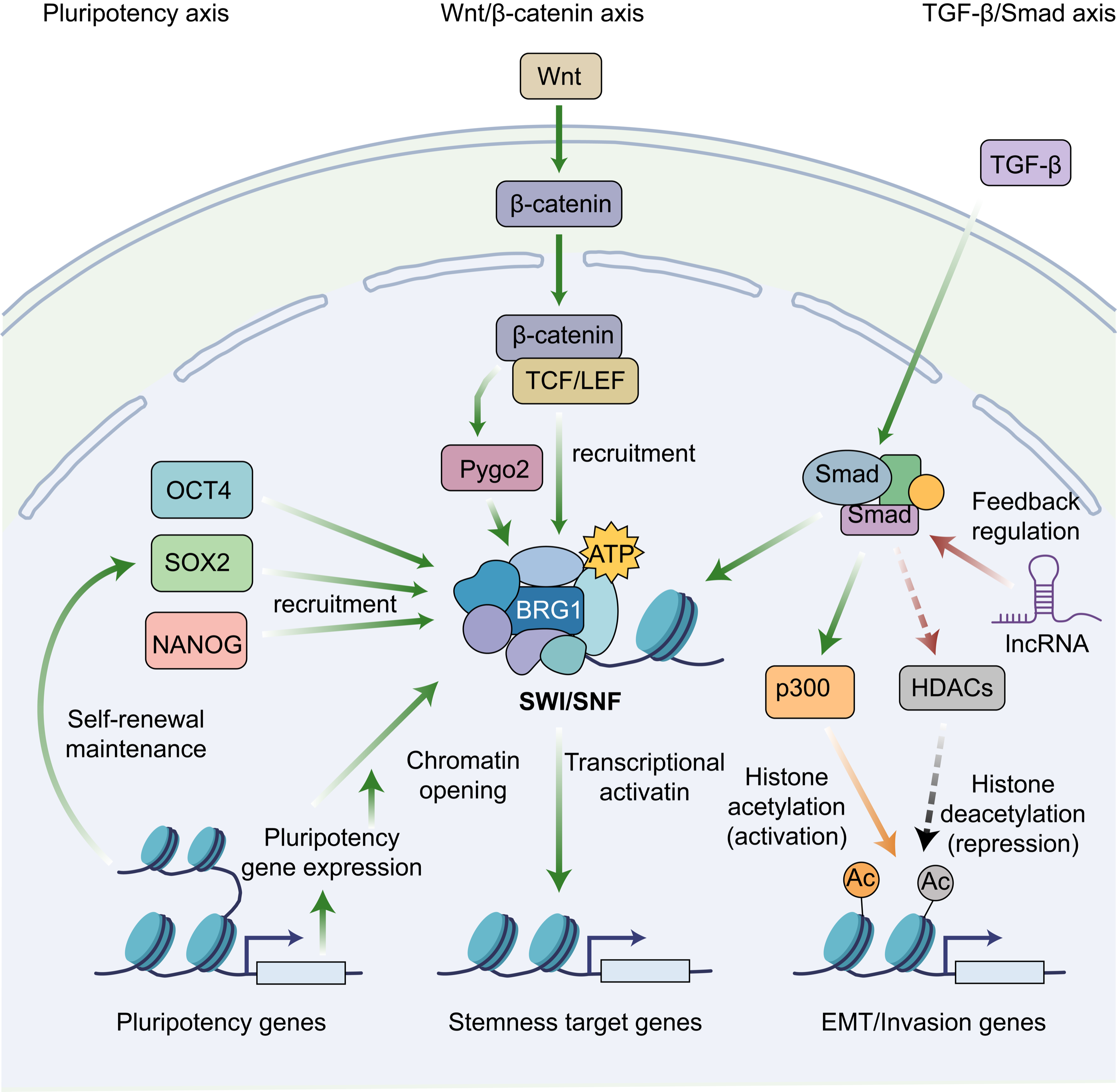
Reciprocal interactions between transcription factors and chromatin regulation can help maintain cellular identity. However, the participating factors, remodeling-complex composition, and direction of regulation vary across tumor lineages and cell states (14, 49). A self-reinforcing OCT4/SOX2/NANOG–SWI/SNF circuit should therefore be treated as a context-dependent model rather than a universal feature of CSCs. Establishing such a circuit requires direct evidence of factor binding, remodeler recruitment, chromatin changes, and functional effects at the relevant endogenous loci. Expression correlations or overlapping genomic occupancy alone cannot establish the direction of recruitment or demonstrate that the proposed interaction is necessary for self-renewal. Experimental reprogramming of differentiated glioblastoma cells demonstrates that tumor-propagating potential can be acquired through changes in transcriptional regulatory networks (8). This supports plasticity of malignant cell identity, while leaving the contribution of individual remodeling complexes to be resolved through targeted perturbation. Comparisons between stem-like and differentiated populations, ideally combined with time-resolved measurements, could distinguish mechanisms that initiate state transitions from those that maintain an established state. The relationships summarized in Figure 3 should therefore be interpreted as a conceptual integration of mechanisms from different experimental systems.
Developmental and malignant dependencies should be distinguished. Depletion of SMARCB1 or BRD7 altered porcine embryo development and pluripotency-associated expression (52), providing developmental evidence rather than proof of selective CSC targeting. Cancer-associated SWI/SNF alterations can be tumor-suppressive or dependency-forming depending on genotype and lineage (14, 36). In glioma-initiating cells, BRG1 perturbation altered stemness-associated properties and chemosensitivity, supporting a functional role for BRG1 in this experimental context (53). These findings provide a rationale for investigating BRG1-associated dependencies, but should not be generalized to breast cancer or interpreted as direct evidence of nucleosome reoccupation at OCT4, SOX2, and NANOG promoters. Therapeutic development must account for tumor genotype, remodeling-complex composition, and normal-tissue requirements (54, 55).
Crosstalk Between Core CSC Signaling Pathways and Chromatin Remodeling
Interaction Between the Wnt/β-catenin Pathway and Chromatin Remodeling
The Wnt/β-catenin signaling pathway is a core regulator of embryonic development and tissue homeostasis. Its aberrant activation plays a key role in the initiation and progression of various cancers, particularly in maintaining CSC stemness (56). β-Catenin stability is regulated by phosphorylation-dependent degradation. Mechanistic experiments demonstrated that casein kinase Iα-mediated priming enables subsequent GSK-3 phosphorylation, linking sequential kinase activity to β-catenin turnover (57). After entering the nucleus, β-catenin not only acts as a classical transcriptional co-activator, binding to TCF/LEF family transcription factors to initiate transcription of downstream target genes (such as c-MYC, CCND1), but also extends its function to epigenetic regulation. Wnt signaling can also influence three-dimensional regulatory architecture. Experimental activation of Wnt signaling induced CTCF binding and loop formation at cis-regulatory elements of target genes, linking pathway activity to changes in chromatin organization (58). For example, in uveal melanoma, the chromatin regulator RUVBL1 enhances CTNNB1 transcription by modulating chromatin remodeling, thereby promoting malignant biological behavior in tumor cells (59). These observations link Wnt-related transcription to chromatin regulation in defined experimental systems but do not establish a universal mechanism of CSC self-renewal.
Structural analysis defined interactions between BCL9–Pygo and LDB–SSBP complexes that contribute to Wnt enhanceosome assembly, providing a molecular basis for the organization of transcriptional cofactors (60). These findings provide a structural basis for cofactor organization, not direct evidence of H3K56 acetylation or SWI/SNF recruitment. The BAF chromatin-remodeling complex, a member of the SWI/SNF family, also cooperates with β-catenin to regulate Wnt-responsive transcription. BRG1, the catalytic subunit of BAF, physically interacts with β-catenin and promotes TCF-dependent target-gene activation. Conversely, ATPase-inactive BRG1 expression inhibits endogenous TCF target-gene expression in colon carcinoma cells, establishing a functional link between chromatin remodeling and Wnt signaling (61).
In colorectal cancer models, BRG1 supports intestinal tumor stem-cell self-renewal and survival (48). These findings suggest that the interaction between chromatin remodeling and Wnt signaling may represent a therapeutic vulnerability, although disrupting the BRG1–β-catenin interaction has not yet been established as a strategy to eradicate colorectal CSCs. In ovarian cancer, alterations in the SWI/SNF subunit ARID1A and the Wnt pathway component CTNNB1 have also been implicated in tumorigenesis, highlighting the context-dependent relationship between chromatin remodeling and Wnt signaling (62).
Crosstalk between Wnt signaling and chromatin-associated regulation provides a rationale for investigating interventions directed at transcriptional cofactors or context-specific remodeling dependencies (63). Their effects on treatment-resistant, tumor-propagating populations require direct evaluation, including functional tumor-initiation assays and comparison with normal-cell dependencies. These approaches remain investigational strategies rather than established methods for selectively eradicating CSCs or preventing recurrence (Figure 3).
Epigenetic Regulation of the TGF-β/Smad Pathway in Stemness Maintenance
The TGF-β/Smad signaling pathway plays a complex and critical role in regulating CSC characteristics, particularly in maintaining stemness. Through its core transcription factors, the Smad proteins, this pathway can directly or indirectly recruit chromatin-remodeling factors and histone-modifying enzymes, thereby reshaping the gene expression profile at the epigenetic level, driving epithelial-mesenchymal transition (EMT), and activating or suppressing stemness-related genes (64). For example, in breast cancer, TGF-β signaling activates p38 MAPK, which phosphorylates the transcription factor ΔNp63 and enhances its DNA-binding ability. Phosphorylated ΔNp63 is recruited to the regulatory regions of TGF-β/Smad target genes, where it promotes recruitment of the histone acetyltransferase p300, increasing H3K27ac while reducing the repressive mark H3K27me3. This creates an open chromatin environment, promoting transcription of genes associated with stem cell characteristics and invasiveness (64). This process shows how TGF-β signaling lays an epigenetic foundation for stemness gene expression by altering local chromatin accessibility. Furthermore, TGF-β signaling can regulate lncRNA expression, which can act as molecular scaffolds or decoys to further influence chromatin state. For instance, LINC00707 can interact with Smad proteins in the cytoplasm, limiting their nuclear translocation and thereby negatively regulating TGF-β signal output. Conversely, TGF-β signaling itself can suppress LINC00707 expression, forming a feedback loop that finely controls epigenetic and transcriptional programs related to cell invasion (65). These studies indicate that the TGF-β/Smad pathway profoundly influences CSCs fate decisions by integrating multiple epigenetic mechanisms (Figure 3).
TGF-β-associated stemness can arise through distinct regulatory mechanisms. In mammary epithelial models, induction of epithelial–mesenchymal transition, including through TGF-β exposure, increased stemness-associated markers and mammosphere formation; transformed cells undergoing EMT also exhibited enhanced tumor-forming capacity (66). In patient-derived glioma-initiating cells, TGF-β promoted self-renewal and inhibited differentiation through Smad-dependent induction of leukemia inhibitory factor (LIF) and subsequent activation of JAK–STAT signaling (67). These findings establish model-specific connections between signaling and stemness-associated behavior, rather than demonstrating a universal mechanism involving recruitment of ATP-dependent chromatin remodelers.
In patient-derived glioblastoma models, TGF-β receptor inhibition reduced a CD44-high/Id1-high population enriched for glioma-initiating activity. This response involved repression of Id1 and Id3 and reduced tumor-initiation capacity, providing preclinical evidence for a TGF-β-dependent vulnerability in a defined cellular population (68). Experimental evidence further links epigenetic regulation of TGF-β/SMAD3 signaling to lung cancer metastasis. The histone methyltransferase SMYD2 promotes SMAD3 expression through epigenetic regulation, whereas SMYD2 depletion suppresses epithelial–mesenchymal transition (EMT)-associated changes, migration, invasion, and metastatic tumor formation in lung cancer models (69). Additionally, in invasive tumors such as glioblastoma, dysregulated TGF-β signaling contributes to tumor progression and therapeutic resistance by promoting EMT-like transitions and stem-like characteristics (70). These processes are accompanied by profound epigenetic reprogramming. For example, TGF-β stimulation can alter the distribution of histone modifications (such as H3K9me3) across the genome. These changes are associated with pro-cancer functions, such as cell migration and adhesion, and may depend on specific transcription factor-binding sites, such as SRY and FOXJ3 (71).
TGF-β-associated transcription and chromatin regulation can influence stemness-related or invasive phenotypes in selected systems, but the contributing cofactors and functional endpoints vary by model (Figure 3).
Chromatin Remodeling and CSCs Plasticity
CSCs are not a fixed or homogeneous cell population; rather, they exhibit plasticity, allowing transitions between stem-like and differentiated states in response to environmental cues and therapeutic pressures. This capacity to acquire or lose stemness-associated properties contributes to intratumoral heterogeneity, tumor recurrence, and therapy resistance (72). Such plasticity complicates the distinction between a stable CSC hierarchy and a changing distribution of tumor-cell states. Hierarchical organization and reversible transitions need not be mutually exclusive: a tumor may contain populations with different tumor-propagating capacities while retaining the potential for selected cells to change state. Stemness-associated properties should therefore be interpreted in relation to the experimental model, microenvironment, and treatment conditions rather than assumed to define an irreversible cellular identity.
Chromatin remodeling provides an important molecular mechanism underlying these transitions by regulating access to genomic regions involved in cell-fate determination. Glioblastoma reprogramming experiments demonstrate that a defined transcription-factor network can restore tumor-propagating potential in differentiated tumor cells, supporting the plasticity of malignant cell identity (8). These findings illustrate how changes in regulatory programs can alter functional tumor-cell properties, although experimentally induced reprogramming does not establish the frequency or direction of spontaneous transitions in patients. They also highlight the importance of distinguishing the acquisition of a stemness-associated expression profile from the acquisition of experimentally demonstrated tumor-propagating capacity. Whether a particular remodeler initiates a transition, stabilizes the resulting state, or supports cell survival during reprogramming requires direct functional investigation.
Through coordinated alterations in chromatin accessibility and gene expression, chromatin-regulatory mechanisms can facilitate transitions between proliferative, differentiated, and stem-like cellular states. In various cancers, genetic alterations or dysregulated activity of chromatin-remodeling complexes disrupt normal transcriptional programs and differentiation pathways, potentially promoting the acquisition or maintenance of stem-like characteristics and contributing to tumor progression (73). However, chromatin accessibility should not be interpreted as a simple measure of stemness: opening or closing particular regulatory regions can have different consequences depending on their target genes and cellular context. Mechanistic studies must therefore connect changes at defined loci with transcriptional responses and functional phenotypes.
This dynamic regulation suggests that, under specific conditions, non-stem tumor cells may regain self-renewal and tumorigenic capacity through epigenetic reprogramming (Figure 4). Consequently, eliminating an initially identified CSC population may be insufficient if surviving cells retain the capacity to regenerate tumor-propagating states. Therapeutic studies should distinguish selection of pre-existing resistant populations from treatment-induced state changes, because an increased proportion of stemness-marker-positive cells after treatment could reflect either process. Changes in marker expression alone cannot establish conversion. Functional tumor-initiation assays, lineage tracing, and longitudinal profiling are needed to distinguish durable acquisition of tumor-propagating capacity from transient stress responses or reversible drug tolerance. Measurements before treatment, during exposure, and after withdrawal would further clarify whether an altered state persists, reverses, or enables tumor repopulation.
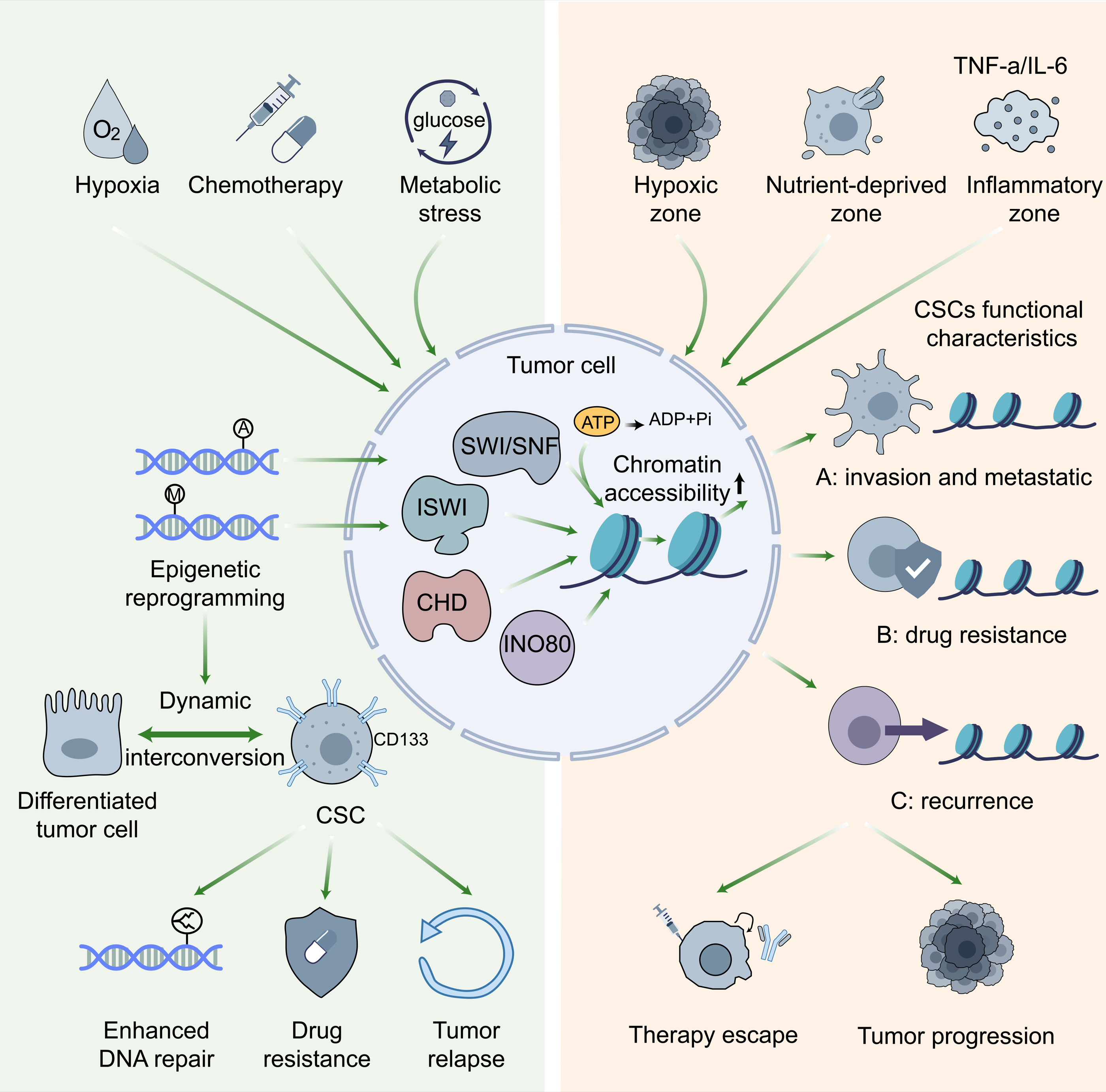
External pressures, such as chemotherapy drugs, hypoxia, or metabolic stress, drive chromatin remodeling and cell-state transitions. These stress signals can directly or indirectly affect chromatin-modifying enzyme activity, metabolite levels, and the localization and function of chromatin-remodeling complexes, rapidly and extensively altering chromatin accessibility at numerous genes (74). For example, under hypoxic conditions, cancer cells undergo widespread epigenetic reprogramming to activate gene expression programs adapted to low-oxygen conditions, including pathways that maintain stemness characteristics (74).
In cancer cell-line models, a small subpopulation survived drug exposure by entering a reversible drug-tolerant state associated with altered chromatin regulation and dependence on the histone demethylase KDM5A (75). This finding supports a role for chromatin-associated adaptation in treatment persistence, but does not establish that drug-tolerant persisters are equivalent to functionally defined CSCs. An unresolved issue is whether therapy-induced chromatin changes generate stable CSC populations or transient drug-tolerant states that only partially overlap with conventional definitions of stemness. Distinguishing these possibilities requires longitudinal lineage tracing and paired chromatin/transcriptomic profiling before, during, and after treatment. Without such temporal evidence, post-treatment enrichment of stemness markers cannot establish conversion to a durable CSC state.
Drug-tolerance experiments and glioblastoma reprogramming studies illustrate distinct routes through which tumor cells can acquire treatment-persistent or tumor-propagating properties (75, 8). Neither study alone establishes that chemotherapy, hypoxia, or metabolic stress universally induces self-renewal, drug efflux, and enhanced DNA repair in the same cell population. Determining whether treatment-induced states acquire durable tumor-propagating capacity requires longitudinal and functional validation (Figure 4).
Chromatin Remodeling and CSCs Heterogeneity
Tumor-cell heterogeneity reflects genetic variation and differences in regulatory state. Breast cancer experiments demonstrate that ZEB1-associated chromatin regulation can facilitate acquisition of CSC properties (3), while broader epigenetic mechanisms contribute to variation in tumor-cell identity (30). SHARE-seq demonstrated simultaneous measurement of chromatin accessibility and gene expression in individual cells and provided a framework for examining relationships between regulatory accessibility and cell-state transitions (76). However, this methodological study was not a direct demonstration of CSC heterogeneity in human tumors. Applying paired profiling to CSC-enriched populations requires careful cell annotation and functional assessment of tumor-propagating capacity.
Invasion/metastasis, treatment persistence, and post-treatment repopulation are used in Figure 4 as conceptual functional outcomes rather than three established, mutually exclusive CSC states. In melanoma models, a slow-cycling JARID1B-positive population supported sustained tumor growth, while JARID1B-negative cells could acquire the positive state, demonstrating dynamic population behavior rather than a fixed, unidirectional CSC hierarchy (77). Direct experiments in mammary epithelial models demonstrated that nonstem cells could spontaneously acquire stem-like properties, with oncogenic transformation enhancing this conversion (78). These findings support plasticity between cellular states, but do not validate the three functional categories depicted in Figure 4 as a universal CSC classification. In glioblastoma, single-cell analyses identified four major malignant cellular states whose distributions were influenced by genetic alterations and the microenvironment, with evidence of state plasticity (79). In breast cancer cell-line models, sorted phenotypic populations re-established characteristic population proportions through stochastic state transitions (80). These studies support plasticity and state interconversion, but do not demonstrate that ISWI inhibition induces differentiation or that SWI/SNF overexpression drives an invasive/metastatic state.
This epigenetic heterogeneity forms through a dynamic process shaped by multiple factors, with spatial heterogeneity in chromatin remodeling complex activity and differences in tumor microenvironment signals playing central roles. Chromatin remodeling complexes, such as the SWI/SNF, ISWI, CHD, and INO80 families, are key regulators of chromatin accessibility. They determine whether specific genomic regions are open or closed by consuming ATP to alter nucleosome positioning and composition (4). Within a tumor, uneven distribution of oxygen and nutrients and differences in cell-cell interactions across regions can lead to spatial heterogeneity in remodeling complex activity. For example, hypoxic regions of the tumor may affect the expression or recruitment of remodeling complex subunits through specific signaling pathways (such as HIF), leading to changes in the chromatin state of CSCs in that region and activating gene programs related to hypoxia adaptation and invasion. Determining how immune-cell and fibroblast-derived signals alter specific remodeling complexes in CSCs remains an important experimental objective. This reversible epigenetic reprogramming, driven by microenvironmental signals, endows CSCs with high plasticity, enabling them to quickly adapt to therapeutic pressure or pre-metastatic microenvironmental changes (30).
However, this also greatly increases the complexity of targeted therapy because therapies targeting a specific epigenetic state or signaling pathway may not effectively eliminate all heterogeneous CSC subpopulations and may even select for more resistant clones. Therefore, future therapeutic strategies need to comprehensively consider the multidimensional drivers of intratumoral epigenetic heterogeneity and develop combination therapies that can simultaneously target multiple key nodes or induce CSC differentiation (Figure 4).
4. The Role of Chromatin Remodeling in CSCs Therapy Resistance
Mediating Chemotherapy and Radiotherapy Resistance
Resistance of CSC-associated populations to chemotherapy and radiotherapy can contribute to tumor recurrence and treatment failure, with chromatin regulation influencing several underlying mechanisms. Some CSC populations exhibit relatively quiescent states or increased expression of drug efflux pumps, particularly members of the ATP-binding cassette (ABC) transporter family; however, these properties vary by tumor type and cellular state. Epigenetic mechanisms can regulate multidrug-resistance programs, including expression of ABC transporters such as ABCB1 (MDR1). In human cell models, SMARCB1 loss increased ABCB1 expression and doxorubicin resistance through a SMARCA4-dependent mechanism, demonstrating that SWI/SNF composition can influence drug-efflux-associated resistance (81). This mechanism is drug-dependent and should be distinguished from resistance arising through altered drug targets, enhanced survival signaling, or DNA damage responses.
LncRNAs can influence chemotherapy resistance through transcriptional, chromatin-associated, and post-transcriptional mechanisms. Depending on the experimental context, these activities may alter ABC transporter expression, apoptosis, or other resistance-associated pathways. Studies summarized in the doxorubicin-resistance literature implicate multiple lncRNA-dependent mechanisms, rather than a single universal pathway involving recruitment of chromatin-remodeling complexes to transporter promoters (82). Establishing such a direct mechanism requires evidence connecting lncRNA binding, remodeler recruitment, local chromatin changes, transporter expression, and altered drug sensitivity.
In colorectal cancer, CSC-associated markers such as CD44 and CD133 and signaling pathways including Wnt/β-catenin participate in regulatory networks linked to tumor maintenance and treatment resistance. Epigenetic dysregulation can support these programs, although marker expression alone does not establish a chromatin-remodeling dependency or predict therapeutic response (56). Additionally, high mobility group box 3 (HMGB3), a chromatin-associated protein implicated in DNA damage responses, has been linked to stemness-associated phenotypes and therapy resistance in several malignancies. Reported mechanisms include effects on survival signaling and DNA repair, with their relative contributions depending on the tumor model and treatment examined (83). These observations provide a rationale for investigating chromatin-associated resistance mechanisms, while the relationships summarized in Figure 5 should be interpreted as context-dependent rather than a single experimentally established pathway.
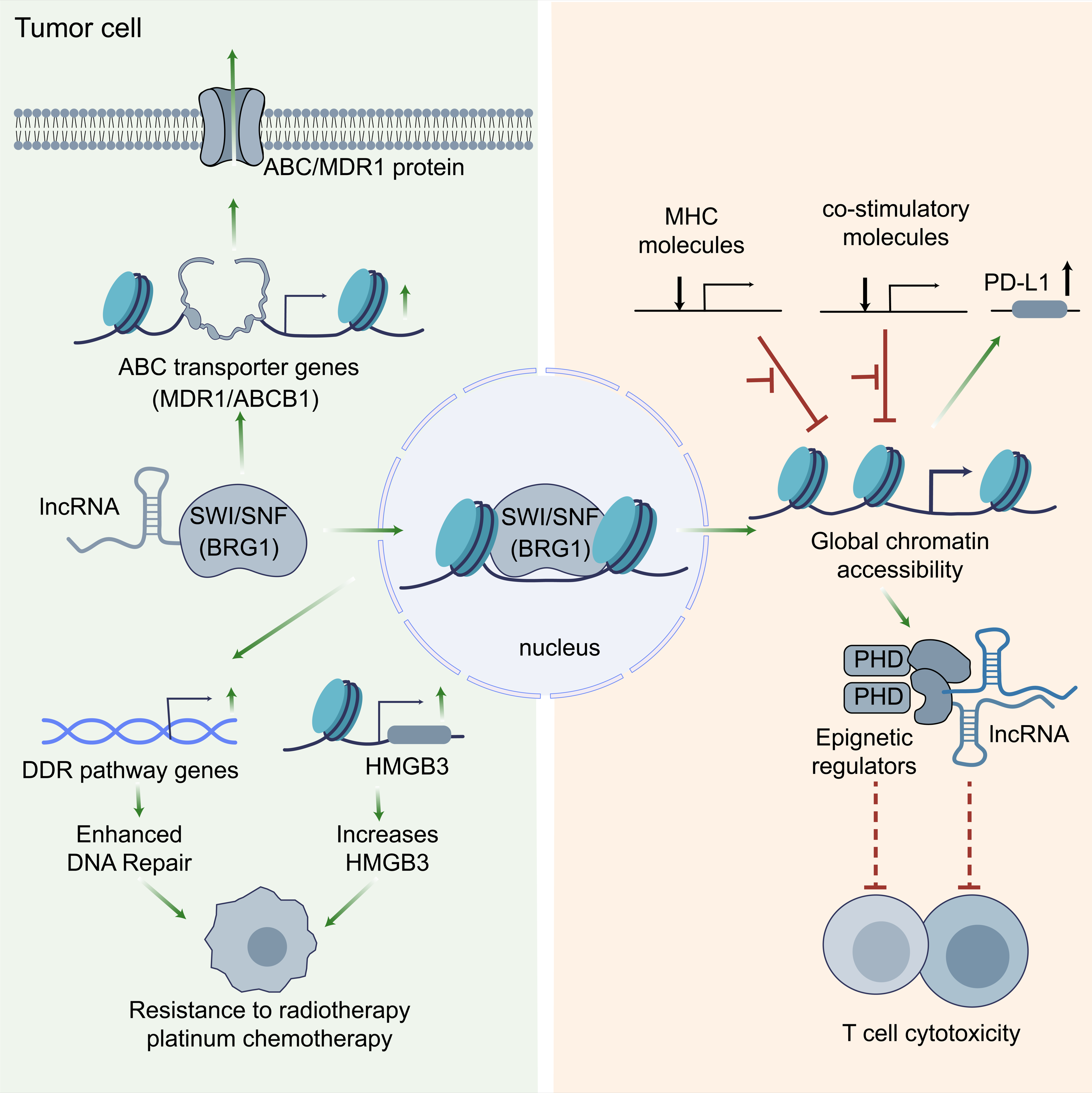
Epigenetic regulation can also influence radiation responses through mechanisms that vary across experimental systems (84). In gliomas, altered chromatin insulation can permit abnormal regulatory interactions and oncogene activation, but these findings do not directly establish a mechanism of resistance to temozolomide or radiation (22). In colorectal cancer, Brg1 depletion impaired tumor stem-cell maintenance and survival (48). Separate acute-leukemia experiments demonstrated a BRG1-associated transcriptional dependency involving MYC regulation (6). In colorectal cancer models, BRG1 suppression activated adaptive JNK signaling, providing a rationale for combined BRG1/JNK pathway inhibition rather than demonstrating that BRG1 inhibition alone reverses radioresistance (85). These findings support investigation of context-specific dependencies, with treatment sensitization requiring direct testing for each therapeutic combination.
Chromatin-associated mechanisms may contribute to treatment resistance by affecting transcription, cellular adaptation, and DNA damage responses. Their relative importance depends on the tumor model and treatment, and therapeutic studies must distinguish general cytotoxicity from selective effects on tumor-propagating populations (Figure 5).
Maintaining the Immune Evasion Phenotype
Colorectal CSC-associated populations interact with immune and stromal cells through mechanisms that can influence tumor maintenance and immune escape. Reported features include changes in antigen presentation, checkpoint signaling, and the surrounding immune microenvironment, although their direction and functional significance vary across experimental settings (56). These interactions should be evaluated within the relevant tumor context, since stemness-associated markers do not necessarily identify populations with uniform immune properties. Differences between marker-enriched populations may also reflect lineage identity, treatment exposure, or microenvironmental conditions.
Epigenetic regulation provides a framework for investigating these differences, but expression patterns alone do not establish a causal role for a particular ATP-dependent chromatin-remodeling complex (30). Direct perturbation and immune-function assays are therefore needed to connect chromatin regulation with immune evasion. Such experiments should distinguish altered tumor-cell viability from changes in susceptibility to immune-mediated killing and determine whether effects persist after accounting for proliferation and cell-state differences.
Perturbation of specific chromatin-remodeling complexes can alter tumor-cell sensitivity to immune attack. In melanoma models, loss of PBAF components increased sensitivity to T-cell-mediated killing and enhanced interferon-γ-responsive programs, providing direct evidence for a context-dependent connection between chromatin regulation and antitumor immunity (86). These experiments did not establish selective targeting of CSCs or a universal effect of BRG1 inhibition on antigen presentation and PD-L1 expression. Translational evaluation must therefore specify the affected complex, tumor genotype, and immune context rather than treating all SWI/SNF-associated interventions as equivalent. Figure 5 should be interpreted as a conceptual summary of potential relationships rather than a single established immune-evasion pathway.
5. Targeted Chromatin Remodeling Strategies for Anti-CSCs Therapy
Direct Targeting of Remodeling Complex ATPase Activity
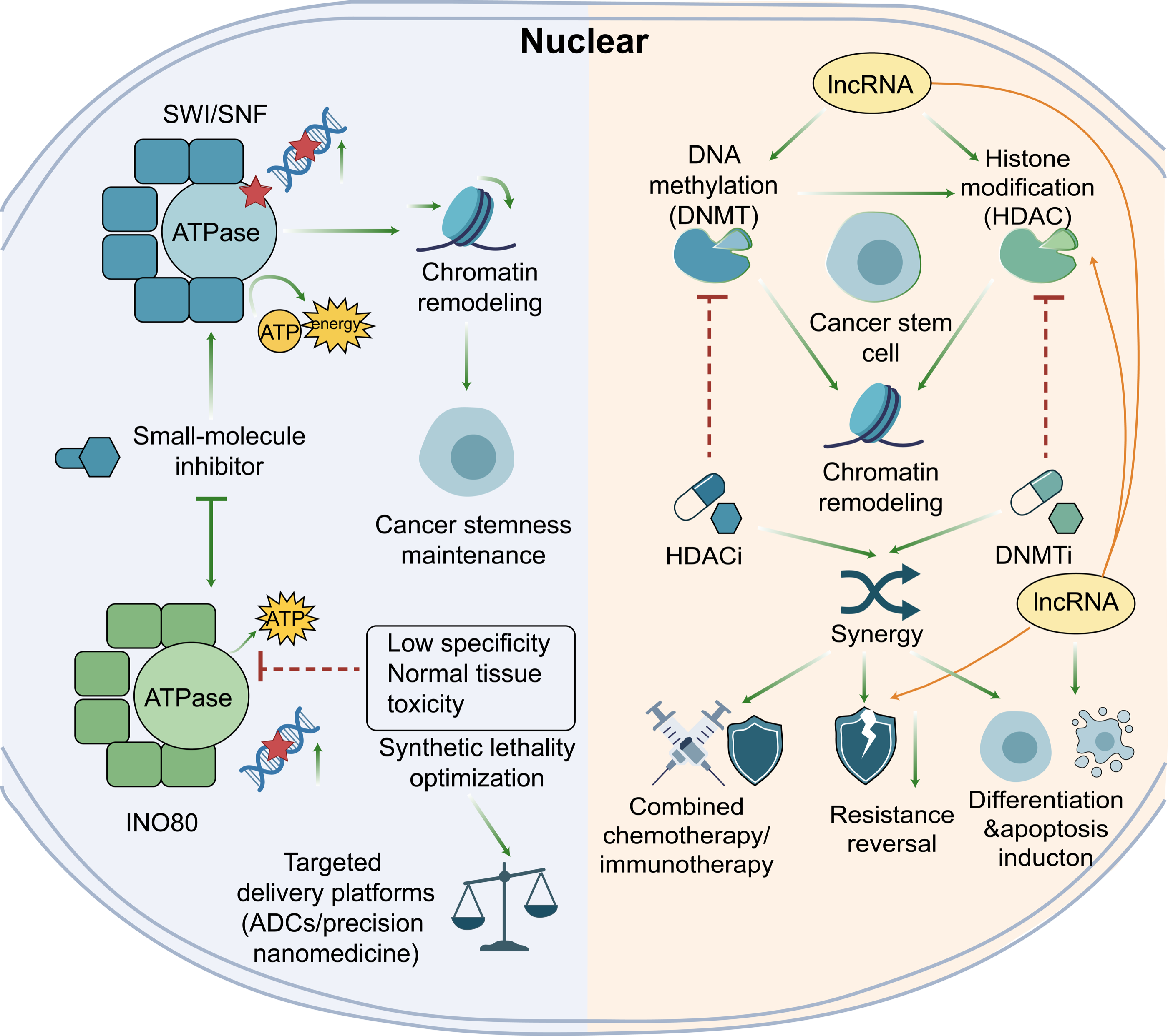
Developing small-molecule inhibitors that directly target the ATPase activity of chromatin-remodeling complexes is an important direction in cancer therapy, including strategies to disrupt CSC-associated dependencies (14). Chromatin-remodeling complexes, such as SWI/SNF, INO80, ISWI, and members of the CHD family, rely on conserved SNF2-like catalytic ATPase subunits to hydrolyze ATP, providing energy for nucleosome sliding, eviction, assembly, or editing and thereby regulating chromatin structure and gene expression (18, 87). Experimental CHD1 studies connect ATP-dependent remodeling with chromatin organization and pluripotency in embryonic stem cells (13), while broader CSC literature describes context-dependent relationships between cell state and tumor propagation (19). In cancer, alterations in genes encoding these complexes can disrupt transcriptional programs and differentiation, although their consequences depend on tumor lineage, genotype, and complex composition (14). Direct ATPase inhibition may therefore promote differentiation or cell death in tumors that retain a functional dependence on the targeted remodeler. Identifying this dependence is essential because loss of remodeling activity can itself contribute to tumorigenesis in other contexts. Target expression alone is not sufficient evidence of dependence; functional perturbation and confirmation of the relevant molecular response are needed to justify therapeutic targeting.
FHD-286 is an orally bioavailable allosteric dual inhibitor of BRG1/BRM ATPase activity with antitumor activity in preclinical models (88). These findings support the pharmacological tractability of these ATPases, while leaving CSC selectivity and the determinants of treatment response to be established. Evaluation should connect drug exposure and target inhibition with changes in transcription, cellular state, and tumor-propagating function. INO80 represents a separate investigational direction: experiments in HCT116 cells demonstrated reciprocal regulation between INO80-complex components and miR-372, but did not evaluate a selective small-molecule INO80 inhibitor (89). Structural and mechanistic analyses provide additional insight into INO80-family complexes and their substrate interactions without establishing clinical inhibition (90). Revumenib, which inhibits the menin–KMT2A interaction rather than a chromatin-remodeler ATPase, has shown clinical activity in genetically defined acute leukemias (91). Its therapeutic activity illustrates the value of targeting a specific epigenetic dependency, while remaining mechanistically distinct from direct ATPase inhibition.
Selectivity and tolerability remain major challenges because chromatin-remodeling ATPases support essential cellular processes (14, 92). Biochemical selectivity must therefore be distinguished from a therapeutic window between malignant and normal tissues: inhibiting an intended target can still cause toxicity by disrupting its physiological functions. SRCAP depletion impaired replication dynamics in PC3 cells, demonstrating a replication-associated function rather than clinical toxicity in normal tissues (93). A phase I study of FHD-286 in 40 patients with advanced myeloid malignancies reported differentiation and reductions in leukemic burden, but no objective responses; differentiation syndrome was an important safety finding (94). These results emphasize the need to distinguish pharmacodynamic activity from durable clinical benefit and to selectively deplete tumor-propagating populations. Future assessments should also examine response duration and whether treatment-associated differentiation persists after drug withdrawal.
Targeted delivery remains a separate preclinical strategy. CD133-targeted PLGA nanoparticles carrying paclitaxel reduced mammosphere and colony formation in vitro and suppressed tumor regrowth compared with free paclitaxel in a breast cancer xenograft model, providing preclinical evidence for marker-directed drug delivery (95). Nanoparticle delivery of HDAC inhibitors provides additional formulation rationale, although HDAC inhibitors are mechanistically distinct from ATP-dependent remodeler inhibitors (96). Another CSC-associated vulnerability involves CD44 variant-dependent stabilization of xCT, which supports redox defense and tumor growth; this finding does not demonstrate targeted delivery of a remodeler inhibitor (97). Establishing a therapeutic window will require payload-specific delivery studies, confirmation of target engagement, and direct comparison of malignant and normal-cell dependencies. Preferential uptake into marker-positive cells should also be distinguished from selective activity against functionally defined CSCs. Functional and longitudinal assessments will be needed to determine whether treatment eliminates tumor-propagating capacity or produces a temporary change in cell state (Figure 6).
Combination Therapy Strategies
CSC-associated populations can contribute to tumor maintenance, treatment resistance, and recurrence through self-renewal and cell-state plasticity (98). Epigenetic regulation provides a rationale for investigating combinations that alter these properties, although different drug classes act through distinct molecular mechanisms (12). Reviews of colorectal epigenetic therapy describe potential therapeutic approaches (99), while glioblastoma chromatin-insulation biology provides a separate framework for investigating transcriptional vulnerabilities (22). These observations support experimental evaluation of combinations, but do not establish universal synergy, selective CSC elimination, or prevention of recurrence (84) (Figure 6).
Epigenetic therapies differ in their molecular targets and levels of clinical validation. HDAC inhibitors have clinical roles in selected hematologic malignancies (100), while decitabine-based treatment is used in myelodysplastic syndromes and related myeloid disorders (101). Dual BRG1/BRM ATPase inhibition has shown preclinical activity in AML models (102), whereas menin inhibition has demonstrated clinical activity through a separate molecular mechanism (91). Reviews of colorectal epigenetic strategies and DNMT inhibitors provide a rationale for combination studies, but do not establish a universal synergistic or CSC-selective effect of HDACi/DNMTi coadministration (99, 103). Such combinations require direct evaluation of dose, schedule, target engagement, tumor-propagating capacity, and normal-tissue effects.
Epigenetic interventions have been investigated in pediatric nervous-system tumors and CSC-associated experimental systems, but the evidence is heterogeneous and largely preclinical (104). Combining lncRNA-directed approaches with other epigenetic agents is therefore a testable therapeutic hypothesis. Demonstrating benefit will require distinguishing selective effects on tumor-propagating populations from broader cytotoxic or differentiation responses and establishing an acceptable therapeutic window (Figure 6).
A further distinction is needed between clinically validated epigenetic therapy and clinically validated CSC-directed chromatin-remodeling therapy. HDAC and DNMT inhibitors have established clinical roles in selected hematologic malignancies, and other epigenetic agents have entered clinical development, but these observations do not demonstrate that their clinical activity results from CSC depletion. For most ATP-dependent chromatin remodelers discussed here, evidence remains at the mechanistic or preclinical stage. Future trials will therefore need pharmacodynamic biomarkers that demonstrate modulation of the intended remodeling complex, along with longitudinal measures that determine whether stem-like tumor populations are selectively depleted.
Clinical translation is further complicated by the absence of a validated biomarker that uniquely identifies CSCs across tumor types. Markers such as CD44, CD133, and ALDH activity can enrich for tumor-propagating populations in particular settings but are neither universal nor exclusive to CSCs. Consequently, reducing marker-positive cells alone cannot establish CSC eradication. Translational studies should combine phenotypic markers with functional assays, molecular evidence of target engagement, and longitudinal assessment of recurrence or tumor-repopulating capacity.
Discussion
The frequency, proliferative state, and phenotypic definition of functionally identified CSCs vary across tumors and experimental systems, complicating their isolation and comparison. First, isolating CSCs with high purity from complex tumor cell populations is challenging. Traditional surface marker-based sorting methods often fail to capture all CSC subpopulations, and the sorting process may alter the cells' original state (19). Paired single-cell profiling can connect chromatin accessibility with gene expression. SHARE-seq demonstrated this capability and provided a framework for investigating regulatory changes associated with cell-state transitions (76). Its application to CSCs requires appropriate tumor-cell annotation and functional validation. For example, single-cell ATAC-seq can reveal heterogeneity in chromatin accessibility, but when applied to rare cells like CSCs, it still requires sufficient sequencing depth and coverage to capture key regulatory elements (29). Additionally, CSC chromatin states are not static; they undergo rapid reprogramming in response to therapeutic pressure, microenvironmental signals, or different stages of the cell cycle. This requires research methods with high temporal resolution to capture these dynamic changes (5). Therefore, developing platforms capable of non-destructive, high-precision CSC sorting, combined with time-resolved single-cell multi-omics analysis, is an important direction for overcoming technical bottlenecks and gaining a deeper understanding of the chromatin basis of CSCs in the future.
To target CSCs more precisely, it is essential to analyze mechanisms of action, upstream and downstream regulatory networks, and the association of different chromatin-remodeling complexes with three-dimensional chromatin conformation in specific CSC types. Research indicates that distinct remodeling complexes play unique and intricate roles in maintaining CSC characteristics. In TNBC models, MUC1-C-associated induction of NuRD components illustrates how oncogenic signaling can engage chromatin-regulatory machinery to suppress differentiation-associated transcription (26).
Chromatin-remodeling complexes participate in regulatory networks involving transcription factors and non-coding RNAs. In liver cancer stem-cell models, lncBRM promoted self-renewal through BRM/BRG1-associated remodeling and activation of YAP1 signaling (16). More importantly, chromatin remodeling is closely related to changes in three-dimensional genome structure. Three-dimensional genome organization provides another layer of gene regulation, but changes in accessibility do not automatically demonstrate altered TADs or enhancer–promoter contacts. Establishing the contribution of a specific remodeling complex requires appropriate perturbation and chromosome-conformation measurements (29). Genome-wide RNAi screens identified PHF5A as differentially required for the viability of patient-derived glioblastoma stem cells through its role in splice-site recognition, with additional effects on tumor formation and maintenance (105). This identifies a splicing-associated vulnerability rather than demonstrating that PHF5A regulates CSC growth by remodeling three-dimensional chromatin. Combining perturbation screens with accessibility and chromosome-conformation measurements could help distinguish these mechanisms.
Defining a therapeutic window requires comparison of malignant and nonmalignant dependencies. Inhibition of an essential normal-cell function can produce on-target toxicity even when an agent is biochemically selective; this should be distinguished from inhibition of unintended molecular targets. PHF5A experiments illustrate the value of comparing glioblastoma stem cells with untransformed cells, without establishing systemic clinical safety (105). For direct BRG1/BRM inhibition, the FHD-286 phase I study demonstrates the importance of evaluating tolerability and differentiation syndrome alongside pharmacodynamic activity and clinical response (94). Low abundance and limited sequence conservation can also complicate the development of lncRNA-directed interventions (32).
Synthetic lethality provides a framework for identifying dependencies created by defined genetic alterations (106). The relevant residual remodeling complex, subunit or paralog must be specified, and any therapeutic window requires comparison with normal populations. Tumor-specific dependency and localized delivery are complementary research strategies, but neither should be assumed to spare normal stem cells or selectively eliminate CSCs without direct evidence. Upstream oncogenic regulators may provide additional routes for investigating chromatin-associated dependencies. In TNBC models, MUC1-C promoted NuRD-component expression and dedifferentiation-associated transcriptional regulation (26). These findings provide a mechanistic rationale for further investigation, but do not establish selective clinical depletion of CSCs. Tumor-localized delivery is a potential strategy for modifying drug exposure, but its benefits must be demonstrated for the specific payload and formulation. A CD133-targeted paclitaxel nanoparticle study provides a preclinical delivery example, although its findings do not establish delivery or reduced toxicity of a chromatin-remodeler inhibitor (95). Similarly, HDAC-inhibitor nanomedicine offers formulation examples for a distinct drug class (96). Delivery of direct remodeler inhibitors will require payload-specific studies of distribution, target engagement, efficacy, and safety, including assessment of brain exposure where relevant.
Clinical development will require molecular stratification rather than treating chromatin remodeling as a tumor-agnostic vulnerability. CSC maintenance is closely linked to functional abnormalities in chromatin remodeling complexes. Mutations or expression-profile differences in these complexes are an important basis for tumor heterogeneity. In IDH-mutant gliomas, DNA hypermethylation can disrupt chromatin insulation and alter oncogenic regulatory interactions (22). These observations support mechanism-specific investigation of molecular stratification rather than establishing IDH status as a predictor of response to every CSC-directed chromatin therapy. Distinct mechanisms require distinct therapeutic interpretations. PBAF perturbation increased sensitivity to T-cell-mediated killing in melanoma models (86), whereas PHF5A dependence in glioblastoma stem cells reflected a vulnerability in RNA splicing (105). Menin inhibition represents a separate strategy supported by clinical evidence in KMT2A-rearranged or NPM1-mutant leukemia (91). These findings should not be presented as interchangeable examples of PHD-reader or ATP-dependent chromatin-remodeler inhibition.
Circulating epigenetic signals and lncRNA profiles remain candidate biomarkers rather than established universal measures of CSC burden. Literature linking lncRNAs with epithelial–mesenchymal plasticity and stemness provides a rationale for investigation, but clinical biomarker performance requires separate validation (32). Detecting a circulating methylation pattern or lncRNA does not by itself establish the abundance or tumor-propagating capacity of CSCs. Proposed assays should be evaluated against matched tissue states, functional tumor-propagating capacity, and clinical outcomes before being used to direct treatment (98).
Single-cell and spatial approaches offer opportunities to investigate tumor-state regulation and microenvironmental interactions, as illustrated by studies of Ras-driven tumors and colorectal cancer metastasis (107, 108). In glioblastoma, ATAC-seq of isolated stem-cell populations identified regulatory signatures associated with migration and established a functional role for TEAD1; this was not a single-cell ATAC-seq study or the source of patient-survival subgrouping (109). Targeted protein degradation provides a separate preclinical strategy: the PROTAC ACBI1 degraded the BAF-associated proteins SMARCA2, SMARCA4, and PBRM1 and exposed context-dependent cancer vulnerabilities, without demonstrating CSC-selective clinical efficacy (110). In bladder cancer models, the HDAC inhibitor entinostat promoted antitumor immune responses through immune editing of tumor neoantigens and supported investigation of combinations with immune checkpoint blockade (111). Related prostate cancer work examined ketogenic-diet-associated epigenetic and immune changes in combination-treatment settings (112). These findings support further investigation of distinct therapeutic approaches, but do not establish selective clinical CSC depletion by ATP-dependent chromatin-remodeler inhibitors.
The evidence reviewed here spans biochemical studies, established cell lines, tumorsphere models, genetically engineered systems, patient-derived cultures, xenografts, and early clinical studies. These levels of evidence should not be interpreted equivalently. Direct perturbation followed by functional assessment of tumor initiation provides stronger support for a CSC dependency than expression correlations or marker enrichment, whereas clinical activity of an epigenetic drug does not establish CSC-specific targeting without appropriate pharmacodynamic evidence. Maintaining this distinction will be important as chromatin-remodeling biology moves from mechanistic studies toward therapeutic development.
Conclusion
Chromatin remodeling contributes to transcriptional programs associated with cancer stemness, plasticity, and treatment resistance. These effects depend on tumor lineage, genotype and remodeling-complex composition. Genetic alterations and epigenetic regulation interact in shaping malignant cell states; functional perturbation is needed to distinguish a therapeutically relevant dependency from expression or accessibility associations. Targeting context-specific chromatin dependencies may offer opportunities to influence tumor-propagating states and treatment resistance. However, preclinical activity and early clinical pharmacodynamic effects should be distinguished from durable clinical benefit or selective CSC depletion. Therapeutic development must account for differences in tumor genotype, remodeling-complex composition, normal-tissue requirements, drug delivery, and adaptive cell-state transitions.
Future studies should integrate longitudinal molecular profiling with functional assessment of tumor-propagating capacity in appropriate experimental models and clinical samples. Direct ATPase inhibitors, targeted protein degraders, and combinations with other epigenetic or immune therapies represent distinct strategies requiring mechanism-specific validation. Their clinical value will depend on demonstrating meaningful benefit within an acceptable therapeutic window.
References
1. Schepers AG, Snippert HJ, Stange DE, van den Born M, van Es JH, van de Wetering M, et al. Lineage tracing reveals Lgr5+ stem cell activity in mouse intestinal adenomas. Science. 2012;337(6095):730–735. https://doi.org/10.1126/science.1224676
2. Dakal TC, Bhushan R, Xu C, Gadi BR, Cameotra SS, Yadav V, et al. Intricate relationship between cancer stemness, metastasis, and drug resistance. MedComm (2020). 2024;5(10):e710. https://doi.org/10.1002/mco2.710
3. Chaffer CL, Marjanovic ND, Lee T, Bell G, Kleer CG, Reinhardt F, et al. Poised chromatin at the ZEB1 promoter enables breast cancer cell plasticity and enhances tumorigenicity. Cell. 2013;154(1):61–74. https://doi.org/10.1016/j.cell.2013.06.005
4. Eustermann S, Patel AB, Hopfner KP, He Y, Korber P. Energy-driven genome regulation by ATP-dependent chromatin remodellers. Nat Rev Mol Cell Biol. 2024;25(4):309–332. https://doi.org/10.1038/s41580-023-00683-y
5. Chen HM, Nikolic A, Singhal D, Gallo M. Roles of chromatin remodelling and molecular heterogeneity in therapy resistance in glioblastoma. Cancers (Basel). 2022;14(19):4942. https://doi.org/10.3390/cancers14194942
6. Shi J, Whyte WA, Zepeda-Mendoza CJ, Milazzo JP, Shen C, Roe JS, et al. Role of SWI/SNF in acute leukemia maintenance and enhancer-mediated Myc regulation. Genes Dev. 2013;27(24):2648–2662. https://doi.org/10.1101/gad.232710.113
7. Portney BA, Arad M, Gupta A, Brown RA, Khatri R, Lin PN, et al. ZSCAN4 facilitates chromatin remodeling and promotes the cancer stem cell phenotype. Oncogene. 2020;39(26):4970–4982. https://doi.org/10.1038/s41388-020-1333-1
8. Suvà ML, Rheinbay E, Gillespie SM, Patel AP, Wakimoto H, Rabkin SD, et al. Reconstructing and reprogramming the tumor-propagating potential of glioblastoma stem-like cells. Cell. 2014;157(3):580–594. https://doi.org/10.1016/j.cell.2014.02.030
9. Lu X, Maturi NP, Jarvius M, Yildirim I, Dang Y, Zhao L, et al. Cell-lineage controlled epigenetic regulation in glioblastoma stem cells determines functionally distinct subgroups and predicts patient survival. Nat Commun. 2022;13(1):2236. https://doi.org/10.1038/s41467-022-29912-2
10. Bhattacharya A, Fushimi A, Yamashita N, Hagiwara M, Morimoto Y, Rajabi H, et al. MUC1-C dictates JUN and BAF-mediated chromatin remodeling at enhancer signatures in cancer stem cells. Mol Cancer Res. 2022;20(4):556–567. https://doi.org/10.1158/1541-7786.MCR-21-0672
11. Hagiwara M, Fushimi A, Yamashita N, Bhattacharya A, Rajabi H, Long MD, et al. MUC1-C activates the PBAF chromatin remodeling complex in integrating redox balance with progression of human prostate cancer stem cells. Oncogene. 2021;40(30):4930–4940. https://doi.org/10.1038/s41388-021-01899-y
12. Ghasemi S, Xu S, Nabavi SM, Amirkhani MA, Sureda A, Tejada S, et al. Epigenetic targeting of cancer stem cells by polyphenols (cancer stem cells targeting). Phytother Res. 2021;35(7):3649–3664. https://doi.org/10.1002/ptr.7059
13. Gaspar-Maia A, Alajem A, Polesso F, Sridharan R, Mason MJ, Heidersbach A, et al. Chd1 regulates open chromatin and pluripotency of embryonic stem cells. Nature. 2009;460(7257):863–868. https://doi.org/10.1038/nature08212
14. Malone HA, Roberts CWM. Chromatin remodellers as therapeutic targets. Nat Rev Drug Discov. 2024;23(9):661–681. https://doi.org/10.1038/s41573-024-00978-5
15. Sun F, Zou B, Li H, Xu C, Luo Q, Wang C, et al. Structural basis for BCL7B-mediated ncBAF-nucleosome engagement. Nucleic Acids Res. 2026;54(4):gkag092. https://doi.org/10.1093/nar/gkag092
16. Zhu P, Wang Y, Wu J, Huang G, Liu B, Ye B, et al. LncBRM initiates YAP1 signalling activation to drive self-renewal of liver cancer stem cells. Nat Commun. 2016;7:13608. https://doi.org/10.1038/ncomms13608
17. Bonilla B, Patty BJ, Sambare SV, Dekker J, Fazzio TG, Hainer SJ. esBAF and INO80C fine-tune subcompartments and differentially regulate enhancer-promoter interactions. Genetics. 2026;232(4):iyag037. https://doi.org/10.1093/genetics/iyag037
18. Sahu RK, Singh S, Tomar RS. The mechanisms of action of chromatin remodelers and implications in development and disease. Biochem Pharmacol. 2020;180:114200. https://doi.org/10.1016/j.bcp.2020.114200
19. Oliver L, Lalier L, Salaud C, Heymann D, Cartron PF, Vallette FM. Drug resistance in glioblastoma: are persisters the key to therapy? Cancer Drug Resist. 2020;3(3):287–301. https://doi.org/10.20517/cdr.2020.29
20. Bao S, Wu Q, McLendon RE, Hao Y, Shi Q, Hjelmeland AB, et al. Glioma stem cells promote radioresistance by preferential activation of the DNA damage response. Nature. 2006;444(7120):756–760. https://doi.org/10.1038/nature05236
21. Li Y, Gong H, Wang P, Zhu Y, Peng H, Cui Y, et al. The emerging role of ISWI chromatin remodeling complexes in cancer. J Exp Clin Cancer Res. 2021;40(1):346. https://doi.org/10.1186/s13046-021-02151-x
22. Sesé B, Ensenyat-Mendez M, Iñiguez S, Llinàs-Arias P, Marzese DM. Chromatin insulation dynamics in glioblastoma: challenges and future perspectives of precision oncology. Clin Epigenetics. 2021;13(1):150. https://doi.org/10.1186/s13148-021-01139-w
23. Cui T, Bell EH, McElroy J, Liu K, Sebastian E, Johnson B, et al. A novel miR-146a-POU3F2/SMARCA5 pathway regulates stemness and therapeutic response in glioblastoma. Mol Cancer Res. 2021;19(1):48–60. https://doi.org/10.1158/1541-7786.MCR-20-0353
24. Mills AA. The chromodomain helicase DNA-binding chromatin remodelers: family traits that protect from and promote cancer. Cold Spring Harb Perspect Med. 2017;7(4):a026450. https://doi.org/10.1101/cshperspect.a026450
25. Li S, Ma Q, Lian K, Jiang Z, Ma Y. Role of CHD4 in tumor progression, DNA damage response and treatment resistance (Review). Oncol Rep. 2026;55(6):107. https://doi.org/10.3892/or.2026.9112
26. Hata T, Rajabi H, Takahashi H, Yasumizu Y, Li W, Jin C, et al. MUC1-C activates the NuRD complex to drive dedifferentiation of triple-negative breast cancer cells. Cancer Res. 2019;79(22):5711–5722. https://doi.org/10.1158/0008-5472.CAN-19-1034
27. Xiao G, Lu W, Yuan J, Liu Z, Wang P, Fan H. Fbxw7 suppresses carcinogenesis and stemness in triple-negative breast cancer through CHD4 degradation and Wnt/β-catenin pathway inhibition. J Transl Med. 2024;22(1):99. https://doi.org/10.1186/s12967-024-04897-2
28. Barjij I, Meliani M. Cancer stem cells and post-therapy tumour recurrence: a systematic review of mechanistic pathways and translational gaps. Ecancermedicalscience. 2025;19:2016. https://doi.org/10.3332/ecancer.2025.2016
29. Feng Y, Liu X, Pauklin S. 3D chromatin architecture and epigenetic regulation in cancer stem cells. Protein Cell. 2021;12(6):440–454. https://doi.org/10.1007/s13238-020-00819-2
30. Zhou W, Liu X, Li Z, Jia B, Lei X, Sun K, et al. Epigenetic crosstalk between stem cells and tumors: mechanisms and emerging perspectives. Am J Stem Cells. 2025;14(3):98–133. https://doi.org/10.62347/gqzh3508
31. Yu H, Wang J, Lackford B, Bennett B, Li JL, Hu G. INO80 promotes H2A.Z occupancy to regulate cell fate transition in pluripotent stem cells. Nucleic Acids Res. 2021;49(12):6739–6755. https://doi.org/10.1093/nar/gkab476
32. Yuan Y, Tang Y, Fang Z, Wen J, Wicha MS, Luo M. Long non-coding RNAs: key regulators of tumor epithelial/mesenchymal plasticity and cancer stemness. Cells. 2025;14(3):227. https://doi.org/10.3390/cells14030227
33. Wang Y, Zhu P, Luo J, Wang J, Liu Z, Wu W, et al. LncRNA HAND2-AS1 promotes liver cancer stem cell self-renewal via BMP signaling. EMBO J. 2019;38(17):e101110. https://doi.org/10.15252/embj.2018101110
34. Rawal Y, Qiu H, Hinnebusch AG. Distinct functions of three chromatin remodelers in activator binding and preinitiation complex assembly. PLoS Genet. 2022;18(7):e1010277. https://doi.org/10.1371/journal.pgen.1010277
35. Loesch R, Chenane L, Colnot S. ARID2 chromatin remodeler in hepatocellular carcinoma. Cells. 2020;9(10):2152. https://doi.org/10.3390/cells9102152
36. Dreier MR, de la Serna IL. SWI/SNF chromatin remodeling enzymes in melanoma. Epigenomes. 2022;6(1):10. https://doi.org/10.3390/epigenomes6010010
37. Chakraborty P, Kurkalang S, Ghatak S, Das S, Palodhi A, Sarkar S, et al. Deep sequencing reveals recurrent somatic mutations and distinct molecular subgroups in gastric cancer in Mizo population, North East India. Genomics. 2023;115(6):110741. https://doi.org/10.1016/j.ygeno.2023.110741
38. Shah AM, Guo L, Morales MG, Jaichander P, Chen K, Huang H, et al. TWIST2-mediated chromatin remodeling promotes fusion-negative rhabdomyosarcoma. Sci Adv. 2023;9(17):eade8184. https://doi.org/10.1126/sciadv.ade8184
39. Bayona-Feliu A, Barroso S, Muñoz S, Aguilera A. The SWI/SNF chromatin remodeling complex helps resolve R-loop-mediated transcription-replication conflicts. Nat Genet. 2021;53(7):1050–1063. https://doi.org/10.1038/s41588-021-00867-2
40. Hozumi C, Iizuka A, Ikeya T, Miyata H, Maeda C, Ashizawa T, et al. Impact of mutations in subunit genes of the mammalian SWI/SNF complex on immunological tumor microenvironment. Cancer Genomics Proteomics. 2024;21(1):88–101. https://doi.org/10.21873/cgp.20432
41. Zhou D, Alver BM, Li S, Hlady RA, Thompson JJ, Schroeder MA, et al. Distinctive epigenomes characterize glioma stem cells and their response to differentiation cues. Genome Biol. 2018;19(1):43. https://doi.org/10.1186/s13059-018-1420-6
42. Gimple RC, Kidwell RL, Kim LJY, Sun T, Gromovsky AD, Wu Q, et al. Glioma stem cell-specific superenhancer promotes polyunsaturated fatty-acid synthesis to support EGFR signaling. Cancer Discov. 2019;9(9):1248–1267. https://doi.org/10.1158/2159-8290.CD-19-0061
43. Rheinbay E, Suvà ML, Gillespie SM, Wakimoto H, Patel AP, Shahid M, et al. An aberrant transcription factor network essential for Wnt signaling and stem cell maintenance in glioblastoma. Cell Rep. 2013;3(5):1567–1579. https://doi.org/10.1016/j.celrep.2013.04.021
44. Hall AW, Battenhouse AM, Shivram H, Morris AR, Cowperthwaite MC, Shpak M, et al. Bivalent chromatin domains in glioblastoma reveal a subtype-specific signature of glioma stem cells. Cancer Res. 2018;78(10):2463–2474. https://doi.org/10.1158/0008-5472.CAN-17-1724
45. Singh DK, Kollipara RK, Vemireddy V, Yang XL, Sun Y, Regmi N, et al. Oncogenes activate an autonomous transcriptional regulatory circuit that drives glioblastoma. Cell Rep. 2017;18(4):961–976. https://doi.org/10.1016/j.celrep.2016.12.064
46. Kurani H, Razavipour SF, Harikumar KB, Dunworth M, Ewald AJ, Nasir A, et al. DOT1L is a novel cancer stem cell target for triple-negative breast cancer. Clin Cancer Res. 2022;28(9):1948–1965. https://doi.org/10.1158/1078-0432.CCR-21-1299
47. Guilhamon P, Chesnelong C, Kushida MM, Nikolic A, Singhal D, MacLeod G, et al. Single-cell chromatin accessibility profiling of glioblastoma identifies an invasive cancer stem cell population associated with lower survival. Elife. 2021;10:e64090. https://doi.org/10.7554/eLife.64090
48. Yoshikawa T, Fukuda A, Omatsu M, Namikawa M, Sono M, Fukunaga Y, et al. Brg1 is required to maintain colorectal cancer stem cells. J Pathol. 2021;255(3):257–269. https://doi.org/10.1002/path.5759
49. Kadoch C, Crabtree GR. Mammalian SWI/SNF chromatin remodeling complexes and cancer: mechanistic insights gained from human genomics. Sci Adv. 2015;1(5):e1500447. https://doi.org/10.1126/sciadv.1500447
50. Singh A, Modak SB, Chaturvedi MM, Purohit JS. SWI/SNF chromatin remodelers: structural, functional and mechanistic implications. Cell Biochem Biophys. 2023;81(2):167–187. https://doi.org/10.1007/s12013-023-01140-5
51. Wagner FR, Dienemann C, Wang H, Stützer A, Tegunov D, Urlaub H, et al. Structure of SWI/SNF chromatin remodeller RSC bound to a nucleosome. Nature. 2020;579(7799):448–451. https://doi.org/10.1038/s41586-020-2088-0
52. Tseng YC, Crodian JS, Cabot R. Depletion of SMARCB1 and BRD7, two SWI/SNF chromatin remodelling complex subunits, differentially impact porcine embryo development. Reprod Fertil Dev. 2022;34(7):549–559. https://doi.org/10.1071/RD21251
53. Ganguly D, Sims M, Cai C, Fan M, Pfeffer LM. Chromatin remodeling factor BRG1 regulates stemness and chemosensitivity of glioma initiating cells. Stem Cells. 2018;36(12):1804–1815. https://doi.org/10.1002/stem.2909
54. Wanior M, Krämer A, Knapp S, Joerger AC. Exploiting vulnerabilities of SWI/SNF chromatin remodelling complexes for cancer therapy. Oncogene. 2021;40(21):3637–3654. https://doi.org/10.1038/s41388-021-01781-x
55. Centore RC, Sandoval GJ, Soares LMM, Kadoch C, Chan HM. Mammalian SWI/SNF chromatin remodeling complexes: emerging mechanisms and therapeutic strategies. Trends Genet. 2020;36(12):936–950. https://doi.org/10.1016/j.tig.2020.07.011
56. Zhao Q, Zong H, Zhu P, Su C, Tang W, Chen Z, et al. Crosstalk between colorectal CSCs and immune cells in tumorigenesis, and strategies for targeting colorectal CSCs. Exp Hematol Oncol. 2024;13(1):6. https://doi.org/10.1186/s40164-024-00474-x
57. Liu C, Li Y, Semenov M, Han C, Baeg GH, Tan Y, et al. Control of beta-catenin phosphorylation/degradation by a dual-kinase mechanism. Cell. 2002;108(6):837–847. https://doi.org/10.1016/S0092-8674(02)00685-2
58. Nordin A, Chakraborty C, Jonasson M, Dano O, Zambanini G, Pagella P, et al. Wnt signaling activation induces CTCF binding and loop formation at cis-regulatory elements of target genes. Genome Res. 2025;35(8):1701–1716. https://doi.org/10.1101/gr.279684.124
59. Zhang C, Wu S. RUVBL1-modulated chromatin remodeling alters the transcriptional activity of oncogenic CTNNB1 in uveal melanoma. Cell Death Discov. 2023;9(1):132. https://doi.org/10.1038/s41420-023-01429-7
60. Wang H, Bienz M, Yan XX, Xu W. Structural basis of the interaction between BCL9-Pygo and LDB-SSBP complexes in assembling the Wnt enhanceosome. Nat Commun. 2023;14(1):3702. https://doi.org/10.1038/s41467-023-39439-9
61. Barker N, Hurlstone A, Musisi H, Miles A, Bienz M, Clevers H. The chromatin remodelling factor Brg-1 interacts with β-catenin to promote target gene activation. EMBO J. 2001;20(17):4935–4943. https://doi.org/10.1093/emboj/20.17.4935
62. Vaicekauskaitė I, Dabkevičienė D, Šimienė J, Žilovič D, Čiurlienė R, Jarmalaitė S, et al. ARID1A, NOTCH and WNT signature in gynaecological tumours. Int J Mol Sci. 2023;24(6):5854. https://doi.org/10.3390/ijms24065854
63. Bhal S, Kundu CN. Targeting crosstalk of signaling pathways in cancer stem cells: a promising approach for development of novel anti-cancer therapeutics. Med Oncol. 2023;40(2):82. https://doi.org/10.1007/s12032-022-01905-7
64. Vasilaki E, Bai Y, Ali MM, Sundqvist A, Moustakas A, Heldin CH. ΔNp63 bookmarks and creates an accessible epigenetic environment for TGFβ-induced cancer cell stemness and invasiveness. Cell Commun Signal. 2024;22(1):411. https://doi.org/10.1186/s12964-024-01794-5
65. Gélabert C, Papoutsoglou P, Golán I, Ahlström E, Ameur A, Heldin CH, et al. The long non-coding RNA LINC00707 interacts with Smad proteins to regulate TGFβ signaling and cancer cell invasion. Cell Commun Signal. 2023;21(1):271. https://doi.org/10.1186/s12964-023-01273-3
66. Mani SA, Guo W, Liao MJ, Eaton EN, Ayyanan A, Zhou AY, et al. The epithelial-mesenchymal transition generates cells with properties of stem cells. Cell. 2008;133(4):704–715. https://doi.org/10.1016/j.cell.2008.03.027
67. Peñuelas S, Anido J, Prieto-Sánchez RM, Folch G, Barba I, Cuartas I, et al. TGF-beta increases glioma-initiating cell self-renewal through the induction of LIF in human glioblastoma. Cancer Cell. 2009;15(4):315–327. https://doi.org/10.1016/j.ccr.2009.02.011
68. Anido J, Sáez-Borderías A, Gonzàlez-Juncà A, Rodón L, Folch G, Carmona MA, et al. TGF-β receptor inhibitors target the CD44(high)/Id1(high) glioma-initiating cell population in human glioblastoma. Cancer Cell. 2010;18(6):655–668. https://doi.org/10.1016/j.ccr.2010.10.023
69. Kim K, Ryu TY, Jung E, Han TS, Lee J, Kim SK, et al. Epigenetic regulation of SMAD3 by histone methyltransferase SMYD2 promotes lung cancer metastasis. Exp Mol Med. 2023;55(5):952–964. https://doi.org/10.1038/s12276-023-00987-1
70. Birch JL, Coull BJ, Spender LC, Watt C, Willison A, Syed N, et al. Multifaceted transforming growth factor-beta (TGFβ) signalling in glioblastoma. Cell Signal. 2020;72:109638. https://doi.org/10.1016/j.cellsig.2020.109638
71. Naik A, Dalpatraj N, Thakur N. Global gene expression regulation mediated by TGFβ through H3K9me3 mark. Cancer Inform. 2022;21:11769351221115135. https://doi.org/10.1177/11769351221115135
72. Sun S, Sun Y, Lan L, Luan S, Zhou J, Deng J, et al. Tumor cell plasticity in cancer: signaling pathways and pharmaceutical interventions. MedComm (2020). 2025;6(12):e70541. https://doi.org/10.1002/mco2.70541
73. Johann PD. Invited review: dysregulation of chromatin remodellers in paediatric brain tumours – SMARCB1 and beyond. Neuropathol Appl Neurobiol. 2020;46(1):57–72. https://doi.org/10.1111/nan.12616
74. Verdikt R, Thienpont B. Epigenetic remodelling under hypoxia. Semin Cancer Biol. 2024;98:1–10. https://doi.org/10.1016/j.semcancer.2023.10.005
75. Sharma SV, Lee DY, Li B, Quinlan MP, Takahashi F, Maheswaran S, et al. A chromatin-mediated reversible drug-tolerant state in cancer cell subpopulations. Cell. 2010;141(1):69–80. https://doi.org/10.1016/j.cell.2010.02.027
76. Ma S, Zhang B, LaFave LM, Earl AS, Chiang Z, Hu Y, et al. Chromatin potential identified by shared single-cell profiling of RNA and chromatin. Cell. 2020;183(4):1103–1116.e20. https://doi.org/10.1016/j.cell.2020.09.056
77. Roesch A, Fukunaga-Kalabis M, Schmidt EC, Zabierowski SE, Brafford PA, Vultur A, et al. A temporarily distinct subpopulation of slow-cycling melanoma cells is required for continuous tumor growth. Cell. 2010;141(4):583–594. https://doi.org/10.1016/j.cell.2010.04.020
78. Chaffer CL, Brueckmann I, Scheel C, Kaestli AJ, Wiggins PA, Rodrigues LO, et al. Normal and neoplastic nonstem cells can spontaneously convert to a stem-like state. Proc Natl Acad Sci U S A. 2011;108(19):7950–7955. https://doi.org/10.1073/pnas.1102454108
79. Neftel C, Laffy J, Filbin MG, Hara T, Shore ME, Rahme GJ, et al. An integrative model of cellular states, plasticity, and genetics for glioblastoma. Cell. 2019;178(4):835–849.e21. https://doi.org/10.1016/j.cell.2019.06.024
80. Gupta PB, Fillmore CM, Jiang G, Shapira SD, Tao K, Kuperwasser C, et al. Stochastic state transitions give rise to phenotypic equilibrium in populations of cancer cells. Cell. 2011;146(4):633–644. https://doi.org/10.1016/j.cell.2011.07.026
81. Dubey R, Lebensohn AM, Bahrami-Nejad Z, Marceau C, Champion M, Gevaert O, et al. Chromatin-remodeling complex SWI/SNF controls multidrug resistance by transcriptionally regulating the drug efflux pump ABCB1. Cancer Res. 2016;76(19):5810–5821. https://doi.org/10.1158/0008-5472.CAN-16-0716
82. Zhang HB, Hu Y, Deng JL, Fang GY, Zeng Y. Insights into the involvement of long non-coding RNAs in doxorubicin resistance of cancer. Front Pharmacol. 2023;14:1243934. https://doi.org/10.3389/fphar.2023.1243934
83. Zhang J, Sun Y, Wang L, Gu J, Hua Y, Shao J, et al. HMGB3: a pivotal orchestrator of therapy resistance and cancer stemness in human malignancies (Review). Oncol Rep. 2026;55(6):104. https://doi.org/10.3892/or.2026.9109
84. Cabrera-Licona A, Pérez-Añorve IX, Flores-Fortis M, Moral-Hernández OD, González-de la Rosa CH, Suárez-Sánchez R, et al. Deciphering the epigenetic network in cancer radioresistance. Radiother Oncol. 2021;159:48–59. https://doi.org/10.1016/j.radonc.2021.03.012
85. Yoshikawa T, Fukuda A, Omatsu M, Namikawa M, Sono M, Fukunaga Y, et al. JNK pathway plays a critical role for expansion of human colorectal cancer in the context of BRG1 suppression. Cancer Sci. 2022;113(10):3417–3427. https://doi.org/10.1111/cas.15520
86. Pan D, Kobayashi A, Jiang P, Ferrari de Andrade L, Tay RE, Luoma AM, et al. A major chromatin regulator determines resistance of tumor cells to T cell-mediated killing. Science. 2018;359(6377):770–775. https://doi.org/10.1126/science.aao1710
87. Reyes AA, Marcum RD, He Y. Structure and function of chromatin remodelers. J Mol Biol. 2021;433(14):166929. https://doi.org/10.1016/j.jmb.2021.166929
88. Vaswani RG, Huang DS, Anthony N, Xu L, Centore R, Schiller S, et al. Discovery of FHD-286, a first-in-class, orally bioavailable, allosteric dual inhibitor of the Brahma homologue (BRM) and Brahma-related gene 1 (BRG1) ATPase activity for the treatment of SWItch/Sucrose Non-Fermentable (SWI/SNF) dependent cancers. J Med Chem. 2025;68(2):1772–1792. https://doi.org/10.1021/acs.jmedchem.4c02535
89. Shah JA, Miao Y, Chu J, Chen W, Zhao Q, Cai C, et al. Feedback modulation between human INO80 chromatin remodeling complex and miR-372 in HCT116 cells. Int J Mol Sci. 2023;24(13):10685. https://doi.org/10.3390/ijms241310685
90. Bowman GD. Mechanistic insights into INO80-type chromatin remodelers. Curr Opin Struct Biol. 2025;92:103030. https://doi.org/10.1016/j.sbi.2025.103030
91. Issa GC, Aldoss I, DiPersio J, Cuglievan B, Stone R, Arellano M, et al. The menin inhibitor revumenib in KMT2A-rearranged or NPM1-mutant leukaemia. Nature. 2023;615(7954):920–924. https://doi.org/10.1038/s41586-023-05812-3
92. Clapier CR, Iwasa J, Cairns BR, Peterson CL. Mechanisms of action and regulation of ATP-dependent chromatin-remodelling complexes. Nat Rev Mol Cell Biol. 2017;18(7):407–422. https://doi.org/10.1038/nrm.2017.26
93. Dzhokova SK, Hristova RH, Botev PS, Guecheva TN, Gospodinov AG. Srcap chromatin remodeler is required for efficient replication dynamics in mammalian cells. Int J Mol Sci. 2025;26(24):12189. https://doi.org/10.3390/ijms262412189
94. DiNardo CD, Fathi AT, Kishtagari A, Bhalla KN, Quintás-Cardama A, Reilly SA, et al. A phase I study of FHD-286, a dual BRG1/BRM (SMARCA4/SMARCA2) inhibitor, in patients with advanced myeloid malignancies. Clin Cancer Res. 2025;31(12):2327–2338. https://doi.org/10.1158/1078-0432.CCR-24-3790
95. Swaminathan SK, Roger E, Toti U, Niu L, Ohlfest JR, Panyam J. CD133-targeted paclitaxel delivery inhibits local tumor recurrence in a mouse model of breast cancer. J Control Release. 2013;171(3):280–287. https://doi.org/10.1016/j.jconrel.2013.07.014
96. Hafez DA, Hassanin IA, Teleb M, Khattab SN, Elkhodairy KA, Elzoghby AO. Recent advances in nanomedicine-based delivery of histone deacetylase inhibitors for cancer therapy. Nanomedicine (Lond). 2021;16(25):2305–2325. https://doi.org/10.2217/nnm-2021-0196
97. Ishimoto T, Nagano O, Yae T, Tamada M, Motohara T, Oshima H, et al. CD44 variant regulates redox status in cancer cells by stabilizing the xCT subunit of system xc(-) and thereby promotes tumor growth. Cancer Cell. 2011;19(3):387–400. https://doi.org/10.1016/j.ccr.2011.01.038
98. Batlle E, Clevers H. Cancer stem cells revisited. Nat Med. 2017;23(10):1124–1134. https://doi.org/10.1038/nm.4409
99. Deka D, Scarpa M, Das A, Pathak S, Banerjee A. Current understanding of epigenetics driven therapeutic strategies in colorectal cancer management. Endocr Metab Immune Disord Drug Targets. 2021;21(10):1882–1894. https://doi.org/10.2174/1871530321666210219155544
100. Parveen R, Harihar D, Chatterji BP. Recent histone deacetylase inhibitors in cancer therapy. Cancer. 2023;129(21):3372–3380. https://doi.org/10.1002/cncr.34974
101. Patel AA, Cahill K, Saygin C, Odenike O. Cedazuridine/decitabine: from preclinical to clinical development in myeloid malignancies. Blood Adv. 2021;5(8):2264–2271. https://doi.org/10.1182/bloodadvances.2020002929
102. Rago F, Rodrigues LU, Bonney M, Sprouffske K, Kurth E, Elliott G, et al. Exquisite sensitivity to dual BRG1/BRM ATPase inhibitors reveals broad SWI/SNF dependencies in acute myeloid leukemia. Mol Cancer Res. 2022;20(3):361–372. https://doi.org/10.1158/1541-7786.MCR-21-0390
103. Michael DC, Mehdipour P. DNA methyltransferase inhibitors in oncology: clinical progress, limitations and future directions. Epigenomics. 2026;18(4):463–491. https://doi.org/10.1080/17501911.2026.2637418
104. Freire NH, Jaeger MDC, de Farias CB, Nör C, Souza BK, Gregianin L, et al. Targeting the epigenome of cancer stem cells in pediatric nervous system tumors. Mol Cell Biochem. 2023;478(10):2241–2255. https://doi.org/10.1007/s11010-022-04655-2
105. Hubert CG, Bradley RK, Ding Y, Toledo CM, Herman J, Skutt-Kakaria K, et al. Genome-wide RNAi screens in human brain tumor isolates reveal a novel viability requirement for PHF5A. Genes Dev. 2013;27(9):1032–1045. https://doi.org/10.1101/gad.212548.112
106. Setton J, Zinda M, Riaz N, Durocher D, Zimmermann M, Koehler M, et al. Synthetic lethality in cancer therapeutics: the next generation. Cancer Discov. 2021;11(7):1626–1635. https://doi.org/10.1158/2159-8290.CD-20-1503
107. Yuan S, Stewart KS, Yang Y, Abdusselamoglu MD, Parigi SM, Feinberg TY, et al. Ras drives malignancy through stem cell crosstalk with the microenvironment. Nature. 2022;612(7940):555–563. https://doi.org/10.1038/s41586-022-05475-6
108. Ogden S, Metic N, Leylek O, Smith EA, Berner AM, Baker AM, et al. Phenotypic heterogeneity and plasticity in colorectal cancer metastasis. Cell Genom. 2025;5(7):100881. https://doi.org/10.1016/j.xgen.2025.100881
109. Tome-Garcia J, Erfani P, Nudelman G, Tsankov AM, Katsyv I, Tejero R, et al. Analysis of chromatin accessibility uncovers TEAD1 as a regulator of migration in human glioblastoma. Nat Commun. 2018;9(1):4020. https://doi.org/10.1038/s41467-018-06258-2
110. Farnaby W, Koegl M, Roy MJ, Whitworth C, Diers E, Trainor N, et al. BAF complex vulnerabilities in cancer demonstrated via structure-based PROTAC design. Nat Chem Biol. 2019;15(7):672–680. https://doi.org/10.1038/s41589-019-0294-6
111. Truong AS, Zhou M, Krishnan B, Utsumi T, Manocha U, Stewart KG, et al. Entinostat induces antitumor immune responses through immune editing of tumor neoantigens. J Clin Invest. 2021;131(16):e138560. https://doi.org/10.1172/JCI138560
112. Murphy S, Rahmy S, Gan D, Liu G, Zhu Y, Manyak M, et al. Ketogenic diet alters the epigenetic and immune landscape of prostate cancer to overcome resistance to immune checkpoint blockade therapy. Cancer Res. 2024;84(10):1597–1612. https://doi.org/10.1158/0008-5472.CAN-23-2742
Declarations
Funding Statement
This work was supported by the Scientific and Technological Innovation Project of Higher Education Institutions in Shanxi (2025L078), the Doctoral Research Startup Foundation Project of Changzhi Medical College (BS202509), and the Shanxi Provincial Basic Research Program Project (202503021212271).
Competing Interest
The authors declare no financial or personal relationships with other individuals or organizations that could inappropriately influence or bias the content of this work. All authors have read the final version of the manuscript and confirm that there are no competing interests.
Consent for Publication
Consent for publication: All authors have approved the final version of the manuscript.
Use of Artificial Intelligence Disclosure
This article was written by human contributors. Artificial intelligence-based tools were used to improve grammar, language, and readability without affecting the article's scientific content, data interpretation, or conclusions. The authors reviewed and verified all content to ensure its accuracy and integrity.
Data Availability Statement
No datasets were generated or analyzed in the current study.
Ethics approval and consent to participate
Not applicable, as this study did not involve the conduct of research.
Authors’ affiliations
1. School of Basic Medicine, Changzhi Medical College, Changzhi, Shanxi, China.
2. Department of Human Anatomy, School of Basic Medicine, Tongji Medical College, Huazhong University of Science and Technology, Wuhan, China.
3. Institute of Precision Cancer Medicine and Pathology, School of Medicine, MOE Key Laboratory of Tumor Molecular Biology, State Key Laboratory of Bioactive Molecules and Druggability Assessment, Jinan University, Guangzhou, China.
4. Department of Radiotherapy, The Third Affiliated Hospital of Kunming Medical University, Yunnan Cancer Hospital, Peking University Cancer Hospital Yunnan; Xishan District, Kunming, Yunnan, China.
5. School of Basic Medicine, Changzhi Medical College, Changzhi, Shanxi, China; MOE Key Laboratory of Tumor Molecular Biology, State Key Laboratory of Bioactive Molecules and Druggability Assessment, Institute of Precision Cancer Medicine and Pathology, School of Medicine, Jinan University, Guangzhou, China.
CRediT authorship contribution statement
Shasha Li: Conceptualization, Writing – original draft, Writing – review & editing. Huixia Zhang: Visualization, Writing – review & editing. Mengyuan Hu: Writing – review & editing. Xiaolei Huo: Investigation. Wei Xiong: Writing – original draft. Hao Zhang: Supervision, Writing – review & editing, Project administration. Yiqiang Zhang: Supervision, Writing – review & editing. All authors reviewed and approved the final manuscript.
ORCID ID
Shasha Li: https://orcid.org/0009-0002-0826-8311
Huixia Zhang: https://orcid.org/0000-0002-1223-0634
Mengyuan Hu: https://orcid.org/0009-0009-7255-8365
Xiaolei Huo: https://orcid.org/0009-0004-6742-6572
Wei Xiong: https://orcid.org/0000-0002-4899-6332
Hao Zhang: https://orcid.org/0000-0002-2860-5912
Yiqiang Zhang: https://orcid.org/0000-0001-7400-7991