Microbiome-Modulating Nanoplatforms for Breast Cancer Therapy
1 Next-Generation Translational Nanomedicine Laboratory, Department of Pharmaceutical Sciences, Dr. Harisingh Gour Vishwavidyalaya (A Central University), Sagar, Madhya Pradesh, 470003, India
2 Graphic Era Hill University, Dehradun, 248002, India
3 School of Allied Medical Sciences, Lovely Professional University, Phagwara, Punjab, India
4 Faculty of Data Science and Information Technology, INTI International University, Nilai, Malaysia
5 Department of Pharmacognosy, Delhi Pharmaceutical Sciences and Research University, Pushp Vihar, New Delhi 110017, India
6 Biotechnology Research Center, Pharmaceutical Technology Institute, Mashhad University of Medical Sciences, Mashhad, Iran
7 Applied Biomedical Research Center, Basic Sciences Research Institute, Mashhad University of Medical Sciences, Mashhad, Razavi Khorasan, Iran
Correspondence: Prashant Kesharwani (prashantdops@gmail.com)
Received: May 8, 2026
Accepted: August 11, 2026
Published: November 15, 2026
© 2026 The Author(s). Published by GCINC Press, Spokane, Washington, United States. Open Access licensed under a Creative Commons Attribution 4.0 International License (CC BY 4.0), which permits unrestricted use, distribution, and reproduction in any medium, provided the original author(s) and source are credited. To view a copy of this license, visit: Creative Commons Attribution 4.0 International License (CC BY 4.0)
Abstract
Breast tumors comprise interacting malignant, stromal, immune, and microbial compartments, while gut microbial communities can influence systemic immunity, drug metabolism, and therapeutic response. Emerging evidence therefore positions the microbiome as a potential modifier of breast cancer progression and treatment efficacy rather than merely a passive correlate of disease. Nanotechnology offers experimental approaches to couple tumor-directed drug delivery with selective manipulation of intratumoral or intestinal microbiota. Liposomal, lipid-based, inorganic, biomimetic, and biohybrid platforms have been used to reduce tumor-associated bacteria, alter microbial communities or metabolites, and modify antitumor immune responses. Several platforms further combine microbiota modulation with chemotherapy, immunotherapy, ferroptosis induction, or externally activated treatment. However, improved tumor control cannot necessarily be attributed to microbial modulation because many of these systems independently alter drug exposure, tumor-cell viability, oxidative stress, or immune activity. Evidence for microbiota dependence remains largely preclinical and is frequently derived from experimentally colonized or bacteria-enriched tumor models. Low microbial biomass, contamination risk, interpatient microbiome heterogeneity, methodological variability, ecological consequences of microbial depletion, manufacturing complexity, and the absence of clinical validation further constrain translation. This review critically evaluates microbiome-modulating nanoplatforms in breast cancer, distinguishes microbiota-associated from microbiota-dependent therapeutic effects, and defines the mechanistic, analytical, and clinical evidence needed to establish microbiome targeting as a viable component of precision breast cancer therapy.
Keywords
Breast cancer; intratumoral microbiome; gut microbiome; nanomedicine; microbiome-targeted therapy; therapeutic resistance; tumor microenvironment; precision oncology.
1. Introduction
Breast cancer remains a major contributor to cancer morbidity and mortality worldwide. Therapeutic challenges are particularly evident in triple-negative breast cancer (TNBC), where the absence of estrogen receptor, progesterone receptor, and HER2 expression limits established receptor-directed treatment options and contributes to substantial molecular and clinical heterogeneity (1–3). Traditionally, tumor biology has been interpreted primarily through genetic alterations and dysregulated cellular processes (4). Tumors comprise interacting malignant, stromal, immune, and microbial components that can influence disease progression and therapeutic response (5, 6).
The role of microorganisms in carcinogenesis has been recognized for centuries (7). Historical observations of tumor regression following bacterial infections prompted William B. Coley to investigate bacterial preparations as experimental cancer treatments. His early clinical reports described the use of bacterial products in patients with inoperable malignancies, providing a historical foundation for subsequent investigations of bacteria-based cancer therapy (7–9). In contrast, the identification of microorganisms capable of causing cancer, such as the Rous Sarcoma Virus and Epstein-Barr virus, indicates that microorganisms may initiate and promote tumorigenesis (10–12).
Next-generation sequencing and culture-independent methods have identified microbial signatures in multiple tumor types, challenging the historical view that tumor tissues are sterile (13, 14). Microbiota consisting of bacteria, fungi, and viruses have now been identified across a wide range of cancer types, including breast cancer (15, 16). Intratumoral microbiota may originate through translocation across disrupted epithelial barriers, migration from adjacent tissues, or hematogenous dissemination (17, 18). Notably, intratumoral microbiota exhibit tumor-type-specific heterogeneity and spatial organization within the tumor microenvironment (19, 20).
Experimental evidence indicates that at least some intratumoral microorganisms can influence tumor biology rather than functioning solely as passive residents (18). Mechanistically, microbes influence cancer progression through various pathways, such as epigenetic reprogramming, metabolic alterations, DNA damage, and modulation of oncogenic signaling (16, 21). They also shape the tumor immune microenvironment (TIME) by promoting immune evasion, regulating inflammatory responses, or enhancing antitumor immunity (22). In breast cancer, microbial dysbiosis has been increasingly associated with metastasis, tumor initiation, and resistance to chemotherapy and immunotherapy (23). Studies of human breast tissue have detected bacterial DNA and cultivable bacteria, including in samples from women without breast cancer (24, 25). Human breast-tissue profiling identified microbial differences between women with and without breast cancer, and selected bacterial isolates induced DNA damage in vitro (26, 27). Microbial differences associated with breast cancer have been reported, and selected tumor-resident bacteria promoted metastatic colonization in mouse models; these findings do not establish that microbial changes initiate breast cancer in patients (28, 29). Diet, adiposity, antibiotic exposure, hormonal status, aging, environmental exposures, and other host factors can influence microbial composition, although their specific contribution to breast-associated microbial communities remains incompletely defined (26, 28, 30).
The intratumoral microbiome has also been implicated in treatment resistance, although the magnitude and clinical relevance of this contribution appear to vary by tumor type, microbial taxa, and therapeutic context. Intratumoral bacteria can directly metabolize certain chemotherapeutic agents, as demonstrated by bacterial inactivation of gemcitabine and the resulting drug resistance in experimental tumor models (31). Microorganisms may also alter drug bioavailability and influence therapeutic penetration through additional mechanisms (32). Furthermore, gut microorganisms can disrupt immune response pathways, limiting immunotherapy efficacy (33). This emphasizes the need to discover innovative ways to specifically control microorganisms associated with malignancies. Nanotechnology has enabled the development of drug-delivery systems that can improve the pharmacokinetic properties, biodistribution, and therapeutic performance of selected anticancer agents (34). Various smart nano-delivery systems, such as lipid-, inorganic, metallic, and polymer-based nanoparticles and liposomes, can deliver drugs, regulate drug release, and optimize pharmacokinetic properties (35, 36). Beyond conventional drug delivery, nanocarrier systems offer a unique opportunity to integrate multiple therapeutic modalities such as photothermal therapy, photodynamic therapy, and immunotherapy (37, 38). Importantly, nanotechnology has provided a versatile toolkit for interacting with the tumor microbiota (23, 39).
Microbiome-directed nanoplatforms therefore represent an emerging experimental strategy in breast cancer, but their therapeutic value remains clinically unestablished (23). Engineered nanoparticles have been investigated for localized antimicrobial delivery, biofilm disruption, and modulation of microbial communities (23, 40). Whether these interventions can reproducibly overcome microbiome-associated therapeutic resistance in patients remains unknown. Preclinical platforms have combined microbiota modulation with chemotherapy, ferroptosis induction, or immune activation, with several models reporting greater antitumor activity than individual interventions (41, 42). Such results do not yet establish that microbiota modulation will provide comparable therapeutic benefit in clinically heterogeneous breast cancers. Despite these advances, challenges remain, including accurate characterization of low-biomass intratumoral microbiota, potential contamination during analysis, interpatient heterogeneity, and safety concerns associated with microbiome-targeted interventions (13, 15). Addressing these challenges will require multidisciplinary approaches integrating microbiology, oncology, immunology, and nanotechnology.
This review examines the contribution of intratumoral and gut microbiota to breast cancer progression, metastasis, and therapeutic resistance, and evaluates emerging nanoplatforms designed to manipulate these microbial compartments. This review emphasizes the strength of mechanistic evidence, the distinction between microbiota-associated and microbiota-dependent effects, and the biological and technical barriers that currently limit clinical translation.
2. Role of Microbiota in Breast Cancer
Beyond their impact on the tumor microenvironment, the gut-mammary axis expands our understanding of microbiota effects on breast cancer as a two-way communication pathway between gut microbes and host immunity and tumor biology (43, 44). Microbial short-chain fatty acids can modulate CD8+ T-cell function and enhance adoptive T-cell therapy in experimental cancer models (45, 46). Selected microbial species and their products can activate Toll-like receptors and downstream inflammatory signaling pathways, although these effects depend on the microbial species, host, and tumor context (47, 48). In breast cancer models, small extracellular vesicles derived from Fusobacterium nucleatum have been shown to promote cancer cell proliferation, migration, and invasion, as well as tumor growth and metastasis, through a TLR4-dependent mechanism (49). These findings illustrate how specific microbial products may contribute to breast cancer progression but should not be generalized to the intratumoral microbiome as a whole. Some bacterial strains, such as F. nucleatum, can stimulate epithelial-mesenchymal transition, increasing tumor invasiveness and metastasis (50, 51). In contrast, intratumoral microbiota can significantly modify the immune microenvironment. These bacteria attract and expand immunosuppressive populations, such as MDSCs and M2-like macrophages, and decrease the cytotoxic activity of CD8⁺ T cells (17, 52). These immunosuppressive changes may contribute to an immunologically “cold” tumor microenvironment and reduced responsiveness to immunotherapy (53). Experimental studies in pancreatic cancer models have demonstrated that microbial depletion can reprogram the tumor immune microenvironment, reduce immunosuppressive myeloid populations, enhance antitumor T-cell responses, and improve responsiveness to immune checkpoint blockade (54). Together with broader evidence of microbiome-immunotherapy interactions (55), these findings support microbiota-directed interventions as a potential therapeutic strategy, although their applicability to breast cancer requires further investigation.
The therapeutic relevance of breast-associated microbiota may differ across molecular subtypes and disease stages. Triple-negative, hormone receptor-positive, and HER2-positive breast cancers exhibit distinct tumor microenvironments, immune landscapes, metabolic characteristics, and treatment sensitivities, which may influence microbial colonization and host–microbe interactions. Differences between primary tumors and metastatic lesions may further affect microbial distribution, nanoparticle delivery, and therapeutic response. However, evidence establishing reproducible subtype-specific microbial targets or microbiome-dependent treatment responses remains limited. Future investigations should therefore integrate molecular subtype, disease stage, metastatic site, and treatment history into microbiome profiling and therapeutic evaluation rather than assuming that findings from individual TNBC models apply broadly to breast cancer.
Together, these observations support a functional contribution of selected intratumoral microorganisms to tumor progression and treatment response in experimental models. However, microbial effects are unlikely to be uniform across breast cancer subtypes or microbial communities, and causal evidence in patients remains limited. A central limitation of this literature is the difficulty of distinguishing microbial causation from microbial selection by the tumor microenvironment. Tumor hypoxia, altered nutrients, immune suppression, necrosis, and treatment exposure may themselves reshape microbial communities. Consequently, microbial enrichment within breast tumors does not establish that the enriched organism initiated or sustained the malignant phenotype. Longitudinal human studies and experimental systems incorporating microbial depletion, recolonization, and defined microbial consortia are needed to resolve directionality.
3. Gut Microbiota-Tumor Axis
Beyond the local tumor niche, the gut microbiome contributes to systemic regulation of breast cancer via the gut-tumor axis, which involves bidirectional communication through microbial, metabolic, and immune signaling pathways that shape tumor progression and therapeutic response (44, 56). In experimental tumor models, gut commensals enhanced dendritic-cell function and antitumor T-cell responses (57), while microbiota disruption impaired therapy-associated myeloid-cell responses (57, 58).
Key signaling molecules produced by the microbiota, such as tryptophan metabolites, bile acid metabolites, and SCFA metabolites, mediate this communication (59, 60). Microbial metabolites can also influence immune regulation: butyrate promoted peripheral regulatory T-cell generation in mice (61, 62). Moreover, recent studies have shown that microbial metabolites regulate oxidative stress and ferroptosis pathways, linking gut microbiota to cancer cell fate determination (63, 64).
Preclinical evidence links gut microbiota perturbation to nanomedicine disposition and treatment response, but the mechanisms should not be generalized across platforms. In mice bearing 4T1 breast tumors, antibiotic-mediated gut microbiota depletion increased tumor vascular permeability and intratumoral accumulation of liposomal doxorubicin, accompanied by improved therapeutic outcomes (65). Broader interactions among microbial metabolites, the tumor microenvironment, and nanomedicine have been discussed in reviews (66, 67), but direct evidence that specific metabolites determine nanoparticle penetration or distribution remains limited. Microbiota-associated immune regulation and macrophage polarization may link to nanocarrier behavior (68, 69); however, direct experimental testing is needed to assess their effects on nanoparticle uptake, retention, and clearance. Similarly, microbial effects on anticancer drug metabolism should not be interpreted as demonstrating microbial control of nanocarrier stability or payload release (70). These uncertainties support evaluating selective microbiota modulation while monitoring its ecological consequences (71). Combining microbial interventions with drug-delivery systems remains an experimental strategy (23, 44, 66), whose value will depend on demonstrating benefits beyond altered drug exposure and the independent activities of the therapeutic cargo.
Apart from breast cancer, there is a growing amount of evidence suggesting that alteration of the gut microbiota can have far-reaching systemic impact on the biology of tumors in a variety of cancers (72). Approaches like probiotics, prebiotics, diet, fecal microbiota transplantation, selective antibiotics, and microbiome-based nanotherapies can alter microbial composition and function, thereby affecting immune responses, metabolism, and inflammation (73, 74).
Specific microbiota-directed interventions have altered dendritic-cell, T-cell, NK-cell, and myeloid-cell responses in experimental cancer models, with effects varying by intervention and biological context (33, 72). These changes have accompanied improved responses to selected anticancer treatments, but increased diversity or altered taxonomic abundance does not, by itself, establish restoration of a healthy microbial state. Although this review focuses on breast cancer, related observations in other solid tumors provide mechanistic context for microbiota-directed interventions. Their relevance to breast cancer should nevertheless be established independently rather than extrapolated across tumor types.
The ecological consequences of microbiota modulation require careful consideration. Increased microbial diversity or enrichment of selected taxa should not automatically be interpreted as restoration of a healthy microbiome, because microbial functions and host effects depend on community composition, metabolic activity, and biological context. Broad microbial depletion may eliminate beneficial organisms, alter metabolite production, and promote antimicrobial resistance or opportunistic colonization. Conversely, selective microbial targeting may preserve beneficial microbial functions, although this advantage remains to be demonstrated for most microbiome-modulating nanoplatforms. Future studies should therefore assess microbial functional capacity, ecological stability, and recovery following treatment rather than relying exclusively on changes in relative taxonomic abundance.
4. Nanocarriers for Microbiota Modulation in Breast Cancer
Experimental evidence that tumor-resident bacteria can promote metastatic colonization has motivated microbiota-directed therapeutic strategies in breast cancer (29, 56). Nevertheless, traditional treatment methods that rely on broad-spectrum antibiotics remain constrained by their lack of specificity. Unnecessary use of these antibiotics destroys both harmful and harmless microorganisms, disrupts the bacterial flora, weakens the immune system, and increases resistance risk (75, 76). Repeated antibiotic exposure can produce individualized and incompletely reversible changes in the human gut microbiota (77). Nanocarrier-based approaches may offer greater spatial control over antimicrobial or microbiota-modulating interventions than systemic microbial depletion (23). Tumor-directed antimicrobial delivery may reduce exposure of non-target microbial communities compared with conventional systemic antibiotic administration. In a preclinical breast cancer model, liposomal mupirocin demonstrated activity against intratumoral F. nucleatum without an immediate detectable effect on the gut microbiome (78). Whether this selectivity persists during repeated treatment or preserves microbiome function in patients remains to be established.
Another experimental strategy integrates bacteria-assisted tumor targeting with nanoparticle-mediated chemotherapy. In breast cancer models, Bifidobacterium infantis-nanoparticle biohybrids preferentially accumulated in hypoxic tumor regions, enhanced doxorubicin delivery, and improved therapeutic outcomes (79). For example, nanoplatforms that integrate microbial targeting and ferroptosis induction can simultaneously disrupt bacterial survival and induce oxidative stress-mediated cancer cell death (23, 41). Similarly, microbiota-targeted systems can enhance immunotherapy by alleviating bacterial-induced immunosuppression and promoting immune cell activation (33). Some preclinical nanocarriers alter both intratumoral and intestinal microbial communities, raising the possibility of coordinated local and systemic effects. This dual activity may be advantageous, but it also complicates attribution of therapeutic benefit to a specific microbial compartment.
Liposomal Nanocarriers
Liposomes are clinically established anticancer drug-delivery platforms, as demonstrated by randomized trials of pegylated liposomal doxorubicin in breast cancer (80). Their application to microbiota-directed treatment remains experimental, although preclinical studies have demonstrated the feasibility of liposomal antibiotic delivery against tumor-associated bacteria (66, 78). Their lipid bilayer structure and tunable surface properties enable the encapsulation and targeted delivery of antibiotics and other bioactive agents, providing opportunities to modulate tumor-associated microbial populations (81, 82). However, preferential tumor accumulation does not necessarily confer bacterial selectivity. Although microbiota-directed nanoplatforms have demonstrated targeted intratumoral bacterial depletion and enhanced antitumor responses in preclinical breast cancer models, their ability to preserve beneficial microbial communities during repeated treatment remains to be established (23, 83).
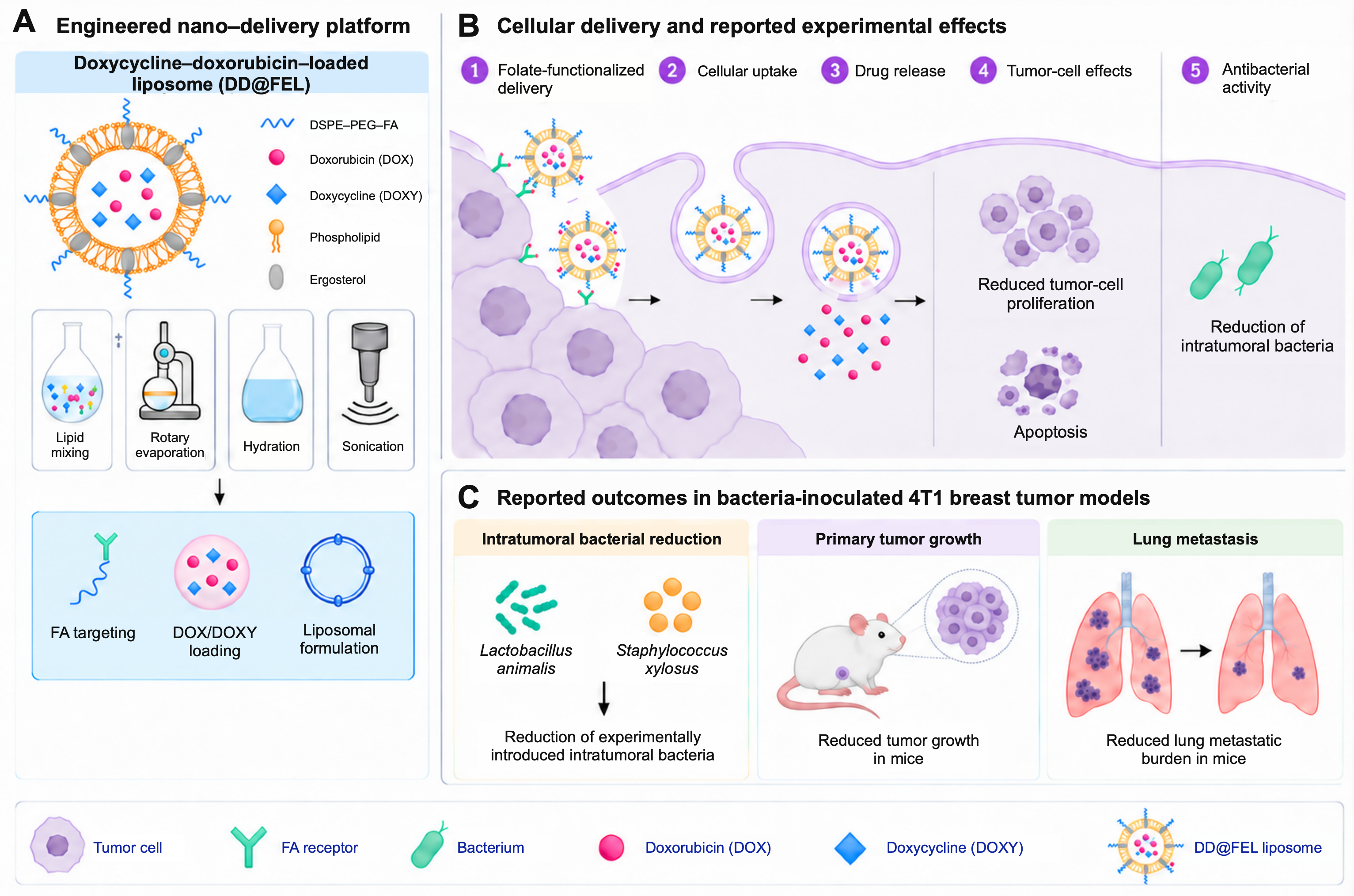
Controlled release and surface functionalization may improve local drug exposure and support microbiota-directed therapeutic strategies, although their contribution to treatment efficacy must be distinguished from improvements in drug pharmacokinetics and direct tumor-cell cytotoxicity (36, 84). An ergosterol-containing, folate-targeted liposomal nanoplatform (DD@FEL) has been developed to simultaneously target tumor cells and the intratumoral microbiota in triple-negative breast cancer (TNBC). The platform co-delivers doxycycline and doxorubicin (DOX), combining microbiota-directed antibacterial activity with DOX-mediated tumor-cell cytotoxicity. The replacement of conventional liposomal cholesterol with ergosterol introduces an additional bioactive component with reported antitumor activity. This study focused on the intratumoral microbiota and did not assess effects on the gut microbial community.
In 4T1 cells, DD@FEL showed enhanced cellular uptake associated with folate-receptor targeting and produced greater cytotoxicity and apoptosis, along with reduced proliferation and migration, compared with the corresponding single-treatment controls (85). DD@FEL also reduced the abundance of tumor-associated S. xylosus and L. animalis in the reported model, consistent with antibacterial activity against the microbial compartment examined. In the reported in vivo model, DD@FEL reduced primary tumor growth and lung metastatic burden, with reported inhibition rates of 81.6% and 80.6%, respectively (85). No major toxicity was detected using the parameters examined; however, these short-term measurements do not establish systemic or long-term safety (Figure 1).
Tumor-associated F. nucleatum has been implicated in breast cancer chemoresistance through Gal-GalNAc-mediated tumor colonization and activation of autophagy and epithelial–mesenchymal transition (EMT) programs. To counter this microbiota-associated resistance, F. nucleatum-mimicking nanovehicles (LipoFM) were engineered by incorporating bacterial membrane components into antibiotic-loaded liposomes, enabling preferential tumor targeting and localized antibacterial activity. Colistin-loaded LipoFM selectively reduced intratumoral F. nucleatum and restored chemotherapy responsiveness in experimental breast cancer models (86). Doxycycline-loaded LipoFM was developed to target additional tumor-associated bacteria. Combined with conventional chemotherapy, it reduced lung metastasis and produced complete regression of established tumors in some mice (86). However, deliberately colonized or bacteria-rich experimental models may amplify microbial contributions relative to naturally occurring human breast cancers. The prevalence, abundance, and functional importance of F. nucleatum in unselected breast tumors therefore remain important determinants of clinical applicability.
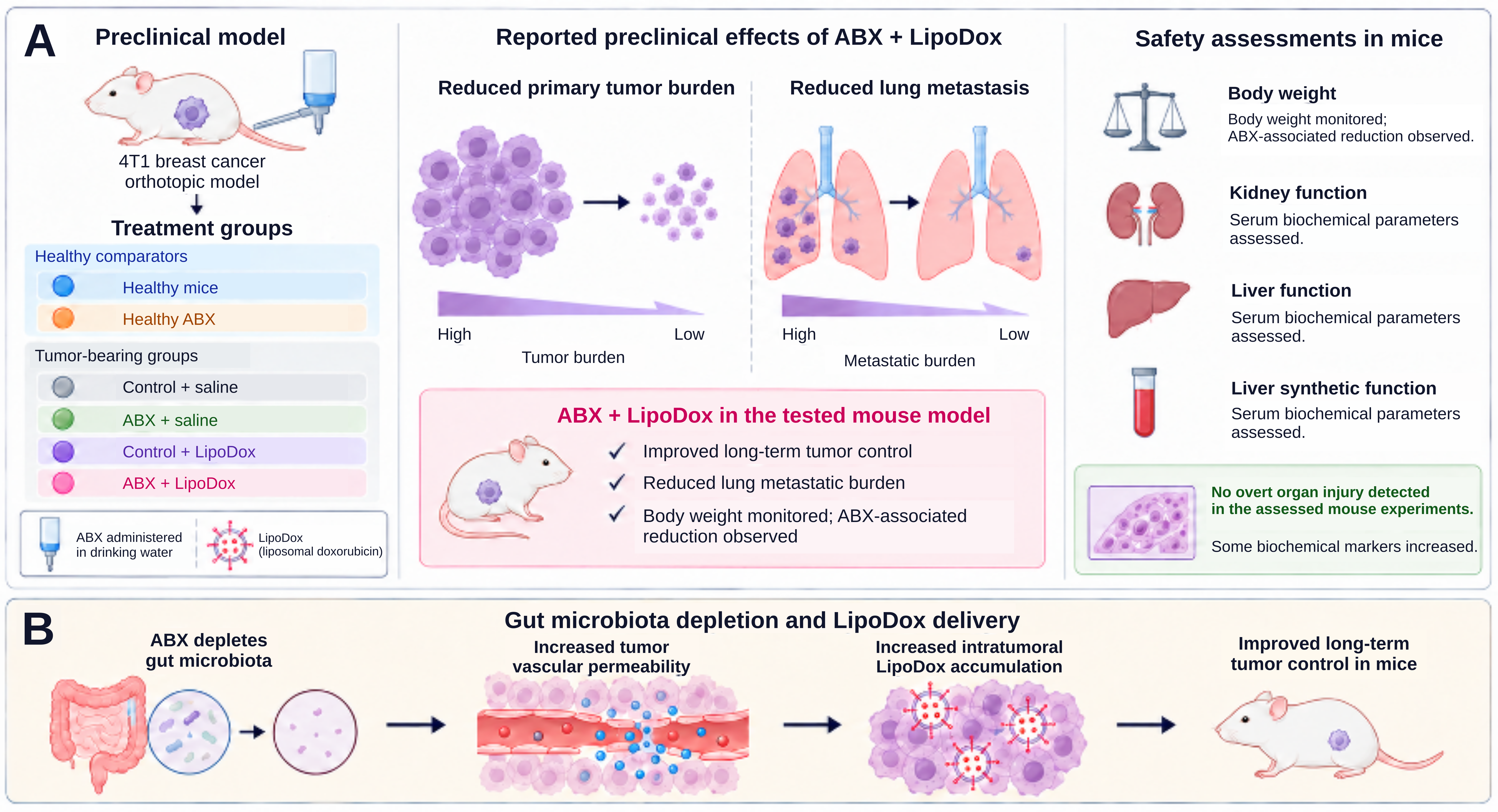
Depletion of the gut microbiota with antibiotics (ABX) has been shown to alter host interactions with nanotherapeutics and increase the intratumoral accumulation of liposomal doxorubicin (LipoDox). Microbiota depletion following ABX treatment was confirmed by RT-qPCR and 16S rRNA sequencing and was accompanied by increased tumor vascular permeability and enhanced nanoparticle penetration. These changes were associated with greater antitumor activity and improved survival following LipoDox treatment. Notably, the intratumoral microbiota remained largely unchanged, supporting a predominantly gut microbiota-associated effect on host physiology and nanoparticle delivery rather than direct remodeling of the tumor-associated microbial community (65) (Figure 2).
Nano-mupirocin is a nanoliposomal formulation of the antibiotic mupirocin designed to enable systemic administration and improve drug exposure beyond that achievable with conventional topical formulations (78). The formulation has been investigated for selective targeting of tumor-associated bacteria while limiting perturbation of the gut microbiota. Nanoparticle encapsulation improved mupirocin's pharmacokinetic profile, resulting in prolonged systemic exposure, increased plasma concentrations, and sustained drug release.
Following tumor accumulation, Nano-mupirocin exerted antibacterial activity against tumor-associated F. nucleatum and substantially reduced intratumoral bacterial burden. Microbiome profiling showed no significant changes in the assessed gut microbial diversity following treatment, in contrast to the broader microbiota alterations associated with conventional antibiotic exposure (78). A complementary approach targets bacterial mechanisms of chemotherapy resistance rather than relying exclusively on bacterial depletion. Xi et al. developed carbon nanozymes to counteract intratumoral bacteria-mediated gemcitabine resistance by inhibiting bacterial cytidine deaminase activity. In experimental tumor models, this strategy enhanced gemcitabine-associated antitumor activity, illustrating the potential of nanomaterials to interfere with microbial drug metabolism (87). However, these findings were not established in breast cancer and should be considered mechanistic evidence from other tumor models rather than direct validation of microbiome-targeted nanotherapy in breast cancer. Separately, nano-mupirocin liposomes retained in vitro activity against several multidrug-resistant bacterial strains and maintained plasma antibiotic concentrations above relevant minimum inhibitory concentrations for more than 24 hours in rats, supporting their potential for systemic antibacterial delivery (88).
Lipid-Based Nanoparticles
Lipid-based drug-delivery systems encompass solid lipid nanoparticles, nanostructured lipid carriers, and lipid–polymer hybrid nanoparticles, which can improve the stability, encapsulation, and controlled release of selected bioactive compounds (89–91). Lipid composition, surface charge, and functionalization can influence membrane interactions and cargo delivery (36, 92–94). These properties may support microbiota-directed applications, but formulation-specific evidence is needed to demonstrate microbial selectivity, physiological stability, and therapeutic benefit. Lipid-based delivery alone does not establish control of microbial communities or improved microbiome-mediated treatment outcomes (23, 44).
An RGD-functionalized solid lipid nanoparticle platform produced using microfluidic technology (MF-SLNs) has been developed for oral doxorubicin delivery. In addition to enhancing antitumor activity, MF-SLN treatment was associated with substantial changes in gut microbial composition, as assessed by 16S rRNA sequencing. These changes included increased relative abundance of genera such as Faecalibacterium and Bacillus and reduced abundance of Staphylococcus, Enterococcus, and Shigella compared with conventional doxorubicin treatment. The resulting microbial profile was consistent with attenuation of chemotherapy-associated dysbiosis, although the functional contribution of individual taxa to treatment response remains to be established. In vivo, these microbiota changes accompanied greater tumor suppression, improved survival, and reduced treatment-associated toxicity. The parallel changes in microbial composition and treatment response establish an association but do not determine whether microbiota remodeling mediated the therapeutic benefit or occurred secondarily to altered drug exposure (95).
Inorganic and Hybrid Nanoparticles
Inorganic and hybrid nanoparticles provide a structurally unique platform for microbiota-based breast cancer approaches because they can combine nanocatalytic, physicochemical, and bioresponsive functionalities (23, 78). Unlike purely lipid-based approaches, inorganic nanomaterials offer opportunities for both bacterial disruption and tumor targeting by generating reactive oxygen species, releasing ions, and enabling externally induced stimulation (96, 97). Selected inorganic and hybrid platforms combine antibacterial activity with oxidative tumor-cell injury, including ferroptosis; effects on microbial metabolites require platform-specific mechanistic evidence (41).
Hybrid nanoplatforms combining inorganic and lipid-based components can integrate targeted delivery with immunomodulatory functions. For example, liposome-coated gold nanocages carrying tumor antigens and immune adjuvants enhanced dendritic-cell activation and antitumor immune responses in preclinical melanoma models (98). Other inorganic–organic hybrid systems may provide complementary tumor-targeting and therapeutic functions (99). A pH-responsive nanoplatform (PEPCA@SPA/Fe³⁺ NPs) has been developed to integrate intratumoral microbiota modulation with ferroptosis-based therapy and antitumor immune activation. In preclinical breast cancer models, treatment remodeled the intratumoral microbial community, altering overall microbial diversity and taxonomic composition, including reduced Proteobacteria abundance and changes in genera such as Lactobacillus and Akkermansia. Treatment-associated microbiota changes accompanied ferroptotic tumor-cell injury; combination with anti-CD47 treatment additionally enhanced antitumor immune responses in the tested models (Figure 3) (100).
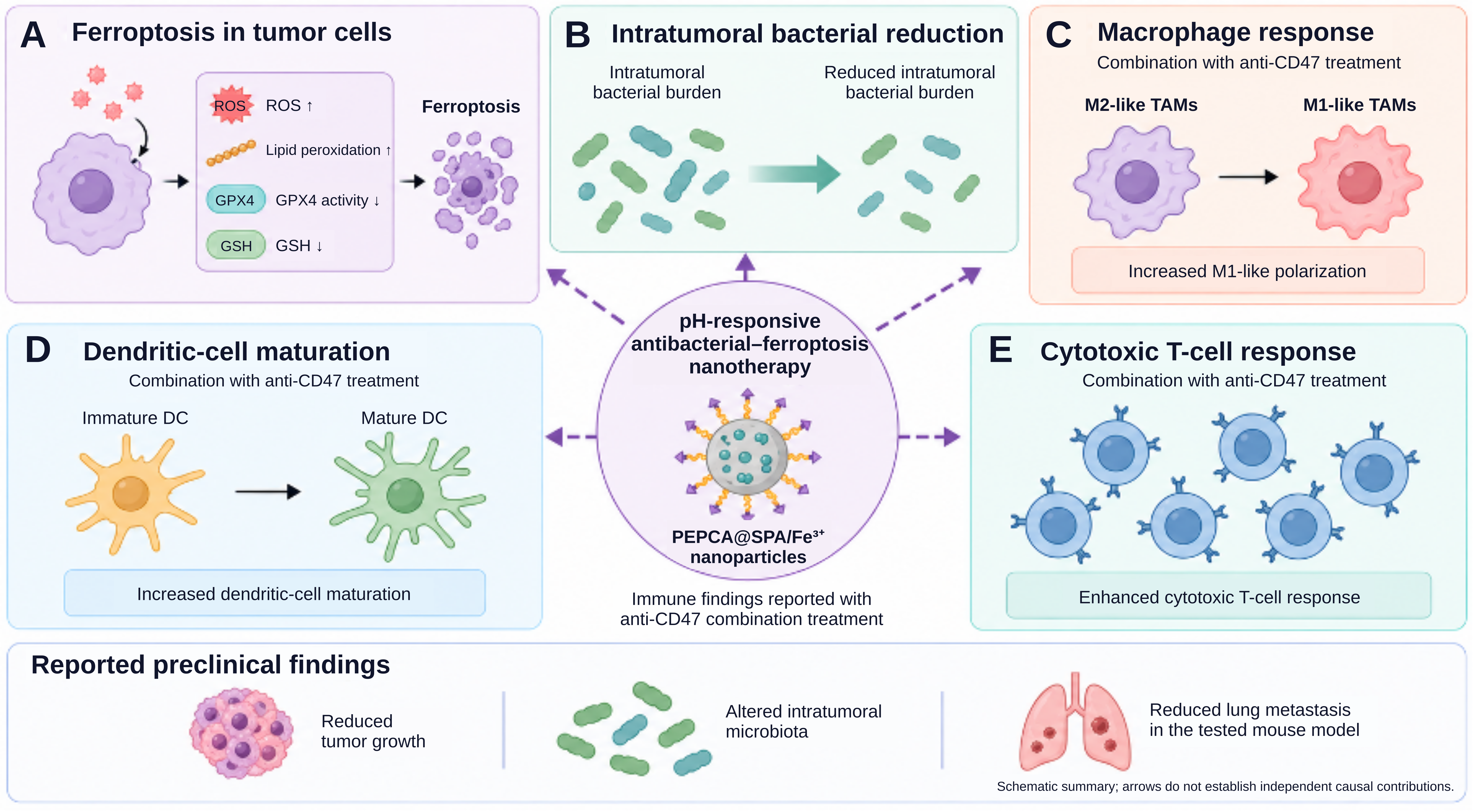
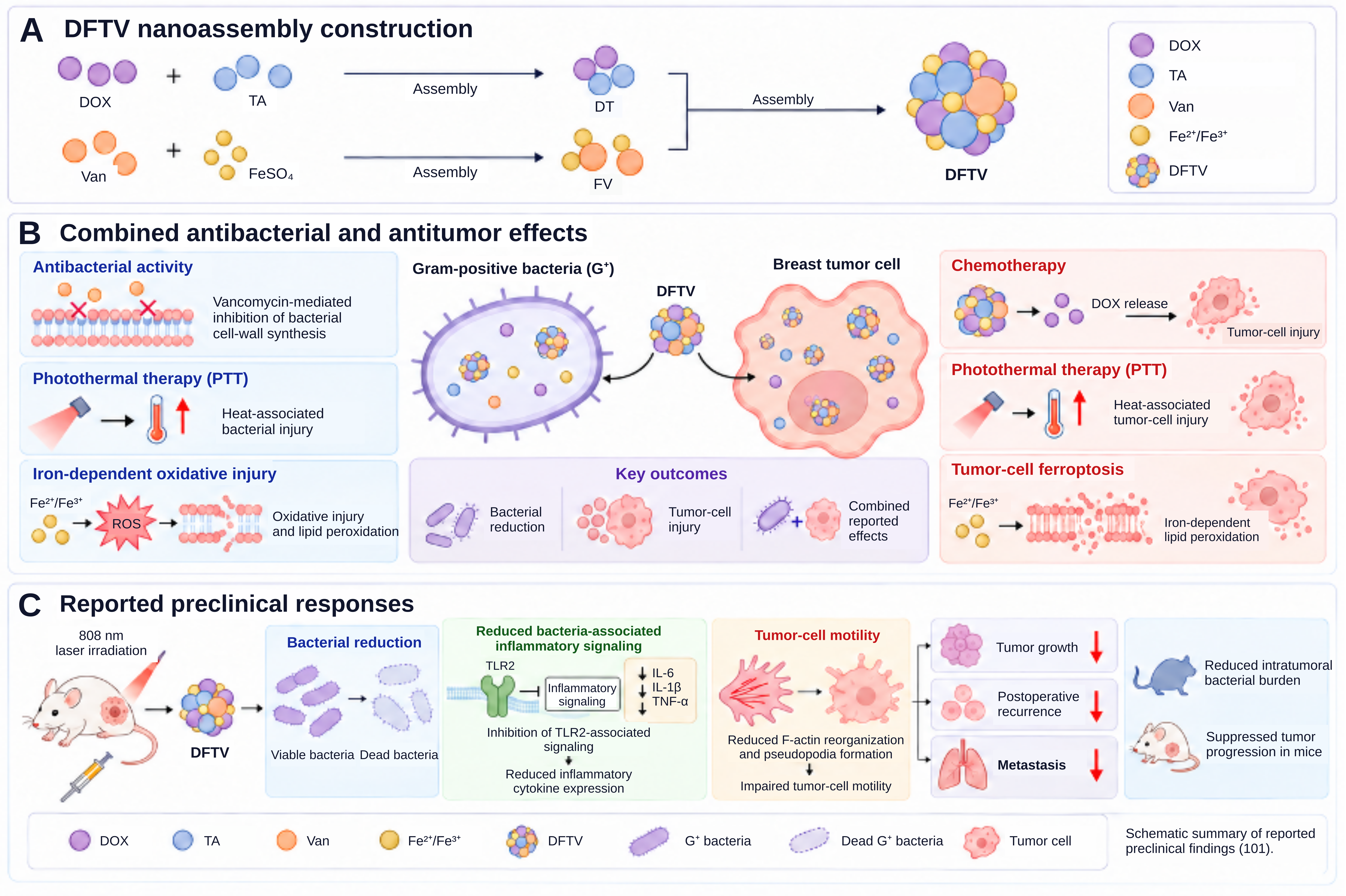
Nanoplatform treatment suppressed primary tumor growth and reduced lung metastasis; however, these experiments do not isolate the contribution of bacterial reduction from direct ferroptotic injury or accompanying immune effects (100). Mechanistically, the nanoparticles showed potent antibacterial activity against E. coli, Pseudomonas aeruginosa, and F. nucleatum while inducing ferroptosis by disrupting the GSH/GPX4 axis, increasing ROS, and promoting lipid peroxidation. Furthermore, microbiota elimination synergized with α-CD47 immune checkpoint blockade to enhance antitumor immunity by promoting dendritic cell maturation, M1 macrophage polarization, increased CD8⁺ T-cell infiltration, and reduction of regulatory T cells (100). A multifunctional amorphous nanoassembly (DFTV) has been developed to simultaneously target intratumoral Gram-positive (G⁺) bacteria and breast tumor cells. The platform reduced intratumoral bacterial burden and was associated with attenuated inflammatory signaling and disrupted cytoskeletal remodeling linked to tumor-cell motility, thereby addressing limitations of conventional antibiotic delivery within tumors. In preclinical models, DFTV treatment inhibited tumor growth, metastatic progression, and postoperative recurrence. Mechanistically, DFTV promoted iron-dependent lipid peroxidation and ferroptotic tumor-cell death, enhanced by laser irradiation, while also contributing to antibacterial activity and remodeling of the tumor immune microenvironment (Figure 4) (101).
A multifunctional ultrasound (US)-activatable piezoelectric nanoplatform (HN-T/BT@Lip) has been developed for triple-negative breast cancer (TNBC) by integrating ferroptosis induction with modulation of intratumoral microbial metabolism. The platform incorporates toyocamycin-loaded CaCO₃–carboxymethyl chitosan hybrid nanoparticles and barium titanate within folate-modified liposomes, enabling targeted delivery and US-responsive activation. Upon US stimulation, the nanoplatform generates localized electric fields, promotes Ca²⁺ influx, and increases reactive oxygen species (ROS) production. Treatment was also associated with remodeling of the intratumoral microbiota and its metabolic profile, with trimethylamine N-oxide (TMAO) identified as a key metabolite linked to the therapeutic response. Mechanistically, TMAO was implicated in potentiating ferroptotic cell death by enhancing lipid peroxidation and suppressing antioxidant defenses involving the Nrf2–GSH/GPX4 axis, thereby providing a potential mechanistic link between microbiota-derived metabolism and ferroptosis-based therapy (102).
Although these findings suggest a mechanistic relationship between microbiota-associated metabolic remodeling and ferroptosis, the contribution of TMAO should be distinguished from the direct effects of ultrasound activation, reactive oxygen species generation, and the therapeutic cargo. Establishing metabolite-dependent efficacy requires experiments that independently manipulate TMAO availability and assess whether metabolite supplementation or inhibition alters lipid peroxidation, ferroptosis, and tumor control. Such studies should also determine whether the relevant metabolic changes originate from intratumoral microorganisms, intestinal microbiota, or host metabolism. External laser activation can provide spatial control over selected photothermal nanoplatforms, potentially limiting treatment to the irradiated region (23, 103). In experimental breast cancer cell models harboring intracellular bacteria, near-infrared-activated silver nanoparticles demonstrated photothermal cytotoxicity and reduced intracellular bacterial burden (104). However, the extent of microbiota modulation depends on the platform and experimental context, and localized activation does not necessarily translate into reduced systemic toxicity. Camptothecin-loaded nanoparticles (PCRHNs) have been developed to mitigate treatment-associated alterations in the gut microbiota while enhancing anticancer activity. In the reported preclinical model, high-dose free camptothecin (CPT) induced gut microbial dysbiosis, including increased abundance of potentially pathogenic Escherichia and Shigella, whereas PCRHN treatment combined with laser irradiation preserved microbial richness and diversity and increased the abundance of potentially beneficial species, including Lactobacillus reuteri. Mechanistically, the photothermal activity of PCRHNs enhanced catalase-like activity and oxygen generation, thereby altering the intestinal microenvironment in a manner proposed to favor the growth of facultative anaerobic probiotic bacteria (105).
DNA aptamers provide programmable molecular recognition and can be incorporated into nanocarriers to improve binding to selected cellular or microbial targets (106). However, their in vivo performance depends on target accessibility, nuclease stability, pharmacokinetics, and retention of binding specificity in complex biological environments. Researchers developed a DNA tetrahedron-based nanodelivery system functionalized with a bacteria-targeting aptamer (TA@B) for targeted berberine delivery. The nanostructure exhibited high drug-loading capacity and selective binding to intratumoral bacteria, integrating antibacterial, anticancer, and immunomodulatory activities. In preclinical models, TA@B suppressed tumor-cell proliferation both in vitro and in vivo and was associated with reduced TNF-α and IL-6 levels and remodeling of the immune microenvironment (107).
Metallic Nanoparticles and Bacteria-Metal Biohybrids
Metallic nanoparticles offer physicochemical properties that have been widely investigated for antimicrobial applications, including surface functionalization, catalytic activity, ion release, and the generation of reactive oxygen species (108). Their integration with engineered bacteria has enabled the development of bacteria–nanomaterial biohybrids that combine bacterial tumor-homing capabilities with nanomaterial-mediated cargo delivery and therapeutic functions (109). In cancer therapy, metallic nanomaterials can also provide photothermal, catalytic, and immunomodulatory functions that may be exploited within the tumor microenvironment (110). However, the biological behavior and therapeutic performance of these systems depend on the specific nanomaterial, bacterial strain, route of administration, and platform design. A bacteria-nanoparticle biohybrid system has been shown to function as an efficient peptide-delivery platform while concurrently modulating the tumor-associated microbiota and immune microenvironment (111). A quorum-sensing-engineered E. coli strain TSYPU was used as a bacterial delivery system for the scorpion venom-derived peptide (BmK AGAP). This enabled synchronized lysis and sustained intratumoral drug release. TSYPU colonized the tumor microenvironment and released AGAP, resulting in considerable inhibition of tumor growth through anti-proliferative and anti-angiogenic effects, along with TME modulation, as E. coli K12 induced immune activation and macrophage polarization toward the anti-tumor M1 phenotype, thereby enhancing therapeutic efficacy. To further improve delivery efficiency and stability, TSYPU was integrated with gold nanoparticles (TSYPU@Au), which protected the bacteria from gastrointestinal degradation and enabled effective oral administration. TSYPU@Au produced greater tumor suppression than the bacterial system alone in the reported experimental models (111), although the safety and controllability of orally administered live engineered bacteria–nanoparticle hybrids remain major translational considerations.
Biomimetic and Biohybrid Nanocarriers
Biomimetic platforms and nano–bacterial biohybrids use biological components or live bacteria with nanomaterials for targeted drug delivery and microbiota modulation (109, 112). The nano–bacterial biohybrid LYC@Bac combines IL-15-expressing engineered Escherichia coli with pH-responsive lysozyme nanoparticles targeting F. nucleatum. Treatment reduced F. nucleatum colonization and altered microbial composition, including increased Escherichia abundance detected by 16S rRNA sequencing. These changes accompanied reduced immunosuppression and enhanced antitumor immune activity. Bacterial clearance and pathogen-associated molecular pattern (PAMP) release promoted dendritic-cell activation, while bacteria-derived IL-15 enhanced NK-cell activity through mTOR signaling, suggesting a reinforcing interaction between microbial clearance and immune activation (113).
The biomimetic nanoplatform DPI@HMV-RS17 targets F. nucleatum-colonized triple-negative breast cancer (TNBC). It integrates doxorubicin (DOX), IR820, polymetformin, and RS17-functionalized hybrid membrane vesicles for combined chemotherapy, phototherapy, and immunomodulation. In preclinical models, treatment achieved a reported 99.86% reduction in intratumoral F. nucleatum abundance and was associated with reduced bacteria-associated chemoresistance and immunosuppression. Experiments using infected 4T1 cells and colonized 4T1 tumor-bearing mice demonstrated antitumor activity, including suppression of primary tumor growth, lung metastasis, and recurrence in mice (114) (Figure 5).
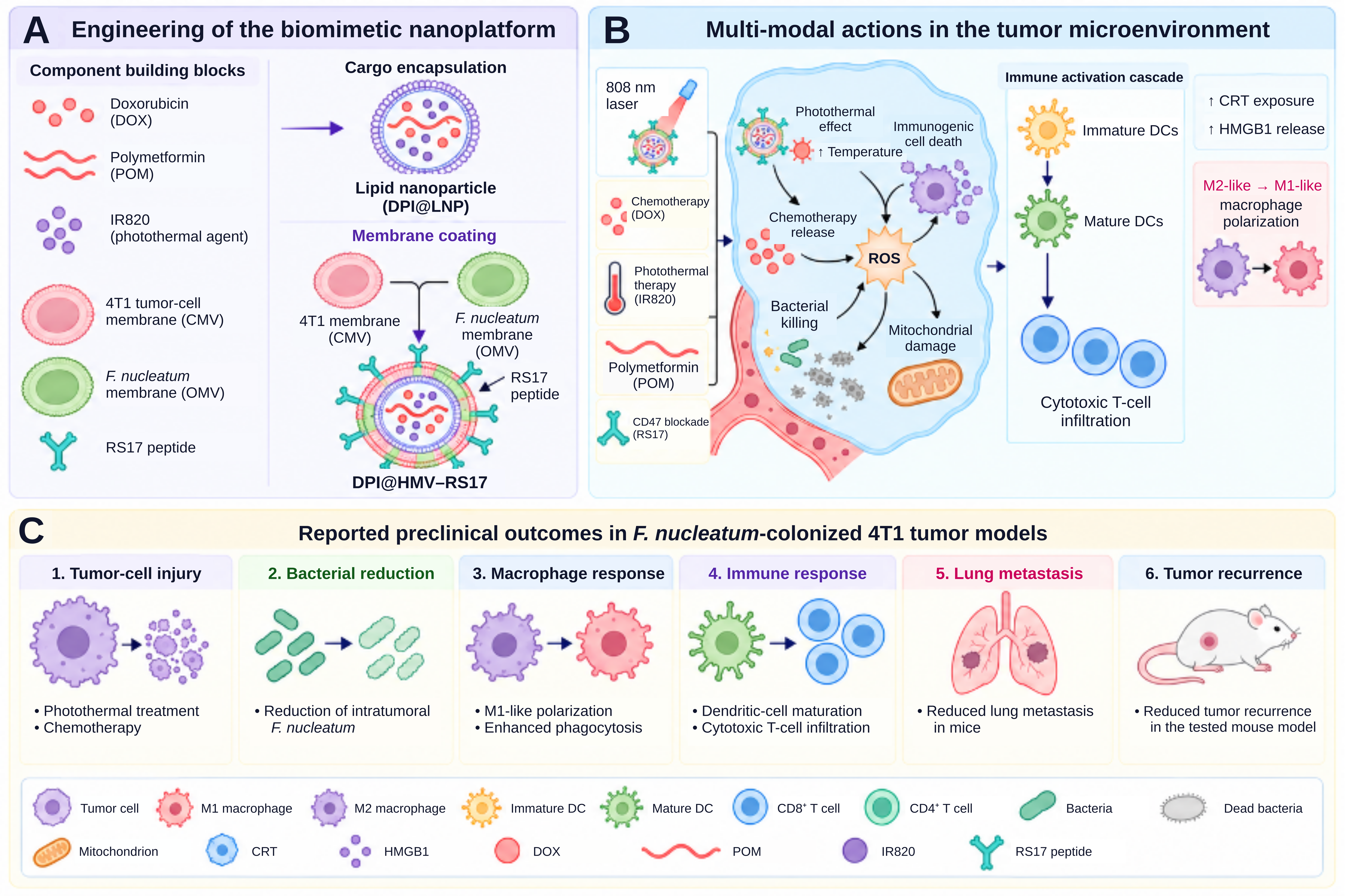
These antitumor effects were accompanied by remodeling of the immune microenvironment, including enhanced dendritic-cell maturation, increased cytotoxic T-cell infiltration, and macrophage polarization toward an M1-like phenotype through inhibition of CD47–SIRPα signaling (114). The magnitude of bacterial depletion and tumor control in this model is notable, but both outcomes were obtained in F. nucleatum-colonized experimental systems. To establish broader relevance, comparable targeting in tumors with naturally occurring, lower-biomass, heterogeneous microbial communities is needed. Various studies elucidate the role of intratumoral microbiota, particularly F. nucleatum, in driving immunosuppression and limiting the efficacy of immune checkpoint blockade (ICB) therapy in breast cancer (48). F. nucleatum has been implicated in an immunosuppressive tumor microenvironment characterized by increased infiltration of myeloid-derived suppressor cells (MDSCs) and M2-like macrophages, along with reduced CD8⁺ T-cell activity and resistance to anti-PD-L1 therapy (115).
Biomimetic nanovehicles have been investigated for targeted depletion of intratumoral F. nucleatum in breast cancer. In preclinical models, selective bacterial depletion was combined with PD-L1 blockade to enhance antitumor activity, providing a microbiota-directed strategy for simultaneously targeting tumor-associated bacteria and immunosuppression (83). Nevertheless, the relative contribution of bacterial depletion to therapeutic efficacy should be distinguished from the independent effects of immune checkpoint blockade. An alternative biohybrid strategy integrates intratumoral bacterial elimination with activation of innate immune signaling. Zhou et al. developed nanodrug–bacteria biohybrids designed to eliminate intratumoral bacteria while activating the cyclic GMP–AMP synthase–stimulator of interferon genes (cGAS–STING) pathway. In preclinical tumor models, the platform combined antibacterial activity with immune stimulation, providing a distinct approach to enhancing cancer immunotherapy (116). Unlike approaches combining bacterial depletion with immune checkpoint blockade, this system couples bacterial targeting to cGAS–STING activation. Nevertheless, the relative contributions of bacterial elimination, biohybrid-associated immunostimulation, and other therapeutic components require careful interpretation when evaluating microbiota-dependent antitumor efficacy.
A cold tumor microenvironment is characterized by low immune-cell infiltration, reduced antigen presentation, and minimal inflammatory signaling, making tumors less responsive to immunotherapy (117). This immunosuppressive state limits effective antitumor immune activation and represents a major challenge in cancer treatment (118). Microbiota-targeted outer membrane vesicle (OMV)-coated nanoparticles (OMV@Fe-ZIF-90@M) have been developed to remodel the intratumoral microbiota and enhance antitumor immunity in triple-negative breast cancer (TNBC). The platform exploits homologous bacterial targeting and Fap2-Gal-GalNAc interactions to selectively target F. nucleatum, achieving a reported 99.7% reduction in bacterial burden. Beyond bacterial depletion, treatment was designed to convert microbiota-associated signals into immunostimulatory cues: disruption of F. nucleatum releases pathogen-associated molecular patterns (PAMPs), including lipopolysaccharide (LPS), which act together with immunostimulatory OMV components to promote dendritic-cell maturation and antigen presentation. In preclinical models, these effects were accompanied by increased TNF-α and IFN-γ, reduced IL-6 and IL-10, and enhanced infiltration of CD4⁺ and CD8⁺ T cells, consistent with remodeling of the immunosuppressive tumor microenvironment toward a more immune-active state (119).
Collectively, these studies support the experimental feasibility of integrating microbiota modulation with immune activation in breast cancer models. However, increased immune-cell infiltration, cytokine production, or improved responses to immune checkpoint blockade do not independently establish microbiota-mediated immune restoration, particularly when nanoplatforms contain immunostimulatory components or cytotoxic agents that can induce immunogenic cell death. Furthermore, clinical evidence supporting microbiome-based immunotherapy modulation in other malignancies should not be directly extrapolated to breast cancer. Future studies should determine whether microbial depletion or remodeling contributes independently to immune checkpoint blockade efficacy using platform-matched controls, microbial recolonization, and clinically representative breast cancer models.
Natural Nanocarriers
Naturally derived nanocarriers, including plant-derived extracellular vesicle-like particles, have emerged as experimental delivery systems that can affect both tumor cells and host microbial communities (120–123). Their biological composition may confer distinct uptake and biodistribution properties, but evidence that their natural origin provides a specific advantage for microbiome modulation remains limited (120). For example, plant-based exosome-like nanoparticles and vesicles loaded with bioactive molecules can simultaneously exert direct cytotoxicity against cancer cells and alter gut microbial composition and diversity, with potentially beneficial effects that require functional validation (121). Plant-derived extracellular vesicles have demonstrated the potential to modulate gut microbiota and reshape the tumor immune microenvironment. In preclinical triple-negative breast cancer models, Platycodon grandiflorum-derived extracellular vesicles altered gut microbial composition, promoted M1-like macrophage polarization and cytotoxic T-cell infiltration, and suppressed tumor growth (122). Likewise, researchers have developed nano formulations based on natural compounds to enable controlled release and distribution throughout the body, thereby modulating the microbiota in the intestine and tumor tissues (123).
Natural nanocarriers can influence microbial composition, microbial metabolites, and host immune responses while also directly affecting tumor cells. These overlapping activities make mechanistic attribution difficult, and short-term tolerability in preclinical models should not be interpreted as evidence of no toxicity. Tea flower-derived exosome-like nanoparticles (TFENs), containing bioactive constituents including polyphenols, flavonoids, proteins, and lipids, have been investigated as a plant-derived nanoplatform for breast cancer therapy. TFENs were efficiently internalized by breast cancer cells and induced reactive oxygen species (ROS) accumulation, mitochondrial dysfunction, cell-cycle arrest, and apoptosis, with G2-phase arrest observed in MCF-7 cells and S/G2-phase arrest in 4T1 cells. In preclinical models, both intravenous and oral administration suppressed primary tumor growth and lung metastasis, with preferential nanoparticle accumulation at tumor and metastatic sites. Notably, oral administration produced substantial antitumor activity despite lower pulmonary accumulation and was associated with gut microbiota remodeling and enhanced systemic antitumor immune responses. TFEN activity therefore coincided with both ROS-mediated tumor-cell injury and microbiota-associated immune changes (121); the available experiments do not establish the relative contribution of these mechanisms to tumor control.
Tea leaf-derived exosome-like nanotherapeutics (TLNTs) have also been investigated for breast cancer treatment, with direct anticancer activity accompanied by changes in gut microbial composition and immune responses (124). The TLNTs were efficiently internalized by 4T1 breast cancer cells, resulting in a 2.5-fold increase in ROS levels, mitochondrial dysfunction, cell cycle arrest at the G0/G1 and S phases, and apoptosis. Notably, TLNT treatment altered gut microbial composition and increased microbial diversity, accompanied by a higher Bacteroidetes/Firmicutes ratio, enrichment of Alloprevotella, and reduced abundance of Alistipes, Oscillibacter, and Desulfovibrio. Although these changes coincided with enhanced antitumor immune activity, their functional significance and contribution to tumor suppression require further validation (124). More broadly, microbiota–cancer interactions involve interconnected metabolic, inflammatory, and immune pathways that vary with microbial composition, anatomical location, and host context (125). Accordingly, microbiota changes observed following nanocarrier treatment require functional validation before they can be considered responsible for improved antitumor activity.
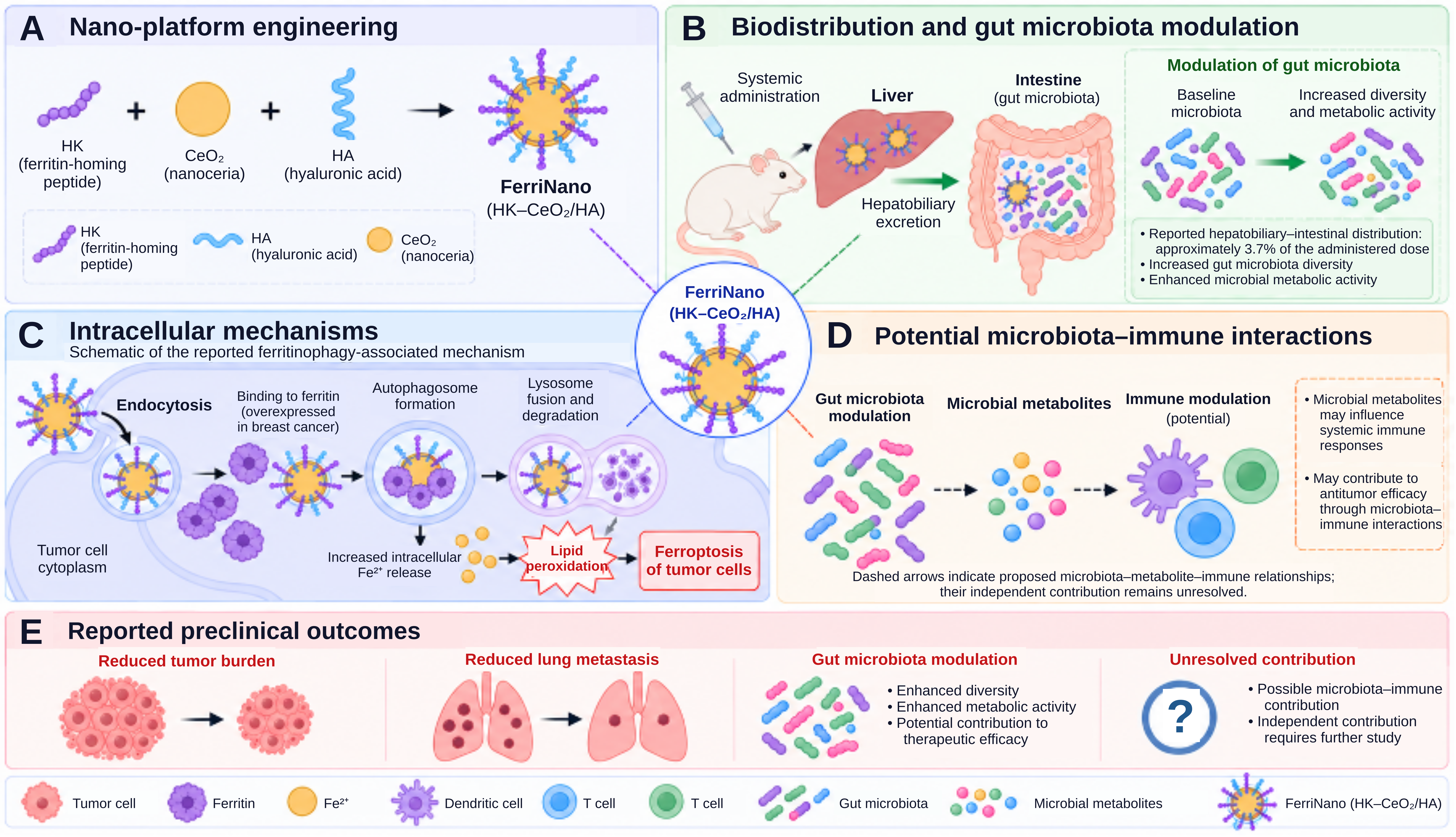
FerriNano provides a preclinical example of a nanoplatform combining tumor-directed ferroptotic stress with modulation of intestinal and intratumoral microbiota. Following systemic administration, a fraction of the formulation underwent hepatobiliary excretion and reached the intestinal compartment, where treatment was associated with changes in microbial diversity, community composition, and metabolite profiles. These changes included reduced abundance of potentially pathogenic genera, such as Streptococcus and Staphylococcus, and enrichment of Bifidobacterium and Blautia, accompanied by reduced tumor progression. Metabolomic analysis also identified increased prostaglandin E3 levels, suggesting a possible association between microbiota remodeling, metabolic changes, and antitumor responses; however, its microbial origin and independent functional contribution require confirmation. FerriNano additionally reduced intratumoral bacterial burden, suggesting activity in both intestinal and tumor-associated microbial compartments.
Importantly, antibiotic-mediated microbiota depletion attenuated therapeutic efficacy, supporting a microbiota-dependent component of the response. Nevertheless, because antibiotics can independently alter host metabolism and immunity, these findings do not establish which microbial taxa or metabolites mediate tumor suppression. Studies using defined microbial communities, metabolite-depletion and rescue experiments, and compartment-specific microbial manipulation are needed to resolve these causal relationships (126) (Figure 6).
PEGylated antibiotic nanoparticles (MTI-PEG) have been developed to selectively target intratumoral bacteria, including F. nucleatum, while limiting perturbation of the intestinal microbiota. In preclinical models, MTI-PEG preferentially accumulated in tumors and markedly reduced intratumoral bacterial burden. Bacterial depletion attenuated microbiota-driven mechanisms implicated in therapeutic resistance, including TLR4-MYD88-associated autophagy, TIGIT-associated suppression of NK-cell activity, and PI3K-AKT-mTOR signaling. Accordingly, MTI-PEG enhanced responses to multiple therapeutic modalities, including oxaliplatin, NK-cell-based therapy, and trastuzumab, while producing comparatively limited alterations in intestinal microbial composition and reducing treatment-associated toxicity.
These findings suggest that selectively targeting intratumoral bacteria may mitigate microbiota-associated therapeutic resistance while producing fewer detectable changes in intestinal microbial composition than less selective antimicrobial approaches; however, intestinal microbial function and long-term ecological stability remain to be established (127). Zhang et al. developed a programmed nanocloak incorporating commensal bacteria-derived nanovesicles to enhance antitumor immune responses. In experimental tumor models, this strategy suppressed tumor growth and metastatic progression, demonstrating the potential of engineered bacterial vesicles as immunotherapeutic components (128).
The potential value of selective bacterial targeting is further supported by evidence that intratumoral bacterial species beyond F. nucleatum can influence breast cancer progression. Gerbec et al. identified intratumoral bacteria capable of enhancing breast tumor metastasis in experimental models (129). These findings underscore the importance of identifying functionally relevant bacterial targets rather than assuming that a single antimicrobial strategy will be appropriate across heterogeneous breast tumors. Across these studies, microbiota modulation accompanies antitumor activity through substantially different delivery systems and mechanisms. Table 1 summarizes representative platforms, emphasizing that most evidence comes from model-specific preclinical studies rather than comparative or clinical evaluations.
Table 1. Summary of Microbiota-Modulating Nanoplatforms in Breast Cancer Treatment.
| Nanoplatform | Microbiota target/modulation | Therapeutic strategy | Reported preclinical outcome | Ref |
|---|---|---|---|---|
| DD@FEL | Decrease tumor-associated bacteria | Antibacterial activity + DOX chemotherapy | Reduced primary tumor growth and metastasis | (85) |
| Colistin-LipoFM / Doxy-LipoFM | F. nucleatum / broader intratumoral microbiota | Targeted bacterial depletion combined with chemotherapy | Improved chemotherapy responsiveness; reduced lung metastasis and complete tumor regression in some mice with Doxy-LipoFM combination | (86) |
| MF-SLNs | Gut microbiota remodeling | DOX delivery + attenuation of dysbiosis | Improved tumor suppression and reduced toxicity | (95) |
| PEPCA@SPA/Fe³⁺ | Intratumoral microbiota remodeling | Ferroptosis + immune remodeling | Reduced tumor progression/metastasis | (100) |
| HN-T/BT@Lip | Microbiota metabolic remodeling/TMAO | Ultrasound activation + ferroptosis | TMAO-associated potentiation of ferroptosis | (102) |
| DPI@HMV-RS17 | Intratumoral F. nucleatum | Targeted bacterial eradication, chemotherapy and immune microenvironment reprogramming | Reduced tumor growth and metastasis; enhanced dendritic-cell maturation, CD8+ T-cell infiltration and M1-like macrophage polarization | (114) |
| OMV@Fe-ZIF-90@M | Decrease F. nucleatum | Microbial targeting + immune activation | Increased antitumor immune activity | (119) |
| TFENs | Gut microbiota remodeling | ROS-mediated tumor injury + systemic immune modulation | Reduced tumor growth and metastasis | (121) |
| FerriNano | Gut + intratumoral microbiota remodeling | Ferritinophagy/ferroptosis + microbiota modulation | Microbiota-dependent antitumor activity | (126) |
| MTI-PEG | Decrease F. nucleatum | Selective antibacterial targeting + therapy sensitization | Enhanced responses to multiple therapeutic modalities | (127) |
Abbreviations: DD@FEL, doxycycline and doxorubicin co-loaded in a folate-functionalized, ergosterol-based liposome. Colistin-LipoFM, colistin-loaded liposomes fused with Fusobacterium nucleatum cytoplasmic membrane. Doxy-LipoFM, doxycycline-loaded liposomes fused with F. nucleatum cytoplasmic membrane. MF-SLNs, microfluidics-prepared, RGD-modified solid lipid nanoparticles. PEPCA@SPA/Fe³⁺, sparfloxacin–ferric ion nanoparticles encapsulated in a polyethylene glycol–poly (cinnamyl aldehyde) copolymer. HN-T/BT@Lip, folate-modified liposomes co-encapsulating toyocamycin-loaded calcium carbonate–carboxymethyl chitosan hybrid nanoparticles and barium titanate. DPI@HMV-RS17, lipid nanoparticles co-loaded with doxorubicin, polymetformin, and IR820, functionalized with F. nucleatum–4T1 hybrid cell membrane vesicles and decorated with the RS17 peptide. OMV@Fe-ZIF-90@M, F. nucleatum outer membrane vesicle-coated, metronidazole-loaded iron-containing zeolitic imidazolate framework-90. TFENs, tea flower-derived exosome-like nanovesicles. FerriNano, ferritin nanochaperone composed of ferritin-homing peptide-functionalized nanoceria coated with hyaluronic acid. MTI-PEG, polyethylene glycol-conjugated metronidazole. CD8⁺, cluster of differentiation 8-positive. DOX, doxorubicin. F. nucleatum, Fusobacterium nucleatum. RGD, arginine–glycine–aspartic acid. ROS, reactive oxygen species. TMAO, trimethylamine N-oxide.
Comparative Therapeutic Value of Microbiome-Modulating Nanoplatforms
The therapeutic relevance of microbiome-modulating nanoplatforms should be assessed against existing treatment strategies, not solely based on their multifunctional design or superior efficacy relative to untreated controls. Conventional antibiotics offer established antibacterial activity but may disrupt beneficial microbial communities and promote antimicrobial resistance. Tumor-directed antibiotic nanocarriers may provide greater spatial control and reduce systemic exposure; however, preferential tumor accumulation does not necessarily confer selective bacterial targeting or preserve intestinal microbial function.
Multifunctional nanoplatforms integrate antimicrobial activity with chemotherapy, ferroptosis induction, phototherapy, or immunomodulation. Although these combinations have produced promising results in preclinical breast cancer models, their added therapeutic value must be distinguished from improvements in drug delivery and the independent anticancer activities of their constituent components. Similarly, microbiota-derived metabolite modulation and engineered bacterial biohybrids offer alternative approaches to modifying tumor–microbe interactions, but uncertainties about mechanistic specificity, biological reproducibility, microbial persistence, and safety constrain their clinical development.
The central question is therefore whether microbiome-directed engineering provides a reproducible therapeutic advantage over simpler treatment strategies. Future comparative studies should incorporate free-drug controls, conventional nanocarriers, platform-matched formulations without antimicrobial components, and microbiota-depleted or recolonized experimental models. Comparative endpoints should include tumor control, microbial selectivity, systemic toxicity, ecological stability, and the incremental contribution of microbiota modulation. These considerations are summarized in Table 2.
Table 2. Comparative Therapeutic Advantages and Unresolved Challenges of Microbiome-Modulating Strategies in Breast Cancer
| Strategy | Potential advantage | Principal unresolved question |
|---|---|---|
| Systemic antibiotics | Established antibacterial activity and clinical accessibility | Can harmful ecological effects be avoided? |
| Tumor-directed antibiotic nanoparticles | Greater spatial control and potentially reduced systemic exposure | Is microbial selectivity demonstrated? |
| Multifunctional nanoplatforms | Simultaneous targeting of microbial and tumor-associated pathways | Does added complexity provide incremental therapeutic benefit? |
| Engineered bacterial biohybrids | Localized therapeutic production and responsive delivery | Can colonization, persistence and systemic safety be controlled? |
| Microbiota-derived metabolite modulation | Potentially more selective biological intervention | Which metabolites have reproducible causal effects? |
5. Mechanistic Basis of Microbiome-Modulating Nanotherapy
Microbiome-directed nanoplatforms may influence therapeutic responses through complementary mechanisms, including modulation of intratumoral bacteria, enhanced chemotherapy delivery, and remodeling of local antitumor immunity. In preclinical triple-negative breast cancer models, a doxycycline- and doxorubicin-loaded liposomal platform demonstrated that microbiota modulation can complement chemotherapy to suppress tumor growth and metastasis (85). In contrast, bacteria-derived nanovesicle platforms, discussed in Section 4.6, exploit microbial components to stimulate antitumor immunity rather than primarily depleting tumor-associated bacteria (128). These distinct approaches illustrate why microbiota-dependent effects must be distinguished from the direct pharmacological and immunological activities of nanoplatforms.
Other experimental approaches have investigated changes in drug distribution and immune activity (65, 83, 130). Nanocarriers can increase intratumoral drug exposure and protect therapeutic cargo from premature degradation, but these pharmacological effects can themselves improve tumor control independently of microbiota modulation. Similarly, changes in dendritic cells, cytotoxic T cells, NK cells, macrophages, Tregs, and MDSCs may reflect direct nanomaterial or drug effects as well as microbial remodeling. Mechanistic studies therefore need to distinguish microbiota-dependent effects from the intrinsic pharmacological and immunological activity of the nanoplatform. For platforms involving microbial metabolites, targeted metabolomics combined with metabolite-depletion and rescue experiments will be particularly important for distinguishing causal mediators from treatment-associated metabolic changes.
Understanding these mechanisms may eventually support microbiome-informed treatment selection, although their application to personalized nanotherapy remains investigational. Substantial interpatient variability in microbiome composition complicates, rather than yet enables, microbiome-guided nanotherapy (131). Advances in 16S rRNA sequencing, metagenomic analysis, and multi-omics approaches have enabled detailed characterization of microbiome profiles, helping identify microbial markers of response and disease progression (131, 132). Combining microbial markers with nanotechnology may support patient stratification, rational use of microbiome-specific nanocarriers, and tailoring drug combinations based on individual microbiome and tumor profiles (131, 133). Whether microbiome-based stratification improves treatment selection, efficacy, or toxicity beyond established tumor and host biomarkers remains to be tested prospectively (134).
Integrating microbiome, metabolome, immunome, and other multi-omic data with machine-learning approaches could assist patient stratification and modeling of nanocarrier–microbiome–host interactions (135). Such models will require external validation, adequate sample sizes, standardized input data, and prospective demonstration that their predictions improve therapeutic decision-making.
Despite promising preclinical results, many obstacles remain before microbiome-regulating nanotechnologies can be implemented in clinical practice. Key prerequisites for successful implementation include standardizing microbiome analysis tools, validating microbial biomarkers with high predictive value, and conducting well-planned clinical trials (136, 137). Until standardized microbial measurements, reproducible biomarkers, and prospective clinical evidence are available, microbiome-guided nanotherapy should be regarded as an experimental precision-medicine strategy rather than an established treatment paradigm.
6. Translational Barriers and Clinical Development.
The clinical translation of microbiome-regulating nanomedicine for breast cancer faces considerable biological and technological limitations. Interpatient heterogeneity in both gut and intratumoral microbiota impedes reproducibility (28). Safety remains a critical concern because disrupting the microbial ecosystem could cause chronic dysbiosis, immune imbalance, and inflammation. In addition, preclinical models of the tumor-microbiome interface have insufficient fidelity to humans (138). The lack of a standard process for evaluating the microbiome, nanoparticles, and pharmacokinetics-microbiome interactions reduces comparability and slows regulatory progress (139). Moreover, detecting and quantifying tumor-associated bacteria or metabolites in the microbiome further hinders the development of effective treatments (16). Tumor heterogeneity, including differences in vasculature and microbial niches, also affects nanoparticle delivery and retention.
A major analytical challenge is reliably characterizing low-biomass intratumoral microbiota, where microbial signals may be difficult to distinguish from contamination introduced during tissue collection, processing, DNA extraction, and sequencing (13, 15). Analytical workflows for low-biomass samples should include appropriate negative controls and assessment of reagent-derived contamination (140). Importantly, sequencing-based detection of microbial DNA does not establish bacterial viability, active colonization, or functional involvement in tumor biology (13). Differences in extraction reagents and laboratory processing can generate misleading microbial profiles, particularly in low-biomass samples (140). Accordingly, claims of intratumoral microbial persistence or functional activity should be supported by complementary spatial, microbiological, or transcriptomic evidence. Standardized microbial measurements and independently validated biomarkers are essential to distinguish biologically relevant microbial communities from technical artifacts and establish the clinical relevance of microbiome-targeted therapeutic strategies (136, 137).
Moreover, significant interindividual variability in gut and breast microbiome composition, influenced by age, diet, ethnicity, antibiotic exposure, obesity, hormonal status, and environmental factors, makes developing universally applicable microbiome-based therapeutic strategies challenging (131, 141). Although preclinical models report microbiome remodeling and improved antitumor responses, clinical evidence for microbiome-directed interventions in breast cancer remains sparse, and clinical evidence for microbiome-modulating nanomedicine remains unestablished. Fecal microbiota transplantation (FMT) carries documented pathogen-transmission risks: donor-derived drug-resistant Escherichia coli caused bloodstream infections in recipients (142). Similarly, clinical evidence from gastrointestinal conditions indicates that probiotic benefits vary across indications, strains, and formulations, limiting the generalizability of findings between preparations (143).
Furthermore, emerging microbiome-based nanomedicine approaches encounter additional challenges, including targeted delivery to specific microbial or tumor-associated sites, nanoparticle stability, long-term biosafety evaluation, scalable manufacturing, and regulatory approval. The absence of standardized regulatory frameworks and ethical guidelines for microbiome-derived therapeutics further restricts their clinical implementation (144, 145). Therefore, future studies should develop standardized analytical protocols, conduct well-designed multicenter clinical trials, and establish personalized microbiome-based strategies to support the safe and effective translation of microbiome-targeted therapies into clinical practice.
Clinical investigation of the microbiome in breast cancer remains in its early stages (Table 3). Existing studies primarily examine probiotics or microbiome profiling, whereas FMT as an immunotherapy-resensitization strategy has been investigated principally outside breast cancer. Small cohorts, heterogeneous populations, non-standardized microbiome assays, and limited efficacy endpoints restrict cross-study interpretation. Clinical evidence supporting microbiome-modulating nanomedicines in breast cancer remains unestablished, leaving a substantial translational gap between the preclinical platforms discussed here and demonstrated patient benefit.
A major limitation across the current literature is that microbiome modulation is frequently measured as a treatment-associated endpoint rather than tested as a causal determinant of therapeutic response. Many nanoplatforms simultaneously alter drug pharmacokinetics, tumor-cell viability, oxidative stress, vascular permeability, and immune activity, making it difficult to attribute improved efficacy specifically to microbial remodeling. Antibiotic-depletion experiments provide useful evidence but are themselves confounded by systemic metabolic and immunological effects.
Stronger causal inference will require platform-matched controls, gnotobiotic or defined-community models, microbial depletion followed by recolonization, and rescue experiments linking specific taxa or metabolites to therapeutic response. Without such evidence, changes in microbial abundance are described as treatment-associated rather than microbiome-mediated.
Table 3. Clinical studies of microbiome-directed interventions in breast cancer and relevant evidence from other solid tumors.
| Clinical strategy | Clinical Trial ID | Disease | Intervention | Main objective | Major limitation | Ref |
|---|---|---|---|---|---|---|
| Fecal microbiota transplantation (FMT) + anti-PD-1 | NCT03353402 | Melanoma |
FMT + anti-PD-1 immunotherapy |
Restore immunotherapy response | Safety, donor variability, and lack of standardization | (146) |
| Engineering gut microbiome using probiotics | NCT03358511 | Breast cancer | Primal Defense Ultra® probiotic | Evaluate probiotic supplementation on the gut microbiome, immune function | Small sample size, short intervention period, limited evidence of therapeutic efficacy | NCT03358511 |
| Probiotic intervention | NCT04362826 | Breast cancer | BIOHM probiotic versus placebo | Assess the effects of probiotics on gut and breast microbiome composition and systemic inflammation | Microbiome and inflammatory endpoints do not establish improved breast-cancer treatment outcomes. | NCT04362826 |
| Microbiome biomarker profiling | Various observational studies. No NCT | Breast cancer | 16S rRNA sequencing and microbiome analysis | Identify microbial biomarkers associated with breast cancer diagnosis, prognosis, and treatment response | Lack of standardized sampling, sequencing, and bioinformatics methods | (15) |
Abbreviations: 16S rRNA, 16S ribosomal RNA. Anti-PD-1, antibody targeting programmed cell death protein 1. FMT, fecal microbiota transplantation. NCT, ClinicalTrials.gov trial registration identifier prefix.
7. Critical Perspectives and Research Priorities
Future development should prioritize establishing the clinical utility of microbiome-targeted nanotherapy rather than increasing platform complexity alone. Clinical microbiome studies have demonstrated the potential of strain-resolved microbial signatures to predict immunotherapy response, while also revealing important limitations in reproducibility across treatment regimens (147). Prospective studies in breast cancer are therefore needed to determine whether microbial biomarkers provide predictive value beyond established clinical variables and whether deliberate microbiome modulation improves treatment outcomes. Multi-omic and computational approaches may help model nanocarrier–microbiome–host interactions, but their utility will depend on standardized datasets and external validation (148, 149).
Stimuli-responsive and theranostic platforms could improve spatial or temporal control of treatment (150, 151), although additional engineering complexity must be balanced against manufacturability, reproducibility, safety, and regulatory feasibility. Another research priority is to determine whether tumor-associated bacteria can serve as reproducible targets for precision nanomedicine. Song et al. developed bacteria-targeted mesoporous silica nanoparticles and demonstrated tumor targeting in mouse models of colon, lung, and breast cancer (152). Clinical translation will require confirmation that the targeted bacterial features are reproducible, biologically relevant, and accessible in naturally occurring human breast tumors.
A further challenge is determining when microbiota modulation contributes enough to justify its inclusion in a therapeutic nanoplatform. If tumor accumulation or the anticancer cargo accounts for most of the observed benefit, adding a microbiome-directed component may increase biological and regulatory complexity while offering little clinical value. Studies should use platform-matched comparators that deliver the same anticancer cargo at comparable tumor exposure. Microbiota depletion and controlled recolonization can help determine whether the treatment response depends on specific organisms or microbial functions. Patient stratification should account for breast cancer molecular subtype, disease stage, treatment history, and baseline microbial features. Candidate microbial biomarkers should be validated in independent cohorts to establish whether they predict added benefit from microbial targeting.
These priorities place comparative therapeutic value at the center of nanoplatform development. Clinically representative tumor models, rigorous microbial controls, and standardized analytical methods can help establish which approaches warrant further study. Comparisons with conventional drug delivery should assess the magnitude and durability of any improvement in tumor control and determine whether it comes at the cost of greater toxicity or persistent disruption of commensal communities. For clinical translation, the strongest case would be a reproducible benefit in a defined patient population that is large enough to justify the additional demands of manufacturing, safety monitoring, and regulatory evaluation.
Discussion
Evidence reviewed here supports interactions among tumor-associated microbiota, gut microbial communities, host immunity, and therapeutic response in breast cancer, although the strength of evidence varies across mechanisms and experimental systems. Nanocarriers can modify these interactions while delivering cytotoxic, ferroptosis-inducing, photothermal, or immunomodulatory interventions. This versatility creates an attribution problem because the same formulation can alter microbial abundance, drug exposure, tumor-cell viability, and immune activity. To determine whether microbial changes contribute to tumor control, studies should compare formulations with matched anticancer cargo and delivery properties that differ in microbial targeting. Microbial depletion and reconstitution experiments can then help establish whether specific organisms or microbial products influence the response to treatment.
The translational significance of these findings depends on whether microbiota-associated effects observed in experimental models can be reproduced across clinically heterogeneous breast cancers. Low microbial biomass in tumor specimens complicates distinguishing resident organisms from contamination, while differences in sampling and processing can limit comparisons across studies. Gut and intratumoral microbial changes should also be measured separately because shifts in one compartment provide limited information about the other. Longitudinal sampling could help determine whether microbial changes precede tumor regression or occur after treatment alters the tumor environment. In clinical studies, baseline microbial profiles, breast cancer subtype, and prior antibiotic exposure may help explain why patients respond differently to the same intervention. Changes in microbial composition and immune markers can inform mechanistic interpretation, while comparative clinical outcomes are needed to establish therapeutic benefit.
In conclusion, microbiome-modulating nanoplatforms offer a way to integrate microbial targeting with breast cancer therapy. Preclinical studies report effects on microbial communities, drug delivery, immune responses, and tumor control, but the independent contribution of microbiota modulation remains incompletely established, and clinical validation is lacking. Progress will depend on reproducible microbial biomarkers, mechanistic studies that isolate the contribution of microbial targeting, and long-term safety assessment, including the persistence of treatment-induced changes in microbial communities. The decisive clinical question is whether adding microbial targeting to a comparable treatment regimen improves response depth or durability, reduces recurrence, or lowers treatment-related toxicity.
References
1. Mandapati A, Lukong KE. Triple negative breast cancer: approved treatment options and their mechanisms of action. J Cancer Res Clin Oncol. 2023;149(7):3701–3719. https://doi.org/10.1007/s00432-022-04189-6
2. Pont M, Marqués M, Sorolla A. Latest therapeutical approaches for triple-negative breast cancer: from preclinical to clinical research. Int J Mol Sci. 2024;25(24):13518. https://doi.org/10.3390/ijms252413518
3. Zhou Z, Zhou Q. Immunotherapy resistance in triple-negative breast cancer: molecular mechanisms, tumor microenvironment, and therapeutic implications. Front Oncol. 2025;15:1630464. https://doi.org/10.3389/fonc.2025.1630464
4. Nam AS, Chaligne R, Landau DA. Integrating genetic and non-genetic determinants of cancer evolution by single-cell multi-omics. Nat Rev Genet. 2021;22(1):3–18. https://doi.org/10.1038/s41576-020-0265-5
5. Zhang W, Xiang Y, Ren H, Liu Y, Wang Q, Ran M, et al. The tumor microbiome in cancer progression: mechanisms and therapeutic potential. Mol Cancer. 2025;24(1):195. https://doi.org/10.1186/s12943-025-02403-w
6. Zheng J, Chen H. Effects of intratumoral microbiota on tumorigenesis, anti-tumor immunity, and microbe-based cancer therapy. Front Oncol. 2024;14:1429722. https://doi.org/10.3389/fonc.2024.1429722
7. Coley WB. The treatment of malignant tumors by repeated inoculations of erysipelas. With a report of ten original cases. 1893. Clin Orthop Relat Res. 1991;(262):3–11. PMID: 1984929.
8. Carlson RD, Flickinger JC Jr, Snook AE. Talkin' toxins: from Coley's to modern cancer immunotherapy. Toxins (Basel). 2020;12(4):241. https://doi.org/10.3390/toxins12040241
9. Trivanović D, Pavelić K, Peršurić Ž. Fighting cancer with bacteria and their toxins. Int J Mol Sci. 2021;22(23):12980. https://doi.org/10.3390/ijms222312980
10. Torres MKDS, Pereira Neto GDS, Cayres Vallinoto IMV, Reis LO, Vallinoto ACR. The impact of oncogenic viruses on cancer development: a narrative review. Biology (Basel). 2025;14(7):797. https://doi.org/10.3390/biology14070797
11. Xiao Q, Liu Y, Li T, Wang C, He S, Zhai L, et al. Viral oncogenesis in cancer: from mechanisms to therapeutics. Signal Transduct Target Ther. 2025;10(1):151. https://doi.org/10.1038/s41392-025-02197-9
12. Eladwy RA, Vu HT, Shah R, Li CG, Chang D, Bhuyan DJ. The fight against the carcinogenic Epstein-Barr virus: gut microbiota, natural medicines, and beyond. Int J Mol Sci. 2023;24(2):1716. https://doi.org/10.3390/ijms24021716
13. Wang Z, Zhang T, Liu Y. Emerging technologies and current challenges in intratumoral microbiota research. Front Cell Infect Microbiol. 2026;15:1685862. https://doi.org/10.3389/fcimb.2025.1685862
14. Nejman D, Livyatan I, Fuks G, Gavert N, Zwang Y, Geller LT, et al. The human tumor microbiome is composed of tumor type-specific intracellular bacteria. Science. 2020;368(6494):973–980. https://doi.org/10.1126/science.aay9189
15. Xu Y, Wang M-C. The intratumoral microbiota in breast cancer: from basic research to clinical translation. Gut Microbes. 2025;17(1):2560695. https://doi.org/10.1080/19490976.2025.2560695
16. Lin A, Xiong M, Jiang A, Huang L, Wong HZH, Feng S, et al. The microbiome in cancer. iMeta. 2025;4(5):e70070. https://doi.org/10.1002/imt2.70070
17. Zhang J, You Z, Li X, Hu J, Li J, Jing Z. Harnessing intratumoral microbiota: new horizons in immune microenvironment and immunotherapy. J Transl Med. 2025;23:897. https://doi.org/10.1186/s12967-025-06916-2
18. Lombardo C, Fazio R, Sinagra M, Gattuso G, Longo F, Lombardo C, et al. Intratumoral microbiota: insights from anatomical, molecular, and clinical perspectives. J Pers Med. 2024;14(11):1083. https://doi.org/10.3390/jpm14111083
19. Galeano Niño JL, Wu H, LaCourse KD, Kempchinsky AG, Baryiames A, Barber B, et al. Effect of the intratumoral microbiota on spatial and cellular heterogeneity in cancer. Nature. 2022;611(7937):810–817. https://doi.org/10.1038/s41586-022-05435-0
20. Tan Q, Cao X, Zou F, Wang H, Xiong L, Deng S. Spatial heterogeneity of intratumoral microbiota: a new frontier in cancer immunotherapy resistance. Biomedicines. 2025;13(5):1261. https://doi.org/10.3390/biomedicines13051261
21. Ramadan YN, Alatawi MN, Albalawi AS, Hetta HF. Bacterial contributions to cancer development: mechanisms, dysbiosis, and cross-cancer associations. Infect Agent Cancer. 2026;21(1):11. https://doi.org/10.1186/s13027-025-00722-7
22. Tao YL, Wu XX, Wang JR, Liu M, Liu YN, Lian YQ, et al. The microbiome: regulating anti-tumor immunity. Front Immunol. 2026;17:1781872. https://doi.org/10.3389/fimmu.2026.1781872
23. Sabeel Z, Yang Z. Microbiome-targeted nanoplatforms and engineering approaches in breast cancer therapy. Mol Cancer. 2025;24(1):276. https://doi.org/10.1186/s12943-025-02456-x
24. Rad SK, Yeo KKL, Wu F, Li R, Nourmohammadi S, Tomita Y, et al. A systematic review and meta-analysis of 16S rRNA and Cancer Microbiome Atlas datasets to characterize microbiota signatures in normal breast, mastitis, and breast cancer. Microorganisms. 2025;13(2):467. https://doi.org/10.3390/microorganisms13020467
25. Urbaniak C, Cummins J, Brackstone M, Macklaim JM, Gloor GB, Baban CK, et al. Microbiota of human breast tissue. Appl Environ Microbiol. 2014;80(10):3007–3014. https://doi.org/10.1128/AEM.00242-14
26. Al-Ansari MM, Mahmood SM, Al-Alwan M. The human breast microbiome: from homeostasis to malignancy, mechanistic insights and therapeutic perspectives. Int J Mol Sci. 2026;27(11):4723. https://doi.org/10.3390/ijms27114723
27. Urbaniak C, Gloor GB, Brackstone M, Scott L, Tangney M, Reid G. The microbiota of breast tissue and its association with breast cancer. Appl Environ Microbiol. 2016;82(16):5039–5048. https://doi.org/10.1128/AEM.01235-16
28. Bose C, Uczkowski NG, Sukla K, Raval N, Haque MM, Zhang Y, et al. Breast cancer and microbiome: a systematic review highlighting challenges for clinical translation. BMC Womens Health. 2025;25(1):416. https://doi.org/10.1186/s12905-025-03843-8
29. Fu A, Yao B, Dong T, Chen Y, Yao J, Liu Y, et al. Tumor-resident intracellular microbiota promotes metastatic colonization in breast cancer. Cell. 2022;185(8):1356–1372.e26. https://doi.org/10.1016/j.cell.2022.02.027
30. Kang L, Chen H, He C, Li J. Effects of gut microbiota in breast cancer. Front Oncol. 2025;15:1617410. https://doi.org/10.3389/fonc.2025.1617410
31. Geller LT, Barzily-Rokni M, Danino T, Jonas OH, Shental N, Nejman D, et al. Potential role of intratumor bacteria in mediating tumor resistance to the chemotherapeutic drug gemcitabine. Science. 2017;357(6356):1156–1160. https://doi.org/10.1126/science.aah5043
32. Che C, Zhou Y, Chen L, Li S, Fan D, Lu Y, et al. The role of the microbiota in cancer drug resistance and emerging therapeutic strategies. Cancer Lett. 2026;642:218305. https://doi.org/10.1016/j.canlet.2026.218305
33. Xie M, Li X, Lau HCH, Yu J. The gut microbiota in cancer immunity and immunotherapy. Cell Mol Immunol. 2025;22(9):1012–1031. https://doi.org/10.1038/s41423-025-01326-2
34. Gradishar WJ, Tjulandin S, Davidson N, Shaw H, Desai N, Bhar P, et al. Phase III trial of nanoparticle albumin-bound paclitaxel compared with polyethylated castor oil-based paclitaxel in women with breast cancer. J Clin Oncol. 2005;23(31):7794–7803. https://doi.org/10.1200/JCO.2005.04.937
35. Giri PM, Banerjee A, Layek B. A recent review on cancer nanomedicine. Cancers (Basel). 2023;15(8):2256. https://doi.org/10.3390/cancers15082256
36. Dhiman R, Bazad N, Mukherjee R, Himanshu, Gunjan, Leal E, et al. Enhanced drug delivery with nanocarriers: a comprehensive review of recent advances in breast cancer detection and treatment. Discov Nano. 2024;19(1):143. https://doi.org/10.1186/s11671-024-04086-6
37. Bartusik-Aebisher D, Przygórzewska A, Woźnicki P, Aebisher D. Nanoparticles for photodynamic therapy of breast cancer: a review of recent studies. Molecules. 2025;30(7):1571. https://doi.org/10.3390/molecules30071571
38. Li T, Xiao D, Yin Y, Hu L. Research progress on nano-drug delivery systems for photothermal cancer therapy. Int J Nanomedicine. 2026;21:567551. https://doi.org/10.2147/IJN.S567551
39. Yang J, Wang J, Li J, Cui J. Nanotechnology-microbiota synergy in cancer immunotherapy. Crit Rev Oncol Hematol. 2026;217:105018. https://doi.org/10.1016/j.critrevonc.2025.105018
40. Zhang Y, Zhang H, Xu J, Xu Z, Lou J, Zheng Q, et al. Recent advances in nanomedicine for modulating intratumoral bacteria in cancer therapy. Mol Pharm. 2025;22(10):5767–5790. https://doi.org/10.1021/acs.molpharmaceut.5c00677
41. Mohapatra A, Mohanty A, Park IK. Inorganic nanomedicine-mediated ferroptosis: a synergistic approach to combined cancer therapies and immunotherapy. Cancers (Basel). 2024;16(18):3210. https://doi.org/10.3390/cancers16183210
42. Yin W, Chang J, Sun J, Zhang T, Zhao Y, Li Y, et al. Nanomedicine-mediated ferroptosis targeting strategies for synergistic cancer therapy. J Mater Chem B. 2023;11(6):1171–1190. https://doi.org/10.1039/D2TB02161G
43. Buchta Rosean C, Bostic RR, Ferey JCM, Feng TY, Azar FN, Tung KS, et al. Preexisting commensal dysbiosis is a host-intrinsic regulator of tissue inflammation and tumor cell dissemination in hormone receptor-positive breast cancer. Cancer Res. 2019;79(14):3662–3675. https://doi.org/10.1158/0008-5472.CAN-18-3464
44. Arnone AA, Ansley K, Heeke AL, Howard-McNatt M, Cook KL. Gut microbiota interact with breast cancer therapeutics to modulate efficacy. EMBO Mol Med. 2025;17(2):219–234. https://doi.org/10.1038/s44321-024-00185-0
45. Luu M, Riester Z, Baldrich A, Reichardt N, Yuille S, Busetti A, et al. Microbial short-chain fatty acids modulate CD8+ T cell responses and improve adoptive immunotherapy for cancer. Nat Commun. 2021;12(1):4077. https://doi.org/10.1038/s41467-021-24331-1
46. Liu H, Xiong X, Zhu W, Wang S, Huang W, Zhu G, et al. Gut microbial metabolites in cancer immunomodulation. Mol Cancer. 2026;25(1):8. https://doi.org/10.1186/s12943-025-02521-5
47. Guo H. Interactions between the tumor microbiota and breast cancer. Front Cell Infect Microbiol. 2025;14:1499203. https://doi.org/10.3389/fcimb.2024.1499203
48. Little A, Tangney M, Tunney MM, Buckley NE. Fusobacterium nucleatum: a novel immune modulator in breast cancer? Expert Rev Mol Med. 2023;25:e15. https://doi.org/10.1017/erm.2023.9
49. Li G, Sun Y, Huang Y, Lian J, Wu S, Luo D, et al. Fusobacterium nucleatum-derived small extracellular vesicles facilitate tumor growth and metastasis via TLR4 in breast cancer. BMC Cancer. 2023;23(1):473. https://doi.org/10.1186/s12885-023-10844-z
50. Huang Y, Guo Z, Zeng Z, Shang C, Zhang Y, Ran Z, et al. Fusobacterium nucleatum promotes metastasis of breast cancer via the miR-21-3p/FOXO3 axis. Front Oncol. 2025;15:1530269. https://doi.org/10.3389/fonc.2025.1530269
51. Parhi L, Alon-Maimon T, Sol A, Nejman D, Shhadeh A, Fainsod-Levi T, et al. Breast cancer colonization by Fusobacterium nucleatum accelerates tumor growth and metastatic progression. Nat Commun. 2020;11(1):3259. https://doi.org/10.1038/s41467-020-16967-2
52. Tingting Y, Xiaoling Z, Yu Z, Yan L, Yang L, Xueqin S, et al. Interaction of intratumoral microbiota with the tumor immune microenvironment and its impact on cancer progression. Front Oncol. 2025;15:1609889. https://doi.org/10.3389/fonc.2025.1609889
53. Xia F, Yi Q, Xu Z, Zhou Z, Tang H, Zhang K, et al. Microbial modulation as a game changer: boosting immunotherapy efficacy in breast cancer. Semin Cancer Biol. 2025;117:152–167. https://doi.org/10.1016/j.semcancer.2025.10.006
54. Pushalkar S, Hundeyin M, Daley D, Zambirinis CP, Kurz E, Mishra A, et al. The pancreatic cancer microbiome promotes oncogenesis by induction of innate and adaptive immune suppression. Cancer Discov. 2018;8(4):403–416. https://doi.org/10.1158/2159-8290.CD-17-1134
55. Routy B, Le Chatelier E, Derosa L, Duong CPM, Alou MT, Daillère R, et al. Gut microbiome influences efficacy of PD-1-based immunotherapy against epithelial tumors. Science. 2018;359(6371):91–97. https://doi.org/10.1126/science.aan3706
56. Liu Y, Ning H, Li Y, Li Y, Ma J. The microbiota in breast cancer: dysbiosis, microbial metabolites, and therapeutic implications. Am J Cancer Res. 2025;15(4):1384–1409. https://doi.org/10.62347/ZJCF2843
57. Sivan A, Corrales L, Hubert N, Williams JB, Aquino-Michaels K, Earley ZM, et al. Commensal Bifidobacterium promotes antitumor immunity and facilitates anti-PD-L1 efficacy. Science. 2015;350(6264):1084–1089. https://doi.org/10.1126/science.aac4255
58. Iida N, Dzutsev A, Stewart CA, Smith L, Bouladoux N, Weingarten RA, et al. Commensal bacteria control cancer response to therapy by modulating the tumor microenvironment. Science. 2013;342(6161):967–970. https://doi.org/10.1126/science.1240527
59. Yan J, Qian L, Chen S, Karekad MMA, Wu X, Xu M, et al. Gut microbiota-regulated tryptophan metabolism in breast cancer: mechanisms and therapeutic perspectives. Front Oncol. 2026;16:1765550. https://doi.org/10.3389/fonc.2026.1765550
60. Mikó E, Vida A, Kovács T, Ujlaki G, Trencsényi G, Márton J, et al. Lithocholic acid, a bacterial metabolite reduces breast cancer cell proliferation and aggressiveness. Biochim Biophys Acta Bioenerg. 2018;1859(9):958–974. https://doi.org/10.1016/j.bbabio.2018.04.002
61. Wang J, Xu D, Hu S, Zheng B, Chen Y, Pan T. The impact of microbiome on breast cancer and regulatory strategies. Microorganisms. 2026;14(1):75. https://doi.org/10.3390/microorganisms14010075
62. Arpaia N, Campbell C, Fan X, Dikiy S, van der Veeken J, deRoos P, et al. Metabolites produced by commensal bacteria promote peripheral regulatory T-cell generation. Nature. 2013;504(7480):451–455. https://doi.org/10.1038/nature12726
63. Rah B, Shafarin J, Janeeh AS, Rafiq R, Kurabi TMS, Muhammad JS, et al. Short-chain fatty acids modulate iron metabolism and induce oxidative stress and ferroptosis in breast cancer cells. J Biochem Mol Toxicol. 2026;40(3):e70771. https://doi.org/10.1002/jbt.70771
64. Wen J, Liang H, Zhao M, Xuan B, Meng X, Liu Y, et al. Harnessing the gut microbiome to modulate ferroptosis: a metabolic strategy for the treatment of digestive tract cancers. Front Immunol. 2025;16:1703964. https://doi.org/10.3389/fimmu.2025.1703964
65. Prajnamitra RP, Cheng YY, Beh CY, Lu CY, Lin JH, Ruan SC, et al. Depletion of gut microbiota improves the therapeutic efficacy of cancer nanomedicine. Theranostics. 2022;12(17):7390–7403. https://doi.org/10.7150/thno.73873
66. Xu Y, Zhu M, Zhao Y, Chai E, Wu M, Zhang M. Recent advances in gut microbiota-based nanotherapies for cancer treatment. Front Pharmacol. 2025;16:1617942. https://doi.org/10.3389/fphar.2025.1617942
67. Tang G, Millar EKA, Graham PH, Beretov J. Interaction between the breast tumor microenvironment and gut microbiome. Gut Microbes. 2025;17(1):2514136. https://doi.org/10.1080/19490976.2025.2514136
68. Zhou D, Li Y. Gut microbiota and tumor-associated macrophages: potential in tumor diagnosis and treatment. Gut Microbes. 2023;15(2):2276314. https://doi.org/10.1080/19490976.2023.2276314
69. Trejo J, Koegeboehn H, Faizuddin F, Logan R, Toutoungy M, Sheikh A, et al. Microbial modulation: unraveling the influence of gut microbiota on macrophage polarization in tumor microenvironments. Cells. 2026;15(2):136. https://doi.org/10.3390/cells15020136
70. Zhao LY, Mei JX, Yu G, Lei L, Zhang WH, Liu K, et al. Role of the gut microbiota in anticancer therapy: from molecular mechanisms to clinical applications. Signal Transduct Target Ther. 2023;8(1):201. https://doi.org/10.1038/s41392-023-01406-7
71. Jiang Y, Li Y. Nutrition intervention and microbiome modulation in the management of breast cancer. Nutrients. 2024;16(16):2644. https://doi.org/10.3390/nu16162644
72. Nobels A, van Marcke C, Jordan BF, Van Hul M, Cani PD. The gut microbiome and cancer: from tumorigenesis to therapy. Nat Metab. 2025;7(5):895–917. https://doi.org/10.1038/s42255-025-01287-w
73. Meena AS, Singh P, Shukla PK. Cancer and microbiome-targeted therapies. Int Rev Cell Mol Biol. 2025;395:99–132. https://doi.org/10.1016/bs.ircmb.2024.12.006
74. Elkrief A, Pidgeon R, Maleki Vareki S, Messaoudene M, Castagner B, Routy B. The gut microbiome as a target in cancer immunotherapy: opportunities and challenges for drug development. Nat Rev Drug Discov. 2025;24(9):685–704. https://doi.org/10.1038/s41573-025-01211-7
75. Xu K, Cai J, Xing J, Li X, Wu B, Zhu Z, et al. Broad-spectrum antibiotics associated gut microbiome disturbance impairs T cell immunity and promotes lung cancer metastasis: a retrospective study. BMC Cancer. 2022;22(1):1182. https://doi.org/10.1186/s12885-022-10307-x
76. Datta S. The conundrum of bacteria-specific antibiotics. J Antimicrob Chemother. 2023;78(6):1354–1358. https://doi.org/10.1093/jac/dkad130
77. Dethlefsen L, Relman DA. Incomplete recovery and individualized responses of the human distal gut microbiota to repeated antibiotic perturbation. Proc Natl Acad Sci U S A. 2011;108(Suppl 1):4554–4561. https://doi.org/10.1073/pnas.1000087107
78. Cern A, Skoczen SL, Snapp KS, Hod A, Zilbersheid D, Bavli Y, et al. Nano-mupirocin as tumor-targeted antibiotic: physicochemical, immunotoxicological and pharmacokinetic characterization, and effect on gut microbiome. J Control Release. 2024;373:713–726. https://doi.org/10.1016/j.jconrel.2024.07.045
79. Xiao S, Shi H, Zhang Y, Fan Y, Wang L, Xiang L, et al. Bacteria-driven hypoxia targeting delivery of chemotherapeutic drug proving outcome of breast cancer. J Nanobiotechnology. 2022;20(1):178. https://doi.org/10.1186/s12951-022-01373-1
80. Harbeck N, Saupe S, Jäger E, Schmidt M, Kreienberg R, Müller L, et al. A randomized phase III study evaluating pegylated liposomal doxorubicin versus capecitabine as first-line therapy for metastatic breast cancer: results of the PELICAN study. Breast Cancer Res Treat. 2017;161(1):63–72. https://doi.org/10.1007/s10549-016-4033-3
81. Garbati P, Picco C, Magrassi R, Signorello P, Cacopardo L, Dalla Serra M, et al. Targeting the gut: a systematic review of specific drug nanocarriers. Pharmaceutics. 2024;16(3):431. https://doi.org/10.3390/pharmaceutics16030431
82. Ghosh R, De M. Liposome-based antibacterial delivery: an emergent approach to combat bacterial infections. ACS Omega. 2023;8(39):35442–35451. https://doi.org/10.1021/acsomega.3c04893
83. Geng S, Guo P, Li X, Shi Y, Wang J, Cao M, et al. Biomimetic nanovehicle-enabled targeted depletion of intratumoral Fusobacterium nucleatum synergizes with PD-L1 blockade against breast cancer. ACS Nano. 2024;18(12):8971–8987. https://doi.org/10.1021/acsnano.3c12687
84. Torres J, Valenzuela Oses JK, Rabasco-Álvarez AM, González-Rodríguez ML, García MC. Innovations in cancer therapy: endogenous stimuli-responsive liposomes as advanced nanocarriers. Pharmaceutics. 2025;17(2):245. https://doi.org/10.3390/pharmaceutics17020245
85. Gao J, Tang L, Fu C, Cao Y, Liu H, Yin Y, et al. A nano-strategy for advanced triple-negative breast cancer therapy by regulating intratumoral microbiota. Nano Lett. 2025;25(15):6134–6144. https://doi.org/10.1021/acs.nanolett.5c00298
86. Chen L, Shen J, Kang Z, Zhang Z, Zheng Z, Zhang L, et al. Fusobacterium nucleatum-mimicking nanovehicles to overcome chemoresistance for breast cancer treatment by eliminating tumor-colonizing bacteria. Chem. 2024;10(6):1783–1803. https://doi.org/10.1016/j.chempr.2024.01.030
87. Xi J, Wang Y, Gao X, Huang Y, Chen J, Chen Y, et al. Reverse intratumor bacteria-induced gemcitabine resistance with carbon nanozymes for enhanced tumor catalytic-chemo therapy. Nano Today. 2022;43:101395. https://doi.org/10.1016/j.nantod.2022.101395
88. Cern A, Bavli Y, Hod A, Zilbersheid D, Mushtaq S, Michael-Gayego A, et al. Therapeutic potential of injectable nano-mupirocin liposomes for infections involving multidrug-resistant bacteria. Pharmaceutics. 2021;13(12):2186. https://doi.org/10.3390/pharmaceutics13122186
89. Fathi F, Machado TOX, de A C Kodel H, Portugal I, Ferreira IO, Zielinska A, et al. Solid lipid nanoparticles (SLN) and nanostructured lipid carriers (NLC) for the delivery of bioactives sourced from plants: part II - applications and preclinical advancements. Expert Opin Drug Deliv. 2024;21(10):1491–1499. https://doi.org/10.1080/17425247.2024.2410949
90. Mitchell MJ, Billingsley MM, Haley RM, Wechsler ME, Peppas NA, Langer R. Engineering precision nanoparticles for drug delivery. Nat Rev Drug Discov. 2021;20(2):101–124. https://doi.org/10.1038/s41573-020-0090-8
91. Zhang L, Chan JM, Gu FX, Rhee JW, Wang AZ, Radovic-Moreno AF, et al. Self-assembled lipid–polymer hybrid nanoparticles: a robust drug delivery platform. ACS Nano. 2008;2(8):1696–1702. https://doi.org/10.1021/nn800275r
92. Huang Y, Zhang Z, Xue L, Zhang X, He C. Advances in nanomedicine-mediated modulation of the microbiome for cancer therapy. Int J Nanomedicine. 2025;20:13079–13096. https://doi.org/10.2147/IJN.S558099
93. Ahsan A, Thomas N, Barnes TJ, Subramaniam S, Loh TC, Joyce P, et al. Lipid nanocarriers-enabled delivery of antibiotics and antimicrobial adjuvants to overcome bacterial biofilms. Pharmaceutics. 2024;16(3):396. https://doi.org/10.3390/pharmaceutics16030396
94. Waheed I, Ali A, Tabassum H, Khatoon N, Lai WF, Zhou X. Lipid-based nanoparticles as drug delivery carriers for cancer therapy. Front Oncol. 2024;14:1296091. https://doi.org/10.3389/fonc.2024.1296091
95. Jiang X, Xu Y, Shen B, Ji Y, Shi L, Gao J, et al. Boosting oral chemotherapy efficacy and ameliorating intestinal dysbiosis via a microfluidic-engineered RGD-targeted nanoplatform against breast cancer. Mol Pharm. 2026;23(1):409–425. https://doi.org/10.1021/acs.molpharmaceut.5c01256
96. Kalyane D, Choudhary D, Polaka S, Goykar H, Karanwad T, Rajpoot K, et al. Reactive oxygen nano-generators for cancer therapy. Prog Mater Sci. 2022;130:100974. https://doi.org/10.1016/j.pmatsci.2022.100974
97. Rajaram J, Kuthati Y. Metal peroxide nanoparticles for modulating the tumor microenvironment: current status and recent prospects. Cancers (Basel). 2024;16(21):3581. https://doi.org/10.3390/cancers16213581
98. Liang R, Xie J, Li J, Wang K, Liu L, Gao Y, et al. Liposomes-coated gold nanocages with antigens and adjuvants targeted delivery to dendritic cells for enhancing antitumor immune response. Biomaterials. 2017;149:41–50. https://doi.org/10.1016/j.biomaterials.2017.09.029
99. Zeng B, Pian L, Liu Y, Wang S, Wang N, Liu C, et al. Preparation and effects of functionalized liposomes targeting breast cancer tumors using chemotherapy, phototherapy, and immunotherapy. J Nanobiotechnology. 2024;22(1):558. https://doi.org/10.1186/s12951-024-02838-1
100. Sun Z, Xu M, Nie L, Wang C, Wu J, Wang W, et al. pH-responsive sparfloxacin/Fe3+ nanoparticles eradicate intratumoral bacteria and activate ferroptosis for enhanced antitumor immunotherapy in breast cancer. Biomaterials. 2026;328:123804. https://doi.org/10.1016/j.biomaterials.2025.123804
101. Qi Y, Qiu Y, Zhang Y, Zhang L, Xiong H, Yao J. Ferroptosis-based nano-assembly enhanced breast cancer therapy by inhibiting intratumor bacteria. Acta Pharm Sin B. 2026;16(3):1733–1746. https://doi.org/10.1016/j.apsb.2026.01.027
102. Li S, Gao Y, Jiang D, Wu X, Feng Y, Chen F, et al. Ultrasound-activated nanoplatform counteracts triple-negative breast cancer via remodeling intratumoral microbiota-metabolism and inducing ferroptosis. Biomater Res. 2026;30:0317. https://doi.org/10.34133/bmr.0317
103. Hirsch LR, Stafford RJ, Bankson JA, Sershen SR, Rivera B, Price RE, et al. Nanoshell-mediated near-infrared thermal therapy of tumors under magnetic resonance guidance. Proc Natl Acad Sci U S A. 2003;100(23):13549–13554. https://doi.org/10.1073/pnas.2232479100
104. Liu S, Phillips S, Northrup S, Levi N. The impact of silver nanoparticle-induced photothermal therapy and its augmentation of hyperthermia on breast cancer cells harboring intracellular bacteria. Pharmaceutics. 2023;15(10):2466. https://doi.org/10.3390/pharmaceutics15102466
105. Liu Q, Yang Y, Pan M, Shi K, Mo D, Li Y, et al. Camptothecin multifunctional nanoparticles effectively achieve a balance between the efficacy of breast cancer treatment and the preservation of intestinal homeostasis. Bioact Mater. 2024;41:413–426. https://doi.org/10.1016/j.bioactmat.2024.07.032
106. Carrasco-Maure K, González-Olivares M, Lobos-González L. DNA aptamers: reloaded tools for breast cancer therapeutics. Cancers (Basel). 2026;18(5):766. https://doi.org/10.3390/cancers18050766
107. Wang Y, Fu X, Zhu Y, Lin M, Cai R, Zhu Y, et al. An intratumor bacteria-targeted DNA nanocarrier for multifaceted tumor microenvironment intervention. Mater Today Bio. 2024;27:101144. https://doi.org/10.1016/j.mtbio.2024.101144
108. Mehrabi MR, Soltani M, Chiani M, Raahemifar K, Farhangi A. Nanomedicine: new frontiers in fighting microbial infections. Nanomaterials (Basel). 2023;13(3):483. https://doi.org/10.3390/nano13030483
109. Xiao S, Mu M, Feng C, Pan S, Chen N. The application of bacteria-nanomaterial hybrids in antitumor therapy. J Nanobiotechnology. 2024;22(1):536. https://doi.org/10.1186/s12951-024-02793-x
110. Yang R, Chen L, Wang Y, Zhang L, Zheng X, Yang Y, et al. Tumor microenvironment responsive metal nanoparticles in cancer immunotherapy. Front Immunol. 2023;14:1237361. https://doi.org/10.3389/fimmu.2023.1237361
111. Zhang Y, Li X, Tian C, Zhong C, Li W, Shang X, et al. Engineered bacteria-mediated delivery of scorpion venom peptide AGAP for targeted breast cancer therapy. Curr Microbiol. 2025;82(7):323. https://doi.org/10.1007/s00284-025-04289-9
112. Ye Z, Liang L, Lu H, Shen Y, Zhou W, Li Y. Nanotechnology-employed bacteria-based delivery strategy for enhanced anticancer therapy. Int J Nanomedicine. 2021;16:8069–8086. https://doi.org/10.2147/IJN.S329855
113. Pang Y, Kang R, Shen Z, Luo L, Yu S, Liu F, et al. Self-lytic nano-bacterial biohybrid reprograms intratumoral microbiome-immune interactions for cancer immunotherapy. J Control Release. 2026;390:114511. https://doi.org/10.1016/j.jconrel.2025.114511
114. Li M, Li T, Liu Y, Han D, Wu S, Gong J. Dual-targeted biomimetic hybrid nanovesicles for tumor microenvironment reprogramming and intratumoral bacteria eradication in triple-negative breast cancer. Adv Funct Mater. 2026;36(5):e10695. https://doi.org/10.1002/adfm.202510695
115. Guo J, Zhu P, Li J, Xu L, Tang Y, Liu X, et al. Fusobacterium nucleatum promotes PD-L1 expression in cancer cells to evade CD8+ T cell killing in breast cancer. Hum Immunol. 2024;85(6):111168. https://doi.org/10.1016/j.humimm.2024.111168
116. Zhou X, Pan X, Shen Z, Kang R, Pang Y, Luo L, et al. Eliminating intratumoral bacteria with cGAS-STING-activating nanodrug-bacteria biohybrids potentiates cancer immunotherapy. Biomaterials. 2026;325:123629. https://doi.org/10.1016/j.biomaterials.2025.123629
117. Yang L, Hu Q, Huang T. Breast cancer treatment strategies targeting the tumor microenvironment: how to convert "cold" tumors to "hot" tumors. Int J Mol Sci. 2024;25(13):7208. https://doi.org/10.3390/ijms25137208
118. Liu YT, Wang YL, Wang S, Li JJ, He W, Fan XJ, et al. Turning cold tumors into hot tumors to ignite immunotherapy. Mol Cancer. 2025;24(1):254. https://doi.org/10.1186/s12943-025-02477-6
119. Liu X, Sun M, Pu F, Ren J, Qu X. Transforming intratumor bacteria into immunopotentiators to reverse cold tumors for enhanced immuno-chemodynamic therapy of triple-negative breast cancer. J Am Chem Soc. 2023;145(48):26296–26307. https://doi.org/10.1021/jacs.3c09472
120. Zhang X, Fei G, Xiujia S, Ganesan K, Chen J. Natural compound-nanoparticle therapies for breast cancer: a review from 2018-2025. Phytomedicine. 2026;150:157652. https://doi.org/10.1016/j.phymed.2025.157652
121. Chen Q, Li Q, Liang Y, Zu M, Chen N, Canup BSB, et al. Natural exosome-like nanovesicles from edible tea flowers suppress metastatic breast cancer via ROS generation and microbiota modulation. Acta Pharm Sin B. 2022;12(2):907–923. https://doi.org/10.1016/j.apsb.2021.08.016
122. Yang M, Guo J, Li J, Wang S, Sun Y, Liu Y, et al. Platycodon grandiflorum-derived extracellular vesicles suppress triple-negative breast cancer growth by reversing the immunosuppressive tumor microenvironment and modulating the gut microbiota. J Nanobiotechnology. 2025;23(1):92. https://doi.org/10.1186/s12951-025-03139-x
123. Sichetti M, Giuseffi M, Giglio E, Marino G, Mecca M. Effect of natural polyphenols on breast cancer chemoprevention and treatment. Mol Nutr Food Res. 2025;69(16):e70055. https://doi.org/10.1002/mnfr.70055
124. Chen Q, Zu M, Gong H, Ma Y, Sun J, Ran S, et al. Tea leaf-derived exosome-like nanotherapeutics retard breast tumor growth by pro-apoptosis and microbiota modulation. J Nanobiotechnology. 2023;21(1):6. https://doi.org/10.1186/s12951-022-01755-5
125. Yao Y, Zhu Y, Chen K, Chen J, Li Y, Li D, et al. Microbiota in cancer: current understandings and future perspectives. Signal Transduct Target Ther. 2026;11(1):39. https://doi.org/10.1038/s41392-025-02335-3
126. Xu X, Dong Q, Zhao Y, Yang J, Yang K, Fu P, et al. Harnessing nanochaperone-triggered ferritinophagy for targeted protein degradation and breast cancer therapy. Chem Eng J. 2025;525:169802. https://doi.org/10.1016/j.cej.2025.169802
127. Wang X, Wang Y, Guo C, Guo R, Zuo W, Liu C, et al. Targeted depletion of intratumoral bacteria using PEGylated metronidazole to augment cancer treatment efficacy. Biomaterials. 2026;327:123772. https://doi.org/10.1016/j.biomaterials.2025.123772
128. Zhang J, Wan S, Zhou H, Du J, Li Y, Zhu H, et al. Programmed nanocloak of commensal bacteria-derived nanovesicles amplify strong immunoreactivity against tumor growth and metastatic progression. ACS Nano. 2024;18(13):9613–9626. https://doi.org/10.1021/acsnano.3c13194
129. Gerbec ZJ, Serapio-Palacios A, Metcalfe-Roach A, Krekhno Z, Bar-Yoseph H, Woodward SE, et al. Identification of intratumoral bacteria that enhance breast tumor metastasis. mBio. 2025;16(3):e03595-24. https://doi.org/10.1128/mbio.03595-24
130. Hou K, Wu ZX, Chen XY, Wang JQ, Zhang D, Xiao C, et al. Microbiota in health and diseases. Signal Transduct Target Ther. 2022;7(1):135. https://doi.org/10.1038/s41392-022-00974-4
131. Ratiner K, Ciocan D, Abdeen SK, Elinav E. Utilization of the microbiome in personalized medicine. Nat Rev Microbiol. 2024;22(5):291–308. https://doi.org/10.1038/s41579-023-00998-9
132. Arıkan M, Muth T. Integrated multi-omics analyses of microbial communities: a review of the current state and future directions. Mol Omics. 2023;19(8):607–623. https://doi.org/10.1039/D3MO00089C
133. Cho YS, Han K, Xu J, Moon JJ. Novel strategies for modulating the gut microbiome for cancer therapy. Adv Drug Deliv Rev. 2024;210:115332. https://doi.org/10.1016/j.addr.2024.115332
134. Sandbhor P, Palkar P, Bhat S, John G, Goda JS. Nanomedicine as a multimodal therapeutic paradigm against cancer: on the way forward in advancing precision therapy. Nanoscale. 2024;16(13):6330–6364. https://doi.org/10.1039/D3NR06131K
135. Kumar B, Lorusso E, Fosso B, Pesole G. A comprehensive overview of microbiome data in the light of machine learning applications: categorization, accessibility, and future directions. Front Microbiol. 2024;15:1343572. https://doi.org/10.3389/fmicb.2024.1343572
136. Porcari S, Mullish BH, Asnicar F, Ng SC, Zhao L, Hansen R, et al. International consensus statement on microbiome testing in clinical practice. Lancet Gastroenterol Hepatol. 2025;10(2):154–167. https://doi.org/10.1016/S2468-1253(24)00311-X
137. Rodriguez J, Hassani Z, Alves Costa Silva C, Betsou F, Carraturo F, Fasano A, et al. State of the art and the future of microbiome-based biomarkers: a multidisciplinary Delphi consensus. Lancet Microbe. 2025;6(2):100948. https://doi.org/10.1016/j.lanmic.2024.07.011
138. Ciernikova S, Sevcikova A, Mladosievicova B, Mego M. Microbiome in cancer development and treatment. Microorganisms. 2023;12(1):24. https://doi.org/10.3390/microorganisms12010024
139. Jimonet P, Druart C, Blanquet-Diot S, Boucinha L, Kourula S, Le Vacon F, et al. Gut microbiome integration in drug discovery and development of small molecules. Drug Metab Dispos. 2024;52(4):274–287. https://doi.org/10.1124/dmd.123.001605
140. Salter SJ, Cox MJ, Turek EM, Calus ST, Cookson WO, Moffatt MF, et al. Reagent and laboratory contamination can critically impact sequence-based microbiome analyses. BMC Biol. 2014;12:87. https://doi.org/10.1186/s12915-014-0087-z
141. Ross FC, Patangia D, Grimaud G, Lavelle A, Dempsey EM, Ross RP, et al. The interplay between diet and the gut microbiome: implications for health and disease. Nat Rev Microbiol. 2024;22(11):671–686. https://doi.org/10.1038/s41579-024-01068-4
142. DeFilipp Z, Bloom PP, Torres Soto M, Mansour MK, Sater MRA, Huntley MH, et al. Drug-resistant E. coli bacteremia transmitted by fecal microbiota transplant. N Engl J Med. 2019;381(21):2043–2050. https://doi.org/10.1056/NEJMoa1910437
143. Fehily SR, Basnayake C, Wright EK, Yao CK, Godsell J, Gibson PR, et al. Probiotics: are they beneficial? Intern Med J. 2024;54(6):861–870. https://doi.org/10.1111/imj.16388
144. Joyce P, Allen CJ, Alonso MJ, Ashford M, Bradbury MS, Germain M, et al. A translational framework to DELIVER nanomedicines to the clinic. Nat Nanotechnol. 2024;19(11):1597–1611. https://doi.org/10.1038/s41565-024-01754-7
145. Microbiome Therapeutics Innovation Group, Barberio D. Navigating regulatory and analytical challenges in live biotherapeutic product development and manufacturing. Front Microbiomes. 2024;3:1441290. https://doi.org/10.3389/frmbi.2024.1441290
146. Baruch EN, Youngster I, Ben-Betzalel G, Ortenberg R, Lahat A, Katz L, et al. Fecal microbiota transplant promotes response in immunotherapy-refractory melanoma patients. Science. 2021;371(6529):602–609. https://doi.org/10.1126/science.abb5920
147. Gunjur A, Shao Y, Rozday T, Klein O, Mu A, Haak BW, et al. A gut microbial signature for combination immune checkpoint blockade across cancer types. Nat Med. 2024;30:797–809. https://doi.org/10.1038/s41591-024-02823-z
148. Zhou Y, Jiang M, Shen K, He T, Zhou S, Shao A, et al. Optimizing breast cancer chemotherapy by harnessing gut microbiota with insights from artificial intelligence. NPJ Biofilms Microbiomes. 2025;11(1):198. https://doi.org/10.1038/s41522-025-00825-4
149. Bendani H, Boumajdi N, Belyamani L, Ibrahimi A. Revolutionizing breast cancer immunotherapy by integrating AI and nanotechnology approaches: review of current applications and future directions. Bioelectron Med. 2025;11(1):13. https://doi.org/10.1186/s42234-025-00173-w
150. Park D, Ahn KO, Jeong KC, Choi Y. Polypyrrole-based nanotheranostics for activatable fluorescence imaging and chemo/photothermal dual therapy of triple-negative breast cancer. Nanotechnology. 2016;27(18):185102. https://doi.org/10.1088/0957-4484/27/18/185102
151. Deng L, Cai X, Sheng D, Yang Y, Strohm EM, Wang Z, et al. A laser-activated biocompatible theranostic nanoagent for targeted multimodal imaging and photothermal therapy. Theranostics. 2017;7(18):4410–4423. https://doi.org/10.7150/thno.21283
152. Song WF, Zheng D, Zeng SM, Zeng X, Zhang XZ. Targeting to tumor-harbored bacteria for precision tumor therapy. ACS Nano. 2022;16(10):17402–17413. https://doi.org/10.1021/acsnano.2c08555
Declarations
Funding Statement
The author acknowledges financial support from the Indian Council of Medical Research (ICMR), New Delhi, India, and the Central Council for Research in Unani Medicine (CCRUM), New Delhi, India, through Extramural Research Grants.
Competing Interest
The authors declare no financial or personal relationships with other individuals or organizations that could inappropriately influence or bias the content of this work. All authors have read the final version of the manuscript and confirm that there are no competing interests.
Consent for Publication
Consent for publication: All authors have approved the final version of the manuscript.
Use of Artificial Intelligence Disclosure
This article was written by human contributors. Artificial intelligence (AI)-based tools were used to improve grammar, language, and readability without altering the scientific content, data interpretation, or conclusions. Figure illustrations were prepared with assistance from generative AI and subsequently reviewed and edited by the authors, as needed, for scientific accuracy. The authors reviewed and verified the final manuscript, including all AI-assisted content, and take full responsibility for its accuracy and integrity.
Data Availability Statement
No datasets were generated or analyzed in the current study.
Ethics approval and consent to participate
Not applicable. This review reports no new studies involving human participants or animals.
Authors’ affiliations
1. Next-Generation Translational Nanomedicine Laboratory, Department of Pharmaceutical Sciences, Dr. Harisingh Gour Vishwavidyalaya (A Central University), Sagar, Madhya Pradesh, 470003, India
2. Graphic Era Hill University, Dehradun, 248002, India
3. School of Allied Medical Sciences, Lovely Professional University, Phagwara, Punjab, India
4. Faculty of Data Science and Information Technology, INTI International University, Nilai, Malaysia
5. Department of Pharmacognosy, Delhi Pharmaceutical Sciences and Research University, Pushp Vihar, New Delhi 110017, India
6. Biotechnology Research Center, Pharmaceutical Technology Institute, Mashhad University of Medical Sciences, Mashhad, Iran
7. Applied Biomedical Research Center, Basic Sciences Research Institute, Mashhad University of Medical Sciences, Mashhad, Razavi Khorasan, Iran
CRediT authorship contribution statement
Akanksha Pawar, Muskan Leharwani, Garima Gupta, Khang Wen Goh, and Ajay Sharma: Writing - original draft. Akanksha Pawar, Muskan Leharwani and Amirhossein Sahebkar: Visualization (Tables). Prashant Kesharwani: Supervising. Prashant Kesharwani: Conceptualization. Prashant Kesharwani: Writing - review & editing. All authors contributed to the work and approved the final version of the manuscript.
ORCID ID
Akanksha Pawar: https://orcid.org/0009-0005-9360-9767
Muskan Leharwani: https://orcid.org/0009-0003-7069-2487
Garima Gupta: https://orcid.org/0009-0006-9663-0833
Khang Wen Goh: https://orcid.org/0000-0001-6686-7019
Ajay Sharma: https://orcid.org/0000-0002-3623-4445
Amirhossein Sahebkar: https://orcid.org/0000-0002-8656-1444
Prashant Kesharwani: https://orcid.org/0000-0002-0890-769X