The Tumor Microenvironment in Large B-cell Lymphoma: Cellular Ecosystems, Spatial Organization, and Therapeutic Implications
1 Division of Hematology, Medical University of Graz, Auenbruggerplatz 38, 8036 Graz, Austria
Correspondence: Alexander Deutsch (alexander.deutsch@medunigraz.at)
Received: May 1, 2026
Accepted: August 9, 2026
Published: November 15, 2026
© 2026 The Author(s). Published by GCINC Press, Spokane, Washington, United States. Open Access licensed under a Creative Commons Attribution 4.0 International License (CC BY 4.0), which permits unrestricted use, distribution, and reproduction in any medium, provided the original author(s) and source are credited. To view a copy of this license, visit: Creative Commons Attribution 4.0 International License (CC BY 4.0)
Abstract
Large B-cell lymphomas (LBCLs) comprise biologically heterogeneous malignancies whose clinical behavior reflects interactions between malignant B cells and their surrounding immune and stromal compartments. Single-cell sequencing, spatial transcriptomics, and multiplex imaging have revealed distinct microenvironmental ecosystems that conventional measures of immune-cell abundance cannot adequately characterize. These approaches identify heterogeneous T-cell states, macrophage-rich niches, disrupted lymphoid architecture, and stromal programs associated with immune exclusion and treatment response. Tumor-intrinsic genetic and epigenetic alterations further influence antigen presentation, cytokine signaling, and interactions with the microenvironment. However, spatial proximity and transcriptional signatures do not necessarily establish functional interactions, and the prognostic relevance of individual cell populations varies across cohorts, anatomical sites, and treatment settings. Although emerging microenvironmental classifications offer opportunities to refine risk stratification and investigate resistance to immunochemotherapy, bispecific antibodies, and chimeric antigen receptor T-cell therapy, most require prospective validation before clinical implementation. This review critically examines the cellular composition, spatial organization, and molecular regulation of the LBCL microenvironment, alongside the strengths and limitations of current profiling technologies. Particular attention is given to the distinction between descriptive cellular atlases and experimentally validated mechanisms, the influence of tumor subtype and treatment context, and opportunities to develop clinically feasible biomarkers. Integrating longitudinal sampling, functional studies, and standardized spatial analyses may help establish which microenvironmental features can reliably guide therapeutic selection and the development of targeted combinations.
Keywords
Large B-cell lymphoma; LBCL; diffuse large B-cell lymphoma; tumor microenvironment; spatial transcriptomics; immune escape; tumor-associated macrophages; CAR T-cell therapy.
1. Introduction
Large B-cell lymphomas (LBCLs) comprise a heterogeneous group of aggressive mature B-cell neoplasms characterized by large, transformed lymphoid cells with high proliferative activity. The current classification emphasizes the term “large B-cell lymphoma” rather than requiring a strictly diffuse growth pattern, recognizing that some entities lack classic diffuse architecture. Integrated morphologic, immunophenotypic, molecular, genetic, viral, and site-specific criteria define 17 distinct entities. Diffuse large B-cell lymphoma, not otherwise specified (DLBCL, NOS), is the most common subtype, while primary mediastinal large B-cell lymphoma and genetically defined high-grade B-cell lymphomas with MYC rearrangements, often co-occurring with BCL2 and, less frequently, BCL6 rearrangements, constitute clinically relevant subsets (1, 2). Transcriptional profiling further stratifies DLBCL into activated B-cell-like (ABC) and germinal center B-cell-like (GCB) cell-of-origin subtypes (3) and identifies a subset of GCB-like tumors that express a dark-zone gene expression signature (4). In a population-based DLBCL cohort, targeted sequencing identified molecular subgroups with different clinical outcomes (5). The LymphGen classifier uses genomic features to estimate the probability that an individual tumor belongs to a defined genetic subtype (6). An independent genomic analysis identified four subtypes with distinct patterns of genetic alterations (7). DLBclass was subsequently developed to assign individual tumors to the C1-C5 subtypes (8). The genomic study that defined C1-C5 linked these groups to distinct pathogenic mechanisms and clinical outcomes (9).
For patients with newly diagnosed DLBCL, rituximab-based immunochemotherapy remains a principal first-line treatment, while polatuzumab-containing regimens offer an alternative for selected patients. First-line immunochemotherapy can produce durable disease control in newly diagnosed DLBCL, although outcomes vary across clinical and biological risk groups (10). Frontline treatment decisions also account for disease stage and patient fitness (11, 12). The POLARIX trial compared polatuzumab-R-CHP with R-CHOP in previously untreated DLBCL (13). However, outcomes remain poor for patients with relapsed or refractory disease, who receive salvage chemoimmunotherapy followed by high-dose therapy and autologous stem-cell transplantation, or, more recently, CD19-directed chimeric antigen receptor (CAR) T cell therapy. CD19-directed CAR T-cell therapy can produce durable responses in relapsed or refractory LBCL (14, 15). In ZUMA-1, responses were ongoing in 31% of treated patients at the five-year analysis (16). Treatment options after relapse also include bispecific antibodies and other targeted therapies (17).
More than a century ago, histopathological studies of Hodgkin lymphoma revealed that lymphomas comprise not only malignant cells but also abundant non-malignant immune cells and extracellular matrix, collectively forming the tumor microenvironment (TME). Subsequent work established that this microenvironment is not merely a bystander but an active driver of lymphoma pathogenesis and progression (18). Molecular studies have identified genetic and signaling abnormalities that distinguish DLBCL subgroups (19). These findings have informed diagnostic classification and the development of targeted treatment strategies (20). Across human B-cell lymphomas, the transforming pathways differ by disease entity and developmental context (21). However, the contribution of the tumor microenvironment has historically received less attention. This relative neglect was partly driven by the presence of strong oncogenic lesions, such as MYC translocations, that characterize aggressive lymphomas, by their often high tumor cell content, and by the complexity of normal lymphoid tissue architecture, which obscured tumor-specific microenvironmental effects (22).
In this review, we provide an overview of current insights into the cellular, spatial, and genetic regulation of the LBCL tumor microenvironment and discuss their biological and clinical implications. In addition, we discuss and compare emerging methodological approaches that have significantly advanced our understanding of the tumor microenvironment and enable more comprehensive investigation.
2. Methodological Approaches to Investigate the TME
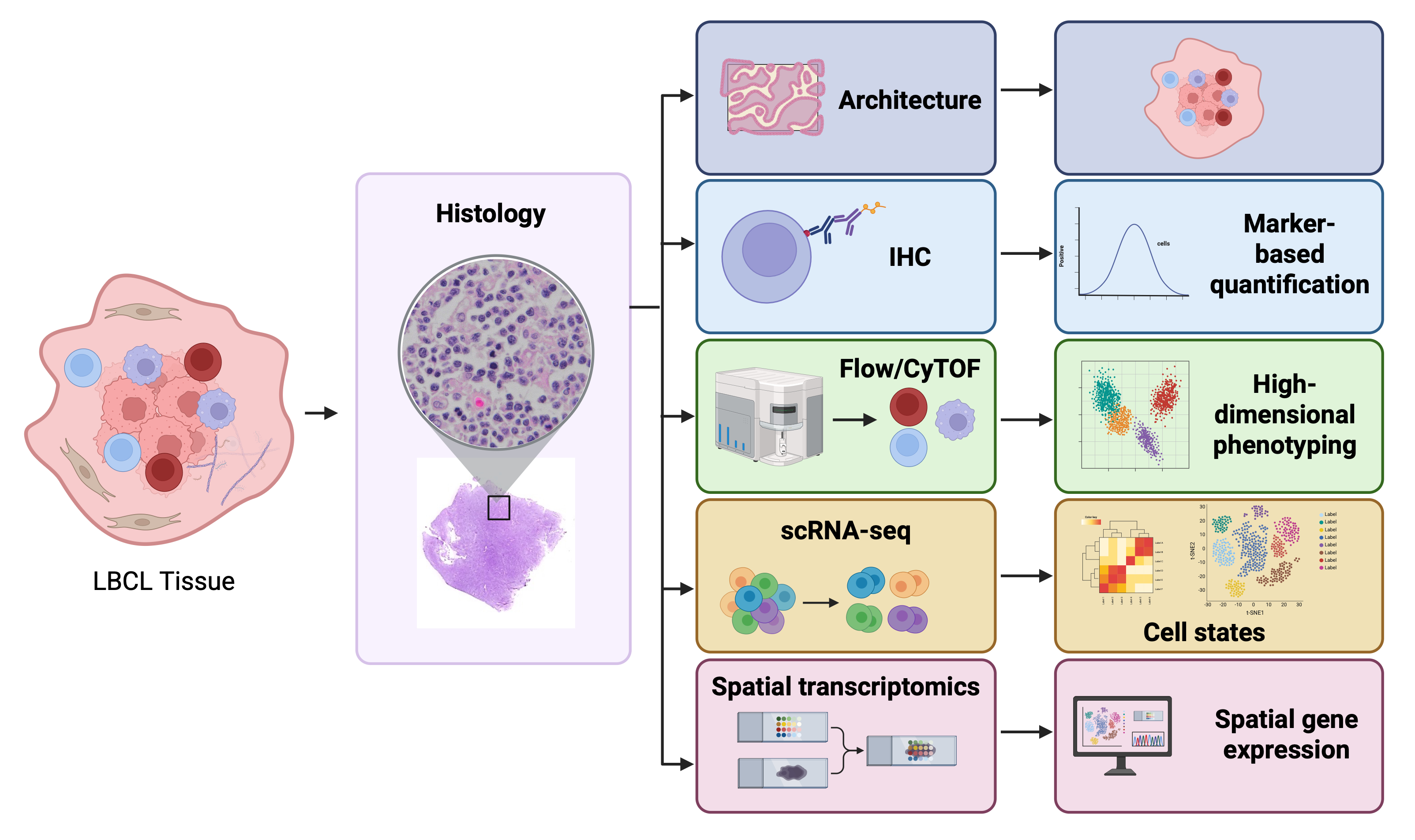
A broad range of complementary methodological approaches is currently available to investigate the TME in LBCL, spanning conventional histopathology, protein-based spatial profiling, cytometric single-cell methods, and transcriptomic technologies (Figure 1).
Conventional Histopathology and Architectural Context of the TME
Formalin-fixed paraffin-embedded (FFPE) hematoxylin and eosin (H&E) sections are essential for assessing tissue architecture and cellular composition in LBCL, DLBCL, and related entities. Stromal gene-expression signatures have been associated with survival in DLBCL (23). The lymphoma microenvironment includes immune, stromal, and vascular components that can be characterized alongside malignant cells (24, 25). Histopathologic and immunophenotypic features remain central to lymphoma classification (26). In nodal LBCL, evaluation includes architectural effacement, residual follicles, fibrosis, necrosis, and abundant non-neoplastic immune cells, including lymphocytes, histiocytes, plasma cells, and eosinophils (25, 27). Histology allows semi-quantitative assessment of immune-rich versus immune-depleted stroma, a finding later corroborated by transcriptomic studies (28, 29). Macrophage-rich areas, including interstitial or sinusoidal histiocytes and ‘starry-sky’ patterns, are readily identified and reflect myeloid biology (30). Spatial imaging in DLBCL has identified recurrent cellular neighborhoods and clinically relevant cell proximities (31). However, despite its indispensable architectural context, the limited cellular specificity and qualitative nature of routine histology necessitate complementary immunophenotypic approaches.
Single-marker immunohistochemistry (IHC) on FFPE tissue has long been the standard method for phenotyping TME components in large LBCL cohorts and for correlating immune infiltrates with clinical outcome (27). Typical panels include markers for T cells (CD3, CD4, CD8), regulatory T cells (FOXP3), macrophages and monocytes (CD68, CD163), and immune checkpoints such as PD-1 and PD-L1. Additional markers include follicular dendritic cells (CD21, CD23), cancer-associated fibroblasts (CAFs; often identified by α-SMA expression), which contribute to extracellular matrix remodeling and immune modulation, and endothelial cells (CD34) to assess non-immune compartments (25, 27, 32). Studies have used both whole sections and tissue microarrays (TMAs). Although TMAs enable high-throughput analysis, intratumoral heterogeneity remains a concern, though this is partly mitigated by the diffuse growth pattern of most DLBCLs. Interpretation has traditionally relied on semi-quantitative scoring, including percentage estimates, hotspot analysis, and dichotomization using empirical cut-offs (32). More recently, digital pathology and automated image analysis have improved reproducibility and enabled continuous quantification of immune cell densities (33).
2.2. Spatial proteomics and multiplex imaging approaches for TME analysis
Multiplex immunofluorescence and imaging mass cytometry are now central tools for mapping the LBCL TME at single-cell resolution while preserving spatial context, but they differ in plex level, readout technology, and typical use cases. Transcriptomic profiling has identified malignant-cell states and associated cellular ecosystems in DLBCL (29). Spatial imaging has identified recurrent cellular neighborhoods and clinically relevant cell proximities (31). Imaging mass cytometry identified phosphorylated S6 as a candidate prognostic and therapeutic marker (34). Quantitative immunofluorescence has also been used to examine the prognostic association of PD-1 and PD-L1 expression (35). Multiplex immunohistochemistry and immunofluorescence enable several markers to be measured in a single tissue section (36). Tyramide signal amplification is one approach to sequential staining and signal detection (37). A published protocol describes how multiplex staining can map immune-cell populations within the TME (38). In DLBCL, optimized Opal-based panels permit analysis of six or more markers per slide, enabling quantitative assessment of immune subsets and their spatial organization (39). High-dimensional mIHC platforms with large antibody panels have been validated for robust, quantitative spatial profiling of tumor and immune phenotypes across cancer types using FFPE tissue (40). By preserving architectural context, mIF reveals in situ cell-cell interactions and spatial immune niches, overcoming limitations of single-plex IHC and enabling comprehensive spatial metrics (41). Quantitative multiplex immunofluorescence combined with automated image analysis, including Automated Quantitative Analysis (AQUA), has been applied in DLBCL to assess T-cell infiltration, PD-1/PD-L1 expression, and spatial relationships between immune-cell populations. These approaches enable quantitative characterization of immune-checkpoint expression and have identified associations between selected immune phenotypes and patient outcomes.
Imaging mass cytometry (IMC) and related techniques, such as multiplexed ion beam imaging (MIBI), combine metal-tagged antibodies with laser ablation and time-of-flight mass spectrometry, enabling simultaneous measurement of 30–40+ markers per pixel while preserving spatial context (42, 43). Imaging mass cytometry has been used to identify candidate signaling markers in DLBCL, including phosphorylated S6 (34). In a separate study, high-multiplex single-cell imaging identified immune features associated with outcomes after CAR T-cell therapy (44). Spatial profiling mapped the organization of immune cells in DLBCL tissue (45). High-plex neighborhood analysis further identified several patterns of immune escape and suppression (46). Panels typically include markers for tumor, T cells, B cells, myeloid cells, stromal cells, and signaling pathways, enabling in situ quantification of cell types, functional states, and pathway activation. Single-cell segmentation supports clustering and neighborhood analysis, generating spatial metrics such as cell-cell contacts and niche composition, thereby enhancing mechanistic understanding of tumor immunity (47). Methodological reviews highlight IMC’s ability to detect >40 markers with minimal spectral overlap at subcellular resolution, making it valuable for detailed dissection of the TME (48).
2.3. Flow and Mass Cytometry for TME Profiling in LBCL
Flow and mass cytometry have been used to (1) quantify TME composition in LBCL at high dimensionality, (2) derive prognostic signatures, and (3) complement tissue‑based spatial methods. Although powerful for deep phenotyping, these methods lose spatial information and require fresh or viably preserved material (49, 50). Routine multi-parameter flow cytometry of fresh lymphoma biopsies provides quantitative data on malignant B cells and non-neoplastic immune populations, including residual B cells, T cells, NK cells, monocytes, granulocytes, and plasma cells, enabling assessment of the TME. Flow-derived immune composition, particularly the relative abundance of T cells, NK cells, and normal B cells versus malignant cells, correlates with clinical outcome in patients with DLBCL treated with immunochemotherapy. These findings align with the “inflamed” and “depleted” TME categories defined by gene expression profiling and immunohistochemistry, supporting the biological relevance of flow-based TME analysis (50, 51). High-dimensional cytometry has further identified expanded activated and regulatory T cell subsets that refine functional immune states (52). However, interpretation is limited by the need for fresh or viable tissue, dissociation-related bias, inter-laboratory variability in panels and gating, and the complete loss of spatial information.
Mass cytometry, or cytometry by time-of-flight (CyTOF), extends flow cytometry by using metal-tagged antibodies to simultaneously measure up to about 40 parameters per cell with minimal signal overlap, enabling deep phenotypic and functional profiling of lymphoid and myeloid compartments. Multimodal profiling identified distinct patterns of T-cell infiltration across nodal B-cell lymphoma entities (49). Mass cytometry has been used to characterize immune-cell profiles in germinal-center B-cell lymphomas (53). In follicular lymphoma, specific intratumoral CD4⁺ T-cell subsets were associated with patient survival (54). CyTOF can resolve rare immune populations and assess T cell exhaustion and regulatory states, with antibody barcoding enabling longitudinal and multi-site sampling (55, 56). However, its application is constrained by the need for fresh or viable tissue, limited accessibility, high cost, complex analytical workflows, and a lack of spatial information, necessitating integration with tissue-based spatial approaches (57).
2.4. Single-cell and spatial transcriptomic approaches for the characterization of the TME
The TME is a highly complex and dynamic ecosystem composed of malignant cells, immune cells, stromal components, vasculature, and the extracellular matrix. It plays an active role in tumor initiation, progression, immune evasion, metastasis, and therapeutic resistance (58, 59). In DLBCL, spatial transcriptomics identified recurrent immune ecosystems associated with patient survival (60). Single-cell RNA sequencing has also been used to study malignant-cell heterogeneity and immune-cell states in DLBCL (61). The broader applications of single-cell profiling to tumor-infiltrating immune cells have been reviewed (62). Primary studies have profiled tumor and immune populations in breast cancer (63), examined immune regulatory programs in multiple myeloma (64), and resolved cell subpopulations in hepatocellular carcinoma (65). A bladder cancer case study illustrates how single-cell profiling can generate hypotheses about treatment resistance (66).
Single-cell RNA sequencing enables transcriptional profiling of individual cells within a tumor, enabling unbiased identification of distinct cell populations and states. Applied to the TME, scRNA-seq has revealed extensive heterogeneity among immune cells, including diverse subsets of T cells, B cells, macrophages, dendritic cells, and innate lymphoid cells, each with specialized functional programs (67). Importantly, scRNA-seq has shown that immune cells within tumors often adopt dysfunctional or reprogrammed states, such as T cell exhaustion or pro-tumorigenic macrophage polarization, that are not readily detectable with bulk transcriptomic approaches. Similarly, stromal cells, such as cancer-associated fibroblasts, consist of multiple transcriptionally distinct subsets with divergent roles in immune regulation, extracellular matrix remodeling, and therapy response. In DLBCL, combined single-cell and bulk RNA sequencing has been used to characterize intratumoral heterogeneity and TME composition (67). Reviews describe the use of these approaches to study DLBCL-specific biology (61) and to resolve tumor-associated immune-cell states across cancers (68).
Spatial transcriptomics maps gene expression in intact tissue sections and preserves information about where cells are located. In pancreatic ductal adenocarcinoma, integrating spatial transcriptomics with single-cell RNA sequencing helped resolve tissue architecture (69). In cutaneous squamous cell carcinoma, combining these methods with imaging identified distinct cellular neighborhoods (70). A review summarizes how spatial transcriptomics has been applied across cancer types (71). For example, immune-infiltrated regions enriched in cytotoxic lymphocytes can coexist with immune-excluded or immunosuppressive areas dominated by stromal cells and myeloid populations. The application of spatial transcriptomics to lymphoma has been reviewed (72). High-parameter imaging independently demonstrated spatial organization of the DLBCL immune microenvironment (73).
Beyond defining cellular identities, scRNA-seq has been pivotal in uncovering dynamic cellular programs within the TME, including activation, differentiation, stress responses, and metabolic reprogramming. These insights have deepened our understanding of how tumors shape their microenvironment to support growth and suppress anti-tumor immunity, complementing conceptual frameworks such as the hallmarks of cancer and the tumor immune microenvironment. In LBCL, single-cell studies have begun to delineate signaling pathways and transcriptional programs associated with immune escape, chemoresistance, and transformation, thereby highlighting potential biomarkers and therapeutic targets. Dissociating tissue for conventional single-cell RNA sequencing removes the original positions of cells. Spatial transcriptomics preserves that context and has been used to examine TME heterogeneity in EBV-positive DLBCL (74). Spatial profiling has also distinguished macrophage populations with different localization and prognostic associations (75).
2.5. Integrated Approaches
Single-cell RNA sequencing identifies cell states, while spatial profiling shows where those states occur in tissue. The two approaches have been combined to resolve pancreatic tumor architecture (69) and to map cellular neighborhoods in cutaneous squamous cell carcinoma (70). Computational integration can help relate cell states across datasets (76). In DLBCL, spatial profiling has identified recurrent immune ecosystems (60), characterized heterogeneity in EBV-positive disease (74), and distinguished macrophage populations with different spatial and prognostic features (75). As these technologies evolve and integrate with proteomic, epigenomic, and imaging-based approaches, they are expected to play a central role in biomarker discovery, patient stratification, and the development of more effective, microenvironment-informed therapies for LBCL and other malignancies. Integrating single-cell and spatial data can generate hypotheses about which cells communicate within tissue (77). Spatial multi-omics can relate immune-cell location to molecular features measured in the same tissue context (78). These approaches are also being developed to study how the TME changes over time or after treatment (79).
2.6. LBCL Biology: Spatial immune organization and its functional consequences
The tumor microenvironment in LBCL is not a random mixture of immune and stromal cells but a highly organized ecosystem in which spatial architecture actively shapes cellular phenotypes, intercellular communication, and therapeutic response. Recent large-scale single-nucleus multiome profiling of 232 LBCL biopsies has defined three reproducible microenvironmental archetypes (LymphoMAPs) that capture fundamental modes of spatial immune organization. The fibroblast/macrophage-rich (FMAC) archetype is characterized by scarce T cells and abundant cancer-associated fibroblasts and tumor-associated macrophages, reflecting an immune-excluded stroma that likely impedes T cell trafficking and engagement with malignant B cells. The lymph node-like (LN) archetype is enriched for naive and memory T cells, supported by follicular dendritic cells and other lymphoid architectural elements, corresponding to an immune-inflamed, well-organized milieu conducive to effective T cell priming and function. In contrast, the T cell-exhausted (TEX) archetype is dominated by activated macrophages and exhausted CD8+ T cells, representing an immune-inflamed but functionally suppressed setting in which T cells are abundant yet transcriptionally imprinted with exhaustion and dysfunction (80).
The clinical relevance of these archetypes appears to depend on treatment context. In the reported CD19-directed CAR T-cell cohorts, LymphoMAPs were strongly associated with progression-free survival, with the most favorable outcomes observed in patients with lymph node-like (LN) tumors, intermediate outcomes in those with fibroblast/macrophage-rich (FMAC) tumors, and the least favorable outcomes in those with T-cell-exhausted (TEX) tumors (80). These findings suggest that T-cell abundance alone is insufficient to characterize the pretreatment immune environment and that cellular organization, functional state, and stromal composition may provide additional prognostic information. Although treatment-comparator analyses suggest that the clinical relevance of these archetypes may differ between CAR T-cell therapy and standard treatments, the available evidence does not establish LymphoMAPs as independently validated predictive biomarkers. Their ability to identify patients who derive additional benefit from CAR T-cell therapy requires confirmation in independent cohorts with appropriate treatment comparators and adjustment for established clinical and molecular risk factors. Complementary spatial transcriptomic and proteomic analyses of 78 DLBCL samples further resolved seven distinct cellular niches, each defined by unique cellular compositions, spatial organizations, and niche-specific programs of intercellular communication that shape the phenotypic states of both tumor B cells and resident immune cells. Notably, DLBCLs arising in immune-privileged sites (e.g., CNS, testis) displayed diffuse niches with extensive intermingling of T cells and malignant B cells, together with transcriptional hallmarks of T cell activation and effector function, suggesting that these T cells may be primed for anti-tumor immunity despite the clinically aggressive behavior of such lymphomas. At the same time, many of these T cells showed features of exhaustion, highlighting a dynamic balance between anti-tumor immunity and immune escape dictated by local niche architecture (28).
These findings mark a conceptual shift from traditional cell-centric or marker-based classifications toward a spatially resolved, ecosystem-level understanding of LBCL biology. Whereas bulk and single-cell approaches primarily capture cellular composition, spatial multi-omics shows that the functional states of both malignant and immune cells are emergent properties of their microenvironmental context. This redefines lymphoma classification not only by intrinsic tumor programs but also by the organization of interacting cellular neighborhoods that govern immune accessibility and dysfunction. However, whether these spatial archetypes represent stable biological entities or dynamic, therapy-induced states remains an open question.
2.7. Limitations
Despite their transformative impact, spatial transcriptomics and multiplex imaging approaches have important technical and analytical limitations. In spot-based spatial transcriptomics, a capture spot can contain transcripts from multiple cells, limiting direct cell-type assignment (81). Variability in RNA preservation, particularly in FFPE-derived specimens, may further reduce transcript detection and introduce cohort-specific biases (82). In addition, the high dimensionality of spatial multi-omics data necessitates complex computational pipelines for integration, deconvolution, and spatial domain inference, which remain sensitive to parameter choices and algorithmic assumptions. Published assessments of imaging-based spatial transcriptomics provide benchmarks for reproducibility (83). Related technical limitations have also been discussed in musculoskeletal research (84). For single-cell RNA-seq, regularized negative binomial regression is one approach to normalization and variance stabilization (85).
Finally, current cost and throughput limitations restrict large-scale clinical application and may introduce selection biases in spatially profiled cohorts. Spatial analyses are inherently correlative and do not directly establish functional interactions between neighboring cells. Spatial transcriptomics can distinguish anatomical domains, as demonstrated in the human dorsolateral prefrontal cortex (86). Spatial resolution and transcript capture vary by platform (87). Inferred cell types and niche boundaries also depend on the computational methods used (88). These limitations should be considered when comparing spatial maps across tissues or technologies (89). A further challenge is distinguishing reproducible biological organization from patterns introduced by sampling and analytical choices. A single tissue section may capture only part of a heterogeneous lymphoma, while biopsy site, tumor-cell content, tissue processing, and computational segmentation can influence the apparent composition of spatial niches. Moreover, cell-cell proximity and inferred ligand-receptor interactions generate mechanistic hypotheses rather than direct evidence of signaling. Orthogonal protein-level measurements, perturbation experiments, and matched longitudinal samples are needed to determine which spatial features reflect stable biological programs and which change with treatment or disease progression. Without such validation, increasingly detailed cellular atlases risk producing classifications that are difficult to compare across cohorts or translate into clinically useful assays.
3. TME and LBCL
Cellular composition and functional diversity of the LBCL microenvironment
Gene expression profiling studies of DLBCL, a major subtype of LBCL, have shifted attention from predominantly lymphoma cell-intrinsic mechanisms to the tumor microenvironment (TME). Subsequent studies have revealed a heterogeneous ecosystem of myeloid and lymphoid populations whose abundance, activation state, and functional phenotype vary considerably across tumors. These populations can exert opposing effects on lymphoma biology, ranging from antitumor cytotoxicity and antigen presentation to immune suppression, stromal remodeling, and support of malignant-cell survival.
Macrophages can adopt inflammatory or immunoregulatory states within the TME (90). In DLBCL models, KIF2C has been linked to M2-like polarization through STAT3 and IL-10 signaling (91). An M2-associated gene signature has also been evaluated as a prognostic model (92). In a rituximab-treated DLBCL cohort, increased M2-like macrophage infiltration was associated with poorer prognosis (93). Other DLBCL profiling studies have identified macrophage populations with strong inflammatory programs (94). Their prognostic interpretation nevertheless depends on the markers used, spatial localization, lymphoma subtype, and accompanying immune populations. Functional and spatial profiling may therefore be more informative than assigning macrophages to a binary polarization category. A DLBCL profiling study linked autophagy-related gene expression in pro-inflammatory macrophages to BCL2 overexpression (95).
Neutrophils are another component of the myeloid compartment. Tumor-associated neutrophils expressing ELANE and CXCR1/2 can participate in cytokine-rich myeloid niches, co-localize with macrophages, and exert tumor-supportive and immunosuppressive functions, including APRIL-mediated survival signaling. Circulating neutrophil counts have been examined in relation to outcomes in newly diagnosed DLBCL (96). A separate study developed a prognostic model based on neutrophil-related genes (97). Tissue infiltration by tumor-associated neutrophils was associated with poorer prognosis in a DLBCL cohort (98). A higher pretreatment neutrophil-to-lymphocyte ratio was associated with poorer survival in separate cohorts (99, 100). Tissue eosinophilia has been described in B-cell lymphomas (101, 102). Eosinophils can have diverse functions in other tumor settings (103). Mast cells can influence inflammation and angiogenesis in tumors (104). Their presence and potential roles in lymphomas have been reviewed (105).
Myeloid-derived suppressor cells (MDSCs) contribute an immunoregulatory component to this compartment. CD11b-positive, CD33-positive, HLA-DR-low/negative MDSCs can suppress T-cell proliferation and effector activity and promote regulatory immune states. Circulating monocytic MDSCs have been associated with advanced disease, high-risk features, inferior survival, and poor CAR T-cell response (106, 107). Dendritic cells (DCs), by contrast, provide antigen-presenting activity and support cytotoxic T-cell priming. Tumor-infiltrating CD11c-positive DCs can occupy T-cell-rich mixed myeloid/DC neighborhoods, although PD-L1 expression may impose local inhibitory effects. Myeloid-derived suppressor cells and systemic inflammation have been evaluated as prognostic features in newly diagnosed DLBCL (108). In tissue, reduced CD11c-positive dendritic cell infiltration was associated with adverse features and outcomes in a DLBCL cohort (109). A separate study developed a composite microenvironmental gene-expression predictor for formalin-fixed tissue (110). In head and neck squamous cell carcinoma, the proportion of circulating dendritic cells was inversely correlated with the proportion of circulating CD4⁺CD25⁺ T cells (111). An NK-associated gene-expression model has been explored for risk stratification in DLBCL (112). In an R-CHOP-treated cohort, NK-cell count was associated with survival (113). Studies in other tumor settings describe how the TME can impair NK-cell function (114), including experimentally observed reversible dysfunction (115).
T cells are a central adaptive immune component of the LBCL microenvironment. Tumor-infiltrating CD3-positive populations include CD8-positive cytotoxic cells, CD4-positive helper cells, and regulatory T cells, with variable expression of exhaustion-associated receptors such as PD-1, TIM-3, and LAG-3. These cells can organize into tumor-proximal, T-cell-rich neighborhoods alongside DCs and macrophages. Greater T-cell infiltration generally characterizes immune-inflamed tumors, whereas low T-cell abundance is associated with immune-depleted or “cold” microenvironments (116). DLBCL immune classifications have incorporated CD8⁺ T-cell and NK-cell features (116). A low T-cell proportion has been associated with immune escape and poorer survival (117). Broader microenvironmental signatures have also been used to define biologically and clinically distinct lymphoma groups (118). Activated CD8⁺ T cells can be present despite checkpoint signaling (119). In HIV-associated DLBCL, HIV status and antiretroviral exposure provide additional context for interpreting T-cell composition (120). CD4-positive T cells provide helper activity and participate in interactions with DCs and macrophages; their abundance and functional state also vary between GCB- and ABC-DLBCL (116, 121). Computational studies have been reviewed as a means of characterizing the DLBCL microenvironment (122). Studies of Th1/Th2 markers have addressed peripheral T-cell lymphoma (123) and, separately, Th1/Th2 imbalance in B-cell diffuse large-cell lymphoma (124). Th9 cells produce IL-9, which can have context-dependent effects in tumors (125, 126). The DLBCL literature has discussed a potential role for IRF8 in IL-9-mediated immune regulation (127). Experimental work in other cancer settings has linked type-17 immunity to CD8⁺ T-cell exhaustion (128).
Regulatory T cells add further complexity because their clinical significance depends on phenotype and context. FOXP3-positive Tregs can suppress antitumor responses through CTLA-4, IL-10, TGF-β, and other mechanisms, and can co-localize with exhausted CD8-positive T cells and M2-like macrophages. The association between FOXP3-positive infiltration and survival in DLBCL varies with the method used to quantify these cells (129). Th17 biology is a separate consideration, with studies examining its regulation and clinical associations in lymphoma and other tumors (130, 131). The composition of the immune infiltrate has been associated with outcome in DLBCL cohorts (132). Within the regulatory compartment, infiltration by effector Tregs has been associated with poorer prognosis (133). A separate study examined the adverse prognostic association of TIM-3⁺FOXP3⁺ Tregs (134). The functions of Tregs and the effects of emerging therapies on these cells have been reviewed in lymphoid malignancies (135). In DLBCL, Vδ1⁺ γδ T cells have been characterized in blood and tumor tissue (136). Reviews describe the functional plasticity of γδ T cells (137, 138), their potential use in B-cell malignancy immunotherapy (139), and their interactions with other cells in the TME (140). NKT-cell recognition of B cells depends in part on CD1d-mediated antigen presentation (141). In peripheral blood from patients with DLBCL, CD73-positive CD8-positive T cells produced more IFN-γ and TNF-α after in vitro stimulation than their CD73-negative counterparts (142). The principal cellular and structural compartments of the LBCL microenvironment are summarized in Table 1. An earlier study also examined the relationship between CD4-positive T-cell responses and outcomes in large B-cell non-Hodgkin lymphoma (143).
Table 1. Immune-cell ecosystem of the LBCL microenvironment: phenotypic signatures, functional states, spatial niches, and clinical relevance.
| Major compartment | Representative populations or states | Principal functions in LBCL | Clinical/biological relevance |
|---|---|---|---|
| Myeloid | M1-/M2-associated programs; TANs; MDSCs; DCs | Inflammation, antigen presentation, immune suppression, angiogenic and tumor-supportive signaling | Composition and functional polarization are associated with immune state, disease aggressiveness, survival, and treatment response (90–110) |
| Innate cytotoxic cells | NK cells | Tumor-cell recognition and cytotoxicity; cytokine production | Functional exhaustion can occur; greater NK-cell abundance has been associated with favorable outcomes and therapeutic response (111–115) |
| Conventional T cells | CD3+, CD8+, and CD4+ T cells | Cytotoxicity, helper activity, immune surveillance, and organization of immune-rich neighborhoods | Abundance and exhaustion state distinguish immune-inflamed from immune-depleted microenvironments and are associated with outcome and treatment response (116–121) |
| Helper/regulatory T-cell states | Th1, Th2, Th9, Th17, Tregs | Cytokine production, immune activation or suppression, macrophage modulation | Functional effects are state dependent; clinical significance is established for some populations but remains uncertain for others (121–134) |
| Other lymphoid populations | γδ T cells; NKT cells | Innate-like cytotoxicity and immune regulation | γδ T cells may contribute to antitumor activity; the role of NKT cells remains poorly defined (133–141) |
| Stromal populations | CAFs; FRCs; FDCs | Tissue organization, ECM remodeling, immune-cell trafficking, immune exclusion, B-cell support | Stromal reprogramming accompanies disruption of normal lymphoid architecture and can generate tumor-supportive niches (144) |
| Extracellular matrix | Stromal-1/ECM-dense; stromal-2/angiogenic; ECM-high regions | Structural organization, regulation of cellular access, angiogenesis and tissue remodeling | Distinct ECM states have been associated with favorable versus adverse biological and clinical features (23) |
|
Vascular/ lymphatic |
Vascular endothelial cells; lymphatic endothelial cells | Angiogenesis, lymphangiogenesis, vascular permeability and dissemination | Vascular and lymphatic structures form part of the stromal architecture of the lymphoma microenvironment (145–150) |
Abbreviations: CAFs, cancer-associated fibroblasts; DCs, dendritic cells; ECM, extracellular matrix; FDCs, follicular dendritic cells; FRCs, fibroblastic reticular cells; LBCL, large B-cell lymphoma; MDSCs, myeloid-derived suppressor cells; NK, natural killer; NKT, natural killer T; TANs, tumor-associated neutrophils; Th, T helper; TME, tumor microenvironment; Tregs, regulatory T cells.
From individual cell states to interacting microenvironmental ecosystems
The biological effects of the LBCL microenvironment cannot be explained solely by the abundance of individual cell populations. Instead, immune, stromal, extracellular matrix, and vascular components interact within spatially organized microenvironments that can either support immune surveillance or favor immune escape and lymphoma-cell survival. For example, tumor-infiltrating T cells occur in neighborhoods containing DCs and macrophages, while exhausted CD8-positive T cells and regulatory T cells can co-localize with immunoregulatory macrophages to form suppressive cellular communities. Thus, similar immune-cell populations may have different biological consequences depending on their activation state and cellular neighbors. NKT-cell responses to B-cell lymphoma have been reviewed in the context of CD1d-dependent recognition (145). Stromal organization also varies across lymphoma entities. In classic Hodgkin lymphoma, cancer-associated fibroblasts show spatial heterogeneity associated with patient survival (146). In B-cell lymphomas, follicular dendritic cells can show altered marker expression (147). These cells help organize the display of antigens to B cells (148). In DLBCL, stromal and immune heterogeneity has been discussed in relation to prognosis and therapeutic targeting (149). The association between fibroblast abundance and survival differs across B-cell lymphomas and carcinomas (150). In experimental DLBCL systems, tumor-activated lymph node fibroblasts altered chemokine expression and impaired T-cell motility (144). The broader roles of cancer-associated fibroblasts in B-cell lymphoma have been reviewed (151). Fibroblastic reticular cells also provide a niche for lymph node-resident macrophages in experimental models (152). Disruption of normal follicular organization is also evident in the Tfh-Tfr-FDC axis. Classical Tfh cells normally provide B-cell help through CD40L-CD40 and ICOS-ICOSL interactions and help maintain germinal-center architecture. T follicular helper and regulatory populations vary across lymphoma contexts. In follicular lymphoma, follicular helper T-cell interactions have been linked to immune evasion (153). Experimental work has also examined the ability of cytotoxic CD4-positive follicular T cells to kill lymphoma cells (154). In DLBCL, experimental work has implicated Tfh-derived IL-21 in regulating FOXP3 expression in Tfr-like cells (155). Consistent with this architectural disruption, CD21-positive/CD23-positive/CD35-positive FDC meshworks are frequently absent or fragmented in de novo DLBCL, with preserved structures occurring predominantly in residual follicles or transformed follicular lymphoma (156, 157).
The extracellular matrix further shapes these ecosystems. The stromal-1 signature is associated with extracellular matrix deposition and favorable survival, whereas the stromal-2 signature reflects angiogenesis and is associated with poorer survival (23). ECM-high regions at tumor borders and perivascular sites can contain CAFs, FRCs, macrophages, and PD-L1-positive myeloid cells and may restrict T-cell infiltration. TAM- and CAF-mediated matrix remodeling further alters collagen and fibronectin organization and can contribute to permissive stromal environments. Fibroblastic reticular cells can also provide a supportive niche for lymph node-resident macrophages (152). These observations support a model in which the LBCL TME is organized into recurring functional ecosystems rather than a uniform admixture of independent cell populations. Table 2 summarizes the major spatially defined configurations emerging from the available evidence.
Table 2. Integrated spatial and functional ecosystems of the LBCL tumor microenvironment.
| Spatial/functional ecosystem | Principal components | Defining organization and interactions | Associated or proposed biological effects |
|---|---|---|---|
| Immune-inflamed/T-cell-rich niche | CD8+ and CD4+ T cells, DCs, NK cells, macrophages | T-cell-rich regions containing antigen-presenting and cytotoxic populations; DCs and NK cells can co-localize with effector T cells | Supports antigen presentation and antitumor immune activity; greater immune-cell density is generally associated with favorable outcome |
| Tumor-proximal immunosuppressive niche | Exhausted CD8+ T cells, Tregs, M2-associated macrophages, MDSCs | Checkpoint-expressing/exhausted lymphocytes occur with suppressive myeloid and regulatory populations near lymphoma cells | Reduced cytotoxic activity, immune evasion, and tumor persistence |
| Disrupted follicular niche | Residual Tfh-like cells, Tfr populations, fragmented FDC networks | Loss of classical Tfh activity and FDC architecture disrupts normal germinal-center T-B-cell interactions | Loss of organized follicular immune regulation and increasing dependence on non-cognate stromal/myeloid support |
| ECM-dense/stromal-1 niche | Collagen, SPARC, fibronectin, fibroblasts, CAFs/FRCs, macrophages | Dense matrix particularly at tumor borders and perivascular regions; relatively low angiogenesis | Restriction of dissemination and angiogenesis; associated with more favorable clinical features |
| ECM-remodeling/stromal-2 niche | Activated fibroblasts, CAFs, M2-associated macrophages, endothelial cells, MMP-rich ECM | Active matrix degradation and remodeling with increased microvessel density and reduced dense collagen | Angiogenesis, tumor spread, immune alteration, and adverse prognosis |
| Stromal immune-exclusion niche | CAFs, FRCs, ECM, macrophages and PD-L1+ myeloid cells | ECM-rich stromal regions form physical and signaling barriers that limit effective T-cell infiltration | Immune exclusion and reduced cytotoxic access to lymphoma cells |
| Perivascular/angiogenic niche | Vascular endothelial cells, CAFs, ECM, M2-associated macrophages | Endothelial remodeling and angiogenesis occur within fibroblast- and macrophage-rich perivascular regions | Vascular remodeling, immune alteration, tumor support, and therapeutic resistance |
| Lymphatic/FRC dissemination niche | LECs, FRCs, CXCL12, BAFF, lymphatic vessels | Perivascular FRC networks guide lymphoma cells toward lymphatic structures while providing survival signals; LECs increase permeability and provide cytokine support | Lymphoma-cell survival, intravasation, lymphangiogenesis, and dissemination |
Abbreviations: BAFF, B-cell activating factor; CAFs, cancer-associated fibroblasts; CXCL12, C-X-C motif chemokine ligand 12; DCs, dendritic cells; ECM, extracellular matrix; FDC, follicular dendritic cell; FRCs, fibroblastic reticular cells; LECs, lymphatic endothelial cells; MDSCs, myeloid-derived suppressor cells; MMP, matrix metalloproteinase; NK, natural killer; PD-L1, programmed death-ligand 1; SPARC, secreted protein acidic and rich in cysteine; Tfh, T follicular helper; Tfr, T follicular regulatory; Tregs, regulatory T cells.
Shared Immunosuppressive Stroma: Lessons from Pancreatic Ductal Adenocarcinoma for DLBCL
Although diffuse large B-cell lymphoma (DLBCL) and pancreatic ductal adenocarcinoma (PDAC) arise in distinct tissue environments, PDAC studies provide a useful framework for investigating how stromal remodeling and myeloid-cell interactions may influence antitumor immunity. PDAC is characterized by an extensive desmoplastic stroma that can restrict immune-cell trafficking and therapeutic penetration. In contrast, DLBCL generally lacks this degree of desmoplasia but exhibits heterogeneous stromal programs, including extracellular matrix-rich stromal-1 and angiogenesis-associated stromal-2 signatures that have been linked to clinical outcomes. A DLBCL cohort study evaluated immunohistochemical markers of stromal-1 and stromal-2 signatures in relation to prognosis (158). Vascular and lymphatic remodeling intersects with these stromal programs. In a three-dimensional DLBCL model, microvascular structures were used to study interactions between lymphoma cells and their vascular environment (159). In a separate experimental study, lymphoma-derived WNT10A promoted endothelial-to-mesenchymal transition through β-catenin and Snail signaling (160). Endothelial-to-mesenchymal transition has also been reviewed as a potential source of cancer-associated fibroblasts (161). In a lymphoma-on-chip model, lymph node stromal cells supported DLBCL cell survival and migration (162). In a clinical DLBCL cohort, stromal fibronectin and SPARC expression were associated with survival (163). In solid-tumor models, desmoplastic stroma restricted T-cell entry into tumor tissue (164). Reviews of upper gastrointestinal cancers have also examined how fibroblast heterogeneity and matrix remodeling may contribute to drug resistance (165).
Myeloid-mediated immune suppression represents another potential point of mechanistic convergence. In PDAC, tumor-associated macrophages, myeloid-derived suppressor cells, and neutrophils can inhibit antitumor T-cell activity through immunoregulatory cytokines, metabolic pathways, and checkpoint signaling. DLBCL studies have also identified macrophage populations with distinct inflammatory and immunoregulatory programs (94). Hypoxia-associated signaling may further influence angiogenesis, metabolic adaptation, and myeloid-cell recruitment, but its role in organizing immunosuppressive niches is more extensively characterized in PDAC than in DLBCL. Thus, mechanisms established in PDAC provide hypotheses for functional investigation in DLBCL rather than direct evidence of shared stromal architecture or equivalent therapeutic vulnerabilities.
Genetic Control of the TME in LBCL
Genetic and epigenetic alterations in LBCLs actively instruct the TME, generating distinct immune and stromal ecosystems rather than passively reflecting tissue context, as illustrated in Figure 2. Integrative genomic and transcriptomic studies show that recurrent somatic mutations, copy number alterations, and structural variants in malignant B cells shape immune composition, cytokine signaling, and stromal organization within the lymphoma niche (7, 9).
A central mechanism of genetically encoded TME remodeling is immune evasion via impaired antigen presentation. Lesions affecting B2M and HLA class I and II genes, together with epigenetic repression driven by mutations in EZH2, CREBBP, and EP300, blunt antigen presentation and interferon-responsive programs, resulting in immune-depleted or immune-excluded microenvironments associated with adverse outcomes (161, 162). In addition, genetic activation of immune checkpoint pathways provides a complementary mechanism of suppression. Copy number gains at 9p24.1, encompassing PD-L1, PD-L2, and JAK2, drive constitutive checkpoint ligand expression and JAK/STAT signaling, promoting T cell exhaustion despite immune infiltration, particularly in primary mediastinal B cell lymphoma and subsets of DLBCL (163, 164). In upper gastrointestinal cancers, fibroblast heterogeneity and extracellular matrix remodeling have been discussed as potential contributors to drug resistance (165).
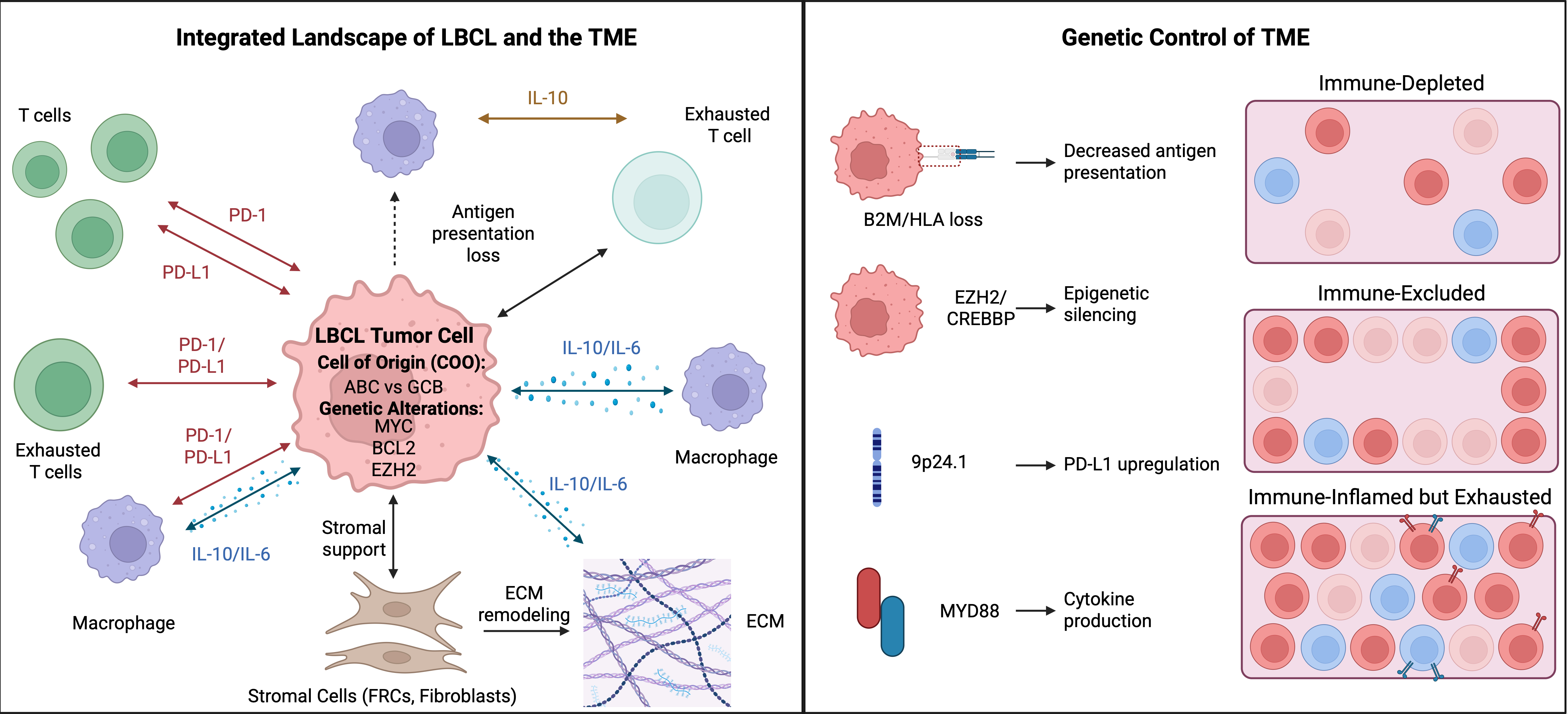
Beyond immune recognition, oncogenic signaling reshapes cytokine and chemokine networks, including IL-6 and IL-10, thereby influencing the recruitment, differentiation, and functional state of immune populations within the tumor microenvironment. These signaling programs can also promote fibroblast activation, extracellular matrix deposition and remodeling, angiogenesis, and the formation of mesenchymal or fibrotic niches that physically and functionally restrict immune-cell access to tumor regions (24). Collectively, these alterations can bias the development of distinct immune and stromal states and contribute to spatial heterogeneity within the TME. However, tumor genotype alone does not determine the resulting phenotypes, which can be substantially modified by anatomical site, disease stage, prior therapeutic exposure, and reciprocal interactions with surrounding non-malignant cells. Moreover, selective pressures imposed during tumor progression and treatment may further reshape these microenvironmental programs. Consequently, the extent to which genetically associated TME phenotypes remain stable, evolve, or converge toward common states over the course of disease remains incompletely established.
Functional Organization and Spatial Heterogeneity in LBCL
In LBCL, a separate analysis described microenvironmental archetypes and examined their associations with treatment outcomes (80). Recent spatial studies further demonstrate that the LBCL TME comprises recurrent cellular neighborhoods, or immune niches, characterized by distinct cellular compositions, transcriptional programs, and ligand-receptor interactions. These findings suggest that the spatial organization of immune and stromal cells provides biological and prognostic information beyond cellular abundance alone, although its independent contribution to disease progression and clinical outcomes requires further validation (45).
A major concept emerging from these studies is spatial immune exclusion, in which cytotoxic lymphocytes are prevented from efficiently infiltrating malignant B cell clusters despite being present within the tumor tissue. Imaging mass cytometry identified immune-poor tumor regions with limited T cell infiltration, revealing a structural mechanism of immune escape that conventional immune-cell quantification cannot capture (45). The coexistence of proliferative lymphoma cells and T cells expressing multiple inhibitory receptors identifies spatially restricted immune states associated with aggressive disease, although spatial profiling alone cannot establish that these neighborhoods directly cause progression or therapeutic resistance (28). Genomic profiling has identified recurrent genetic and functional drivers of DLBCL (166). Genetic inactivation of B2M and CD58 can impair immune recognition of DLBCL cells (167). In primary mediastinal large B-cell lymphoma and nodular sclerosing Hodgkin lymphoma, 9p24.1 amplification was associated with increased PD-1 ligand expression (168). Selective JAK2 inhibition reduced lymphoma growth in experimental models of these diseases (169). Single-cell profiling has identified transcriptional and genetic heterogeneity within nodal B-cell lymphomas (170). In DLBCL, the spatial arrangement of immune cells has also been examined in relation to clinical outcomes (171).
Malignant B cells can reshape the surrounding myeloid compartment. In DLBCL models, CREBBP or EP300 mutations altered macrophage polarization through the FBXW7-NOTCH-CCL2/CSF1 pathway (172). Spatial profiling identified C1q-positive tumor-associated macrophages in DLBCL tissue, although their effects on neighboring cells require functional validation (75). In a different lymphoma setting, tumor-associated macrophage abundance was associated with survival in classic Hodgkin lymphoma (173). Stromal features also carry prognostic information. In rituximab-treated DLBCL, stromal SPARC expression was associated with survival (174). In a murine lymphoma model, CCR7 and lymphotoxin acted together to create a stromal niche that supported lymphoma growth in secondary lymphoid organs (175). The location of immune cells can also matter. In a DLBCL cohort, closer proximity between PD-L1-positive B-cell-rich neighborhoods and CD8-positive T-cell-rich neighborhoods was associated with shorter progression-free survival (31). This association highlights the importance of tissue organization when interpreting T-cell infiltration. Spatial profiling identified C1q-positive tumor-associated macrophages in DLBCL tissue. Their effects on nearby cells require functional validation (75).
Lymphoma cells further exploit stromal compartments by adhering to extracellular matrix components such as fibronectin and SPARC, thereby activating integrin- and focal adhesion-associated survival pathways (158, 168). These extracellular matrix-rich regions frequently co-localize with macrophages and fibroblasts, creating physical and biochemical barriers that restrict immune-cell infiltration and contribute to spatial immune exclusion. Fibroblastic reticular cells (FRCs) are a critical stromal partner. FRCs primed by lymphoma cells reduce the motility of CD8⁺ T cells and CAR T cells, limiting immune-tumor contact; targeting these programs restores T cell cytotoxicity in LBCL models (145). Single-cell profiling has identified transcriptional and genetic heterogeneity within nodal B-cell lymphomas (170). In DLBCL, the spatial arrangement of immune cells has been examined in relation to clinical outcomes (171). In DLBCL models, CREBBP or EP300 mutations altered macrophage polarization through the FBXW7-NOTCH-CCL2/CSF1 pathway (172). In classic Hodgkin lymphoma, tumor-associated macrophage abundance has also been associated with survival (173). Stromal SPARC expression has been associated with survival in rituximab-treated DLBCL (174).
Although CD8⁺ T cells can infiltrate LBCL tissues, their antitumor activity is often constrained by inhibitory signaling within the tumor microenvironment. In particular, engagement of the PD-1/PD-L1 axis can promote local T-cell dysfunction and exhaustion, thereby limiting effective cytotoxic responses despite the presence of tumor-infiltrating lymphocytes. However, recent spatial analyses indicate that the extent of immune infiltration alone does not necessarily predict effective antitumor immunity, as the functional state and spatial organization of infiltrating cells are equally important. In immune-inflamed phenotypes, T cells residing within inflammatory niches frequently exhibit activated or memory-like states and greater cytotoxic potential, whereas adjacent macrophage-rich regions can contain cells expressing inhibitory checkpoint molecules (28). This spatial coexistence of immune activation and suppression illustrates the functional heterogeneity of the LBCL microenvironment and may help explain why tumors with substantial lymphocytic infiltration do not invariably exhibit effective immune control.
Collectively, these findings indicate that LBCL behavior depends not only on the cellular composition of the TME but also on its spatial architecture. Spatial immune exclusion, immune-inflamed phenotypes, and specialized cellular niches represent distinct microenvironmental configurations associated with immune surveillance, immune escape, and therapeutic response. Further investigation is needed to assess their functional stability, causal contributions, and potential utility as treatment-specific biomarkers. A separate analysis associated specific spatial immune ecosystems with patient survival (60). These findings support testing whether spatial biomarkers improve patient stratification in prospective studies.
Microbial Components of the TME in LBCL
Reviews describe how intratumoral microorganisms and microbial metabolites may influence local immune signaling (176, 177). In solid tumors, microbiota-associated remodeling has also been discussed in relation to immunotherapy response (178). In DLBCL, an exploratory analysis identified microbiome-associated subgroups with differences in estimated immune composition and survival (179). The study also generated hypotheses about MIF-associated signaling. MIF has broader roles in inflammation and cancer (180). Whether microorganisms directly alter MIF signaling in DLBCL remains unknown. These observations illustrate the differing strengths of evidence across proposed components of the LBCL microenvironment. Whereas patient cohorts and spatial profiling studies have established associations between immune-stromal organization and clinical outcomes, the functional contributions of putative intratumoral microbial communities remain uncertain. Studies in other malignancies provide useful mechanistic hypotheses but cannot substitute for direct investigation in LBCL.
Future research should combine contamination-controlled microbial detection, spatial localization, independent patient cohorts, and functional experiments to distinguish genuine tumor-associated microorganisms from technical artifacts and determine whether microbial signals contribute causally to immune remodeling or therapeutic resistance. More broadly, candidate microenvironmental interactions should be prioritized according to convergent spatial, molecular, and functional evidence rather than treating every detected cell state or signaling pathway as an equivalent therapeutic target.
4. Relevance, Therapeutic Implications and Perspectives of TME Characterization
TME interactions in LBCL critically shape disease progression, immune escape, and therapeutic resistance. Genetic and epigenetic alterations impair antigen presentation through loss of MHC class II and combined inactivation of β2-microglobulin and CD58, disrupting immune recognition by CD4⁺ T cells (via MHC class II), CD8⁺ T cells (via MHC class I), and NK cells (162, 175, 176). These tumor-intrinsic mechanisms synergize with macrophage-mediated immune suppression and stromal immune exclusion, and are further influenced by epigenetic regulators such as BAP1, which has been implicated in regulating chromatin states at MHC class II loci (177). Clinically, immune infiltration alone does not predict outcome because T cells in the TME are often dysfunctional due to chronic antigen exposure and checkpoint engagement. In DLBCL cohorts, the prognostic associations of immune markers vary with the cell population measured and its spatial context (31, 132). Co-expression of inhibitory receptors may identify dysfunctional T-cell states, but individual markers should not be interpreted as universal indicators of poor prognosis. Lymphoma-TME interactions further drive therapeutic resistance. Stromal niches blunt antibody-dependent and immune-mediated cytotoxicity, contributing to refractory disease (162).
These observations highlight the potential contribution of immune-stromal interactions to therapeutic resistance and provide a rationale for investigating how the composition and spatial organization of the LBCL microenvironment influence CAR T-cell trafficking, persistence, and antitumor activity. Stromal barriers, immunosuppressive macrophage states, and tumor-associated epigenetic programs may collectively create microenvironmental conditions that limit effective T-cell infiltration and function. Accordingly, experimental strategies targeting stromal programs, modulating macrophage polarization, or inhibiting epigenetic regulators such as EZH2 have been shown to remodel tumor microenvironment components and enhance T-cell-based therapeutic activity in LBCL models (145, 178). These findings support further investigation of microenvironment-directed approaches as potential complements to CAR T-cell therapy, while their clinical relevance, optimal timing, and effects across heterogeneous LBCL microenvironmental states remain to be established.
In summary, reciprocal interactions between malignant B cells and their microenvironment drive LBCL progression and treatment resistance. Spatially organized immune-stromal ecosystems govern immune control and are key therapeutic targets. Reflecting this functional heterogeneity, we provide a hierarchical overview linking immune cell types to functional states and clinical relevance, emphasizing that cellular function, rather than abundance, determines clinical outcome (Table 3).
Table 3. Immune Cell Phenotypes, Functional States, and Clinical Associations in the LBCL Tumor Microenvironment
| Cell type / compartment | Functional state / mechanism | Clinical impact / significant endpoints | Relevance for DLBCL | Refs |
|---|---|---|---|---|
| CD68⁺ TAMs | Abundant myeloid infiltrate; mediates lymphoma-stroma crosstalk | Better OS (HR 0.945 per 1% increase; p=0.032); lower stage (p=0.02); associations vary across studies | Total CD68⁺ burden alone has limited prognostic specificity | (179–183) |
| CD163⁺/M2-like TAMs | Immunosuppressive, tumor-supportive; promotes immune escape | Higher stage (p=0.03); worse OS/PFS (p=0.05); enriched in non-GCB DLBCL | Adverse immunosuppressive TME marker | (182, 184–188) |
| SPARC-M⁺ macrophages | SPARC-expressing tumor-associated macrophage population | Associated with more favorable PFS and OS in DLBCL; prognostic significance reported independently of MYC rearrangement and double-/triple-hit status | Candidate prognostic biomarker requiring further validation | (186) |
| Inflammatory TAMs | M0/M1-skewed; IFN/inflammasome and IL1B/CXCL programs | Promotes immune-cell recruitment | Defines an inflammatory TME niche | (93) |
| Noncanonical TAMs | Hybrid M1/M2 inflammatory-suppressive programs | May explain inconsistent prognostic value of total TAM counts | Highlights TAM-state heterogeneity | (183) |
| Recruited monocyte-derived TAMs | CCL5-CCR1/CCR5-mediated recruitment and local differentiation | Promotes TAM accumulation in lymphoma lesions | Mechanism of TME remodeling | (182–184) |
| CD3⁺ T cells | General adaptive immune infiltration | CD3⁺ T-cell density was associated with progression-free survival in an R-CHOP-treated DLBCL cohort. | Favorable immune-infiltration marker | (32) |
| CD8⁺ T cells | Perforin/granzyme-mediated cytotoxicity | Improved response and survival; higher CR odds (p=0.003) | Major antitumor effector population | (117, 179) |
| CD4⁺ T cells | Immune-coordinating; orchestration of cytotoxic, B-cell, and myeloid responses | Prognostic role is unclear; context-dependent | Modulates adaptive and myeloid interactions | (189) |
| FOXP3⁺ lymphocytes | Immunoregulatory/suppressive | FOXP3⁺ cell density was associated with progression-free survival in univariate analysis of an R-CHOP-treated DLBCL cohort. | Context-dependent prognostic marker | (32) |
| Checkpoint-exhausted T cells (PD-1⁺/TIM-3⁺/LAG-3⁺) | Chronic stimulation with reduced effector function | Generally inferior OS/PFS; strongest adverse signals with TIM-3high and PD-1⁺LAG-3⁺ states | Marker of T-cell exhaustion/immune evasion | (188, 190) |
| Tumor/TME interface-PD-L1⁺ | PD-L1-PD-1 checkpoint signaling | Associated with immune infiltration; potential checkpoint-therapy relevance | Immune-evasion/therapeutic marker | (188) |
| Tumor/TME interface—immune infiltration | TAM-T-cell spatial/quantitative interactions | Multiplex immunohistochemistry was used to assess associations between T-cell–macrophage spatial relationships and survival in DLBCL | Defines prognostic immune contexture | (171) |
| Tumor/TME interface—integrated immune contexture | Composite TAM, T-cell, and cytokine states | Associated with progression, treatment response, and survival | Integrated TME architecture | (93) |
| nPD-L1⁺/mPD-L1⁺ | PD-L1 on non-tumor or malignant cells | Associated with IPI and immune infiltration; mPD-L1 negativity associated with worse OS/PFS | Checkpoint/potential predictive biomarker | (188) |
| TIM-3 on lymphoma cells | Checkpoint protein expression on malignant cells | High expression was associated with inferior PFS and OS in one DLBCL cohort | Candidate prognostic association | (191) |
Abbreviations: ADCP, antibody-dependent cellular phagocytosis; CCL5, C-C motif chemokine ligand 5; CCR1, C-C chemokine receptor 1; CCR5, C-C chemokine receptor 5; CR, complete response; DLBCL, diffuse large B-cell lymphoma; ECOG, Eastern Cooperative Oncology Group; GCB, germinal center B-cell-like; HR, hazard ratio; IFN, interferon; IPI, International Prognostic Index; LAG-3, lymphocyte activation gene 3; mPD-L1, malignant-cell programmed death-ligand 1; nPD-L1, nonmalignant-cell programmed death-ligand 1; OS, overall survival; PD-1, programmed cell death protein 1; PD-L1, programmed death-ligand 1; PFS, progression-free survival; SIRPα, signal regulatory protein alpha; SPARC, secreted protein acidic and rich in cysteine; TAMs, tumor-associated macrophages; TILs, tumor-infiltrating lymphocytes; TIM-3, T-cell immunoglobulin and mucin-domain containing-3; TME, tumor microenvironment; Tregs, regulatory T cells.
Therapeutic Implications of the TME in LBCL
These emerging insights into the LBCL tumor microenvironment have fueled the development of TME-directed therapeutic strategies, including immune checkpoint inhibitors, bispecific antibodies, CAR T cell therapies, and immunomodulatory agents such as lenalidomide. These strategies are summarized in Table 3 and illustrated in Figure 4.
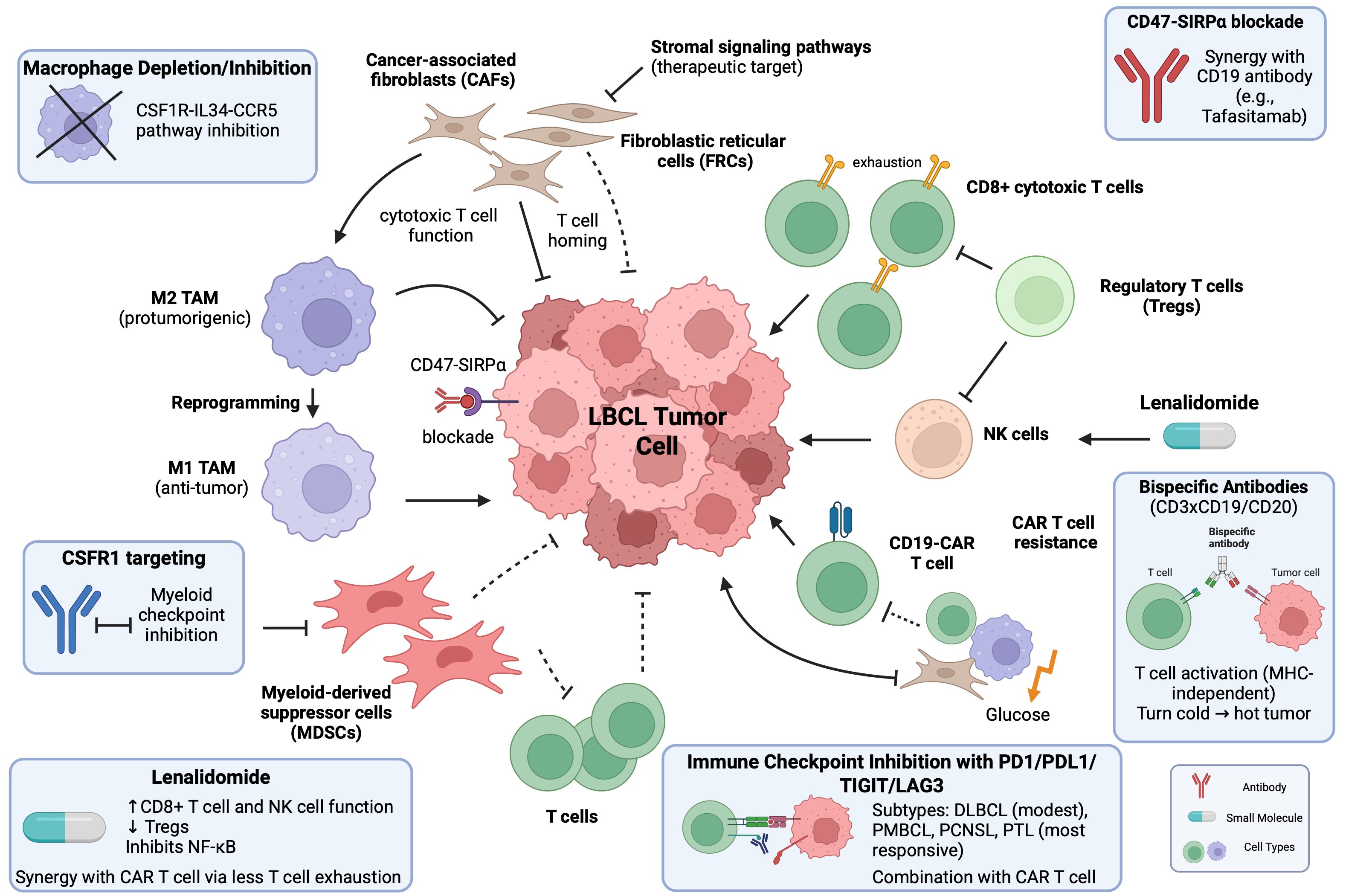
These therapeutic approaches exploit distinct features of the LBCL microenvironment, ranging from inhibitory immune checkpoints and T-cell engagement to modulation of immunosuppressive cellular and signaling networks. Table 4 summarizes the principal TME-directed strategies, their biological targets and mechanisms, and the corresponding clinical implications in LBCL. Published studies describe distinct treatment settings for bispecific antibodies and CAR T cells in LBCL (192, 193). Epcoritamab plus GemOx was evaluated in transplant-ineligible patients with relapsed or refractory DLBCL (194). Correlative analyses examined intratumoral immune populations and potential mechanisms of resistance to odronextamab (195). A separate glofitamab study examined pharmacodynamic changes and molecular correlates of response (196). The lymphoma microenvironment can influence CAR T-cell activity (197). Resistance can also arise from tumor-intrinsic mechanisms (198). A DLBCL study linked an immunosuppressive microenvironment to progression following anti-CD19 CAR T-cell therapy (199). The broader mechanisms of CAR T-cell resistance in DLBCL have been reviewed (200). Metabolic remodeling is being explored as a potential way to improve CAR T-cell activity (201). Treatment after CAR T-cell relapse is a separate clinical question (202). A PD-1-armored CD19 CAR T-cell product has also been investigated (203). Checkpoint-inhibitor studies include early nivolumab experience in hematologic malignancies and a phase II study in relapsed or refractory DLBCL (204, 205). Ibrutinib plus nivolumab was evaluated in relapsed CNS lymphoma (206). Pembrolizumab was studied in relapsed or refractory primary mediastinal large B-cell lymphoma (207). A separate report described nivolumab treatment in primary CNS and testicular lymphoma (208). Atezolizumab plus obinutuzumab was evaluated in relapsed or refractory B-cell lymphoma (209). Other early-phase studies combined checkpoint blockade with a BTK inhibitor (210, 211). Pembrolizumab and durvalumab were each studied with frontline chemoimmunotherapy in DLBCL (212, 213). A phase I study evaluated dual checkpoint blockade in relapsed or refractory lymphoid malignancies (214). A separate trial tested atezolizumab, obinutuzumab, and venetoclax in relapsed or refractory B-cell non-Hodgkin lymphoma (215). LAG-3 is expressed by several lymphocyte subsets in DLBCL (216). TIGIT has also been investigated as an inhibitory receptor in lymphoma (217). Nivolumab plus relatlimab was evaluated in relapsed or progressed B-cell malignancies (218). An exploratory study examined whether LAG3 genetic variation was associated with outcomes after CAR T-cell therapy (219). Macrophage-directed studies include CD47-SIRPα blockade combined with tafasitamab in preclinical and ex vivo models, and observational work has examined circulating MDSCs as prognostic markers in B-cell lymphoma (220, 221). Lenalidomide has been studied as an immunomodulatory agent in DLBCL and other B-cell lymphomas (222, 223). A preclinical study examined whether lenalidomide could overcome resistance to CD19-directed CAR T cells (224). Its clinical use in primary CNS lymphoma has been reviewed (225). A separate report described lenalidomide monotherapy as salvage treatment in recurrent primary CNS lymphoma (226). Table 4 summarizes the principal TME-directed strategies, their biological targets and mechanisms, and the corresponding clinical implications in LBCL.
Table 4. Therapeutic targeting of the LBCL tumor microenvironment: biological rationale, candidate interventions, and clinical implications.
| Category | Target / mechanism | Key TME finding | Therapeutic implication | Intervention / evidence | Refs |
|---|---|---|---|---|---|
| Targeting TAMs | M2-like macrophage polarization | High M2-like TAM density is associated with poor OS/PFS | Macrophage depletion, recruitment blockade, or repolarization may reduce tumor immunosuppression | TLR7 agonist imiquimod and TLR3 agonist poly(I:C); preclinical | (188, 227, 228) |
| CD47-SIRPα | CD47 signaling inhibits macrophage-mediated phagocytosis | Blockade can enhance ADCP, particularly with tumor-targeting antibodies | Anti-CD47 + tafasitamab (anti-CD19); preclinical/translational | (221, 229) | |
| CCL5-CCR1/CCR5 | CCL5 signaling promotes monocyte/TAM recruitment into the TME | Recruitment blockade may reduce tumor-supportive TAM accumulation | CCL5 antagonist; migration and xenograft studies | (230) | |
| IL-34-CSF1R | IL-34 promotes monocyte recruitment and is associated with adverse DLBCL features | Disruption of IL-34-CSF1R signaling represents a potential macrophage-directed strategy | Candidate therapeutic axis; limited DLBCL-specific therapeutic evidence | (231) | |
| Stromal prognostic programs | Stromal-1/stromal-2 programs | Stromal-1 is associated with favorable outcome, whereas angiogenic stromal-2 is adverse | Potential prognostic stratification; therapeutic utility remains unvalidated | Biomarker/mechanistic evidence | (23) |
| PD-L1⁺ CAFs | CAF-associated PD-L1 can suppress T-cell cytotoxicity | Stromal checkpoint signaling may contribute to immune escape | PD-1/PD-L1 blockade; indirect therapeutic rationale | (144) | |
| CCL19/CCL21 reduction | Reduced CCL19/CCL21 impairs T-cell homing | Restoration of stromal immune trafficking may enhance antitumor immunity | Mechanistic/preclinical rationale | (144) | |
| CAF-mediated CAR T resistance | CAF-rich stroma can impede CAR T-cell migration and access | Stromal targeting may improve CAR T-cell efficacy | Preclinical therapeutic rationale | (144) | |
| PCNSL/PTL stromal compartment | CAF abundance and functional evidence remain limited | Therapeutic significance remains undefined | Insufficient disease-specific evidence | (232) | |
| Targeting MDSCs / suppressive myeloid cells | Immunosuppressive activity | MDSCs suppress T- and NK-cell function through multiple pathways | Reducing MDSC abundance or activity may restore antitumor immunity | Therapeutic rationale; limited LBCL-specific clinical evidence | (233) |
| Prognostic relevance | Elevated MDSC levels are associated with adverse outcomes | Potential prognostic and response biomarker | Biomarker evidence | (220) | |
| PCNSL myeloid checkpoints | Myeloid-rich immune states involve PD-L1 and TIM-3/galectin-9 signaling | Supports investigation of myeloid checkpoint targeting | Disease-specific biological/therapeutic rationale | (234, 235) | |
| PTL immune escape | MHC-II loss contributes to immune-privileged biology | Supports targeting mechanisms of immune escape; direct MDSC involvement remains uncertain | Mechanistic evidence | (236, 237) | |
| CAR T resistance | LAMM populations are associated with poor CAR T-cell response | CSF1R blockade may improve CAR T-cell activity by targeting suppressive macrophages | Anti-CSF1R antibody (AFS98); preclinical | (238) | |
| TME-modifying drugs — Lenalidomide | Immune modulation | Enhances T/NK-cell activity, modulates Tregs, and affects NF-κB signaling in ABC-DLBCL | Modulates both malignant and immune compartments | Lenalidomide; mechanistic and clinical evidence | (222, 223) |
| Clinical activity | ORR ~30% in r/r DLBCL, with greater activity reported in ABC/non-GCB disease | Demonstrates activity in selected molecular contexts | Lenalidomide; clinical evidence | (239) | |
| CAR T-cell modulation | Reduces CD8⁺ CAR T-cell exhaustion in murine models | Potential combination strategy to enhance CAR T-cell function | Lenalidomide + CD19 CAR T cells; preclinical | (224) | |
| PCNSL cytokine modulation | CSF IL-10 changes correlate with treatment response | CSF IL-10 changes correlate with treatment response | Lenalidomide-based therapy; clinical evidence | (225, 226) | |
| PTL | Direct disease-specific evidence remains limited | Therapeutic relevance remains uncertain | Limited disease-specific evidence | (237) |
Abbreviations: ABC, activated B-cell-like; ADCP, antibody-dependent cellular phagocytosis; CAFs, cancer-associated fibroblasts; CAR T, chimeric antigen receptor T cell; CCL, C-C motif chemokine ligand; CCR, C-C chemokine receptor; CD, cluster of differentiation; CSF, cerebrospinal fluid; CSF1R, colony-stimulating factor 1 receptor; DLBCL, diffuse large B-cell lymphoma; FRCs, fibroblastic reticular cells; IL, interleukin; LAMMs, lymphoma-associated macrophage and monocyte populations; LBCL, large B-cell lymphoma; MDSCs, myeloid-derived suppressor cells; MHC-II, major histocompatibility complex class II; NF-κB, nuclear factor kappa B; NK, natural killer; ORR, overall response rate; OS, overall survival; PCNSL, primary central nervous system lymphoma; PD-1, programmed cell death protein 1; PD-L1, programmed death-ligand 1; PFS, progression-free survival; PTL, primary testicular lymphoma; r/r, relapsed/refractory; SIRPα, signal regulatory protein alpha; TAMs, tumor-associated macrophages; TIM-3, T-cell immunoglobulin and mucin-domain containing-3; TLR, Toll-like receptor; TME, tumor microenvironment; Tregs, regulatory T cells.
5. Discussion
Fibroblast architecture and its prognostic associations have also been studied in classic Hodgkin lymphoma (146). In DLBCL, PD-L1 expression has been examined in relation to clinicopathologic features and prognosis (240). A systematic review evaluated the prognostic value of non-neoplastic cell populations (241). A review discussed the role of the tumor microenvironment in DLBCL pathogenesis and its therapeutic implications (242). A treatment-correlative study examined tumor microenvironment features in patients receiving CD19-directed CAR T-cell therapy, chemotherapy, or transplantation (243). Candidate prognostic molecular biomarkers in DLBCL have been reviewed in the rituximab era (244). Another review discussed how the microenvironment of lymphoid neoplasms informs the evaluation of immune-based therapies (245). A separate review examined strategies and challenges for targeting the tumor microenvironment in B-cell lymphoma (246). Experimental work has examined how PD-L1-expressing macrophages influence lymphoma growth (247). High-plex imaging revealed distinct patterns of immune escape and suppression (46). High-parameter imaging also demonstrated spatially defined immune microenvironments in DLBCL (73). A methods review describes approaches to spatial profiling in immuno-oncology (248). Multimodal analysis has separately examined TME heterogeneity in relation to immune activity and evasion (249). Outside lymphoma, perivascular fibroblasts have been studied in glioblastoma as a potential correlate of immunotherapy resistance (250). Single-cell multi-omics has been reviewed as an approach to studying lymphoma heterogeneity (251). Broader oncology work describes strategies for integrating multiple data types in clinical research (252). One experimental workflow integrates spatially resolved datasets generated from the same tissue section (253). In DLBCL, multi-omics studies have been reviewed as a source of candidate prognostic biomarkers (254). DIABLO integrates multi-omics measurements, whereas MISO integrates spatial omics with tissue morphology (255, 256). Multimodal artificial intelligence offers further approaches to combining clinical and molecular data (257). Spatial transcriptomics identified recurrent immune ecosystems in DLBCL tissue (60). High-parameter imaging provides a complementary view of their cellular architecture (73). Spatial transcriptomics has characterized heterogeneity in EBV-positive DLBCL (74). A separate study combined single-cell and spatial profiling in testicular DLBCL (258).
An integrative genomic analysis identified four DLBCL immune quadrants. Quadrant assignment was associated with clinical benefit in a CD20 × CD3 bispecific antibody cohort, whereas an association was not detected in the CD19 CAR T-cell cohorts studied (259). Complementary experimental approaches, including three-dimensional B-cell lymphoma culture systems, provide platforms to investigate tumor-microenvironment interactions and evaluate antibody-based therapies (260). These findings highlight the need for functional validation and treatment-specific assessment of candidate biomarkers. A correlative analysis examined pretreatment TME features in LBCL treated with CD19 CAR T cells or with chemotherapy and transplantation (243). A separate study found that DLBCL immune quadrants were associated with clinical benefit in a bispecific-antibody cohort, while an association was not detected in the CAR T-cell cohorts studied (259). Reviews of prognostic biomarkers provide background for risk assessment (244, 254). General multimodal integration strategies (252) and checkpoint-blockade literature (261) provide context for future validation. Spatial profiling provides several ways to measure immune and stromal organization in tissue (248). Deep-learning methods have been developed to integrate spatial omics with tissue morphology (256). Multimodal AI offers additional ways to combine clinical and molecular measurements (257). Reduced gene signatures and multiplex immunohistochemistry panels remain candidate approaches for prospective validation. Studies of odronextamab and glofitamab have examined immune or molecular correlates of response (195, 196). In LBCL, separate analyses have examined TME features in relation to CAR T-cell and chemotherapy/transplant outcomes (243). The DLBCL immune-quadrant study illustrates why candidate biomarkers should be evaluated in the treatment setting where they would be used (259). Biomarker-adapted treatment has also been examined in high-risk LBCL (262). Prospective studies are needed to establish whether spatial immune classifications improve treatment allocation beyond existing clinical and molecular markers.
Spatial and multi-omics profiling has expanded our understanding of how immune-stromal ecosystems interact with tumor-intrinsic programs to shape biological heterogeneity, immune evasion, and therapeutic response in LBCL (74). These advances have generated candidate biomarkers for risk stratification, treatment-response assessment, and identifying potentially actionable immune states (52). However, most proposed signatures remain exploratory, and their clinical utility requires independent validation, standardized analytical methods, and evidence of added value beyond established clinical and molecular predictors. Translating high-dimensional tissue atlases into practical diagnostic assays will also require attention to specimen availability, intratumoral heterogeneity, cost, and turnaround time. Prospective studies should determine whether individual microenvironmental signatures are merely associated with clinical outcomes or can reliably identify patients who benefit from specific interventions. Integrating validated TME biomarkers into clinical trials may ultimately support more precise therapeutic selection and rational combination strategies for patients with high-risk or relapsed LBCL. Comparative immune profiling has been reported in primary CNS and testicular LBCL (232). Primary testicular lymphoma has distinctive clinical features that warrant separate consideration (237). Within primary testicular lymphoma, a T-cell-inflamed microenvironment was associated with favorable prognosis (263). Other work has documented variation in NK and NK-like T-cell populations across B-cell proliferations (264).
Outstanding Challenges and Future Directions
Spatial transcriptomics requires standardization of pre-analytical variables, assay chemistries, and computational workflows. Tissue preservation and other pre-analytical variables can influence gene-expression measurements, while standardized metrics can help assess the reproducibility of imaging-based spatial datasets (83, 265). High-definition spatial transcriptomics improves the resolution of gene-expression mapping in tissue (266). Same-section integration workflows can combine complementary spatial measurements (253). Reviews discuss how spatial multi-omics can be used to characterize immune organization and changes in the TME (78, 79). Translating these data into clinically deployable assays will require prospective validation. A key translational distinction is whether a microenvironmental signature is prognostic, predicts benefit from a particular treatment, or is simply associated with an established molecular subtype. These functions require different validation strategies. A signature associated with poor survival after immunochemotherapy, for example, may not identify patients who benefit from CAR T-cell therapy or macrophage-directed treatment. Treatment-specific analyses, independent validation cohorts, and predefined biomarker thresholds are therefore necessary before spatial classifications can guide therapeutic selection. Longitudinal sampling is equally important because treatment may alter the very cellular states used to assign patients to a microenvironmental category. Metabolic remodeling of the TME has been proposed as a strategy to improve CAR T-cell activity (201). In DLBCL, lipid metabolic reprogramming (267) and potential metabolic vulnerabilities (268) have been reviewed. A separate review focuses on interactions between metabolism and the immune environment (269). Experimental work has also investigated cholesterol metabolic reprogramming in DLBCL (270). Experimental work in solid-tumor models showed that tumor-derived lactic acid can shape macrophage polarization (271). Broader studies of hypoxia and antitumor immunity provide additional context for investigating these interactions in lymphoma (272).
Future directions should prioritize longitudinal profiling of the tumor microenvironment to capture dynamic remodeling under therapeutic pressure, including immune activation and escape mechanisms. In addition, developing clinically applicable surrogate assays derived from high-dimensional spatial data, such as reduced gene expression signatures, multiplex immunohistochemistry panels, or digital pathology-based models, will be essential for translation into routine practice. Ultimately, integrating spatial biology with functional validation and prospective clinical trials will be required to translate TME insights into precision immuno-oncology strategies for patients with LBCL.
References
1. Alaggio R, Amador C, Anagnostopoulos I, Attygalle AD, Araujo IB de O, Berti E, et al. The 5th edition of the World Health Organization classification of haematolymphoid tumours: lymphoid neoplasms. Leukemia. 2022;36(7):1720–1748. https://doi.org/10.1038/s41375-022-01620-2
2. Kurz KS, Ott M, Kalmbach S, Steinlein S, Kalla C, Horn H, et al. Large B-cell lymphomas in the 5th edition of the WHO-classification of haematolymphoid neoplasms-updated classification and new concepts. Cancers (Basel). 2023;15(8):2285. https://doi.org/10.3390/cancers15082285
3. Alizadeh AA, Eisen MB, Davis RE, Ma C, Lossos IS, Rosenwald A, et al. Distinct types of diffuse large B-cell lymphoma identified by gene expression profiling. Nature. 2000;403(6769):503–511. https://doi.org/10.1038/35000501
4. Alduaij W, Collinge B, Ben-Neriah S, Jiang A, Hilton LK, Boyle M, et al. Molecular determinants of clinical outcomes in a real-world diffuse large B-cell lymphoma population. Blood. 2023;141(20):2493–2507. https://doi.org/10.1182/blood.2022018248
5. Lacy SE, Barrans SL, Beer PA, Painter D, Smith AG, Roman E, et al. Targeted sequencing in DLBCL, molecular subtypes, and outcomes: a Haematological Malignancy Research Network report. Blood. 2020;135(20):1759–1771. https://doi.org/10.1182/blood.2019003535
6. Wright GW, Huang DW, Phelan JD, Coulibaly ZA, Roulland S, Young RM, et al. A probabilistic classification tool for genetic subtypes of diffuse large B cell lymphoma with therapeutic implications. Cancer Cell. 2020;37(4):551–568.e14. https://doi.org/10.1016/j.ccell.2020.03.015
7. Schmitz R, Wright GW, Huang DW, Johnson CA, Phelan JD, Wang JQ, et al. Genetics and pathogenesis of diffuse large B-cell lymphoma. N Engl J Med. 2018;378(15):1396–1407. https://doi.org/10.1056/NEJMoa1801445
8. Chapuy B, Wood T, Stewart C, Dunford A, Wienand K, Khan SJ, et al. DLBclass: a probabilistic molecular classifier to guide clinical investigation and practice in diffuse large B-cell lymphoma. Blood. 2025;145(18):2041–2055. https://doi.org/10.1182/blood.2024025652
9. Chapuy B, Stewart C, Dunford AJ, Kim J, Kamburov A, Redd RA, et al. Molecular subtypes of diffuse large B cell lymphoma are associated with distinct pathogenic mechanisms and outcomes. Nat Med. 2018;24(5):679–690. https://doi.org/10.1038/s41591-018-0016-8
10. Abrisqueta P. New insights into first-line therapy in diffuse large B-cell lymphoma: are we improving outcomes? J Clin Med. 2024;13(7):1929. https://doi.org/10.3390/jcm13071929
11. Spinner MA, Advani RH. Current frontline treatment of diffuse large B-cell lymphoma. Oncology (Williston Park). 2022;36(1):51–58. https://doi.org/10.46883/2022.25920940
12. Melchardt T, Egle A, Greil R. How I treat diffuse large B-cell lymphoma. ESMO Open. 2023;8(1):100750. https://doi.org/10.1016/j.esmoop.2022.100750
13. Tilly H, Morschhauser F, Sehn LH, Friedberg JW, Trněný M, Sharman JP, et al. Polatuzumab vedotin in previously untreated diffuse large B-cell lymphoma. N Engl J Med. 2022;386(4):351–363. https://doi.org/10.1056/NEJMoa2115304
14. Abramson JS, Palomba ML, Gordon LI, Lunning MA, Wang M, Arnason J, et al. Lisocabtagene maraleucel for patients with relapsed or refractory large B-cell lymphomas (TRANSCEND NHL 001): a multicentre seamless design study. Lancet. 2020;396(10254):839–852. https://doi.org/10.1016/S0140-6736(20)31366-0
15. Abramson JS, Palomba ML, Gordon LI, Lunning M, Wang M, Arnason J, et al. Two-year follow-up of lisocabtagene maraleucel in relapsed or refractory large B-cell lymphoma in TRANSCEND NHL 001. Blood. 2024;143(5):404–416. https://doi.org/10.1182/blood.2023020854
16. Neelapu SS, Jacobson CA, Ghobadi A, Miklos DB, Lekakis LJ, Oluwole OO, et al. Five-year follow-up of ZUMA-1 supports the curative potential of axicabtagene ciloleucel in refractory large B-cell lymphoma. Blood. 2023;141(19):2307–2315. https://doi.org/10.1182/blood.2022018893
17. Bock AM, Epperla N. Therapeutic landscape of primary refractory and relapsed diffuse large B-cell lymphoma: recent advances and emerging therapies. J Hematol Oncol. 2025;18(1):68. https://doi.org/10.1186/s13045-025-01702-5
18. Hanahan D, Weinberg RA. Hallmarks of cancer: the next generation. Cell. 2011;144(5):646–674. https://doi.org/10.1016/j.cell.2011.02.013
19. Frick M, Dörken B, Lenz G. The molecular biology of diffuse large B-cell lymphoma. Ther Adv Hematol. 2011;2(6):369–379. https://doi.org/10.1177/2040620711419001
20. Patil S, Rajput S, Patil S, Mhaiskar A. B-cell lymphoma: advances in pathogenesis, diagnosis, and targeted therapies. Pathol Res Pract. 2025;271:156036. https://doi.org/10.1016/j.prp.2025.156036
21. Shaffer AL 3rd, Young RM, Staudt LM. Pathogenesis of human B cell lymphomas. Annu Rev Immunol. 2012;30:565–610. https://doi.org/10.1146/annurev-immunol-020711-075027
22. Fowler NH, Cheah CY, Gascoyne RD, Gribben J, Neelapu SS, Ghia P, et al. Role of the tumor microenvironment in mature B-cell lymphoid malignancies. Haematologica. 2016;101(5):531–540. https://doi.org/10.3324/haematol.2015.139493
23. Lenz G, Wright G, Dave SS, Xiao W, Powell J, Zhao H, et al. Stromal gene signatures in large-B-cell lymphomas. N Engl J Med. 2008;359(22):2313–2323. https://doi.org/10.1056/NEJMoa0802885
24. Scott DW, Gascoyne RD. The tumour microenvironment in B cell lymphomas. Nat Rev Cancer. 2014;14(8):517–534. https://doi.org/10.1038/nrc3774
25. Ng WL, Ansell SM, Mondello P. Insights into the tumor microenvironment of B cell lymphoma. J Exp Clin Cancer Res. 2022;41(1):362. https://doi.org/10.1186/s13046-022-02579-9
26. Swerdlow SH, Campo E, Pileri SA, Harris NL, Stein H, Siebert R, et al. The 2016 revision of the World Health Organization classification of lymphoid neoplasms. Blood. 2016;127(20):2375–2390. https://doi.org/10.1182/blood-2016-01-643569
27. Sethi A, Tandon A, Mishra H, Singh I. Diffuse large B-cell lymphoma: an immunohistochemical approach to diagnosis. J Oral Maxillofac Pathol. 2019;23(2):284–288. https://doi.org/10.4103/jomfp.JOMFP_294_18
28. Dai Y, Kizhakeyil A, Chihara D, Li X, Liu Y, Sainz Zuniga TP, et al. Multi-modal spatial characterization of tumor immune microenvironments identifies targetable inflammatory niches in diffuse large B cell lymphoma. Nat Genet. 2025;57(11):2715–2727. https://doi.org/10.1038/s41588-025-02353-5
29. Steen CB, Luca BA, Esfahani MS, Azizi A, Sworder BJ, Nabet BY, et al. The landscape of tumor cell states and ecosystems in diffuse large B cell lymphoma. Cancer Cell. 2021;39(10):1422–1437.e10. https://doi.org/10.1016/j.ccell.2021.08.011
30. Ennishi D. The biology of the tumor microenvironment in DLBCL: targeting the "don't eat me" signal. J Clin Exp Hematop. 2021;61(4):210–215. https://doi.org/10.3960/jslrt.21015
31. Autio M, Leivonen SK, Meriranta L, Karjalainen-Lindsberg ML, Pellinen T, Leppä S. Spatial analysis of the tumor microenvironment in diffuse large B-cell lymphoma reveals clinically relevant cell interactions and recurrent cellular neighborhoods. Cancer Immunol Res. 2025;13(10):1674–1686. https://doi.org/10.1158/2326-6066.CIR-24-1163
32. Coutinho R, Clear AJ, Mazzola E, Owen A, Greaves P, Wilson A, et al. Revisiting the immune microenvironment of diffuse large B-cell lymphoma using a tissue microarray and immunohistochemistry: robust semi-automated analysis reveals CD3 and FoxP3 as potential predictors of response to R-CHOP. Haematologica. 2015;100(3):363–369. https://doi.org/10.3324/haematol.2014.110189
33. Kumar G, Pandurengan RK, Parra ER, Kannan K, Haymaker C. Spatial modelling of the tumor microenvironment from multiplex immunofluorescence images: methods and applications. Front Immunol. 2023;14:1288802. https://doi.org/10.3389/fimmu.2023.1288802
34. Zhang B, Xie Y, Shen M, Sun J, Li Y, Zhang J, et al. Multiplexed imaging mass cytometry elicits p-S6 as a therapeutic and prognostic factor in diffuse large B-cell lymphoma. Blood Adv. 2025;9(13):3249–3262. https://doi.org/10.1182/bloodadvances.2024015469
35. Li L, Sun R, Miao Y, Tran T, Adams L, Roscoe N, et al. PD-1/PD-L1 expression and interaction by automated quantitative immunofluorescent analysis show adverse prognostic impact in patients with diffuse large B-cell lymphoma having T-cell infiltration: a study from the International DLBCL Consortium Program. Mod Pathol. 2019;32(6):741–754. https://doi.org/10.1038/s41379-018-0193-5
36. Tan WCC, Nerurkar SN, Cai HY, Ng HHM, Wu D, Wee YTF, et al. Overview of multiplex immunohistochemistry/immunofluorescence techniques in the era of cancer immunotherapy. Cancer Commun (Lond). 2020;40(4):135–153. https://doi.org/10.1002/cac2.12023
37. Stack EC, Wang C, Roman KA, Hoyt CC. Multiplexed immunohistochemistry, imaging, and quantitation: a review, with an assessment of Tyramide signal amplification, multispectral imaging and multiplex analysis. Methods. 2014;70(1):46–58. https://doi.org/10.1016/j.ymeth.2014.08.016
38. Gorris MAJ, Martynova E, Sweep MWD, van der Hoorn IAE, Sultan S, Claassens MJDE, et al. Multiplex immunohistochemical analysis of the spatial immune cell landscape of the tumor microenvironment. J Vis Exp. 2023;(198):e65717. https://doi.org/10.3791/65717
39. Lee CW, Ren YJ, Marella M, Wang M, Hartke J, Couto SS. Multiplex immunofluorescence staining and image analysis assay for diffuse large B cell lymphoma. J Immunol Methods. 2020;478:112714. https://doi.org/10.1016/j.jim.2019.112714
40. Banik G, Betts CB, Liudahl SM, Sivagnanam S, Kawashima R, Cotechini T, et al. High-dimensional multiplexed immunohistochemical characterization of immune contexture in human cancers. Methods Enzymol. 2020;635:1–20. https://doi.org/10.1016/bs.mie.2019.05.039
41. McGue JJ, Edwards JJ, Griffith BD, Frankel TL. Multiplex fluorescent immunohistochemistry for preservation of tumor microenvironment architecture and spatial relationship of cells in tumor tissues. Methods Mol Biol. 2023;2660:235–246. https://doi.org/10.1007/978-1-0716-3163-8_16
42. Schulz D, Zanotelli VRT, Fischer JR, Schapiro D, Engler S, Lun XK, et al. Simultaneous multiplexed imaging of mRNA and proteins with subcellular resolution in breast cancer tissue samples by mass cytometry. Cell Syst. 2018;6(1):25–36.e5. https://doi.org/10.1016/j.cels.2017.12.001
43. Giesen C, Wang HAO, Schapiro D, Zivanovic N, Jacobs A, Hattendorf B, et al. Highly multiplexed imaging of tumor tissues with subcellular resolution by mass cytometry. Nat Methods. 2014;11(4):417–422. https://doi.org/10.1038/nmeth.2869
44. Jin J, Lin L, Meng J, Jiang L, Zhang M, Fang Y, et al. High-multiplex single-cell imaging analysis reveals tumor immune contexture associated with clinical outcomes after CAR T cell therapy. Mol Ther. 2024;32(5):1252–1265. https://doi.org/10.1016/j.ymthe.2024.03.023
45. Colombo AR, Hav M, Singh M, Xu A, Gamboa A, Lemos T, et al. Single-cell spatial analysis of tumor immune architecture in diffuse large B-cell lymphoma. Blood Adv. 2022;6(16):4675–4690. https://doi.org/10.1182/bloodadvances.2022007493
46. Reiss DJ, Nakayama Y, Weng AP, Stokes ME, Sehn L, Steidl C, et al. High-plex imaging and cellular neighborhood spatial analysis reveals multiple immune escape and suppression patterns in diffuse large B-cell lymphoma. Leukemia. 2024;38(5):1164–1168. https://doi.org/10.1038/s41375-024-02239-1
47. Keren L, Bosse M, Marquez D, Angoshtari R, Jain S, Varma S, et al. A structured tumor-immune microenvironment in triple negative breast cancer revealed by multiplexed ion beam imaging. Cell. 2018;174(6):1373–1387.e19. https://doi.org/10.1016/j.cell.2018.08.039
48. Cotechini T, Jones O, Hindmarch CCT. Imaging mass cytometry in immuno-oncology. Methods Mol Biol. 2023;2614:1–15. https://doi.org/10.1007/978-1-0716-2914-7_1
49. Roider T, Baertsch MA, Fitzgerald D, Vöhringer H, Brinkmann BJ, Czernilofsky F, et al. Multimodal and spatially resolved profiling identifies distinct patterns of T cell infiltration in nodal B cell lymphoma entities. Nat Cell Biol. 2024;26(3):478–489. https://doi.org/10.1038/s41556-024-01358-2
50. Yu T, Xu-Monette ZY, Lagoo A, Shuai W, Wang B, Neff J, et al. Flow cytometry quantification of tumor-infiltrating lymphocytes to predict the survival of patients with diffuse large B-cell lymphoma. Front Immunol. 2024;15:1335689. https://doi.org/10.3389/fimmu.2024.1335689
51. Chen Z, Deng X, Ye Y, Zhang W, Liu W, Zhao S. Flow cytometry-assessed PD1/PDL1 status in tumor-infiltrating lymphocytes: a link with the prognosis of diffuse large B-cell lymphoma. Front Oncol. 2021;11:687911. https://doi.org/10.3389/fonc.2021.687911
52. Díaz Herrero A, Le PH, Renaud L, Meignin V, Thieblemont C, Blanc V, et al. High-dimensional spectral cytometry identifies follicular regulatory CD8+ T cells in diffuse large B-cell lymphoma. Clin Transl Immunology. 2025;14(11):e70062. https://doi.org/10.1002/cti2.70062
53. Roussel M, Lhomme F, Roe CE, Bartkowiak T, Gravelle P, Laurent C, et al. Mass cytometry defines distinct immune profile in germinal center B-cell lymphomas. Cancer Immunol Immunother. 2020;69(3):407–420. https://doi.org/10.1007/s00262-019-02464-z
54. Yang ZZ, Kim HJ, Villasboas JC, Price-Troska T, Jalali S, Wu H, et al. Mass cytometry analysis reveals that specific intratumoral CD4+ T cell subsets correlate with patient survival in follicular lymphoma. Cell Rep. 2019;26(8):2178–2193.e3. https://doi.org/10.1016/j.celrep.2019.01.085
55. Zunder ER, Finck R, Behbehani GK, Amir EAD, Krishnaswamy S, Gonzalez VD, et al. Palladium-based mass tag cell barcoding with a doublet-filtering scheme and single-cell deconvolution algorithm. Nat Protoc. 2015;10(2):316–333. https://doi.org/10.1038/nprot.2015.020
56. Lin D, Maecker HT. CyTOF intracellular cytokine assays for antigen-specific T cells. Methods Mol Biol. 2024;2779:395–405. https://doi.org/10.1007/978-1-0716-3738-8_18
57. Spitzer MH, Nolan GP. Mass cytometry: single cells, many features. Cell. 2016;165(4):780–791. https://doi.org/10.1016/j.cell.2016.04.019
58. Ye X, Wang L, Nie M, Wang Y, Dong S, Ren W, et al. A single-cell atlas of diffuse large B cell lymphoma. Cell Rep. 2022;39(3):110713. https://doi.org/10.1016/j.celrep.2022.110713
59. Ozmen F, Ozmen TY, Ors A, Janghorban M, Rames MJ, Li X, et al. Single-cell RNA sequencing reveals different cellular states in malignant cells and the tumor microenvironment in primary and metastatic ER-positive breast cancer. NPJ Breast Cancer. 2025;11(1):95. https://doi.org/10.1038/s41523-025-00808-w
60. Diaz-Herrero A, Pelaez-Prestel HF, Massenet-Regad L, Veyssiere M, Calvani J, Cristinelli C, et al. Spatial transcriptomics unveils immune cellular ecosystems associated with patient survival in diffuse large B-cell lymphoma. Oncoimmunology. 2026;15(1):2660418. https://doi.org/10.1080/2162402X.2026.2660418
61. Li L, Li Q, Niu R, Sun W, Liang H. Single-cell RNA sequencing in diffuse large B-cell lymphoma: tumor heterogeneity, microenvironment, resistance, and prognostic markers. Front Oncol. 2025;15:1583250. https://doi.org/10.3389/fonc.2025.1583250
62. Ren X, Zhang L, Zhang Y, Li Z, Siemers N, Zhang Z. Insights gained from single-cell analysis of immune cells in the tumor microenvironment. Annu Rev Immunol. 2021;39:583–609. https://doi.org/10.1146/annurev-immunol-110519-071134
63. Chung W, Eum HH, Lee HO, Lee KM, Lee HB, Kim KT, et al. Single-cell RNA-seq enables comprehensive tumour and immune cell profiling in primary breast cancer. Nat Commun. 2017;8:15081. https://doi.org/10.1038/ncomms15081
64. Su Q, Long K, Adu M, Jiang M, Li Q, Wan X, et al. Single-cell RNA sequencing reveals immune regulatory mechanisms and molecular therapeutic strategies in the microenvironment of multiple myeloma. Int J Surg. 2026;112(1):333–351. https://doi.org/10.1097/JS9.0000000000003306
65. Gao J, Li Z, Lu Q, Zhong J, Pan L, Feng C, et al. Single-cell RNA sequencing reveals cell subpopulations in the tumor microenvironment contributing to hepatocellular carcinoma. Front Cell Dev Biol. 2023;11:1194199. https://doi.org/10.3389/fcell.2023.1194199
66. Lee HW, Chung W, Lee HO, Jeong DE, Jo A, Lim JE, et al. Single-cell RNA sequencing reveals the tumor microenvironment and facilitates strategic choices to circumvent treatment failure in a chemorefractory bladder cancer patient. Genome Med. 2020;12(1):47. https://doi.org/10.1186/s13073-020-00741-6
67. Zhao Y, Xu H, Zhang M, Li L. Single-cell RNA-seq and bulk RNA-seq reveal intratumoral heterogeneity and tumor microenvironment characteristics in diffuse large B-cell lymphoma. Front Genet. 2022;13:881345. https://doi.org/10.3389/fgene.2022.881345
68. Chen S, Zhou Z, Li Y, Du Y, Chen G. Application of single-cell sequencing to the research of tumor microenvironment. Front Immunol. 2023;14:1285540. https://doi.org/10.3389/fimmu.2023.1285540
69. Moncada R, Barkley D, Wagner F, Chiodin M, Devlin JC, Baron M, et al. Integrating microarray-based spatial transcriptomics and single-cell RNA-seq reveals tissue architecture in pancreatic ductal adenocarcinomas. Nat Biotechnol. 2020;38(3):333–342. https://doi.org/10.1038/s41587-019-0392-8
70. Ji AL, Rubin AJ, Thrane K, Jiang S, Reynolds DL, Meyers RM, et al. Multimodal analysis of composition and spatial architecture in human squamous cell carcinoma. Cell. 2020;182(2):497–514.e22. https://doi.org/10.1016/j.cell.2020.05.039
71. Cilento MA, Sweeney CJ, Butler LM. Spatial transcriptomics in cancer research and potential clinical impact: a narrative review. J Cancer Res Clin Oncol. 2024;150(6):296. https://doi.org/10.1007/s00432-024-05816-0
72. Pickard K, Stephenson E, Mitchell A, Jardine L, Bacon CM. Location, location, location: mapping the lymphoma tumor microenvironment using spatial transcriptomics. Front Oncol. 2023;13:1258245. https://doi.org/10.3389/fonc.2023.1258245
73. Wright KT, Weirather JL, Jiang S, Kao KZ, Sigal Y, Giobbie-Hurder A, et al. Diffuse large B-cell lymphomas have spatially defined, tumor immune microenvironments revealed by high-parameter imaging. Blood Adv. 2023;7(16):4633–4646. https://doi.org/10.1182/bloodadvances.2023009813
74. He MY, Liu M, Yuan J, Lv J, Li W, Yan Q, et al. Spatial transcriptomics reveals tumor microenvironment heterogeneity in EBV positive diffuse large B cell lymphoma. Sci Rep. 2025;15(1):15878. https://doi.org/10.1038/s41598-025-00410-x
75. Liu M, Bertolazzi G, Sridhar S, Lee RX, Jaynes P, Mulder K, et al. Spatially resolved transcriptomics reveal macrophage heterogeneity and prognostic significance in diffuse large B-cell lymphoma. Nat Commun. 2024;15(1):2113. https://doi.org/10.1038/s41467-024-46220-z
76. Stuart T, Satija R. Integrative single-cell analysis. Nat Rev Genet. 2019;20(5):257–272. https://doi.org/10.1038/s41576-019-0093-7
77. Shi W, Zhang Z, Xu X, Tian Y, Feng L, Huang X, et al. Single-cell and spatial transcriptomics integration: new frontiers in tumor microenvironment and cellular communication. Front Immunol. 2025;16:1649468. https://doi.org/10.3389/fimmu.2025.1649468
78. Hsieh WC, Budiarto BR, Wang YF, Lin CY, Gwo MC, So DK, et al. Spatial multi-omics analyses of the tumor immune microenvironment. J Biomed Sci. 2022;29(1):96. https://doi.org/10.1186/s12929-022-00879-y
79. Nguyen H, Eapen MM, Nguyen Q, Sharma A. Spatial omics for profiling the dynamic tumor microenvironment. Clin Transl Immunology. 2026;15(4):e70084. https://doi.org/10.1002/cti2.70084
80. Li X, Singhal K, Deng Q, Chihara D, Russler-Germain D, Harkins RA, et al. Large B cell lymphoma microenvironment archetype profiles. Cancer Cell. 2025;43(7):1347–1364.e13. https://doi.org/10.1016/j.ccell.2025.06.002
81. Moses L, Pachter L. Museum of spatial transcriptomics. Nat Methods. 2022;19(5):534–546. https://doi.org/10.1038/s41592-022-01409-2
82. Ståhl PL, Salmén F, Vickovic S, Lundmark A, Fernández Navarro J, Magnusson J, et al. Visualization and analysis of gene expression in tissue sections by spatial transcriptomics. Science. 2016;353(6294):78–82. https://doi.org/10.1126/science.aaf2403
83. Plummer JT, Segato Dezem F, Cook DP, Park J, Zhang L, Liu Y, et al. Standardized metrics for assessment and reproducibility of imaging-based spatial transcriptomics datasets. Nat Biotechnol. 2026;44(7):1213–1225. https://doi.org/10.1038/s41587-025-02811-9
84. Delos Santos K, Rockel JS, Kapoor M. Spatial transcriptomics: challenges and future directions in musculoskeletal diseases. Curr Opin Rheumatol. 2026;38(2):143–153. https://doi.org/10.1097/BOR.0000000000001140
85. Hafemeister C, Satija R. Normalization and variance stabilization of single-cell RNA-seq data using regularized negative binomial regression. Genome Biol. 2019;20(1):296. https://doi.org/10.1186/s13059-019-1874-1
86. Maynard KR, Collado-Torres L, Weber LM, Uytingco C, Barry BK, Williams SR, et al. Transcriptome-scale spatial gene expression in the human dorsolateral prefrontal cortex. Nat Neurosci. 2021;24(3):425–436. https://doi.org/10.1038/s41593-020-00787-0
87. Lee J, Yoo M, Choi J. Recent advances in spatially resolved transcriptomics: challenges and opportunities. BMB Rep. 2022;55(3):113–124. https://doi.org/10.5483/BMBRep.2022.55.3.014
88. Fang S, Chen B, Zhang Y, Sun H, Liu L, Liu S, et al. Computational approaches and challenges in spatial transcriptomics. Genomics Proteomics Bioinformatics. 2023;21(1):24–47. https://doi.org/10.1016/j.gpb.2022.10.001
89. Park HE, Jo SH, Lee RH, Macks CP, Ku T, Park J, et al. Spatial transcriptomics: technical aspects of recent developments and their applications in neuroscience and cancer research. Adv Sci (Weinh). 2023;10(16):e2206939. https://doi.org/10.1002/advs.202206939
90. Pansy K, Uhl B, Krstic J, Szmyra M, Fechter K, Santiso A, et al. Immune regulatory processes of the tumor microenvironment under malignant conditions. Int J Mol Sci. 2021;22(24):13311. https://doi.org/10.3390/ijms222413311
91. Qian L, Shi R, Yang J, Zhang J. KIF2C regulates macrophage M2 polarization and DLBCL progression by regulating the STAT3/IL-10 axis. Cell Signal. 2025;136:112155. https://doi.org/10.1016/j.cellsig.2025.112155
92. Guo B, Duan Y, Cen H. Prognostic model based on M2 macrophage-related signatures for predicting outcomes, enhancing risk stratification, and providing therapeutic insights in diffuse large B-cell lymphoma. Heliyon. 2024;10(24):e41007. https://doi.org/10.1016/j.heliyon.2024.e41007
93. Nam SJ, Go H, Paik JH, Kim TM, Heo DS, Kim CW, et al. An increase of M2 macrophages predicts poor prognosis in patients with diffuse large B-cell lymphoma treated with rituximab, cyclophosphamide, doxorubicin, vincristine and prednisone. Leuk Lymphoma. 2014;55(11):2466–2476. https://doi.org/10.3109/10428194.2013.879713
94. Serna L, Azcoaga P, Brahmachary M, Caffarel MM, Braza MS. Diffuse large B-cell lymphoma microenvironment displays a predominant macrophage infiltrate marked by a strong inflammatory signature. Front Immunol. 2023;14:1048567. https://doi.org/10.3389/fimmu.2023.1048567
95. Resanoa M, Delgado A, Rodiño C, Villar M, Aiastui A, Braza MS. In the diffuse large B-cell lymphoma microenvironment, autophagy genes are upregulated in pro-inflammatory macrophages and linked to BCL2 overexpression. Front Immunol. 2025;16:1676563. https://doi.org/10.3389/fimmu.2025.1676563
96. Yang Z, Yu W. Clinical significance of circulating neutrophils and lymphocyte subsets in newly diagnosed patients with diffuse large B-cell lymphoma. Clin Exp Med. 2023;23(3):815–822. https://doi.org/10.1007/s10238-022-00867-4
97. Gao X, Luo X, Ye H, Hu S, Yu F, Xu P, et al. Risk model based on neutrophil-related genes constructs to assess prognosis and immune landscape in diffuse large B-cell lymphoma. Am J Clin Oncol. 2026;49(8):372–382. https://doi.org/10.1097/COC.0000000000001272
98. Manfroi B, Moreaux J, Righini C, Ghiringhelli F, Sturm N, Huard B. Tumor-associated neutrophils correlate with poor prognosis in diffuse large B-cell lymphoma patients. Blood Cancer J. 2018;8(7):66. https://doi.org/10.1038/s41408-018-0099-y
99. Shih MF, Lue KH, Wang TF, Chu SC, Huang CH. Association between the neutrophil-to-lymphocyte ratio and infection and survival in diffuse large B cell lymphoma. In Vivo. 2023;37(2):948–954. https://doi.org/10.21873/invivo.13167
100. Porrata LF, Ristow K, Habermann T, Inwards DJ, Micallef IN, Markovic SN. Predicting survival for diffuse large B-cell lymphoma patients using baseline neutrophil/lymphocyte ratio. Am J Hematol. 2010;85(11):896–899. https://doi.org/10.1002/ajh.21849
101. Watanabe K, Shinbo T, Kojima M, Naito M, Tanahashi N, Nara M. B-cell lymphoma associated with eosinophilia. Cancer. 1989;64(8):1682–1685. https://doi.org/10.1002/1097-0142(19891015)64:8<1682::aid-cncr2820640821>3.0.co;2-x
102. Zhou T, Wang HW, Ng SB, Summers T, Xi L, Raffeld M, et al. Tissue eosinophilia in B-cell lymphoma: an underrecognized phenomenon. Am J Surg Pathol. 2024;48(4):426–436. https://doi.org/10.1097/PAS.0000000000002160
103. Grisaru-Tal S, Itan M, Klion AD, Munitz A. A new dawn for eosinophils in the tumour microenvironment. Nat Rev Cancer. 2020;20(10):594–607. https://doi.org/10.1038/s41568-020-0283-9
104. Komi DEA, Redegeld FA. Role of mast cells in shaping the tumor microenvironment. Clin Rev Allergy Immunol. 2020;58(3):313–325. https://doi.org/10.1007/s12016-019-08753-w
105. Ribatti D. Mast cells in lymphomas. Crit Rev Oncol Hematol. 2016;101:207–212. https://doi.org/10.1016/j.critrevonc.2016.03.016
106. Hedström G, Berglund M, Molin D, Fischer M, Nilsson G, Thunberg U, et al. Mast cell infiltration is a favourable prognostic factor in diffuse large B-cell lymphoma. Br J Haematol. 2007;138(1):68–71. https://doi.org/10.1111/j.1365-2141.2007.06612.x
107. Efstratiou P, Damianaki A, Kavidopoulou A, Ioannidou P, Markaki E, Skianis IM, et al. Myeloid-derived suppressor cells exhibit distinct characteristics in bone marrow and blood of individuals with diffuse large B-cell lymphoma. Front Med (Lausanne). 2025;11:1515097. https://doi.org/10.3389/fmed.2024.1515097
108. Foureau DM, Guo F, Steuerwald NM, Druhan LJ, Avalos BR, Copelan E, et al. Prognostic significance of myeloid-derived suppressor cells and systemic inflammation in newly diagnosed diffuse large B-cell lymphoma treated with chemoimmunotherapy. Exp Hematol. 2024;129:104125. https://doi.org/10.1016/j.exphem.2023.10.004
109. Yuan CT, Chuang SS, Cheng PY, Chang K, Wang H, Tsai JH, et al. Decreased CD11c-positive dendritic cells in the tumor microenvironment predict double-hit/triple-hit genotype and survival in diffuse large B-cell lymphoma. J Pathol Clin Res. 2022;8(5):436–447. https://doi.org/10.1002/cjp2.283
110. Ciavarella S, Vegliante MC, Fabbri M, De Summa S, Melle F, Motta G, et al. Dissection of DLBCL microenvironment provides a gene expression-based predictor of survival applicable to formalin-fixed paraffin-embedded tissue. Ann Oncol. 2018;29(12):2363–2370. https://doi.org/10.1093/annonc/mdy450
111. Sakakura K, Chikamatsu K, Takahashi K, Whiteside TL, Furuya N. Maturation of circulating dendritic cells and imbalance of T-cell subsets in patients with squamous cell carcinoma of the head and neck. Cancer Immunol Immunother. 2006;55(2):151–159. https://doi.org/10.1007/s00262-005-0697-y
112. Xiao W, Yu K, Deng X, Zeng Y. Natural killer cell-associated prognosis model characterizes immune landscape and treatment efficacy of diffuse large B cell lymphoma. Cytokine. 2024;182:156726. https://doi.org/10.1016/j.cyto.2024.156726
113. Kim JK, Chung JS, Shin HJ, Song MK, Yi JW, Shin DH, et al. Influence of NK cell count on the survival of patients with diffuse large B-cell lymphoma treated with R-CHOP. Blood Res. 2014;49(3):162–169. https://doi.org/10.5045/br.2014.49.3.162
114. Hu Z, Xu X, Wei H. The adverse impact of tumor microenvironment on NK-cell. Front Immunol. 2021;12:633361. https://doi.org/10.3389/fimmu.2021.633361
115. Pouxvielh K, Marotel M, Drouillard A, Villard M, Moreews M, Bossan A, et al. Tumor-induced natural killer cell dysfunction is a rapid and reversible process uncoupled from the expression of immune checkpoints. Sci Adv. 2024;10(35):eadn0164. https://doi.org/10.1126/sciadv.adn0164
116. Qi J, Xu L, Huang D, He H, Yao J, Zhang J, et al. Defining diffuse large B-cell lymphoma immunotypes by CD8+ T cells and natural killer cells. J Oncol. 2022;2022:3168172. https://doi.org/10.1155/2022/3168172
117. Song JY, Nwangwu M, He TF, Zhang W, Meawad H, Bedell V, et al. Low T-cell proportion in the tumor microenvironment is associated with immune escape and poor survival in diffuse large B-cell lymphoma. Haematologica. 2023;108(8):2167–2177. https://doi.org/10.3324/haematol.2022.282265
118. Kotlov N, Bagaev A, Revuelta MV, Phillip JM, Cacciapuoti MT, Antysheva Z, et al. Clinical and biological subtypes of B-cell lymphoma revealed by microenvironmental signatures. Cancer Discov. 2021;11(6):1468–1489. https://doi.org/10.1158/2159-8290.CD-20-0839
119. Greenbaum AM, Fromm JR, Gopal AK, Houghton AMG. Diffuse large B-cell lymphoma (DLBCL) is infiltrated with activated CD8+ T-cells despite immune checkpoint signaling. Blood Res. 2022;57(2):117–128. https://doi.org/10.5045/br.2022.2021145
120. Coelho J, Roush SM, Xu AM, Puranam K, Mponda M, Kasonkanji E, et al. HIV and prior exposure to antiretroviral therapy alter tumour composition and tumour: T-cell associations in diffuse large B-cell lymphoma. Br J Haematol. 2024;205(1):194–206. https://doi.org/10.1111/bjh.19531
121. Battella S, Cox MC, La Scaleia R, Di Napoli A, Di Landro F, Porzia A, et al. Peripheral blood T cell alterations in newly diagnosed diffuse large B cell lymphoma patients and their long-term dynamics upon rituximab-based chemoimmunotherapy. Cancer Immunol Immunother. 2017;66(10):1295–1306. https://doi.org/10.1007/s00262-017-2026-7
122. Opinto G, Vegliante MC, Negri A, Skrypets T, Loseto G, Pileri SA, et al. The tumor microenvironment of DLBCL in the computational era. Front Oncol. 2020;10:351. https://doi.org/10.3389/fonc.2020.00351
123. Tsuchiya T, Ohshima K, Karube K, Yamaguchi T, Suefuji H, Hamasaki M, et al. Th1, Th2, and activated T-cell marker and clinical prognosis in peripheral T-cell lymphoma, unspecified: comparison with AILD, ALCL, lymphoblastic lymphoma, and ATLL. Blood. 2004;103(1):236–241. https://doi.org/10.1182/blood-2002-05-1352
124. Mori T, Takada R, Watanabe R, Okamoto S, Ikeda Y. T-helper (Th)1/Th2 imbalance in patients with previously untreated B-cell diffuse large cell lymphoma. Cancer Immunol Immunother. 2001;50(10):566–568. https://doi.org/10.1007/s00262-001-0232-8
125. Chen T, Guo J, Cai Z, Li B, Sun L, Shen Y, et al. Th9 cell differentiation and its dual effects in tumor development. Front Immunol. 2020;11:539666. https://doi.org/10.3389/fimmu.2020.01026
126. Li Y, Yu Q, Zhang Z, Wang J, Li S, Zhang J, et al. TH9 cell differentiation, transcriptional control and function in inflammation, autoimmune diseases and cancer. Oncotarget. 2016;7(43):71001–71012. https://doi.org/10.18632/oncotarget.11681
127. Cai M, Chen N. The roles of IRF-8 in regulating IL-9-mediated immunologic mechanisms in the development of DLBCL: a state-of-the-art literature review. Front Oncol. 2022;12:817069. https://doi.org/10.3389/fonc.2022.817069
128. Kim BS, Kuen DS, Koh CH, Kim HD, Chang SH, Kim S, et al. Type 17 immunity promotes the exhaustion of CD8+ T cells in cancer. J Immunother Cancer. 2021;9(6):e002603. https://doi.org/10.1136/jitc-2021-002603
129. Bai Y, He T, Zhang L, Liu Q, Yang J, Zhao Z, et al. Prognostic value of FOXP3+ regulatory T cells in patients with diffuse large B-cell lymphoma: a systematic review and meta-analysis. BMJ Open. 2022;12(9):e060659. https://doi.org/10.1136/bmjopen-2021-060659
130. Zhong W, Xu X, Zhu Z, Du Q, Du H, Yang L, et al. Increased expression of IRF8 in tumor cells inhibits the generation of Th17 cells and predicts unfavorable survival of diffuse large B cell lymphoma patients. Oncotarget. 2017;8(30):49757–49772. https://doi.org/10.18632/oncotarget.17693
131. Ye J, Livergood RS, Peng G. The role and regulation of human Th17 cells in tumor immunity. Am J Pathol. 2013;182(1):10–20. https://doi.org/10.1016/j.ajpath.2012.08.041
132. Autio M, Leivonen SK, Brück O, Mustjoki S, Mészáros Jørgensen J, Karjalainen-Lindsberg ML, et al. Immune cell constitution in the tumor microenvironment predicts the outcome in diffuse large B-cell lymphoma. Haematologica. 2021;106(3):718–729. https://doi.org/10.3324/haematol.2019.243626
133. Nakayama S, Yokote T, Akioka T, Hiraoka N, Nishiwaki U, Miyoshi T, et al. Infiltration of effector regulatory T cells predicts poor prognosis of diffuse large B-cell lymphoma, not otherwise specified. Blood Adv. 2017;1(8):486–493. https://doi.org/10.1182/bloodadvances.2016000885
134. Zhong W, Liu X, Zhu Z, Li Q, Li K. High levels of Tim-3+Foxp3+Treg cells in the tumor microenvironment is a prognostic indicator of poor survival of diffuse large B cell lymphoma patients. Int Immunopharmacol. 2021;96:107662. https://doi.org/10.1016/j.intimp.2021.107662
135. Maharaj K, Uriepero A, Sahakian E, Pinilla-Ibarz J. Regulatory T cells (Tregs) in lymphoid malignancies and the impact of novel therapies. Front Immunol. 2022;13:943354. https://doi.org/10.3389/fimmu.2022.943354
136. Reboursiere E, Gac AC, Garnier A, Salaun V, Reman O, Pham AD, et al. Increased frequencies of circulating and tumor-resident Vδ1+ T cells in patients with diffuse large B-cell lymphoma. Leuk Lymphoma. 2018;59(1):187–195. https://doi.org/10.1080/10428194.2017.1321751
137. Wang Y, Xu Y, Chen H, Zhang J, He W. Novel insights based on the plasticity of γδ T cells in the tumor microenvironment. Explor Immunol. 2022;2(1):98–132. https://doi.org/10.37349/ei.2022.00039
138. Park JH, Lee HK. Function of γδ T cells in tumor immunology and their application to cancer therapy. Exp Mol Med. 2021;53(3):318–327. https://doi.org/10.1038/s12276-021-00576-0
139. Rimailho L, Faria C, Domagala M, Laurent C, Bezombes C, Poupot M. γδ T cells in immunotherapies for B-cell malignancies. Front Immunol. 2023;14:1200003. https://doi.org/10.3389/fimmu.2023.1200003
140. Chan KF, Duarte JDG, Ostrouska S, Behren A. γδ T cells in the tumor microenvironment—interactions with other immune cells. Front Immunol. 2022;13:894315. https://doi.org/10.3389/fimmu.2022.894315
141. Chaudhry MS, Karadimitris A. Role and regulation of CD1d in normal and pathological B cells. J Immunol. 2014;193(10):4761–4768. https://doi.org/10.4049/jimmunol.1401805
142. Zhang L, Cheng R, Fan Z, Liu Y, Huang J, Peng J. CD73+CD8+ T cells define a subset with anti-tumor potential in DLBCL patients. Front Med (Lausanne). 2025;12:1526772. https://doi.org/10.3389/fmed.2025.1526772
143. Ansell SM, Stenson M, Habermann TM, Jelinek DF, Witzig TE. CD4+ T-cell immune response to large B-cell non-Hodgkin's lymphoma predicts patient outcome. J Clin Oncol. 2001;19(3):720–726. https://doi.org/10.1200/JCO.2001.19.3.720
144. Apollonio B, Spada F, Petrov N, Cozzetto D, Papazoglou D, Jarvis P, et al. Tumor-activated lymph node fibroblasts suppress T cell function in diffuse large B cell lymphoma. J Clin Invest. 2023;133(13):e166070. https://doi.org/10.1172/JCI166070
145. Li J, Sun W, Subrahmanyam PB, Page C, Younger KM, Tiper IV, et al. NKT cell responses to B cell lymphoma. Med Sci (Basel). 2014;2(2):82–97. https://doi.org/10.3390/medsci2020082
146. Leivonen SK, Karihtala K, Pellinen T, Karjalainen-Lindsberg ML, Aoki T, Steidl C, et al. Characterization of cancer-associated fibroblasts and their spatial architecture reveals heterogeneity and survival associations in classic Hodgkin lymphoma. Hemasphere. 2025;9(5):e70145. https://doi.org/10.1002/hem3.70145
147. Jin MK, Hoster E, Dreyling M, Unterhalt M, Hiddemann W, Klapper W. Follicular dendritic cells in follicular lymphoma and types of non-Hodgkin lymphoma show reduced expression of CD23, CD35 and CD54 but no association with clinical outcome. Histopathology. 2011;58(4):586–592. https://doi.org/10.1111/j.1365-2559.2011.03779.x
148. Kranich J, Krautler NJ. How follicular dendritic cells shape the B-cell antigenome. Front Immunol. 2016;7:225. https://doi.org/10.3389/fimmu.2016.00225
149. Koumpis E, Papoudou-Bai A, Papathanasiou K, Kolettas E, Kanavaros P, Hatzimichael E. Unraveling the immune microenvironment in diffuse large B-cell lymphoma: prognostic and potential therapeutic implications. Curr Issues Mol Biol. 2024;46(7):7048–7064. https://doi.org/10.3390/cimb46070420
150. Haro M, Orsulic S. A paradoxical correlation of cancer-associated fibroblasts with survival outcomes in B-cell lymphomas and carcinomas. Front Cell Dev Biol. 2018;6:98. https://doi.org/10.3389/fcell.2018.00098
151. Mukherjee P, Ansell SM, Mondello P. Unraveling the role of cancer-associated fibroblasts in B cell lymphoma. Front Immunol. 2024;15:1451791. https://doi.org/10.3389/fimmu.2024.1451791
152. D'Rozario J, Knoblich K, Lütge M, Pérez Shibayama C, Cheng HW, Alexandre YO, et al. Fibroblastic reticular cells provide a supportive niche for lymph node-resident macrophages. Eur J Immunol. 2023;53(9):e2250355. https://doi.org/10.1002/eji.202250355
153. Rawal S, Chu F, Zhang M, Park HJ, Nattamai D, Kannan S, et al. Cross talk between follicular Th cells and tumor cells in human follicular lymphoma promotes immune evasion in the tumor microenvironment. J Immunol. 2013;190(12):6681–6693. https://doi.org/10.4049/jimmunol.1201363
154. Xiao Y, Haeusl SS, Jethva G, Weber J, Rosenwald A, Berberich-Siebelt F. Cytotoxic CD4+ T-follicular cells may mediate killing against lymphoma cells. Front Immunol. 2025;16:1657046. https://doi.org/10.3389/fimmu.2025.1657046
155. Ma X, Zha J, He J, Chen L, Huang J, Wu W, et al. T follicular helper cell-mediated IL-21 production suppresses FOXP3 expression of T follicular regulatory-like cells in diffuse large B cell lymphoma patients. Hum Immunol. 2020;81(8):452–459. https://doi.org/10.1016/j.humimm.2020.05.008
156. Ishii K, Kamachi K, Okamoto S, Katsuya H, Fujita M, Nagaie T, et al. Diffuse large B-cell lymphoma involving an abundant infiltration of T follicular helper cells. Intern Med. 2023;62(8):1195–1200. https://doi.org/10.2169/internalmedicine.0521-22
157. Choi YS, Kageyama R, Eto D, Escobar TC, Johnston RJ, Monticelli L, et al. ICOS receptor instructs T follicular helper cell versus effector cell differentiation via induction of the transcriptional repressor Bcl6. Immunity. 2011;34(6):932–946. https://doi.org/10.1016/j.immuni.2011.03.023
158. Abdou AG, Asaad N, Kandil M, Shabaan M, Shams A. Significance of stromal-1 and stromal-2 signatures and biologic prognostic model in diffuse large B-cell lymphoma. Cancer Biol Med. 2017;14(2):151–161. https://doi.org/10.20892/j.issn.2095-3941.2017.0007
159. Mannino RG, Santiago-Miranda AN, Pradhan P, Qiu Y, Mejias JC, Neelapu SS, et al. 3D microvascular model recapitulates the diffuse large B-cell lymphoma tumor microenvironment in vitro. Lab Chip. 2017;17(3):407–414. https://doi.org/10.1039/c6lc01204c
160. Sun X, Fang J, Ye F, Zhang S, Huang H, Hou J, et al. Diffuse large B-cell lymphoma promotes endothelial-to-mesenchymal transition via WNT10A/Beta-catenin/Snail signaling. Front Oncol. 2022;12:871788. https://doi.org/10.3389/fonc.2022.871788
161. Han J, Kim EG, Kim BY, Soung NK. Cancer-associated fibroblasts arising from endothelial-to-mesenchymal transition: induction factors, functional roles, and transcriptomic evidence. Biology (Basel). 2025;14(10):1403. https://doi.org/10.3390/biology14101403
162. Jouybar M, Mikula AM, Zuidmeer N, Konijn T, de Jonge AV, Roest HP, et al. Lymphoma-on-chip model reveals that lymph node stromal cells promote diffuse large B-cell lymphoma survival and migration. Mater Today Bio. 2025;31:101544. https://doi.org/10.1016/j.mtbio.2025.101544
163. Brandt S, Montagna C, Georgis A, Schüffler PJ, Bühler MM, Seifert B, et al. The combined expression of the stromal markers fibronectin and SPARC improves the prediction of survival in diffuse large B-cell lymphoma. Exp Hematol Oncol. 2013;2(1):27. https://doi.org/10.1186/2162-3619-2-27
164. Xiao Z, Todd L, Huang L, Noguera-Ortega E, Lu Z, Huang L, et al. Desmoplastic stroma restricts T cell extravasation and mediates immune exclusion and immunosuppression in solid tumors. Nat Commun. 2023;14(1):5110. https://doi.org/10.1038/s41467-023-40850-5
165. Mirzabeigi Y, Youssef J, Gonzalez J, Martinez T, Avellan R, Wong A, et al. Cancer-associated fibroblast heterogeneity and extracellular matrix remodeling as orchestrators of drug resistance in upper gastrointestinal cancers: insights from spatial multi-omics and therapeutic implications. Cancers (Basel). 2026;18(14):2358. https://doi.org/10.3390/cancers18142358
166. Reddy A, Zhang J, Davis NS, Moffitt AB, Love CL, Waldrop A, et al. Genetic and functional drivers of diffuse large B cell lymphoma. Cell. 2017;171(2):481–494.e15. https://doi.org/10.1016/j.cell.2017.09.027
167. Challa-Malladi M, Lieu YK, Califano O, Holmes AB, Bhagat G, Murty VV, et al. Combined genetic inactivation of β2-microglobulin and CD58 reveals frequent escape from immune recognition in diffuse large B cell lymphoma. Cancer Cell. 2011;20(6):728–740. https://doi.org/10.1016/j.ccr.2011.11.006
168. Green MR, Monti S, Rodig SJ, Juszczynski P, Currie T, O'Donnell E, et al. Integrative analysis reveals selective 9p24.1 amplification, increased PD-1 ligand expression, and further induction via JAK2 in nodular sclerosing Hodgkin lymphoma and primary mediastinal large B-cell lymphoma. Blood. 2010;116(17):3268–3277. https://doi.org/10.1182/blood-2010-05-282780
169. Hao Y, Chapuy B, Monti S, Sun HH, Rodig SJ, Shipp MA. Selective JAK2 inhibition specifically decreases Hodgkin lymphoma and mediastinal large B-cell lymphoma growth in vitro and in vivo. Clin Cancer Res. 2014;20(10):2674–2683. https://doi.org/10.1158/1078-0432.CCR-13-3007
170. Roider T, Seufert J, Uvarovskii A, Frauhammer F, Bordas M, Abedpour N, et al. Dissecting intratumour heterogeneity of nodal B-cell lymphomas at the transcriptional, genetic and drug-response levels. Nat Cell Biol. 2020;22(7):896–906. https://doi.org/10.1038/s41556-020-0532-x
171. Autio M, Leivonen SK, Brück O, Karjalainen-Lindsberg ML, Pellinen T, Leppä S. Clinical impact of immune cells and their spatial interactions in diffuse large B-cell lymphoma microenvironment. Clin Cancer Res. 2022;28(4):781–792. https://doi.org/10.1158/1078-0432.CCR-21-3140
172. Huang YH, Cai K, Xu PP, Wang L, Huang CX, Fang Y, et al. CREBBP/EP300 mutations promoted tumor progression in diffuse large B-cell lymphoma through altering tumor-associated macrophage polarization via FBXW7-NOTCH-CCL2/CSF1 axis. Signal Transduct Target Ther. 2021;6(1):10. https://doi.org/10.1038/s41392-020-00437-8
173. Steidl C, Lee T, Shah SP, Farinha P, Han G, Nayar T, et al. Tumor-associated macrophages and survival in classic Hodgkin's lymphoma. N Engl J Med. 2010;362(10):875–885. https://doi.org/10.1056/NEJMoa0905680
174. Meyer PN, Fu K, Greiner T, Smith L, Delabie J, Gascoyne R, et al. The stromal cell marker SPARC predicts for survival in patients with diffuse large B-cell lymphoma treated with rituximab. Am J Clin Pathol. 2011;135(1):54–61. https://doi.org/10.1309/AJCPJX4BJV9NLQHY
175. Rehm A, Mensen A, Schradi K, Gerlach K, Wittstock S, Winter S, et al. Cooperative function of CCR7 and lymphotoxin in the formation of a lymphoma-permissive niche within murine secondary lymphoid organs. Blood. 2011;118(4):1020–1033. https://doi.org/10.1182/blood-2010-11-321265
176. Zhang J, You Z, Li X, Hu J, Li J, Jing Z. Harnessing intratumoral microbiota: new horizons in immune microenvironment and immunotherapy. J Transl Med. 2025;23(1):897. https://doi.org/10.1186/s12967-025-06916-2
177. Ge J, Gao L, Yu L. Intratumoral microbiota: distribution, mechanisms, and therapeutic implications. Crit Rev Oncol Hematol. 2026;219:105128. https://doi.org/10.1016/j.critrevonc.2026.105128
178. Jia J, Gao W, Wang C, Liu L, Li Y, Sun Z, et al. Intratumoral microbiota remodeling of the tumor microenvironment impact solid tumor immunotherapy. Cell Death Dis. 2025;17(1):62. https://doi.org/10.1038/s41419-025-08211-w
179. Yijia Z, Li X, Ma L, Wang S, Du H, Wu Y, et al. Identification of intratumoral microbiome-driven immune modulation and therapeutic implications in diffuse large B-cell lymphoma. Cancer Immunol Immunother. 2025;74(4):131. https://doi.org/10.1007/s00262-025-03972-x
180. Camacho Meza G, Avalos Navarro G, De La Cruz Mosso U, Ramírez Patiño R, Muñoz Valle JF, Bautista Herrera LA. Macrophage migration inhibitory factor: exploring physiological roles and comparing health benefits against oncogenic and autoimmune risks. Int J Mol Med. 2025;56(4):149. https://doi.org/10.3892/ijmm.2025.5590
181. Yang H, Green MR. Harnessing lymphoma epigenetics to improve therapies. Blood. 2020;136(21):2386–2391. https://doi.org/10.1182/blood.2020006908
182. Rimsza LM, Roberts RA, Miller TP, Unger JM, LeBlanc M, Braziel RM, et al. Loss of MHC class II gene and protein expression in diffuse large B-cell lymphoma is related to decreased tumor immunosurveillance and poor patient survival regardless of other prognostic factors: a follow-up study from the Leukemia and Lymphoma Molecular Profiling Project. Blood. 2004;103(11):4251–4258. https://doi.org/10.1182/blood-2003-07-2365
183. Zhang T, Beytullahoglu O, Tulaiha R, Luvisotto A, Szczepanski A, Tsuboyama N, et al. An epigenetic pathway regulates MHC-II expression and function in B cell lymphoma models. J Clin Invest. 2025;135(2):e19703. https://doi.org/10.1172/JCI179703.
184. Isshiki Y, Chen X, Teater M, Karagiannidis I, Nam H, Cai W, et al. EZH2 inhibition enhances T cell immunotherapies by inducing lymphoma immunogenicity and improving T cell function. Cancer Cell. 2025;43(1):49–68.e9. https://doi.org/10.1016/j.ccell.2024.11.006
185. Ravi S, Sree Rekha J, Basu D, Kayal S. Prognostic significance of T-cells and macrophages in the tumour microenvironment of nodal DLBCL. Indian J Hematol Blood Transfus. 2024;40(4):604–612. https://doi.org/10.1007/s12288-024-01770-6
186. Croci GA, Au-Yeung RKH, Reinke S, Staiger AM, Koch K, Oschlies I, et al. SPARC-positive macrophages are the superior prognostic factor in the microenvironment of diffuse large B-cell lymphoma and independent of MYC rearrangement and double-/triple-hit status. Ann Oncol. 2021;32(11):1400–1409. https://doi.org/10.1016/j.annonc.2021.08.1991
187. Hasselblom S, Hansson U, Sigurdardottir M, Nilsson-Ehle H, Ridell B, Andersson PO. Expression of CD68+ tumor-associated macrophages in patients with diffuse large B-cell lymphoma and its relation to prognosis. Pathol Int. 2008;58(8):529–532. https://doi.org/10.1111/j.1440-1827.2008.02268.x
188. Li YL, Shi ZH, Wang X, Gu KS, Zhai ZM. Tumor-associated macrophages predict prognosis in diffuse large B-cell lymphoma and correlation with peripheral absolute monocyte count. BMC Cancer. 2019;19(1):1049. https://doi.org/10.1186/s12885-019-6208-x
189. Koudouna A, Gkioka AI, Gkiokas A, Tryfou TM, Papadatou M, Alexandropoulos A, et al. Serum-soluble CD163 levels as a prognostic biomarker in patients with diffuse large B-cell lymphoma treated with chemoimmunotherapy. Int J Mol Sci. 2024;25(5):2862. https://doi.org/10.3390/ijms25052862
190. Shen L, Li H, Shi Y, Wang D, Gong J, Xun J, et al. M2 tumour-associated macrophages contribute to tumour progression via legumain remodelling the extracellular matrix in diffuse large B cell lymphoma. Sci Rep. 2016;6:30347. https://doi.org/10.1038/srep30347
191. Chen BJ, Dashnamoorthy R, Galera P, Makarenko V, Chang H, Ghosh S, et al. The immune checkpoint molecules PD-1, PD-L1, TIM-3 and LAG-3 in diffuse large B-cell lymphoma. Oncotarget. 2019;10(21):2030–2040. https://doi.org/10.18632/oncotarget.26771
192. Minson AG, Dickinson MJ. New bispecific antibodies in diffuse large B-cell lymphoma. Haematologica. 2025;110(7):1483–1499. https://doi.org/10.3324/haematol.2024.285343
193. Trabolsi A, Arumov A, Schatz JH. Bispecific antibodies and CAR-T cells: dueling immunotherapies for large B-cell lymphomas. Blood Cancer J. 2024;14(1):27. https://doi.org/10.1038/s41408-024-00997-w
194. Brody JD, Jørgensen J, Belada D, Costello R, Trněný M, Vitolo U, et al. Epcoritamab plus GemOx in transplant-ineligible relapsed/refractory DLBCL: results from the EPCORE NHL-2 trial. Blood. 2025;145(15):1621–1631. https://doi.org/10.1182/blood.2024026830
195. Brouwer-Visser J, Fiaschi N, Deering RP, Cygan KJ, Scott D, Jeong S, et al. Molecular assessment of intratumoral immune cell subsets and potential mechanisms of resistance to odronextamab, a CD20×CD3 bispecific antibody, in patients with relapsed/refractory B-cell non-Hodgkin lymphoma. J Immunother Cancer. 2024;12(3):e008338. https://doi.org/10.1136/jitc-2023-008338
196. Bröske AME, Korfi K, Belousov A, Wilson S, Ooi CH, Bolen CR, et al. Pharmacodynamics and molecular correlates of response to glofitamab in relapsed/refractory non-Hodgkin lymphoma. Blood Adv. 2022;6(3):1025–1037. https://doi.org/10.1182/bloodadvances.2021005954
197. Cai F, Zhang J, Gao H, Shen H. Tumor microenvironment and CAR-T cell immunotherapy in B-cell lymphoma. Eur J Haematol. 2024;112(2):223–235. https://doi.org/10.1111/ejh.14103
198. Lemoine J, Ruella M, Houot R. Overcoming intrinsic resistance of cancer cells to CAR T-cell killing. Clin Cancer Res. 2021;27(23):6298–6306. https://doi.org/10.1158/1078-0432.CCR-21-1559
199. Yan Z, Li L, Fu D, Wu W, Qiao N, Huang Y, et al. Immunosuppressive tumor microenvironment contributes to tumor progression in diffuse large B-cell lymphoma upon anti-CD19 chimeric antigen receptor T therapy. Front Med. 2023;17(4):699–713. https://doi.org/10.1007/s11684-022-0972-8
200. Zhang S, Liu J, Li Z. Research progress on resistance mechanisms to CAR-T cell therapy in diffuse large B-cell lymphoma. Front Oncol. 2026;16:1696105. https://doi.org/10.3389/fonc.2026.1696105
201. Yuan L, Wang C, Du L, Chen W, Zhang X. Optimization of CAR-T therapy based on metabolic remodeling of the tumor immune microenvironment in diffuse large B-cell lymphoma. Am J Cancer Res. 2025;15(8):3338–3358. https://doi.org/10.62347/XWSP9750
202. Negishi S, Girsch JH, Siegler EL, Bezerra ED, Miyao K, Sakemura RL. Treatment strategies for relapse after CAR T-cell therapy in B cell lymphoma. Front Pediatr. 2024;11:1305657. https://doi.org/10.3389/fped.2023.1305657
203. Liu X, Zhang Y, Li K, Liu Y, Xu J, Ma J, et al. A novel dominant-negative PD-1 armored anti-CD19 CAR T cell is safe and effective against refractory/relapsed B cell lymphoma. Transl Oncol. 2021;14(7):101085. https://doi.org/10.1016/j.tranon.2021.101085
204. Lesokhin AM, Ansell SM, Armand P, Scott EC, Halwani A, Gutierrez M, et al. Nivolumab in patients with relapsed or refractory hematologic malignancy: preliminary results of a phase Ib study. J Clin Oncol. 2016;34(23):2698–2704. https://doi.org/10.1200/JCO.2015.65.9789
205. Ansell SM, Minnema MC, Johnson P, Timmerman JM, Armand P, Shipp MA, et al. Nivolumab for relapsed/refractory diffuse large B-cell lymphoma in patients ineligible for or having failed autologous transplantation: a single-arm, phase II study. J Clin Oncol. 2019;37(6):481–489. https://doi.org/10.1200/JCO.18.00766
206. Chihara D, Steiner RE, Nair R, Feng L, Ahmed S, Strati P, et al. Phase 2 trial of ibrutinib and nivolumab in patients with relapsed CNS lymphomas. Blood Adv. 2025;9(7):1485–1491. https://doi.org/10.1182/bloodadvances.2024014635
207. Armand P, Rodig S, Melnichenko V, Thieblemont C, Bouabdallah K, Tumyan G, et al. Pembrolizumab in relapsed or refractory primary mediastinal large B-cell lymphoma. J Clin Oncol. 2019;37(34):3291–3299. https://doi.org/10.1200/JCO.19.01389
208. Nayak L, Iwamoto FM, Lacasce A, Mukundan S, Roemer MGM, Chapuy B, et al. PD-1 blockade with nivolumab in relapsed/refractory primary central nervous system and testicular lymphoma. Blood. 2017;129(23):3071–3073. https://doi.org/10.1182/blood-2017-01-764209
209. Palomba ML, Till BG, Park SI, Morschhauser F, Cartron G, Marks R, et al. Combination of Atezolizumab and Obinutuzumab in Patients with Relapsed/Refractory Follicular Lymphoma and Diffuse Large B‐Cell Lymphoma: Results from a Phase 1b Study. Clin Lymphoma Myeloma Leuk. 2022;22(7):e443–51. https://doi.org/10.1016/j.clml.2021.12.010
210. Younes A, Brody J, Carpio C, Lopez-Guillermo A, Ben-Yehuda D, Ferhanoglu B, et al. Safety and activity of ibrutinib in combination with nivolumab in patients with relapsed non-Hodgkin lymphoma or chronic lymphocytic leukaemia: a phase 1/2a study. Lancet Haematol. 2019;6(2):e67–78. https://doi.org/10.1016/S2352-3026(18)30217-5
211. Herrera AF, Goy A, Mehta A, Ramchandren R, Pagel JM, Svoboda J, et al. Safety and activity of ibrutinib in combination with durvalumab in patients with relapsed or refractory follicular lymphoma or diffuse large B-cell lymphoma. Am J Hematol. 2020;95(1):18–27. https://doi.org/10.1002/ajh.25659
212. Smith SD, Till BG, Shadman MS, Lynch RC, Cowan AJ, Wu QV, et al. Pembrolizumab with R-CHOP in previously untreated diffuse large B-cell lymphoma: potential for biomarker driven therapy. Br J Haematol. 2020;189(6):1119–1126. https://doi.org/10.1111/bjh.16494
213. Nowakowski GS, Willenbacher W, Greil R, Larsen TS, Patel K, Jäger U, et al. Safety and efficacy of durvalumab with R-CHOP or R2-CHOP in untreated, high-risk DLBCL: a phase 2, open-label trial. Int J Hematol. 2022;115(2):222–232. https://doi.org/10.1007/s12185-021-03241-4
214. Armand P, Lesokhin A, Borrello I, Timmerman J, Gutierrez M, Zhu L, et al. A phase 1b study of dual PD-1 and CTLA-4 or KIR blockade in patients with relapsed/refractory lymphoid malignancies. Leukemia. 2021;35(3):777–786. https://doi.org/10.1038/s41375-020-0939-1
215. Herbaux C, Bachy E, Bouabdallah R, Guidez S, Casasnovas O, Feugier P, et al. Atezolizumab, obinutuzumab and venetoclax for the treatment of patients with relapsed/refractory B non-Hodgkin lymphoma: final analysis of a phase II trial from the LYSA group. Br J Haematol. 2025;207(1):110–122. https://doi.org/10.1111/bjh.20109
216. Keane C, Law SC, Gould C, Birch S, Sabdia MB, Merida de Long L, et al. LAG3: a novel immune checkpoint expressed by multiple lymphocyte subsets in diffuse large B-cell lymphoma. Blood Adv. 2020;4(7):1367–1377. https://doi.org/10.1182/bloodadvances.2019001390
217. Godfrey J, Chen X, Sunseri N, Cooper A, Yu J, Varlamova A, et al. TIGIT is a key inhibitory checkpoint receptor in lymphoma. J Immunother Cancer. 2023;11(6):e006582. https://doi.org/10.1136/jitc-2022-006582
218. Gopal AK, Armand P, Neelapu SS, Bartlett NL, Spurgeon SE, Kuruvilla J, et al. Nivolumab plus relatlimab for patients with relapsed or progressed B-cell malignancies in RELATIVITY-022. Blood Adv. 2025;9(13):3383–3394. https://doi.org/10.1182/bloodadvances.2024015086
219. Seipel K, Spahr SM, Shaforostova I, Bacher U, Nilius H, Pabst T. Clinical impact of LAG3 single-nucleotide polymorphism in DLBCL treated with CAR-T cell therapy. Int J Mol Sci. 2025;26(20):9905. https://doi.org/10.3390/ijms26209905
220. Wang Y, Wang J, Zhu F, Wang H, Yi L, Huang K, et al. Elevated circulating myeloid-derived suppressor cells associated with poor prognosis in B-cell non-Hodgkin's lymphoma patients. Immun Inflamm Dis. 2022;10(5):e616. https://doi.org/10.1002/iid3.616
221. Biedermann A, Patra-Kneuer M, Mougiakakos D, Büttner-Herold M, Mangelberger-Eberl D, Berges J, et al. Blockade of the CD47/SIRPα checkpoint axis potentiates the macrophage-mediated antitumor efficacy of tafasitamab. Haematologica. 2024;109(12):3928–3940. https://doi.org/10.3324/haematol.2023.284795
222. Thieblemont C, Delfau-Larue MH, Coiffier B. Lenalidomide in diffuse large B-cell lymphoma. Adv Hematol. 2012;2012:861060. https://doi.org/10.1155/2012/861060
223. Gribben JG, Fowler N, Morschhauser F. Mechanisms of action of lenalidomide in B-cell non-Hodgkin lymphoma. J Clin Oncol. 2015;33(25):2803–2811. https://doi.org/10.1200/JCO.2014.59.5363
224. Jin Z, Xiang R, Qing K, Li D, Liu Z, Li X, et al. Lenalidomide overcomes the resistance to third-generation CD19-CAR-T cell therapy in preclinical models of diffuse large B-cell lymphoma. Cell Oncol (Dordr). 2023;46(4):1143–1157. https://doi.org/10.1007/s13402-023-00833-6
225. Tsang M, Cleveland J, Rubenstein JL. On point in primary CNS lymphoma. Hematol Oncol. 2020;38(5):640–647. https://doi.org/10.1002/hon.2761
226. Houillier C, Choquet S, Touitou V, Martin-Duverneuil N, Navarro S, Mokhtari K, et al. Lenalidomide monotherapy as salvage treatment for recurrent primary CNS lymphoma. Neurology. 2015;84(3):325–326. https://doi.org/10.1212/WNL.0000000000001158
227. Lin M, Ma S, Sun L, Qin Z. The prognostic value of tumor-associated macrophages detected by immunostaining in diffuse large B cell lymphoma: a meta-analysis. Front Oncol. 2023;12:1094400. https://doi.org/10.3389/fonc.2022.1094400
228. Maeda A, Digifico E, Andon FT, Mantovani A, Allavena P. Poly(I:C) stimulation is superior than imiquimod to induce the antitumoral functional profile of tumor-conditioned macrophages. Eur J Immunol. 2019;49(5):801–811. https://doi.org/10.1002/eji.201847888
229. Kazama R, Miyoshi H, Takeuchi M, Miyawaki K, Nakashima K, Yoshida N, et al. Combination of CD47 and signal-regulatory protein-α constituting the "don't eat me signal" is a prognostic factor in diffuse large B-cell lymphoma. Cancer Sci. 2020;111(7):2608–2619. https://doi.org/10.1111/cas.14437
230. Manfroi B, De Grandis M, Moreaux J, Tabruyn S, Mayol JF, Quintero M, et al. The microenvironment of DLBCL is characterized by noncanonical macrophages recruited by tumor-derived CCL5. Blood Adv. 2021;5(21):4338–4351. https://doi.org/10.1182/bloodadvances.2021004203
231. Noyori O, Komohara Y, Nasser H, Hiyoshi M, Ma C, Pan C, et al. Expression of IL-34 correlates with macrophage infiltration and prognosis of diffuse large B-cell lymphoma. Clin Transl Immunology. 2019;8(8):e1074. https://doi.org/10.1002/cti2.1074
232. Kim B, Chae SW, Kim HJ, Roh J, Lee YJ, Jo JC, et al. Comparative analysis of the tumor microenvironment in primary CNS and testicular large B-cell lymphomas using digital image analysis and its implications for immunotherapy. Hum Pathol. 2025;163:105874. https://doi.org/10.1016/j.humpath.2025.105874
233. Li K, Shi H, Zhang B, Ou X, Ma Q, Chen Y, et al. Myeloid-derived suppressor cells as immunosuppressive regulators and therapeutic targets in cancer. Signal Transduct Target Ther. 2021;6(1):362. https://doi.org/10.1038/s41392-021-00670-9
234. Alame M, Cornillot E, Cacheux V, Rigau V, Costes-Martineau V, Lacheretz-Szablewski V, et al. The immune contexture of primary central nervous system diffuse large B cell lymphoma associates with patient survival and specific cell signaling. Theranostics. 2021;11(8):3565–3579. https://doi.org/10.7150/thno.54343
235. Cao J, Xiong S, Zhang S, Yue N, Wu C. Targeting PD-L1 for PCNS-DLBCL: from molecular effects to clinical translation. Front Immunol. 2025;16:1647045. https://doi.org/10.3389/fimmu.2025.1647045
236. de Charette M, Houot R. Hide or defend, the two strategies of lymphoma immune evasion: potential implications for immunotherapy. Haematologica. 2018;103(8):1256–1268. https://doi.org/10.3324/haematol.2017.184192
237. Cheah CY, Wirth A, Seymour JF. Primary testicular lymphoma. Blood. 2014;123(4):486–493. https://doi.org/10.1182/blood-2013-10-530659
238. Stahl D, Gödel P, Balke-Want H, Gholamipoorfard R, Segbers P, Tetenborg L, et al. CSF1R+ myeloid-monocytic cells drive CAR-T cell resistance in aggressive B cell lymphoma. Cancer Cell. 2025;43(8):1476–1494.e10. https://doi.org/10.1016/j.ccell.2025.05.013
239. Witzig TE, Vose JM, Zinzani PL, Reeder CB, Buckstein R, Polikoff JA, et al. An international phase II trial of single-agent lenalidomide for relapsed or refractory aggressive B-cell non-Hodgkin's lymphoma. Ann Oncol. 2011;22(7):1622–1627. https://doi.org/10.1093/annonc/mdq626
240. Xie YL, Ke LF, Zhang WW, Kang F, Lu SY, Wu CY, et al. Comprehensive analysis of tumor microenvironment and PD-L1 expression associations with clinicopathological features and prognosis in diffuse large B-Cell lymphoma. Blood Lymphat Cancer. 2025;15:167–179. https://doi.org/10.2147/BLCTT.S545717
241. Zhao M, Wang L, Wang X, He J, Yu K, Li D. Non-neoplastic cells as prognostic biomarkers in diffuse large B-cell lymphoma: a systematic review and meta-analysis. Tumori. 2024;110(4):227–240. https://doi.org/10.1177/03008916231221636
242. Sinkarevs S, Strumfs B, Volkova S, Strumfa I. Tumour microenvironment: the general principles of pathogenesis and implications in diffuse large B cell lymphoma. Cells. 2024;13(12):1057. https://doi.org/10.3390/cells13121057
243. Locke FL, Filosto S, Chou J, Vardhanabhuti S, Perbost R, Dreger P, et al. Impact of tumor microenvironment on efficacy of anti-CD19 CAR T cell therapy or chemotherapy and transplant in large B cell lymphoma. Nat Med. 2024;30(2):507–518. https://doi.org/10.1038/s41591-023-02754-1
244. Papageorgiou SG, Thomopoulos TP, Katagas I, Bouchla A, Pappa V. Prognostic molecular biomarkers in diffuse large B-cell lymphoma in the rituximab era and their therapeutic implications. Ther Adv Hematol. 2021;12:20406207211013987. https://doi.org/10.1177/20406207211013987
245. Perincheri S. Tumor microenvironment of lymphomas and plasma cell neoplasms: broad overview and impact on evaluation for immune based therapies. Front Oncol. 2021;11:719140. https://doi.org/10.3389/fonc.2021.719140
246. Liu Y, Zhou X, Wang X. Targeting the tumor microenvironment in B-cell lymphoma: challenges and opportunities. J Hematol Oncol. 2021;14(1):125. https://doi.org/10.1186/s13045-021-01134-x
247. Cui N, Leary P, Ivanova VS, Stirm K, Kirsche L, Aceto N, et al. PDL1-expressing macrophages infiltrate diffuse large B-cell lymphoma and promote lymphoma growth in a MYC-driven experimental model. Blood Cancer J. 2025;15(1):66. https://doi.org/10.1038/s41408-025-01281-1
248. Hu B, Sajid M, Lv R, Liu L, Sun C. A review of spatial profiling technologies for characterizing the tumor microenvironment in immuno-oncology. Front Immunol. 2022;13:996721. https://doi.org/10.3389/fimmu.2022.996721
249. Lapuente-Santana Ó, Sturm G, Kant J, Ausserhofer M, Zackl C, Zopoglou M, et al. Multimodal analysis unveils tumor microenvironment heterogeneity linked to immune activity and evasion. iScience. 2024;27(8):110529. https://doi.org/10.1016/j.isci.2024.110529
250. Zarodniuk M, Steele A, Lu X, Li J, Datta M. CNS tumor stroma transcriptomics identify perivascular fibroblasts as predictors of immunotherapy resistance in glioblastoma patients. NPJ Genom Med. 2023;8(1):35. https://doi.org/10.1038/s41525-023-00381-w
251. Jin C, Zhou D, Li J, Bi L, Li L. Single-cell multi-omics advances in lymphoma research (Review). Oncol Rep. 2023;50(4):184. https://doi.org/10.3892/or.2023.8621
252. Zhang B, Wan Z, Luo Y, Zhao X, Samayoa J, Zhao W, et al. Multimodal integration strategies for clinical application in oncology. Front Pharmacol. 2025;16:1609079. https://doi.org/10.3389/fphar.2025.1609079
253. Tran T, Wee F, Joseph CR, Zhang W, Lim JCT, Neo ZW, et al. An integrated approach for analyzing spatially resolved multi-omics datasets from the same tissue section. Front Mol Biosci. 2025;12:1614288. https://doi.org/10.3389/fmolb.2025.1614288
254. Liang XJ, Song XY, Wu JL, Liu D, Lin BY, Zhou HS, et al. Advances in multi-omics study of prognostic biomarkers of diffuse large B-cell lymphoma. Int J Biol Sci. 2022;18(4):1313–1327. https://doi.org/10.7150/ijbs.67892
255. Singh A, Shannon CP, Gautier B, Rohart F, Vacher M, Tebbutt SJ, et al. DIABLO: an integrative approach for identifying key molecular drivers from multi-omics assays. Bioinformatics. 2019;35(17):3055–3062. https://doi.org/10.1093/bioinformatics/bty1054
256. Schmauch B, Herpin L, Olivier A, Duboudin T, Dubois R, Gillet L, et al. A deep learning-based multiscale integration of spatial omics with tumor morphology. Nat Commun. 2025;16(1):11674. https://doi.org/10.1038/s41467-025-66691-y
257. Zhang R, Chen Y, Yue W, Zhang Y, Li X, Feng S, et al. Multimodal artificial intelligence in medicine: a task-oriented framework for clinical translation. Front Med (Lausanne). 2026;12:1736272. https://doi.org/10.3389/fmed.2025.1736272
258. Wu X, Shi J, Lu M, Yun D, Gao S, Hu L, et al. The integration of single-cell RNA sequencing and spatial transcriptomics reveals the tumor microenvironment and spatial organization of testicular diffuse large B-cell lymphomas. Genes Dis. 2025;12(4):101475. https://doi.org/10.1016/j.gendis.2024.101475
259. Tumuluru S, Godfrey JK, Cooper A, Yu J, Chen X, MacNabb BW, et al. Integrative genomic analysis of DLBCL identifies immune environments associated with bispecific antibody response. Blood. 2025;145(21):2460–2472. https://doi.org/10.1182/blood.2024025355
260. Foxall R, Narang P, Glaysher B, Hub E, Teal E, Coles MC, et al. Developing a 3D B Cell Lymphoma Culture System to Model Antibody Therapy. Front Immunol. 2021;11:605231. https://doi.org/10.3389/fimmu.2020.605231
261. Petitprez F, Meylan M, de Reyniès A, Sautès-Fridman C, Fridman WH. The tumor microenvironment in the response to immune checkpoint blockade therapies. Front Immunol. 2020;11:533888. https://doi.org/10.3389/fimmu.2020.00784
262. Leppä S, Meriranta L, Arffman M, Jørgensen J, Karjalainen-Lindsberg ML, Beiske K, et al. Biomarker-adapted treatment in high-risk large B-cell lymphoma. Hemasphere. 2025;9(5):e70139. https://doi.org/10.1002/hem3.70139
263. Leivonen SK, Pollari M, Brück O, Pellinen T, Autio M, Karjalainen-Lindsberg ML, et al. T-cell inflamed tumor microenvironment predicts favorable prognosis in primary testicular lymphoma. Haematologica. 2019;104(2):338–346. https://doi.org/10.3324/haematol.2018.200105
264. Gibson SE, Swerdlow SH, Felgar RE. Natural killer cell subsets and natural killer–like T-cell populations in benign and neoplastic B-cell proliferations vary based on clinicopathologic features. Hum Pathol. 2011;42(5):679–687. https://doi.org/10.1016/j.humpath.2010.07.023
265. Tian T, Lai G, He M, Liu X, Luo Y, Guo Y, et al. Exploring the influence of pre-analytical variables on gene expression measurements and relative expression orderings in cancer research. Sci Rep. 2025;15(1):4489. https://doi.org/10.1038/s41598-025-88756-0
266. Vickovic S, Eraslan G, Salmén F, Klughammer J, Stenbeck L, Schapiro D, et al. High-definition spatial transcriptomics for in situ tissue profiling. Nat Methods. 2019;16(10):987–990. https://doi.org/10.1038/s41592-019-0548-y
267. Ding YE, Zhong YR, Zhang LS, Xu L, Li J, Wen Y. Lipid metabolism reprogramming in diffuse large B-cell lymphoma (DLBCL): mechanisms and treatment strategies. Cancers (Basel). 2026;18(4):701. https://doi.org/10.3390/cancers18040701
268. Neumann MA, Frezza C. Metabolic vulnerabilities and therapeutic opportunities in diffuse large B-cell lymphoma. Oncogenesis. 2026;15(1):22. https://doi.org/10.1038/s41389-026-00629-x
269. Wu J, Meng F, Ran D, Song Y, Dang Y, Lai F, et al. The metabolism and immune environment in diffuse large B-cell lymphoma. Metabolites. 2023;13(6):734. https://doi.org/10.3390/metabo13060734
270. Zhou L, Cheng W, Luo D, Peng Z, Mei J, Luo Q, et al. Cholesterol metabolic reprogramming drives the onset of DLBCL and represents a promising therapeutic target. Front Cell Dev Biol. 2025;13:1585521. https://doi.org/10.3389/fcell.2025.1585521
271. Colegio OR, Chu NQ, Szabo AL, Chu T, Rhebergen AM, Jairam V, et al. Functional polarization of tumour-associated macrophages by tumour-derived lactic acid. Nature. 2014;513(7519):559–563. https://doi.org/10.1038/nature13490
272. Noman MZ, Hasmim M, Messai Y, Terry S, Kieda C, Janji B, et al. Hypoxia: a key player in antitumor immune response. A review in the theme: cellular responses to hypoxia. Am J Physiol Cell Physiol. 2015;309(9):C569–C579. https://doi.org/10.1152/ajpcell.00207.2015
Declarations
Funding Statement
This work received no external funding.
Competing Interest
The authors declare no financial or personal relationships with other individuals or organizations that could inappropriately influence or bias the content of this work. All authors have read the final version of the manuscript and confirm that there are no competing interests.
Consent for Publication
Consent for publication: All authors have approved the final version of the manuscript.
Use of Artificial Intelligence Disclosure
This article was written by human contributors. Artificial intelligence-based tools were used to improve grammar, language, and readability without affecting the article's scientific content, data interpretation, or conclusions. The authors reviewed and verified all content to ensure its accuracy and integrity.
Data Availability Statement
No datasets were generated or analyzed in the current study.
Ethics approval and consent to participate
Not applicable. This review reports no new studies involving human participants or animals.
Authors’ affiliations
1. Division of Hematology, Medical University of Graz, Auenbruggerplatz 38, 8036 Graz, Austria
CRediT authorship contribution statement
Katrin Pansy: Writing – original draft; Writing – review & editing. Matthias Zanker: Writing – review & editing. Antonella Zupo: Writing – review & editing. Alexander Deutsch: Writing – original draft; Writing – review & editing; Supervision
ORCID ID
Katrin Pansy: https://orcid.org/0000-0001-6972-4937
Matthias Zanker: https://orcid.org/0009-0008-8481-7208
Antonella Zupo: https://orcid.org/0009-0005-2162-7237
Alexander Deutsch: https://orcid.org/0000-0003-0914-2809