Metabolic-Epigenetic Reprogramming in Cancer: Cell-State Plasticity, Tumor Microenvironment, and Therapeutic Opportunities
1 Institute of Precision Cancer Medicine and Pathology, School of Medicine, Jinan University, Guangzhou, China.
2 Department of Pathology, Gongli Hospital of Shanghai Pudong New Area, Shanghai, China; State Key Laboratory of Bioactive Molecules and Druggability Assessment, MOE Key Laboratory of Tumor Molecular Biology, and Institute of Precision Cancer Medicine and Pathology, School of Medicine, Jinan University, Guangzhou, China; Department of General Surgery, The First Affiliated Hospital of Jinan University, Guangzhou, China.
Correspondence: Lu Wang (luellawang@163.com); Hao Zhang (haolabcancercenter@163.com)
* Equal contributors to this work.
Received: April 29, 2026
Accepted: August 10, 2026
Published: November 15, 2026
© 2026 The Author(s). Published by GCINC Press, Spokane, Washington, United States. Open Access licensed under a Creative Commons Attribution 4.0 International License (CC BY 4.0), which permits unrestricted use, distribution, and reproduction in any medium, provided the original author(s) and source are credited. To view a copy of this license, visit: Creative Commons Attribution 4.0 International License (CC BY 4.0)
Abstract
Changes in metabolism and chromatin regulation help cancer cells sustain proliferation under nutrient limitation and adapt to therapy. Acetyl-CoA and S-adenosylmethionine donate acetyl and methyl groups for histone acetylation and methylation, respectively. Other chromatin-modifying reactions require α-ketoglutarate or NAD⁺, and lactate metabolism contributes to histone lactylation. Regulation also runs in the opposite direction. DNA methylation, histone modifications, chromatin remodeling, and noncoding RNAs influence metabolic pathway use by altering expression of enzymes and transporters. These reciprocal effects can stabilize malignant cell states or alter lineage identity and substrate use, with consequences for antitumor immunity, metastatic adaptation, and drug tolerance. Accumulation of 2-hydroxyglutarate, succinate, or fumarate illustrates how metabolic perturbations can directly affect chromatin regulation and cell fate. Studies outside cancer show that some chromatin changes persist after the initiating metabolic stimulus ends. In residual disease, the question is whether cells that retain these changes after drug withdrawal respond differently to subsequent treatment than cells whose chromatin state returns to baseline. The distinction matters because a state maintained after withdrawal may present a different therapeutic target from one that requires continued drug exposure. This review examines how metabolic flux influences chromatin and how the resulting transcriptional changes shape cell states within the tumor microenvironment. We discuss clinical evidence for mutant IDH inhibition and consider investigational combinations that target metabolic and epigenetic regulators. We also examine how isotope tracing, single-cell and spatial multi-omics, and serial biomarker measurements can identify therapeutic dependencies, inform patient selection, and monitor emerging resistance.
Keywords
Metabolic reprogramming; cancer epigenetics; metabolic-epigenetic crosstalk; oncometabolites; cell-state plasticity; tumor microenvironment; therapeutic resistance; targeted therapy.
1. Introduction
Histone acetyltransferases use a product of central carbon metabolism; DNA and histone methyltransferases use a different methyl donor. These reactions change nutrient availability to the transcriptional regulatory machinery and, in some cases, to cell identity. Chromatin-modifying enzymes use acetyl-CoA, S-adenosylmethionine (SAM), α-ketoglutarate (α-KG) and NAD⁺ as substrates or cofactors (1–4). The relationship is reciprocal: if chromatin-modified genes encode enzymes involved in metabolite replenishment, the effect creates a feedback loop. By changing the expression of metabolic enzymes and transporters, chromatin regulation can alter substrate availability and metabolic pathway utilization, and thus the metabolic state that underpins the original chromatin state.
DNA methylation, histone modifications, and chromatin remodeling programs regulate the expression of metabolic enzymes and transporters to favor certain pathways and to maintain established metabolic states (5, 6). Changes in metabolism and chromatin often go hand in hand with a change in cell fate. A cell leaving quiescence to enter into division has very different metabolic requirements, and a cell leaving self-renewal to enter into differentiation will begin to use a very different set of regulatory regions (7, 8). Nutrient supply, oxygen tension, and the redox balance will all affect the substrates available for chromatin reactions. Which genes will respond will also depend on whether their regulatory regions are accessible. Collectively, these effects can influence whether a cell adapts to stress or dies, and whether a change in gene expression is maintained once the conditions that triggered it have passed (9–11).
In cancer, metabolic reprogramming and epigenetic dysregulation can reinforce each other. Changes in substrate availability can alter chromatin at genes that control differentiation and survival. The transcriptional response to that change may well be to alter the expression of the metabolic enzymes or transporters themselves, thus helping to maintain the very conditions that gave rise to the change. Such a feedback loop can sustain proliferation, enable lineage transitions, and allow malignant cells to adapt to a metastatic site, escape immune surveillance, and survive therapy (12–14). Work outside cancer has helped separate the initial response from the persistent response. Studies of stem cell differentiation, immune activation, metabolic memory, and cellular stress have examined how changes in nutrient supply, mitochondrial activity, or redox balance can lead to chromatin and transcriptional changes that persist long after the initial stimulus is removed (15–18).
For tumor biology, this distinction is important, as a cell that requires continued exposure to maintain a phenotype may respond quite differently to stress withdrawal or drug rechallenge than a cell that maintains the phenotype during recovery. The same metabolic perturbation can lead to very different chromatin responses depending on cell lineage, starting chromatin state, and the timing and location of exposure (19). Conversely, different metabolic perturbations can lead to the same chromatin state. Paired single-cell measurements of metabolites and transcripts can identify the populations in which these changes occur together (20).
An altered metabolic profile may drive lineage transitions or emerge after cells adopt states with distinct metabolic requirements. Determining which scenario applies is essential when interpreting studies of metastasis and drug tolerance. Differentiation and immune activation provide valuable experimental comparisons, particularly when assessing cells after stimulus withdrawal. Mutant IDH inhibition is discussed as a clinical example, alongside the mechanistic basis of other therapeutic combinations targeting metabolism and chromatin. In these contexts, critical evidence includes whether the treatment engages the intended biochemical reaction in the relevant cell population and whether this engagement influences sensitivity to subsequent therapy. Additionally, the potential for sampling during treatment to distinguish target engagement from compensatory mechanisms is evaluated.
2. Metabolism Shapes the Epigenome and Cancer Cell States
Metabolic Substrates and Cofactors Linking Cellular Metabolism to Chromatin Regulation
Metabolic regulation of epigenetic modifications is fundamentally constrained by the availability of intracellular substrates and cofactors required for chromatin-modifying enzyme activity. DNA methylation, histone methylation, histone acetylation, and their corresponding demethylation and deacetylation processes are all dependent on the supply and balance of key metabolic intermediates, linking the cellular metabolic state directly to chromatin regulation (21, 22). The pathways that supply energy and building blocks also determine which metabolites are available for reactions at chromatin. When nutrient supply or oxygen levels change, or when mitochondrial function or biosynthetic demand shifts, the resulting changes in metabolite pools can affect which epigenetic marks are added, maintained, or removed (10, 21, 23). The same change in metabolite availability can therefore affect deposition and removal unequally, since the enzymes that add and remove a given mark may draw on different metabolic pools.
Four metabolites are responsible for a large proportion of the direct biochemical link between cellular metabolism and chromatin regulation (21). Histone acetyltransferases utilize acetyl-CoA to acetylate lysine residues. Alterations in central carbon metabolism can therefore affect histone acetylation and, in turn, chromatin accessibility and transcription at responsive loci (10, 24). DNA and histone methyltransferases use S-adenosylmethionine (SAM) as their methyl donor. The methionine cycle resupplies SAM, and folate-dependent one-carbon metabolism supports methionine regeneration. Alterations in these pathways can have an effect on methylation by affecting the supply of methyl groups to the methyltransferases (23, 25).
The metabolic requirements for removing a mark differ from those for adding it. Ten-eleven translocation (TET) enzymes and Jumonji C (JmjC) domain histone demethylases use α-ketoglutarate (α-KG) as a cosubstrate. Succinate, fumarate, and 2-hydroxyglutarate can inhibit these enzymes when they accumulate, so α-KG abundance alone gives an incomplete picture of demethylation capacity (22, 26). Reduced demethylase activity can favor retention of methylation marks at loci where these enzymes act. Sirtuins utilize oxidized nicotinamide adenine dinucleotide (NAD⁺) to catalyze the removal of acetyl groups from histones (27, 28). Increased acetylation at histone sites targeted by sirtuins may indicate enhanced local acetyl-CoA availability for acetyltransferases, reduced NAD⁺ availability for sirtuins, or a combination of both factors. Whole-cell measurements may obscure alterations in the metabolite pool available at chromatin. Consequently, total metabolite concentration can remain constant while substrate availability decreases within the compartment where the chromatin enzyme functions. During nutrient limitation or changes in mitochondrial activity, the local metabolite pool may fluctuate even if enzyme abundance remains unchanged. The transcriptional consequences depend on the specific chromatin marks affected, their genomic locations, and whether the impacted loci regulate genes essential for cellular adaptation (23, 28). Metabolic flux determines the extent to which each metabolite reaches its target enzyme (Figure 1).
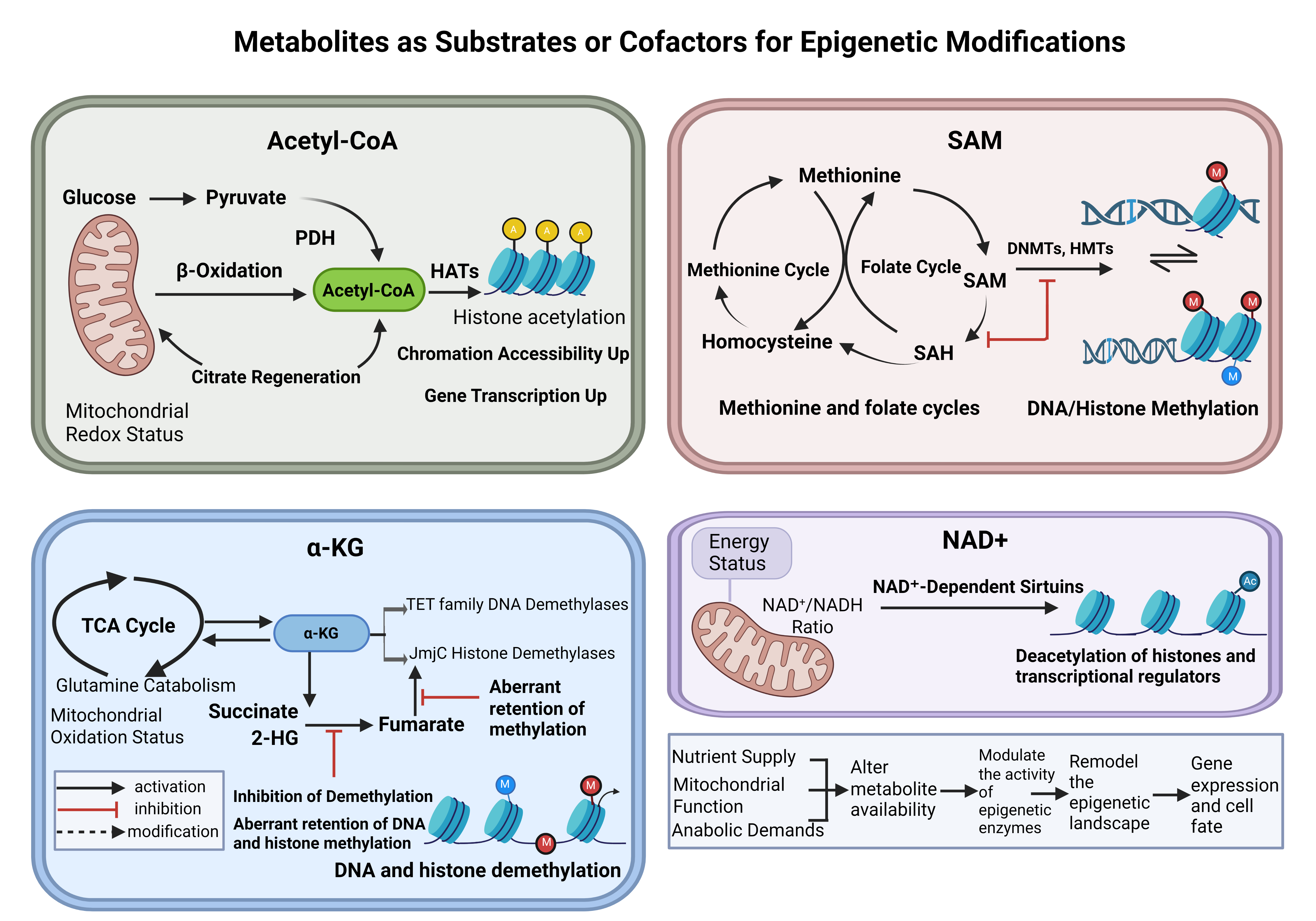
In tumors, oncogenic signaling and biosynthetic demand alter production and consumption. Nutrient limitation, hypoxia, and mitochondrial dysfunction can further shift their availability. Changes in acetyl-CoA, SAM, α-KG, NAD⁺, and related metabolites can affect chromatin regulation at loci involved in proliferation, differentiation, and stress tolerance. This shows one way metabolic reprogramming can influence the ability of malignant cells to maintain or change state during tumor evolution.
Oncometabolites and Metabolic Perturbations
Metabolic disruption in cancer can result in the accumulation of compounds that inhibit chromatin-modifying enzymes (29, 30). These compounds include 2-hydroxyglutarate (2-HG), succinate, and fumarate, which inhibit α-KG-dependent dioxygenases such as TET enzymes and JmjC-domain histone demethylases (4, 31–34). The inhibitory effect depends partly on the concentrations of these metabolites relative to α-KG. Reduced enzyme activity can promote the retention of DNA or histone methylation at loci targeted by these enzymes. In experimental systems, impaired demethylation can limit the expression of differentiation-associated genes and promote maintenance of an undifferentiated state or a shift in lineage (33, 36, 37). Linking enzyme inhibition to phenotype requires identification of the responsive loci and determination of whether methylation changes at these sites affect the relevant differentiation program. Additionally, mitochondria can regulate nuclear transcription through reactive oxygen species, which induce hypoxia-responsive gene expression via a distinct mechanism (35).
Lactate metabolism contributes to histone lysine lactylation, a covalent modification that connects glycolytic activity to chromatin regulation (38, 39). Histone lactylation has been linked to distinct transcriptional responses in astrocytes following experimental subarachnoid hemorrhage (40) and during embryonic genome activation (41). These observations suggest that the same modification can function in different contexts. Bulk measurements reflect changes in total histone lactylation, but attributing a transcriptional effect to modification at a particular locus requires assessing local enrichment and experimentally perturbing lactylation to determine its impact on gene expression. Lactylation introduces a covalent mark to histones, whereas demethylase inhibition impedes the removal of existing methylation marks. Changes in metabolite availability can alter the balance between chromatin mark deposition and removal. Certain metabolites inhibit demethylation or deacetylation, while lactate metabolism promotes histone lactylation (21, 22, 38). Both mechanisms can influence transcription at loci that regulate differentiation, proliferation, or survival under stress.
3. Epigenetic Control of Metabolic Reprogramming and Cancer Cell Plasticity
Epigenetic Regulation of Metabolic Gene Expression
Chromatin regulation can influence cellular metabolic pathway selection by modulating enzyme and transporter expression. This relationship is bidirectional. Differentiation studies have associated metabolite availability with chromatin changes (36, 37), and research on histone lactylation demonstrates a connection between glycolytic activity and transcription (38, 39). Which loci respond, how long the response lasts, and how it affects cell behavior depend on lineage and environmental context. Restoring a candidate chromatin enzyme's activity after perturbation can determine whether recovery of metabolic gene expression coincides with restoration of pathway activity in the same cells. Sequential measurement of chromatin marks, transcript levels, and metabolic flux can clarify the temporal sequence of these changes. Similar measurements after stress withdrawal can reveal whether transcription and pathway activity recover in parallel. Studies in astrocytes after experimental subarachnoid hemorrhage and during embryonic genome activation exemplify the role of histone lactylation in distinct transcriptional programs (40, 41). Alongside DNA methylation and chromatin remodeling, these modifications integrate metabolic conditions with gene expression regulation (42).
Epigenetic Control of Metabolic State Switching
Changes in energy demand and substrate availability can alter the epigenetic regulation of genes encoding metabolic enzymes, transporters, and pathway regulators. The resulting transcriptional response can affect how much substrate enters the cell and how flux is distributed among competing pathways. Studies of obesity and muscle stem cells link chromatin regulation to changes in metabolic function, while cancer studies implicate epigenetic mechanisms in maintaining metabolic programs that support malignant cell survival (43–45). Obesity-associated epigenetic inheritance and human metabolic disease further exemplify the connections between epigenetic regulation and metabolic phenotypes (46, 47). Chromatin remodeling mediates the integration of metabolic signals with transcriptional responses (48). β-Glucan promotes trained innate immunity by inducing glycolysis through mTOR and HIF-1α (49), whereas S-2-hydroxyglutarate influences CD8⁺ T-cell differentiation by modulating α-KG-dependent dioxygenase activity (50). Population studies provide complementary evidence for these mechanisms. An epigenome-wide association study identified correlations between DNA methylation and circulating metabolic measures, yielding candidate loci for functional analysis (51). Experimental perturbation of methylation at a candidate locus, followed by assessment of pathway activity, can determine whether the epigenetic mark contributes to the observed metabolic phenotype. Targeting BAZ2B in hepatic senescence and metabolic dysfunction-associated steatohepatitis further highlights the role of chromatin regulators in metabolic disease (52).
Cancer cells can rapidly adapt substrate utilization, enzyme activity, and metabolite exchange in response to hypoxia, nutrient limitation, or therapeutic intervention. Alterations in chromatin and transcriptional regulation support these adaptive responses. Experimental perturbation of a candidate chromatin regulator, combined with monitoring metabolic pathway activity at multiple time points during and after stress, can differentiate between roles in establishing versus maintaining adaptation. Comparing recovery dynamics with and without the perturbation can further reveal whether sustained pathway activity is dependent on the chromatin regulator.
Non-coding RNAs and Chromatin Architecture in Metabolic Adaptation
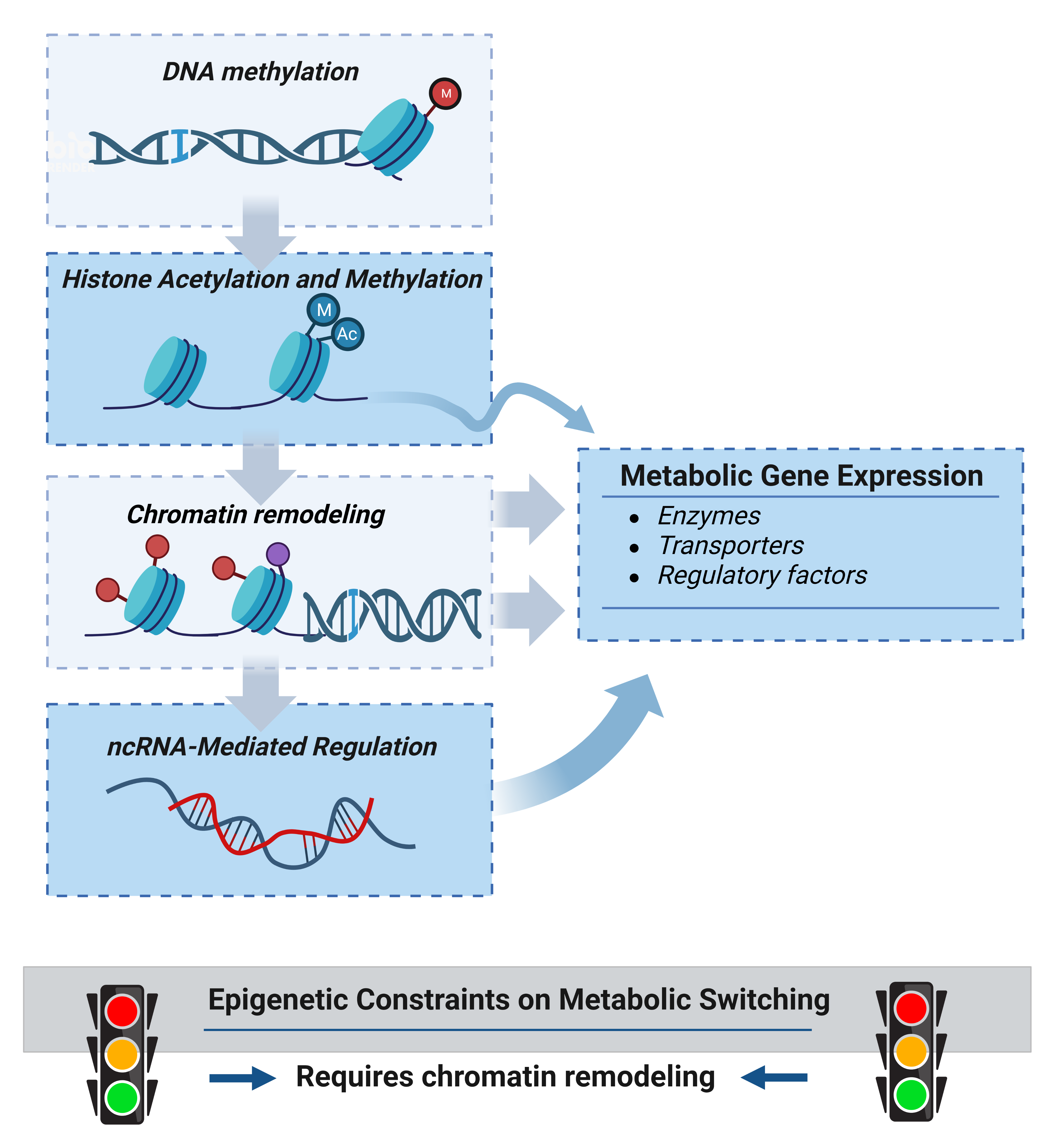
Metabolic gene expression is shaped by higher-order chromatin organization and noncoding RNAs (ncRNAs), as well as by DNA and histone modifications (53, 54). Chromatin-remodeling complexes reposition nucleosomes, changing whether transcription factors can access their binding sites at promoters and enhancers as cellular demands shift (53). Noncoding RNAs act at both transcriptional and post-transcriptional steps. Through these mechanisms, they can change the abundance or activity of the proteins that regulate metabolic pathways. MicroRNAs modulate mitochondrial function and metabolic programs by targeting transcripts that encode metabolic proteins or their regulators (55). Long non-coding RNAs regulate metabolic gene expression through interactions with regulatory proteins as well as through transcriptional and post-transcriptional mechanisms, providing multiple levels of control over metabolic programs (56). Circular RNAs also contribute to cancer-associated metabolic regulation, including the modulation of glycolytic pathways (57).
Distal enhancers influence transcription by forming physical contacts with their target promoters. YY1 mediates a subset of these interactions, and disruption of its binding or expression alters both chromatin looping and transcription (58). Changes in steady-state transcript abundance may result from altered synthesis, decay, or both processes. Simultaneous measurement of enhancer–promoter contacts, nascent transcription, and transcript decay would clarify these mechanisms. In cancer cells subjected to nutrient limitation or therapy, such analyses could determine whether chromatin architecture and noncoding RNAs regulate the same metabolic genes and whether these effects persist after stress withdrawal within the same cell population.
Metabolic gene expression is determined by enzymatic modifications, chromatin organization, and RNA-mediated regulation, which collectively influence gene transcription and transcript utilization (59, 60). In cancer, this integrated regulatory network enables metabolic phenotypes to remain stable enough to support malignant growth while maintaining the plasticity needed to adapt to hypoxia, nutrient limitation, metastatic dissemination, and therapeutic pressure (61). Perturbation of a candidate enhancer or noncoding RNA before and after stress exposure could distinguish between requirements for establishing a new metabolic state and those for maintaining it.
A specific enhancer or noncoding RNA necessary for establishing an adaptive metabolic state may become dispensable once maintenance of that state is achieved through alternative regulatory mechanisms (Figure 2).
4. Metabolic-Epigenetic Reprogramming in Cancer and the Tumor Ecosystem
Tumor Initiation, Progression, Metastasis, and Therapeutic Resistance
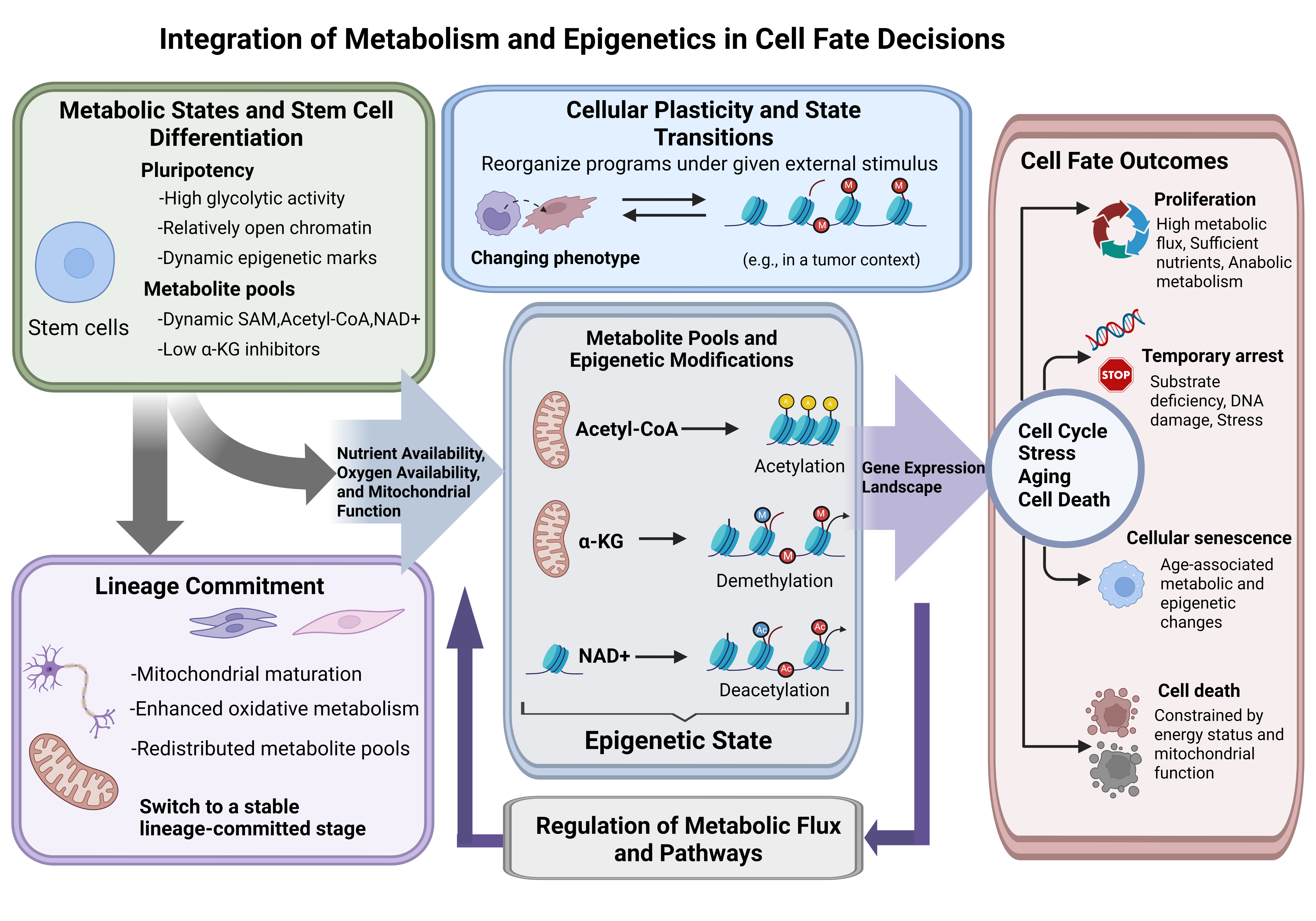
Cancer progression depends on metabolic and epigenetic programs that are stable enough to sustain malignant growth yet plastic enough to accommodate changing microenvironmental and therapeutic pressures. Beyond enhanced glycolysis, tumor cells adapt by altering glutamine utilization, lipid metabolism, mitochondrial function, and redox homeostasis (62). The nuclear pyruvate dehydrogenase complex generates acetyl-CoA for histone acetylation, directly linking metabolic enzyme localization to chromatin regulation (63). Methionine dependence in tumor-initiating cells further exemplifies the connection between nutrient metabolism and methylation-associated cellular regulation (64). Defined cancer models have revealed multiple mechanisms linking metabolic alterations to chromatin regulation and cellular behavior. For example, lactate supports a metabolic-epigenetic axis involving lipid metabolism in prostate cancer (65), while α-ketoglutarate attenuates Wnt signaling and promotes differentiation in colorectal cancer (66). Metastasis-initiating breast cancer cells display specific metabolic dependencies that support their malignant activity (67). Accumulation of fumarate and succinate can inhibit α-KG-dependent histone and DNA demethylases, directly linking metabolic abnormalities to altered methylation (68).
Deficiencies in metabolic enzymes are associated with characteristic molecular profiles. DNA methylation profiling distinguishes SDH-deficient gastrointestinal stromal tumors from KIT-PDGFRA-driven tumors, revealing a distinct epigenetic phenotype (69). Similarly, SDH- and FH-deficient renal cell carcinomas exhibit unique genomic and metabolic features (70). These findings define disease-associated states that can inform investigations into the functional consequences of metabolic disruption. Restoration of enzyme activity could be used to test which metabolic and epigenetic features are dependent on the underlying deficiency.
At super-enhancers, the acetylation reader BRD4 sustains oncogenic transcription (71). In hypoxic esophageal squamous cell carcinoma, DNMT1-dependent methylation of RORA promotes tumor progression by regulating SLC2A3 expression, thereby linking DNA methylation at this locus to metabolic adaptation (72). Enhancer-promoter hubs organize transcriptional networks involved in oncogenesis and drug resistance (73), situating chromatin organization within the broader context of tumor progression and metastasis regulation (74). LKB1 inactivation alters chromatin accessibility and promotes metastatic progression in experimental cancer models (75). Single-cell analysis of colorectal cancer liver metastasis identifies accessibility changes associated with metastatic cellular states and liver tropism (76). Perturbation of candidate regulatory elements in metastatic models could determine whether these accessibility changes contribute to liver colonization or arise after tumor cells enter the hepatic microenvironment. Collectively, these studies demonstrate the connection between chromatin regulation and malignant adaptation across distinct stages of progression.
Therapeutic interventions can select for slow-cycling or dormancy-like tumor cells. Depending on the experimental model, persister cell survival is associated with altered glutamine or lipid utilization, oxidative phosphorylation, or antioxidant metabolism. Alterations in chromatin accessibility and transcription have also been implicated in survival during drug exposure (77, 78). Reversible drug tolerance may precede acquired resistance and is often characterized by reduced expression of lineage markers and increased metabolic flexibility (79). These features can coexist within a tumor and may evolve as drug exposure continues. Resumption of proliferation following drug withdrawal and restoration of drug sensitivity represent distinct endpoints. Lineage tracking during withdrawal and rechallenge can determine whether early persister cells contribute to resistant outgrowth and whether resistance remains restricted to specific lineages. Measurement of pathway activity at each phase can clarify whether metabolic changes associated with initial survival persist as the population resumes growth or whether subsequent outgrowth relies on a different metabolic program.
Metabolic-epigenetic coupling links metabolite availability and chromatin regulation to differentiation, cellular plasticity, and cell-fate outcomes (Figure 3).
Metabolic-Epigenetic Remodeling of the Tumor Immune Microenvironment
Nutrient availability within specific tumor regions is shaped by substrate consumption and release by malignant and stromal cells. Substrate competition can modify metabolism in infiltrating immune cells, while metabolites released by one cell population can affect chromatin structure or effector function in another. As a result, metabolic changes that support malignant growth may also alter the differentiation, persistence, or function of immune populations involved in the antitumor response.
During inflammation and immune responses, metabolic-epigenetic interactions remodel immune cell activation, differentiation, and effector states, thereby influencing tissue injury and repair (80). Immune cells display distinct metabolic requirements across resting, activated, effector, and memory stages, accompanied by dynamic changes in DNA methylation, histone modifications, and chromatin accessibility (81). Recent studies indicate that metabolic reprogramming and epigenetic regulation in immune cells are not independent processes; rather, they jointly determine the intensity and duration of inflammation and the capacity to restore immune homeostasis (82, 83).
This metabolic-epigenetic coupling is especially prominent in innate immune cells. In experimental models of trained immunity, β-glucan stimulation induces mTOR- and HIF-1α-dependent glycolysis, thereby linking metabolic reprogramming to enhanced innate immune responses (49). Additional studies demonstrate epigenetic changes associated with human monocyte-to-macrophage differentiation and trained innate immunity (83).
Adaptive immunity is also regulated by metabolic-epigenetic coupling, but with lineage-specific outcomes (80). Effector T-cell differentiation involves increased glycolysis, one-carbon metabolism, and glutamine utilization, accompanied by chromatin remodeling (81, 84). T-cell metabolic requirements vary according to differentiation state, activation conditions, and tissue context; thus, regulatory and memory populations should not be assigned a single universal metabolic program (81). Metabolites can directly influence T-cell fate; for example, S-2-hydroxyglutarate modulates α-KG-dependent dioxygenase activity and promotes a memory-like differentiation program in CD8+ T cells (50).
β-Glucan studies position mTOR- and HIF-1α-dependent glycolysis as upstream regulators of trained innate immunity (49). Metabolic requirements for immune activation differ by the specific effector function involved (85). In mouse bone marrow-derived dendritic cells, CTCF coordinates enhancer contacts that control inflammatory transcription (86). Macrophages originating from hematopoietic stem cells in autoimmune mice display metabolic and chromatin characteristics linked to trained immunity (87). In regulatory T cells, transketolase deficiency disrupts the non-oxidative pentose phosphate pathway, reduces α-KG availability, and increases DNA methylation. This perturbation impairs suppressive function under the tested conditions, linking disruption of a specific metabolic pathway to an immunoregulatory phenotype (88).
Lactylation represents an additional connection between glycolytic metabolism and protein regulation, with functional outcomes determined by the specific substrate modified and the cellular context (89). In hepatocellular carcinoma, lactylome profiling reveals lactylation-associated mechanisms underlying metabolic adaptation (90). β-Glucan-induced glutaminolysis and fumarate accumulation drive epigenetic reprogramming in monocytes, while mevalonate can induce trained immunity via signaling pathways linked to histone modifications (91, 92). Therefore, inflammation-associated metabolites function not only as byproducts of cellular activation but also as signals that convey metabolic state to persistent chromatin and transcriptional programs.
Within tumors, comparable metabolic-epigenetic programs arise in response to a distinct ecological context. Competition for glucose and amino acids, accumulation of lactate and other metabolites, hypoxia, and tumor-derived signaling factors collectively reshape the metabolic and chromatin states of infiltrating macrophages, T cells, and other immune populations. These alterations can suppress cytotoxic effector programs, promote immunoregulatory or dysfunctional states, and help maintain an immunosuppressive microenvironment. Additionally, metabolic adaptation supports malignant-cell persistence; for example, metabolic changes in residual breast cancer cells facilitate tumor recurrence after oncogene withdrawal (93). Consequently, metabolic-epigenetic regulation of immune and malignant cell fate constitutes a critical interface linking tumor metabolism, cellular plasticity, and therapeutic response.
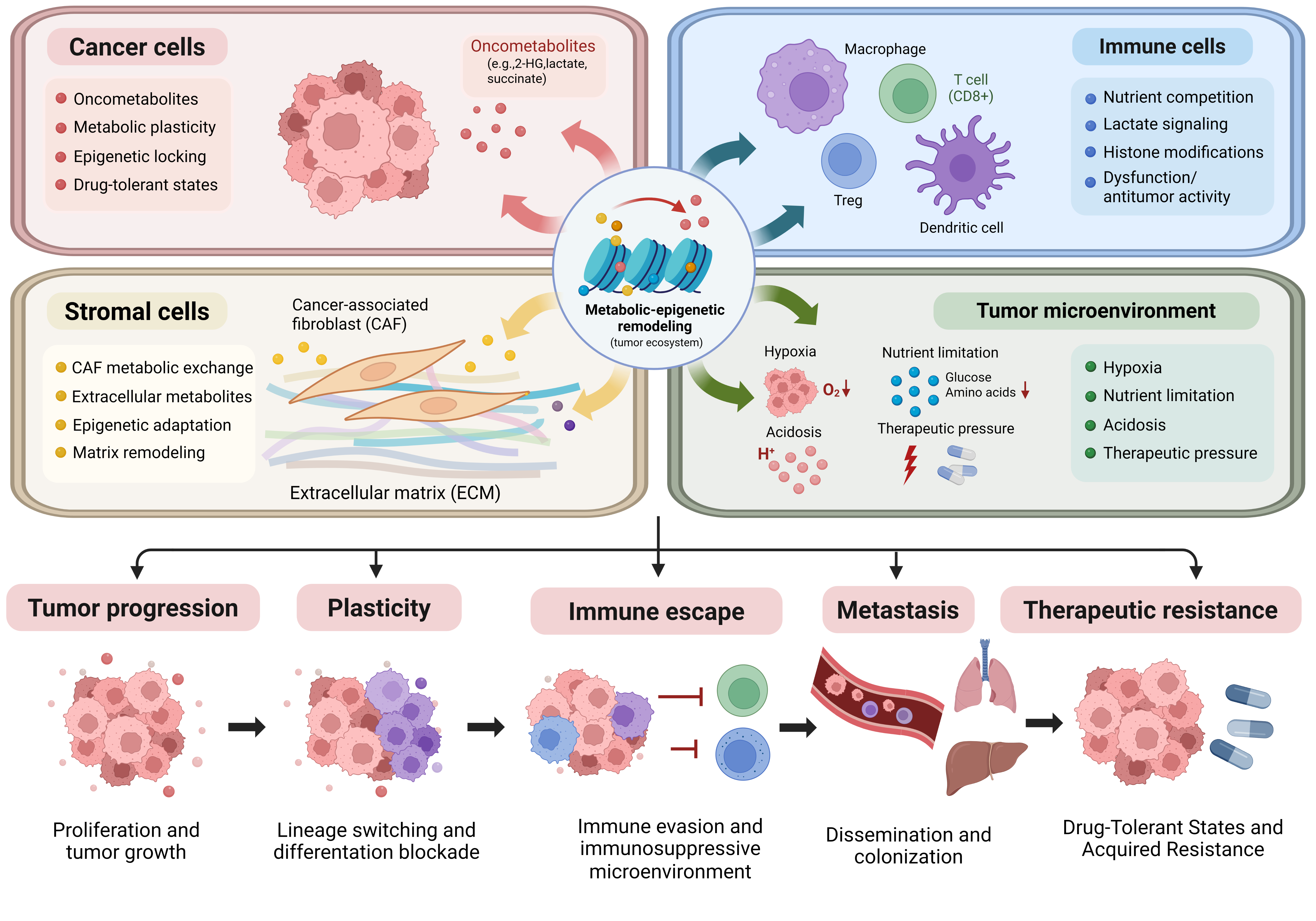
Metabolic conditions within tumors can alter immune-cell function and chromatin regulation. Immune cells, in turn, influence local nutrient availability through substrate consumption and modulate inflammatory signaling through their metabolic activity. Serial measurements of chromatin state and effector function in defined immune-cell populations can reveal whether these responses resolve after the initiating stimulus is withdrawn or persist during recovery. The duration of the response can help determine when to pair tumor-directed therapy with an intervention that supports antitumor immunity (Figure 4).
Metabolic Memory as a Model for Persistent Tumor Cell-State Reprogramming
The molecular effects of a metabolic exposure can outlast the initiating stimulus. In experimental models, transient hyperglycemia induces epigenetic and transcriptional changes that persist after normoglycemia is restored, a phenomenon termed metabolic memory (94). Fatty-acid exposure promotes DNMT3B-dependent methylation of the PGC-1α promoter in muscle cells (95), and epigenetic alterations in human pancreatic islets affect mitochondrial function and insulin secretion (96). After weight loss, adipose tissue in humans and mice retains transcriptional changes, with persistent epigenetic alterations observed in mouse adipocytes (97). Mitochondrial regulation offers another mechanism for altering cellular metabolic phenotypes, as demonstrated by increased hepatic fatty-acid oxidation following MCJ silencing (98). Collectively, these findings differentiate enduring chromatin-associated responses from metabolic adaptations maintained by alternative regulatory mechanisms.
Neural and immune systems demonstrate complementary relationships among metabolite availability, cellular function, and chromatin regulation. SARM1-dependent NAD+ depletion connects metabolite consumption to axonal degeneration (99), while nuclear ACSS2 provides acetyl-CoA for neuronal histone acetylation and gene expression linked to hippocampal memory (100). APOE4-associated lipid-droplet accumulation in microglia exemplifies how altered lipid handling can accompany dysfunctional cellular states (101). A direct metabolic contribution to chromatin modification is shown by the incorporation of alcohol-derived metabolites into brain histone acetylation (102). Histone deacetylation and disease-associated epigenomic changes further influence neural plasticity and immune-related transcriptional programs in the brain (103, 104).
After reoxygenation or nutrient replenishment, metabolite concentrations may return to baseline before transcription or chromatin state does. A similar lag may follow drug withdrawal. Tracking individual cells through recovery and rechallenge can show whether those that retain an altered chromatin state differ in survival, drug sensitivity, or capacity to resume proliferation. Perturbing the mechanism that maintains this state after the original stimulus has been removed can test whether it contributes to residual disease or recurrence.
Metabolic-Epigenetic Coupling across the Tumor Microenvironment
Tumors function as multicellular ecosystems where metabolic-epigenetic states arise from interactions among malignant cells, immune populations, fibroblasts, endothelial cells, and the surrounding extracellular environment. Regional variations in oxygenation, nutrient delivery, extracellular acidity, and metabolite accumulation generate spatially distinct biochemical niches that impose unique constraints on chromatin regulation (105, 106). Consequently, metabolic-epigenetic reprogramming is unlikely to occur uniformly throughout a tumor. Rather, distinct cellular populations may occupy different metabolic and epigenetic states based on their localization and interactions with neighboring cells.
Cancer-associated fibroblasts and other stromal populations influence metabolic-epigenetic regulation through nutrient exchange, extracellular matrix remodeling, cytokine signaling, and metabolite release or consumption (107–109). Conversely, lactate-mediated epigenetic reprogramming can drive the formation of cancer-associated fibroblasts in pancreatic cancer (110). These reciprocal interactions can sustain, buffer, or circumvent malignant-cell metabolic dependencies, highlighting the importance of stromal support when translating findings from isolated cells to intact tumors.
A metabolic signal detected in a bulk tumor specimen may originate from malignant, stromal, or immune cells. Identifying its cellular source is essential for interpreting pharmacological interventions, as inhibiting a pathway in malignant cells can alter substrate availability for neighboring cells. Spatial metabolomics enables localization of the metabolic signal, while single-cell transcriptional and chromatin profiling can identify responsive populations and characterize their transcriptional and epigenetic states during treatment. Collectively, these approaches can differentiate tumor-cell dependencies from those maintained by neighboring populations and determine whether target inhibition also modifies local metabolic conditions that support antitumor immune function.
5. An Integrated Framework for Metabolic-Epigenetic State Transitions in Cancer
Metabolic-epigenetic coupling links biochemical conditions to the transcriptional programs that determine cellular identity and adaptation. Histone acetylation illustrates this relationship: metabolic pathways provide acetyl-CoA for chromatin modification, while transcriptional regulation controls the enzymes and transporters that sustain metabolic activity (111). Single-cell multimodal approaches enable resolution of these relationships across heterogeneous populations and facilitate identification of molecular states that may be masked in bulk measurements (112). Therefore, an integrated framework must consider the timing, location, and functional consequences of metabolic-epigenetic interactions.
Changes in metabolite availability can modulate chromatin-modifying enzyme activity before transcriptional changes. The initial biochemical response and subsequent transcriptional alterations are determined by substrate availability, enzyme characteristics, and cellular context (113, 114). Under conditions of oxygen and serum deprivation, nuclear ACSS2 recycles acetate generated by histone deacetylation to sustain histone acetylation, thereby maintaining chromatin modification during metabolic stress (115). SIRT7-mediated histone desuccinylation further links enzymatic chromatin regulation to chromatin compaction and genome stability (116). These mechanisms enable cellular adaptation to environmental changes, although the persistence of the resulting state is governed by additional transcriptional and regulatory processes.
Spatial organization also influences these interactions. Metabolic-epigenetic signaling in glioblastoma exemplifies the diverse pathways by which metabolic activity and chromatin regulation intersect in cancer (117). At the cellular level, heterogeneous enhancer states facilitate distinct responses to metabolic stress, as observed in pancreatic β cells (118). Spatial multi-omics enables analysis of intact tissues, allowing localization of molecular programs within specific cellular and microenvironmental contexts (119). Integrating these approaches with assessments of nuclear metabolite availability and enzyme localization may clarify why similar metabolic perturbations yield different transcriptional outcomes.
Alterations in cell state can modify metabolic demand, with transcriptional regulation determining how these demands are met. During glutamine deprivation, DDIT3 modulates the balance between glycolysis and oxidative phosphorylation (120). In tumors undergoing lineage transitions, the metabolic pathways available to meet new demands are influenced by genetic background, differentiation status, and local environmental conditions (121). Chromatin accessibility can either facilitate or restrict these transitions by controlling access to regulatory regions (122). The persistence of the adapted state following removal of the initiating stimulus depends, in part, on the continued activity of supporting transcriptional programs.
A multiregional atlas of the human brain has integrated chromatin accessibility and gene expression data to distinguish regulatory differences among cell populations and anatomical regions (123). In the liver, single-cell spatial multi-omics has identified enhancer networks associated with zonation (124). Developmental studies have investigated how environmental exposures influence epigenetic plasticity (125), while integrated analyses of RNA, chromatin, and metabolites have refined the molecular characterization of the human kidney (126). In tumors, these methods may distinguish lineage-associated regulatory differences from those driven by local metabolic conditions, including responses of malignant and stromal cells within the same region.
Molecular profiling can identify chromatin reactions suitable for direct experimental testing. HBO1-catalyzed H3K9 lactylation establishes a direct connection between a specific enzymatic activity and transcriptional regulation (127). In tumor models, genetic or pharmacological perturbation of HBO1 can be combined with metabolite quantification and locus-specific chromatin profiling. Subsequent rescue experiments can assess whether restoration of HBO1 activity reinstates both transcriptional activity and the malignant phenotype. Serial measurements in defined cell populations can establish whether local metabolite availability changes before chromatin state shifts. Following individual cells after stimulus withdrawal can then show whether the altered state returns to baseline or persists across cell divisions. Perturbing the candidate reaction and assessing its impact on differentiation, survival, immune function, or treatment response can identify dependencies that support tumor growth or adaptive resistance (Figure 5).
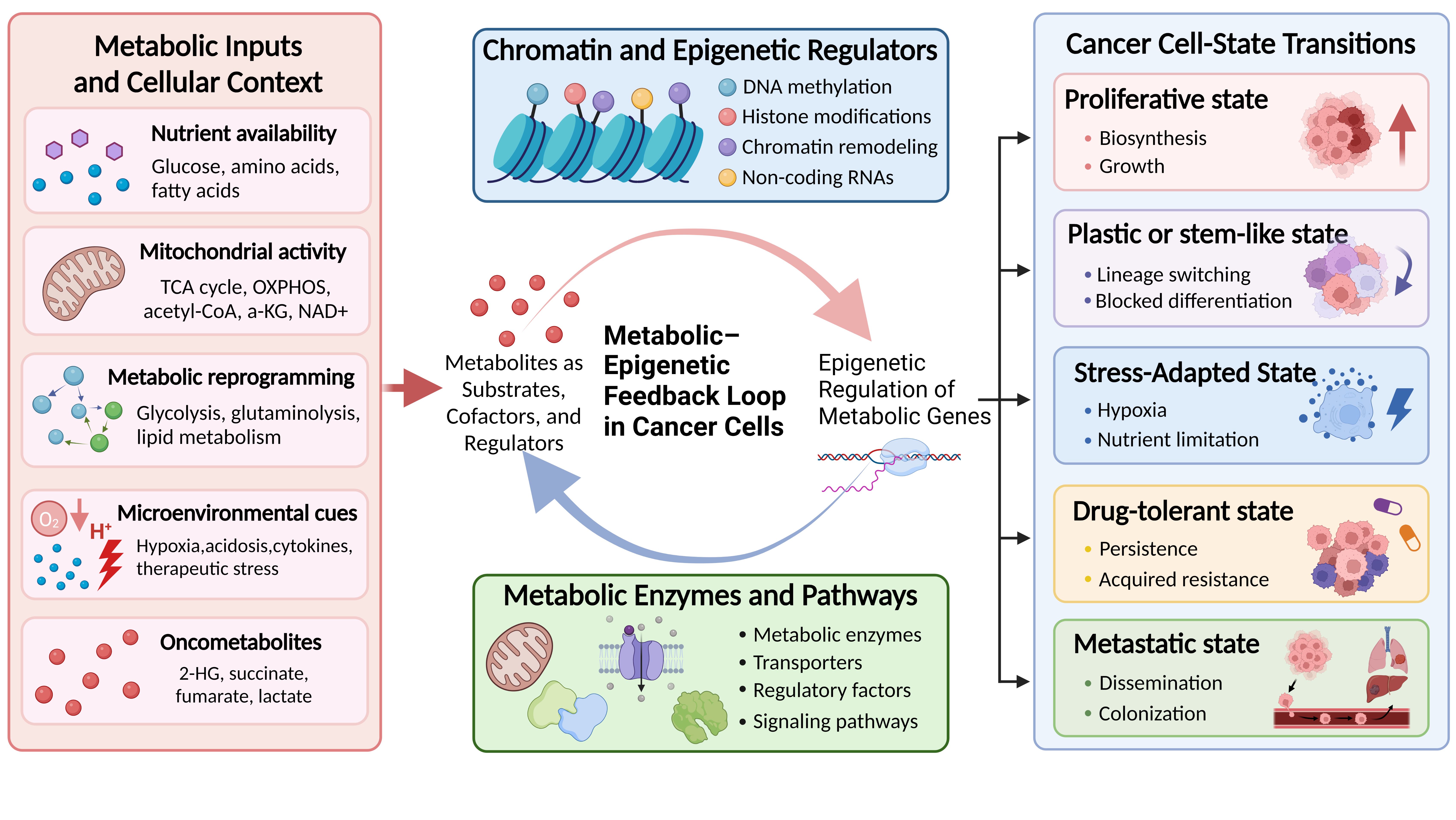
6. Targeting Metabolic-Epigenetic Dependencies in Cancer
Combination strategies require disease-specific mechanistic justification and evidence that the targeted processes contribute to the same malignant state (128). Combined inhibition of HDAC and eIF4A in pancreatic ductal adenocarcinoma exemplifies epigenetic-translation targeting, rather than direct combined inhibition of a metabolic enzyme and a chromatin regulator (129). Metabolic perturbations can modulate epigenetic regulation (130), while HDAC inhibition can remodel metabolic transcriptional programs, as demonstrated in glioblastoma models (131). The most direct clinical example involves inhibition of an abnormal metabolic enzyme that generates an epigenetically active oncometabolite. More indirect strategies, such as targeting glycolysis, one-carbon metabolism, glutamine utilization, or lipid metabolism, remain context-dependent because they also impact normal tissues and can be circumvented through metabolic plasticity (30).
IDH1/2-mutant cancers provide a clinically validated proof of principle that a genetically defined metabolic alteration can create a therapeutically actionable epigenetic dependency. Cancer-associated mutant IDH enzymes acquire neomorphic activity, leading to accumulation of the oncometabolite 2-hydroxyglutarate (2-HG) (132), which inhibits α-KG-dependent TET DNA demethylases and JmjC-domain histone demethylases, resulting in DNA and histone hypermethylation and impaired differentiation. Therapeutic inhibition of mutant IDH has translated this mechanistic insight into clinical benefit. Enasidenib and ivosidenib have produced durable responses and promoted differentiation in patients with IDH2- and IDH1-mutant relapsed or refractory AML, respectively (133, 134). In newly diagnosed IDH1-mutant AML, the addition of ivosidenib to azacitidine significantly improved clinical outcomes compared to azacitidine alone (135). In grade 2 IDH-mutant glioma, the brain-penetrant dual IDH1/2 inhibitor vorasidenib significantly prolonged progression-free survival and delayed the need for subsequent anticancer intervention in the phase III INDIGO trial (136). Collectively, these trials demonstrate the clinical benefit of targeting mutant IDH enzymes. However, the extent to which clinical benefit is attributable to reversal of epigenetic abnormalities or differentiation blockade varies by disease and treatment context (133–136).
Evidence for epigenetic combinations and therapies targeting metabolic–epigenetic dependencies varies by tumor type and mechanism, and many approaches remain investigational (128–131). For interventions targeting histone lactylation, acetyl-CoA-dependent acetylation, NAD⁺-dependent deacylation, or SAM-dependent methylation, clinical development requires validated assays to measure target engagement and evidence that the intended chromatin change occurs at tolerable exposures. After treatment begins, alternative pathways can replenish depleted substrates, and reliance on the targeted pathway may vary across regions of the same tumor. If normal tissues depend on that pathway, toxicity may limit the exposure required for sustained inhibition. A pretreatment biomarker can also miss cell-state transitions that emerge during therapy. Serial measurements of metabolites and chromatin, combined with single-cell or spatial profiling, could guide patient selection and dosing schedules and help detect emerging resistance (137–140).
A paired biomarker may integrate measurements of metabolite abundance or flux with DNA methylation, histone modification, or chromatin accessibility at responsive loci. Early depletion of a substrate may precede reversal of the associated chromatin state. Prospective sampling across tumor regions and treatment time points can assess whether this temporal discordance resolves and whether it predicts subsequent therapeutic response or acquired resistance. A metabolic inhibitor can decrease the supply of a specific metabolite that sustains a malignant chromatin state. If tumor cells compensate by restoring pathway activity through transcriptional mechanisms, a second agent could directly target this compensatory response (128, 141, 142).
Discussion
Metabolic and chromatin changes can establish, stabilize, or redirect malignant cell states during tumor progression. Related changes can occur in adjacent stromal and immune populations. When a metabolic signature coincides with a chromatin state, perturbing the underlying reaction can test whether the cells depend on it to maintain the phenotype. Its value as a therapeutic target also depends on how readily alternative pathways compensate and whether malignant cells are more sensitive to inhibition than normal cells. These questions should be tested across tumor regions, cell populations, and treatment stages, where substrate availability, chromatin accessibility, and enzyme activity may differ.
As a result, identical metabolic perturbations can produce distinct epigenetic outcomes across different tumor regions or cell populations, while similar chromatin states may emerge via separate metabolic pathways. Addressing this heterogeneity necessitates methodologies that surpass bulk-tissue and population-averaged analyses, such as single-cell epigenomic and spatial metabolic profiling capable of delineating cell-state-specific regulatory and metabolic programs (137, 138). Mutant IDH1 establishes a defined biochemical starting point through production of 2-HG (132). Where the evidence begins with co-occurrence of a metabolite and a chromatin mark, timed perturbation can establish temporal order in the responding population. Inhibiting and restoring the candidate reaction can test whether locus-specific chromatin changes and the phenotype depend on it.
A key unresolved issue is whether an adaptive state requires ongoing exposure to therapeutic or microenvironmental stress, or if it persists after such exposure ceases. Evidence shows that a reversible chromatin-mediated drug-tolerant state exists in cancer-cell subpopulations (143), and that ACSS2-mediated metabolic-epigenetic coupling contributes to fulvestrant resistance in breast cancer (144). These observations define distinct forms of chromatin-associated tolerance and metabolic-epigenetic resistance, but do not indicate that all adaptive states inevitably become permanent. Longitudinal perturbation, withdrawal, and rechallenge experiments are necessary to distinguish when these states remain reversible and when additional mechanisms maintain residual disease or acquired resistance.
Time-resolved perturbation studies can determine whether a metabolic alteration sustains a tumor phenotype via chromatin modifications. Paired single-cell and spatial analyses can subsequently identify the specific cell populations and tumor regions where this dependency is present. For clinical application, candidate biomarkers should be monitored serially to assess their correlation with target engagement and resistance development. Treatment studies must also define the optimal timing for pathway inhibition and evaluate whether the necessary exposure can prevent adaptive escape while maintaining normal tissue integrity and antitumor immune responses.
A causal link is strongest when a defined metabolic reaction alters chromatin at specific loci, and those changes contribute to the phenotype in the same cell population. Isotope tracing can identify the metabolic source, and single-cell and spatial profiling can locate the responding cells. Perturbing the affected loci can then test their contribution to survival, dissemination, or immune evasion. Paired patient samples collected before and during therapy can show whether the response also occurs clinically. If the state persists after drug withdrawal, rechallenge can test whether surviving cells retain the dependency and inform whether to inhibit the pathway during initial therapy or after resistance emerges.
References
1. Li X, Yu W, Qian X, Xia Y, Zheng Y, Lee JH, et al. Nucleus-translocated ACSS2 promotes gene transcription for lysosomal biogenesis and autophagy. Mol Cell. 2017;66(5):684–697.e9. https://doi.org/10.1016/j.molcel.2017.04.026
2. Cai L, Sutter BM, Li B, Tu BP. Acetyl-CoA induces cell growth and proliferation by promoting the acetylation of histones at growth genes. Mol Cell. 2011;42(4):426–437. https://doi.org/10.1016/j.molcel.2011.05.004
3. Lu C, Ward PS, Kapoor GS, Rohle D, Turcan S, Abdel-Wahab O, et al. IDH mutation impairs histone demethylation and results in a block to cell differentiation. Nature. 2012;483(7390):474–478. https://doi.org/10.1038/nature10860
4. Kaelin WG Jr, McKnight SL. Influence of metabolism on epigenetics and disease. Cell. 2013;153(1):56–69. https://doi.org/10.1016/j.cell.2013.03.004
5. Zhong L, D’Urso A, Toiber D, Sebastian C, Henry RE, Vadysirisack DD, et al. The histone deacetylase Sirt6 regulates glucose homeostasis via Hif1alpha. Cell. 2010;140(2):280–293. https://doi.org/10.1016/j.cell.2009.12.041
6. DeBerardinis RJ, Thompson CB. Cellular metabolism and disease: what do metabolic outliers teach us? Cell. 2012;148(6):1132–1144. https://doi.org/10.1016/j.cell.2012.02.032
7. Zhang YW, Schönberger K, Cabezas-Wallscheid N. Bidirectional interplay between metabolism and epigenetics in hematopoietic stem cells and leukemia. EMBO J. 2023;42(24):e112348. https://doi.org/10.15252/embj.2022112348
8. Tischler J, Gruhn WH, Reid J, Allgeyer E, Buettner F, Marr C, et al. Metabolic regulation of pluripotency and germ cell fate through α-ketoglutarate. EMBO J. 2019;38(1):e99518. https://doi.org/10.15252/embj.201899518
9. Zhou W, Zhao T, Du J, Ji G, Li X, Ji S, et al. TIGAR promotes neural stem cell differentiation through acetyl-CoA-mediated histone acetylation. Cell Death Dis. 2019;10(3):198. https://doi.org/10.1038/s41419-019-1434-3
10. Yu X, Wu H, Su J, Liu X, Liang K, Li Q, et al. Acetyl-CoA metabolism maintains histone acetylation for syncytialization of human placental trophoblast stem cells. Cell Stem Cell. 2024;31(9):1280–1297.e7. https://doi.org/10.1016/j.stem.2024.07.003
11. Hsieh WC, Sutter BM, Ruess H, Barnes SD, Malladi VS, Tu BP. Glucose starvation induces a switch in the histone acetylome for activation of gluconeogenic and fat metabolism genes. Mol Cell. 2022;82(1):60–74.e5. https://doi.org/10.1016/j.molcel.2021.12.015
12. Xu X, Peng Q, Jiang X, Tan S, Yang Y, Yang W, et al. Metabolic reprogramming and epigenetic modifications in cancer: from the impacts and mechanisms to the treatment potential. Exp Mol Med. 2023;55(7):1357–1370. https://doi.org/10.1038/s12276-023-01020-1
13. Waldschmidt JM, Kloeber JA, Anand P, Frede J, Kokkalis A, Dimitrova V, et al. Single-cell profiling reveals metabolic reprogramming as a resistance mechanism in BRAF-mutated multiple myeloma. Clin Cancer Res. 2021;27(23):6432–6444. https://doi.org/10.1158/1078-0432.CCR-21-2040
14. Baylin SB, Jones PA. A decade of exploring the cancer epigenome - biological and translational implications. Nat Rev Cancer. 2011;11(10):726–734. https://doi.org/10.1038/nrc3130
15. Ling C, Bacos K, Rönn T. Epigenetics of type 2 diabetes mellitus and weight change - a tool for precision medicine? Nat Rev Endocrinol. 2022;18(7):433–448. https://doi.org/10.1038/s41574-022-00671-w
16. Baeken MW, Borlepawar A, Kötzner P, Richly H, Behl C, Moosmann B, et al. Epigenetic regulation of the respiratory chain by a mitochondrial distress-related redox signal. Front Cell Dev Biol. 2025;13:1608400. https://doi.org/10.3389/fcell.2025.1608400
17. Belk JA, Daniel B, Satpathy AT. Epigenetic regulation of T cell exhaustion. Nat Immunol. 2022;23(6):848–860. https://doi.org/10.1038/s41590-022-01224-z
18. Montano EN, Bose M, Huo L, Tumurkhuu G, De Los Santos G, Simental B, et al. α-Ketoglutarate-dependent KDM6 histone demethylases and interferon-stimulated gene expression in lupus. Arthritis Rheumatol. 2024;76(3):396–410. https://doi.org/10.1002/art.42724
19. Zhang C, Meng Y, Han J. Emerging roles of mitochondrial functions and epigenetic changes in the modulation of stem cell fate. Cell Mol Life Sci. 2024;81(1):26. https://doi.org/10.1007/s00018-023-05070-6
20. Mao X, Xia D, Xu M, Gao Y, Tong L, Lu C, et al. Single-cell simultaneous metabolome and transcriptome profiling revealing metabolite-gene correlation network. Adv Sci (Weinh). 2025;12(4):e2411276. https://doi.org/10.1002/advs.202411276
21. Haws SA, Leech CM, Denu JM. Metabolism and the epigenome: a dynamic relationship. Trends Biochem Sci. 2020;45(9):731–747. https://doi.org/10.1016/j.tibs.2020.04.002
22. Martínez-Reyes I, Chandel NS. Mitochondrial TCA cycle metabolites control physiology and disease. Nat Commun. 2020;11(1):102. https://doi.org/10.1038/s41467-019-13668-3
23. Dai Z, Mentch SJ, Gao X, Nichenametla SN, Locasale JW. Methionine metabolism influences genomic architecture and gene expression through H3K4me3 peak width. Nat Commun. 2018;9(1):1955. https://doi.org/10.1038/s41467-018-04426-y
24. Wellen KE, Hatzivassiliou G, Sachdeva UM, Bui TV, Cross JR, Thompson CB. ATP-citrate lyase links cellular metabolism to histone acetylation. Science. 2009;324(5930):1076–1080. https://doi.org/10.1126/science.1164097
25. Mentch SJ, Mehrmohamadi M, Huang L, Liu X, Gupta D, Mattocks D, et al. Histone methylation dynamics and gene regulation occur through the sensing of one-carbon metabolism. Cell Metab. 2015;22(5):861–873. https://doi.org/10.1016/j.cmet.2015.08.024
26. Tran KA, Dillingham CM, Sridharan R. The role of α-ketoglutarate-dependent proteins in pluripotency acquisition and maintenance. J Biol Chem. 2019;294(14):5408–5419. https://doi.org/10.1074/jbc.TM118.000831
27. Imai S, Armstrong CM, Kaeberlein M, Guarente L. Transcriptional silencing and longevity protein Sir2 is an NAD-dependent histone deacetylase. Nature. 2000;403(6771):795–800. https://doi.org/10.1038/35001622
28. Etchegaray JP, Zhong L, Li C, Henriques T, Ablondi E, Nakadai T, et al. The histone deacetylase SIRT6 restrains transcription elongation via promoter-proximal pausing. Mol Cell. 2019;75(4):683–699.e7. https://doi.org/10.1016/j.molcel.2019.06.034
29. Baksh SC, Finley LWS. Metabolic coordination of cell fate by α-ketoglutarate-dependent dioxygenases. Trends Cell Biol. 2021;31(1):24–36. https://doi.org/10.1016/j.tcb.2020.09.010
30. Lanzetti L. Oncometabolites at the crossroads of genetic, epigenetic and ecological alterations in cancer. Cell Death Differ. 2024;31(12):1582–1594. https://doi.org/10.1038/s41418-024-01402-6
31. Pirozzi CJ, Yan H. The implications of IDH mutations for cancer development and therapy. Nat Rev Clin Oncol. 2021;18(10):645–661. https://doi.org/10.1038/s41571-021-00521-0
32. Sciacovelli M, Gonçalves E, Johnson TI, Zecchini VR, Henriques da Costa AS, Gaude E, et al. Fumarate is an epigenetic modifier that elicits epithelial-to-mesenchymal transition. Nature. 2016;537(7621):544–547. https://doi.org/10.1038/nature19353
33. Kusi M, Zand M, Lin LL, Chen M, Lopez A, Lin CL, et al. 2-Hydroxyglutarate destabilizes chromatin regulatory landscape and lineage fidelity to promote cellular heterogeneity. Cell Rep. 2022;38(2):110220. https://doi.org/10.1016/j.celrep.2021.110220
34. Carey BW, Finley LWS, Cross JR, Allis CD, Thompson CB. Intracellular α-ketoglutarate maintains the pluripotency of embryonic stem cells. Nature. 2015;518(7539):413–416. https://doi.org/10.1038/nature13981
35. Chandel NS, Maltepe E, Goldwasser E, Mathieu CE, Simon MC, Schumacker PT. Mitochondrial reactive oxygen species trigger hypoxia-induced transcription. Proc Natl Acad Sci U S A. 1998;95(20):11715–11720. https://doi.org/10.1073/pnas.95.20.11715
36. Schvartzman JM, Reuter VP, Koche RP, Thompson CB. 2-Hydroxyglutarate inhibits MyoD-mediated differentiation by preventing H3K9 demethylation. Proc Natl Acad Sci U S A. 2019;116(26):12851–12856. https://doi.org/10.1073/pnas.1817662116
37. Morris JP 4th, Yashinskie JJ, Koche R, Chandwani R, Tian S, Chen CC, et al. α-Ketoglutarate links p53 to cell fate during tumour suppression. Nature. 2019;573(7775):595–599. https://doi.org/10.1038/s41586-019-1577-5
38. Zhang D, Tang Z, Huang H, Zhou G, Cui C, Weng Y, et al. Metabolic regulation of gene expression by histone lactylation. Nature. 2019;574(7779):575–580. https://doi.org/10.1038/s41586-019-1678-1
39. Hu Y, He Z, Li Z, Wang Y, Wu N, Sun H, et al. Lactylation: the novel histone modification influence on gene expression, protein function, and disease. Clin Epigenetics. 2024;16(1):72. https://doi.org/10.1186/s13148-024-01682-2
40. Zhang F, Zhou J, Lu P, Zhang X, Yang L, Wu J, et al. Lactylation of histone by BRD4 regulates astrocyte polarization after experimental subarachnoid hemorrhage. J Neuroinflammation. 2024;21(1):186. https://doi.org/10.1186/s12974-024-03185-6
41. Zhao Y, Zhang M, Huang X, Liu J, Sun Y, Zhang F, et al. Lactate modulates zygotic genome activation through H3K18 lactylation rather than H3K27 acetylation. Cell Mol Life Sci. 2024;81(1):298. https://doi.org/10.1007/s00018-024-05349-2
42. Sun L, Zhang H, Gao P. Metabolic reprogramming and epigenetic modifications on the path to cancer. Protein Cell. 2022;13(12):877–919. https://doi.org/10.1007/s13238-021-00846-7
43. Gao W, Liu JL, Lu X, Yang Q. Epigenetic regulation of energy metabolism in obesity. J Mol Cell Biol. 2021;13(7):480–499. https://doi.org/10.1093/jmcb/mjab043
44. Yucel N, Wang YX, Mai T, Porpiglia E, Lund PJ, Markov G, et al. Glucose metabolism drives histone acetylation landscape transitions that dictate muscle stem cell function. Cell Rep. 2019;27(13):3939–3955.e6. https://doi.org/10.1016/j.celrep.2019.05.092
45. Wang G, Han JJ. Connections between metabolism and epigenetic modifications in cancer. Med Rev (2021). 2021;1(2):199–221. https://doi.org/10.1515/mr-2021-0015
46. King SE, Skinner MK. Epigenetic transgenerational inheritance of obesity susceptibility. Trends Endocrinol Metab. 2020;31(7):478–494. https://doi.org/10.1016/j.tem.2020.02.009
47. Ling C, Rönn T. Epigenetics in human obesity and type 2 diabetes. Cell Metab. 2019;29(5):1028–1044. https://doi.org/10.1016/j.cmet.2019.03.009
48. Morrison AJ. Chromatin-remodeling links metabolic signaling to gene expression. Mol Metab. 2020;38:100973. https://doi.org/10.1016/j.molmet.2020.100973
49. Cheng SC, Quintin J, Cramer RA, Shepardson KM, Saeed S, Kumar V, et al. mTOR- and HIF-1α-mediated aerobic glycolysis as metabolic basis for trained immunity. Science. 2014;345(6204):1250684. https://doi.org/10.1126/science.1250684
50. Tyrakis PA, Palazon A, Macias D, Lee KL, Phan AT, Veliça P, et al. S-2-hydroxyglutarate regulates CD8+ T-lymphocyte fate. Nature. 2016;540(7632):236–241. https://doi.org/10.1038/nature20165
51. Gomez-Alonso MDC, Kretschmer A, Wilson R, Pfeiffer L, Karhunen V, Seppälä I, et al. DNA methylation and lipid metabolism: an EWAS of 226 metabolic measures. Clin Epigenetics. 2021;13(1):7. https://doi.org/10.1186/s13148-020-00957-8
52. Tu C, Qian C, Li S, Lin DY, Liu ZY, Ouyang WG, et al. Targeting the chromatin remodeler BAZ2B mitigates hepatic senescence and MASH fibrosis. Nat Aging. 2025;5(6):1063–1078. https://doi.org/10.1038/s43587-025-00862-w
53. Markert J, Luger K. Nucleosomes meet their remodeler match. Trends Biochem Sci. 2021;46(1):41–50. https://doi.org/10.1016/j.tibs.2020.08.010
54. Li S, Peng M, Tan S, Oyang L, Lin J, Xia L, et al. The roles and molecular mechanisms of non-coding RNA in cancer metabolic reprogramming. Cancer Cell Int. 2024;24(1):37. https://doi.org/10.1186/s12935-023-03186-0
55. Murri M, El Azzouzi H. MicroRNAs as regulators of mitochondrial dysfunction and obesity. Am J Physiol Heart Circ Physiol. 2018;315(2):H291–H302. https://doi.org/10.1152/ajpheart.00691.2017
56. Wang K, Lu Y, Li H, Zhang J, Ju Y, Ouyang M. Role of long non-coding RNAs in metabolic reprogramming of gastrointestinal cancer cells. Cancer Cell Int. 2024;24(1):15. https://doi.org/10.1186/s12935-023-03194-0
57. Hsu CY, Faisal A, Jumaa SS, Gilmanova NS, Ubaid M, Athab AH, et al. Exploring the impact of circRNAs on cancer glycolysis: insights into tumor progression and therapeutic strategies. Noncoding RNA Res. 2024;9(3):970–994. https://doi.org/10.1016/j.ncrna.2024.05.001
58. Weintraub AS, Li CH, Zamudio AV, Sigova AA, Hannett NM, Day DS, et al. YY1 is a structural regulator of enhancer-promoter loops. Cell. 2017;171(7):1573–1588.e28. https://doi.org/10.1016/j.cell.2017.11.008
59. Su R, Zhou M, Lin J, Shan G, Huang C. A circular RNA-gawky-chromatin regulatory axis modulates stress-induced transcription. Nucleic Acids Res. 2024;52(7):3702–3721. https://doi.org/10.1093/nar/gkae157
60. Sahu V, Lu C. Metabolism-driven chromatin dynamics: molecular principles and technological advances. Mol Cell. 2025;85(2):262–275. https://doi.org/10.1016/j.molcel.2024.12.012
61. Gantner BN, Palma FR, Pandkar MR, Sakiyama MJ, Arango D, DeNicola GM, et al. Metabolism and epigenetics: drivers of tumor cell plasticity and treatment outcomes. Trends Cancer. 2024;10(11):992–1008. https://doi.org/10.1016/j.trecan.2024.08.005
62. Vander Heiden MG, Cantley LC, Thompson CB. Understanding the Warburg effect: the metabolic requirements of cell proliferation. Science. 2009;324(5930):1029–1033. https://doi.org/10.1126/science.1160809
63. Sutendra G, Kinnaird A, Dromparis P, Paulin R, Stenson TH, Haromy A, et al. A nuclear pyruvate dehydrogenase complex is important for the generation of acetyl-CoA and histone acetylation. Cell. 2014;158(1):84–97. https://doi.org/10.1016/j.cell.2014.04.046
64. Wang Z, Yip LY, Lee JHJ, Wu Z, Chew HY, Chong PKW, et al. Methionine is a metabolic dependency of tumor-initiating cells. Nat Med. 2019;25(5):825–837. https://doi.org/10.1038/s41591-019-0423-5
65. Ippolito L, Comito G, Parri M, Iozzo M, Duatti A, Virgilio F, et al. Lactate rewires lipid metabolism and sustains a metabolic-epigenetic axis in prostate cancer. Cancer Res. 2022;82(7):1267–1282. https://doi.org/10.1158/0008-5472.CAN-21-0914
66. Tran TQ, Hanse EA, Habowski AN, Li H, Ishak Gabra MB, Yang Y, et al. α-Ketoglutarate attenuates Wnt signaling and drives differentiation in colorectal cancer. Nat Cancer. 2020;1(3):345–358. https://doi.org/10.1038/s43018-020-0035-5
67. Young CM, Beziaud L, Dessen P, Madurga Alonso A, Santamaria-Martínez A, Huelsken J. Metabolic dependencies of metastasis-initiating cells in female breast cancer. Nat Commun. 2023;14(1):7076. https://doi.org/10.1038/s41467-023-42748-8
68. Xiao M, Yang H, Xu W, Ma S, Lin H, Zhu H, et al. Inhibition of α-KG-dependent histone and DNA demethylases by fumarate and succinate that are accumulated in mutations of FH and SDH tumor suppressors. Genes Dev. 2012;26(12):1326–1338. https://doi.org/10.1101/gad.191056.112
69. Chłopek M, Lasota J, Singh O, Blakely AM, Glod J, Ylaya K, et al. DNA methylation profiling separates SDH-deficient GISTs from KIT-PDGFRA-driven GISTs and identifies predictive biomarkers for targeted therapy. Am J Surg Pathol. 2025;49(11):1105–1113. https://doi.org/10.1097/PAS.0000000000002444
70. Yoo A, Tang C, Zucker M, Fitzgerald K, DiNatale RG, Rappold PM, et al. Genomic and metabolic hallmarks of SDH- and FH-deficient renal cell carcinomas. Eur Urol Focus. 2022;8(5):1278–1288. https://doi.org/10.1016/j.euf.2021.12.002
71. Qian H, Zhu M, Tan X, Zhang Y, Liu X, Yang L. Super-enhancers and the super-enhancer reader BRD4: tumorigenic factors and therapeutic targets. Cell Death Discov. 2023;9(1):470. https://doi.org/10.1038/s41420-023-01775-6
72. Yao W, Shang L, Wang Y, Xu L, Bai Y, Feng M, et al. DNMT1-driven methylation of RORA facilitates esophageal squamous cell carcinoma progression under hypoxia through SLC2A3. J Transl Med. 2024;22(1):1167. https://doi.org/10.1186/s12967-024-05960-8
73. Perlman BS, Burget N, Zhou Y, Schwartz GW, Petrovic J, Modrusan Z, et al. Enhancer-promoter hubs organize transcriptional networks promoting oncogenesis and drug resistance. Nat Commun. 2024;15(1):8070. https://doi.org/10.1038/s41467-024-52375-6
74. Chen JF, Yan Q. The roles of epigenetics in cancer progression and metastasis. Biochem J. 2021;478(17):3373–3393. https://doi.org/10.1042/BCJ20210084
75. Pierce SE, Granja JM, Corces MR, Brady JJ, Tsai MK, Pierce AB, et al. LKB1 inactivation modulates chromatin accessibility to drive metastatic progression. Nat Cell Biol. 2021;23(8):915–924. https://doi.org/10.1038/s41556-021-00728-4
76. Li S, Yang M, Teng S, Lin K, Wang Y, Zhang Y, et al. Chromatin accessibility dynamics in colorectal cancer liver metastasis: uncovering the liver tropism at single cell resolution. Pharmacol Res. 2023;195:106896. https://doi.org/10.1016/j.phrs.2023.106896
77. Dhanyamraju PK, Schell TD, Amin S, Robertson GP. Drug-tolerant persister cells in cancer therapy resistance. Cancer Res. 2022;82(14):2503–2514. https://doi.org/10.1158/0008-5472.CAN-21-3844
78. Rosano D, Sofyali E, Dhiman H, Ghirardi C, Ivanoiu D, Heide T, et al. Long-term multimodal recording reveals epigenetic adaptation routes in dormant breast cancer cells. Cancer Discov. 2024;14(5):866–889. https://doi.org/10.1158/2159-8290.CD-23-1161
79. McDonald PC, Dedhar S. Persister cell plasticity in tumour drug resistance. Semin Cell Dev Biol. 2024;156:1–10. https://doi.org/10.1016/j.semcdb.2023.11.003
80. Liotti A, Ferrara AL, Loffredo S, Galdiero MR, Varricchi G, Di Rella F, et al. Epigenetics: an opportunity to shape innate and adaptive immune responses. Immunology. 2022;167(4):451–470. https://doi.org/10.1111/imm.13571
81. Møller SH, Hsueh PC, Yu YR, Zhang L, Ho PC. Metabolic programs tailor T cell immunity in viral infection, cancer, and aging. Cell Metab. 2022;34(3):378–395. https://doi.org/10.1016/j.cmet.2022.02.003
82. Zhang Q, Cao X. Epigenetic remodeling in innate immunity and inflammation. Annu Rev Immunol. 2021;39:279–311. https://doi.org/10.1146/annurev-immunol-093019-123619
83. Saeed S, Quintin J, Kerstens HHD, Rao NA, Aghajanirefah A, Matarese F, et al. Epigenetic programming of monocyte-to-macrophage differentiation and trained innate immunity. Science. 2014;345(6204):1251086. https://doi.org/10.1126/science.1251086
84. Roy DG, Chen J, Mamane V, Ma EH, Muhire BM, Sheldon RD, et al. Methionine metabolism shapes T helper cell responses through regulation of epigenetic reprogramming. Cell Metab. 2020;31(2):250–266.e9. https://doi.org/10.1016/j.cmet.2020.01.006
85. O'Neill LAJ, Kishton RJ, Rathmell J. A guide to immunometabolism for immunologists. Nat Rev Immunol. 2016;16(9):553–565. https://doi.org/10.1038/nri.2016.70
86. Yang B, Kim S, Jung WJ, Kim K, Kim S, Kim YJ, et al. CTCF controls three-dimensional enhancer network underlying the inflammatory response of bone marrow-derived dendritic cells. Nat Commun. 2023;14(1):1277. https://doi.org/10.1038/s41467-023-36948-5
87. Mills TS, Kain B, Burchill MA, Danis E, Lucas ED, Culp-Hill R, et al. A distinct metabolic and epigenetic state drives trained immunity in HSC-derived macrophages from autoimmune mice. Cell Stem Cell. 2024;31(11):1630–1649.e8. https://doi.org/10.1016/j.stem.2024.09.010
88. Liu Q, Zhu F, Liu X, Lu Y, Yao K, Tian N, et al. Non-oxidative pentose phosphate pathway controls regulatory T cell function by integrating metabolism and epigenetics. Nat Metab. 2022;4(5):559–574. https://doi.org/10.1038/s42255-022-00575-z
89. Li Y, Cao Q, Hu Y, He B, Cao T, Tang Y, et al. Advances in the interaction of glycolytic reprogramming with lactylation. Biomed Pharmacother. 2024;177:116982. https://doi.org/10.1016/j.biopha.2024.116982
90. Yang Z, Yan C, Ma J, Peng P, Ren X, Cai S, et al. Lactylome analysis suggests lactylation-dependent mechanisms of metabolic adaptation in hepatocellular carcinoma. Nat Metab. 2023;5(1):61–79. https://doi.org/10.1038/s42255-022-00710-w
91. Arts RJW, Novakovic B, Ter Horst R, Carvalho A, Bekkering S, Lachmandas E, et al. Glutaminolysis and fumarate accumulation integrate immunometabolic and epigenetic programs in trained immunity. Cell Metab. 2016;24(6):807–819. https://doi.org/10.1016/j.cmet.2016.10.008
92. Bekkering S, Arts RJW, Novakovic B, Kourtzelis I, van der Heijden CDCC, Li Y, et al. Metabolic induction of trained immunity through the mevalonate pathway. Cell. 2018;172(1–2):135–146.e9. https://doi.org/10.1016/j.cell.2017.11.025
93. Havas KM, Milchevskaya V, Radic K, Alladin A, Kafkia E, Garcia M, et al. Metabolic shifts in residual breast cancer drive tumor recurrence. J Clin Invest. 2017;127(6):2091–2105. https://doi.org/10.1172/JCI89914
94. El-Osta A, Brasacchio D, Yao D, Pocai A, Jones PL, Roeder RG, et al. Transient high glucose causes persistent epigenetic changes and altered gene expression during subsequent normoglycemia. J Exp Med. 2008;205(10):2409–2417. https://doi.org/10.1084/jem.20081188
95. Barrès R, Osler ME, Yan J, Rune A, Fritz T, Caidahl K, et al. Non-CpG methylation of the PGC-1alpha promoter through DNMT3B controls mitochondrial density. Cell Metab. 2009;10(3):189–198. https://doi.org/10.1016/j.cmet.2009.07.011
96. Rönn T, Ofori JK, Perfilyev A, Hamilton A, Pircs K, Eichelmann F, et al. Genes with epigenetic alterations in human pancreatic islets impact mitochondrial function, insulin secretion, and type 2 diabetes. Nat Commun. 2023;14(1):8040. https://doi.org/10.1038/s41467-023-43719-9
97. Hinte LC, Castellano-Castillo D, Ghosh A, Melrose K, Gasser E, Noé F, et al. Adipose tissue retains an epigenetic memory of obesity after weight loss. Nature. 2024;636(8042):457–465. https://doi.org/10.1038/s41586-024-08165-7
98. Barbier-Torres L, Fortner KA, Iruzubieta P, Delgado TC, Giddings E, Chen Y, et al. Silencing hepatic MCJ attenuates non-alcoholic fatty liver disease (NAFLD) by increasing mitochondrial fatty acid oxidation. Nat Commun. 2020;11(1):3360. https://doi.org/10.1038/s41467-020-16991-2
99. Gerdts J, Brace EJ, Sasaki Y, DiAntonio A, Milbrandt J. SARM1 activation triggers axon degeneration locally via NAD⁺ destruction. Science. 2015;348(6233):453–457. https://doi.org/10.1126/science.1258366
100. Mews P, Donahue G, Drake AM, Luczak V, Abel T, Berger SL. Acetyl-CoA synthetase regulates histone acetylation and hippocampal memory. Nature. 2017;546(7658):381–386. https://doi.org/10.1038/nature22405
101. Haney MS, Pálovics R, Munson CN, Long C, Johansson PK, Yip O, et al. APOE4/4 is linked to damaging lipid droplets in Alzheimer's disease microglia. Nature. 2024;628(8006):154–161. https://doi.org/10.1038/s41586-024-07185-7
102. Mews P, Egervari G, Nativio R, Sidoli S, Donahue G, Lombroso SI, et al. Alcohol metabolism contributes to brain histone acetylation. Nature. 2019;574(7780):717–721. https://doi.org/10.1038/s41586-019-1700-7
103. Guan JS, Haggarty SJ, Giacometti E, Dannenberg JH, Joseph N, Gao J, et al. HDAC2 negatively regulates memory formation and synaptic plasticity. Nature. 2009;459(7243):55–60. https://doi.org/10.1038/nature07925
104. Gjoneska E, Pfenning AR, Mathys H, Quon G, Kundaje A, Tsai LH, et al. Conserved epigenomic signals in mice and humans reveal immune basis of Alzheimer's disease. Nature. 2015;518(7539):365–369. https://doi.org/10.1038/nature14252
105. Chang CH, Qiu J, O'Sullivan D, Buck MD, Noguchi T, Curtis JD, et al. Metabolic competition in the tumor microenvironment is a driver of cancer progression. Cell. 2015;162(6):1229–1241. https://doi.org/10.1016/j.cell.2015.08.016
106. Ho PC, Bihuniak JD, Macintyre AN, Staron M, Liu X, Amezquita R, et al. Phosphoenolpyruvate is a metabolic checkpoint of anti-tumor T cell responses. Cell. 2015;162(6):1217–1228. https://doi.org/10.1016/j.cell.2015.08.012
107. Sousa CM, Biancur DE, Wang X, Halbrook CJ, Sherman MH, Zhang L, et al. Pancreatic stellate cells support tumour metabolism through autophagic alanine secretion. Nature. 2016;536(7617):479–483. https://doi.org/10.1038/nature19084
108. Fiaschi T, Marini A, Giannoni E, Taddei ML, Gandellini P, De Donatis A, et al. Reciprocal metabolic reprogramming through lactate shuttle coordinately influences tumor-stroma interplay. Cancer Res. 2012;72(19):5130–5140. https://doi.org/10.1158/0008-5472.CAN-12-1949
109. Nieman KM, Kenny HA, Penicka CV, Ladanyi A, Buell-Gutbrod R, Zillhardt MR, et al. Adipocytes promote ovarian cancer metastasis and provide energy for rapid tumor growth. Nat Med. 2011;17(11):1498–1503. https://doi.org/10.1038/nm.2492
110. Bhagat TD, Von Ahrens D, Dawlaty M, Zou Y, Baddour J, Achreja A, et al. Lactate-mediated epigenetic reprogramming regulates formation of human pancreatic cancer-associated fibroblasts. ELife. 2019;8:e50663. https://doi.org/10.7554/eLife.50663
111. Charidemou E, Kirmizis A. A two-way relationship between histone acetylation and metabolism. Trends Biochem Sci. 2024;49(12):1046–1062. https://doi.org/10.1016/j.tibs.2024.10.005
112. Wang X, Wu X, Hong N, Jin W. Progress in single-cell multimodal sequencing and multi-omics data integration. Biophys Rev. 2024;16(1):13–28. https://doi.org/10.1007/s12551-023-01092-3
113. Chen C, Wang Z, Qin Y. Connections between metabolism and epigenetics: mechanisms and novel anti-cancer strategy. Front Pharmacol. 2022;13:935536. https://doi.org/10.3389/fphar.2022.935536
114. Suganuma T, Workman JL. Chromatin and metabolism. Annu Rev Biochem. 2018;87:27–49. https://doi.org/10.1146/annurev-biochem-062917-012634
115. Bulusu V, Tumanov S, Michalopoulou E, van den Broek NJ, MacKay G, Nixon C, et al. Acetate recapturing by nuclear acetyl-CoA synthetase 2 prevents loss of histone acetylation during oxygen and serum limitation. Cell Rep. 2017;18(3):647–658. https://doi.org/10.1016/j.celrep.2016.12.055
116. Li L, Shi L, Yang S, Yan R, Zhang D, Yang J, et al. SIRT7 is a histone desuccinylase that functionally links to chromatin compaction and genome stability. Nat Commun. 2016;7:12235. https://doi.org/10.1038/ncomms12235
117. Markouli M, Strepkos D, Papavassiliou KA, Papavassiliou AG, Piperi C. Crosstalk of epigenetic and metabolic signaling underpinning glioblastoma pathogenesis. Cancers (Basel). 2022;14(11):2655. https://doi.org/10.3390/cancers14112655
118. Wang L, Wu J, Sramek M, Obayomi SMB, Gao P, Li Y, et al. Heterogeneous enhancer states orchestrate β cell responses to metabolic stress. Nat Commun. 2024;15(1):9361. https://doi.org/10.1038/s41467-024-53717-0
119. Liu X, Peng T, Xu M, Lin S, Hu B, Chu T, et al. Spatial multi-omics: deciphering technological landscape of integration of multi-omics and its applications. J Hematol Oncol. 2024;17(1):72. https://doi.org/10.1186/s13045-024-01596-9
120. Li M, Thorne RF, Shi R, Zhang XD, Li J, Li J, et al. DDIT3 directs a dual mechanism to balance glycolysis and oxidative phosphorylation during glutamine deprivation. Adv Sci (Weinh). 2021;8(11):e2003732. https://doi.org/10.1002/advs.202003732
121. Perez LM, Venugopal SV, St Martin A, Freedland SJ, Di Vizio D, Freeman MR. Mechanisms governing lineage plasticity and metabolic reprogramming in cancer. Trends Cancer. 2024;10(11):1009–1022. https://doi.org/10.1016/j.trecan.2024.08.001
122. Yang D, Guo X, Xi R. The chromatin accessibility landscape in cell plasticity and reprogramming: understanding and overcoming the barriers. Bioessays. 2025;47(6):e70005. https://doi.org/10.1002/bies.70005
123. Dong P, Song L, Bendl J, Misir R, Shao Z, Edelstien J, et al. A multi-regional human brain atlas of chromatin accessibility and gene expression facilitates promoter-isoform resolution genetic fine-mapping. Nat Commun. 2024;15(1):10113. https://doi.org/10.1038/s41467-024-54448-y
124. Bravo González-Blas C, Matetovici I, Hillen H, Taskiran II, Vandepoel R, Christiaens V, et al. Single-cell spatial multi-omics and deep learning dissect enhancer-driven gene regulatory networks in liver zonation. Nat Cell Biol. 2024;26(1):153–167. https://doi.org/10.1038/s41556-023-01316-4
125. Tian FY, Marsit CJ. Environmentally induced epigenetic plasticity in development: epigenetic toxicity and epigenetic adaptation. Curr Epidemiol Rep. 2018;5(4):450–460. https://doi.org/10.1007/s40471-018-0175-7
126. Li H, Li D, Ledru N, Xuanyuan Q, Wu H, Asthana A, et al. Transcriptomic, epigenomic, and spatial metabolomic cell profiling redefines regional human kidney anatomy. Cell Metab. 2024;36(5):1105–1125.e10. https://doi.org/10.1016/j.cmet.2024.02.015
127. Niu Z, Chen C, Wang S, Lu C, Wu Z, Wang A, et al. HBO1 catalyzes lysine lactylation and mediates histone H3K9la to regulate gene transcription. Nat Commun. 2024;15(1):3561. https://doi.org/10.1038/s41467-024-47900-6
128. Tolu SS, Viny AD, Amengual JE, Pro B, Bates SE. Getting the right combination to break the epigenetic code. Nat Rev Clin Oncol. 2025;22(2):117–133. https://doi.org/10.1038/s41571-024-00972-1
129. Safari M, Scotto L, Basseville A, Litman T, Xue H, Petrukhin L, et al. Combined HDAC and eIF4A inhibition: a novel epigenetic therapy for pancreatic ductal adenocarcinoma. Drug Resist Updat. 2026;84:101312. https://doi.org/10.1016/j.drup.2025.101312
130. Gao Y, Zhang S, Zhang X, Du Y, Ni T, Hao S. Crosstalk between metabolic and epigenetic modifications during cell carcinogenesis. iScience. 2024;27(12):111359. https://doi.org/10.1016/j.isci.2024.111359
131. Nguyen TTT, Zhang Y, Shang E, Shu C, Torrini C, Zhao J, et al. HDAC inhibitors elicit metabolic reprogramming by targeting super-enhancers in glioblastoma models. J Clin Invest. 2020;130(7):3699–3716. https://doi.org/10.1172/JCI129049
132. Dang L, White DW, Gross S, Bennett BD, Bittinger MA, Driggers EM, et al. Cancer-associated IDH1 mutations produce 2-hydroxyglutarate. Nature. 2009;462(7274):739–744. https://doi.org/10.1038/nature08617
133. Stein EM, DiNardo CD, Pollyea DA, Fathi AT, Roboz GJ, Altman JK, et al. Enasidenib in mutant IDH2 relapsed or refractory acute myeloid leukemia. Blood. 2017;130(6):722–731. https://doi.org/10.1182/blood-2017-04-779405
134. DiNardo CD, Stein EM, de Botton S, Roboz GJ, Altman JK, Mims AS, et al. Durable remissions with ivosidenib in IDH1-mutated relapsed or refractory AML. N Engl J Med. 2018;378(25):2386–2398. https://doi.org/10.1056/NEJMoa1716984
135. Montesinos P, Recher C, Vives S, Zarzycka E, Wang J, Bertani G, et al. Ivosidenib and azacitidine in IDH1-mutated acute myeloid leukemia. N Engl J Med. 2022;386(16):1519–1531. https://doi.org/10.1056/NEJMoa2117344
136. Mellinghoff IK, van den Bent MJ, Blumenthal DT, Touat M, Peters KB, Clarke J, et al. Vorasidenib in IDH1- or IDH2-mutant low-grade glioma. N Engl J Med. 2023;389(7):589–601. https://doi.org/10.1056/NEJMoa2304194
137. Wang C, Huang W, Zhong Y, Zou X, Liu S, Li J, et al. Single-cell multi-modal chromatin profiles revealing epigenetic regulations of cells in hepatocellular carcinoma. Clin Transl Med. 2024;14(9):e70000. https://doi.org/10.1002/ctm2.70000
138. Buglakova E, Ekelöf M, Schwaiger-Haber M, Schlicker L, Molenaar MR, Shahraz M, et al. Spatial single-cell isotope tracing reveals heterogeneity of de novo fatty acid synthesis in cancer. Nat Metab. 2024;6(9):1695–1711. https://doi.org/10.1038/s42255-024-01118-4
139. Hu T, Allam M, Cai S, Henderson W, Yueh B, Garipcan A, et al. Single-cell spatial metabolomics with cell-type specific protein profiling for tissue systems biology. Nat Commun. 2023;14(1):8260. https://doi.org/10.1038/s41467-023-43917-5
140. Nunes JB, Ijsselsteijn ME, Abdelaal T, Ursem R, van der Ploeg M, Giera M, et al. Integration of mass cytometry and mass spectrometry imaging for spatially resolved single-cell metabolic profiling. Nat Methods. 2024;21(10):1796–1800. https://doi.org/10.1038/s41592-024-02392-6
141. Dang Q, Peng L, Niu Y, Pan X, Ye Z, Wang T, et al. Targeting ACSS2 disrupts metabolic-epigenetic crosstalk to restore apoptosis and temozolomide chemosensitivity in pancreatic neuroendocrine tumors. Cancer Lett. 2026;645:218406. https://doi.org/10.1016/j.canlet.2026.218406
142. Zhao Z, Jia W, Li Z, Shi S, Zhou J, Ning C, et al. Targeting the SIN1 mediated TTK/LDHA-H3K18la-GLUT3 axis disrupts metabolic-epigenetic crosstalk and suppresses progression in hyperglycolytic breast cancer. Cancer Lett. 2026;644:218328. https://doi.org/10.1016/j.canlet.2026.218328
143. Sharma SV, Lee DY, Li B, Quinlan MP, Takahashi F, Maheswaran S, et al. A chromatin-mediated reversible drug-tolerant state in cancer cell subpopulations. Cell. 2010;141(1):69–80. https://doi.org/10.1016/j.cell.2010.02.027
144. Mogol AN, Yoo JY, Eve AA, Goel M, Dutton DJ, Schane CP, et al. ACSS2-mediated metabolic-epigenetic crosstalk drives fulvestrant resistance and represents a novel therapeutic target. NPJ Breast Cancer. 2026;12(1):136. https://doi.org/10.1038/s41523-026-00987-0
Declarations
Funding Statement
The work was supported by the National Natural Science Foundation of China (82273183 and 82572981 to H.Z.; 82403413 to Y.L.; 82472858 to L.W.); the First Affiliated Hospital of Henan University of Science and Technology (ZLKFJJ20230104 to H.Z.); the Science Foundation of Guangdong Second Provincial General Hospital (PZ2024-001 to H.Z.); the Guangdong Basic and Applied Basic Research Fund Project (2025A1515012301 and 2026A1515012806 to Y.L., 2024A1515012947 to L.W.).
Competing Interest
The authors declare no financial or personal relationships with other individuals or organizations that could inappropriately influence or bias the content of this work. All authors have read the final version of the manuscript and confirm that there are no competing interests.
Consent for Publication
Consent for publication: All authors have approved the final version of the manuscript.
Use of Artificial Intelligence Disclosure
This article was written by human contributors. Artificial intelligence (AI)-based tools were used to improve grammar, language, and readability without altering the scientific content, data interpretation, or conclusions. The authors reviewed and verified the final manuscript, including all AI-assisted content, and take full responsibility for its accuracy and integrity.
Data Availability Statement
No datasets were generated or analyzed in the current study.
Ethics approval and consent to participate
Not applicable. This review reports no new studies involving human participants or animals.
Authors’ affiliations
1. Institute of Precision Cancer Medicine and Pathology, School of Medicine, Jinan University, Guangzhou, China.
2. Department of Pathology, Gongli Hospital of Shanghai Pudong New Area, Shanghai, China; State Key Laboratory of Bioactive Molecules and Druggability Assessment, MOE Key Laboratory of Tumor Molecular Biology, and Institute of Precision Cancer Medicine and Pathology, School of Medicine, Jinan University, Guangzhou, China; Department of General Surgery, The First Affiliated Hospital of Jinan University, Guangzhou, China.
CRediT authorship contribution statement
Piaoting Fu: Writing – original draft, Visualization. Yiling Chen: Writing – original draft, Visualization. Yusheng Lin: Supervision, Writing – review & editing. Lu Wang: Supervision, Writing – review & editing. Hao Zhang: Supervision, Writing – review & editing. All authors reviewed and approved the final manuscript.
ORCID ID
Piaoting Fu: https://orcid.org/0009-0004-2297-4372
Yiling Chen: https://orcid.org/0009-0009-4514-8846
Yusheng Lin: https://orcid.org/0000-0002-3693-3377
Hao Zhang: https://orcid.org/0000-0002-2860-5912