Targeting immune checkpoint B7-H6 in cancer immunotherapy
1 Cancer Centre, Department of Biomedical Sciences, Faculty of Health Sciences, University of Macau, Macau SAR, 999078, China
2 MoE Frontiers Science Center for Precision Oncology, University of Macau, Taipa, Macau 999078, China
Correspondence: Qi Zhao (qizhao@um.edu.mo)
Received: April 27, 2026
Accepted: May 26, 2026
Published: August 12, 2026
© 2026 The Author(s). Published by GCINC Press, Spokane, Washington, United States. Open Access licensed under a Creative Commons Attribution 4.0 International License (CC BY 4.0), which permits unrestricted use, distribution, and reproduction in any medium, provided the original author(s) and source are credited. To view a copy of this license, visit: Creative Commons Attribution 4.0 International License (CC BY 4.0)
Abstract
Natural killer (NK) cells are central mediators of antitumor immunity and possess the unique ability to recognize and eliminate malignant cells independently of major histocompatibility complex (MHC) restriction. Compared with T-cell-based approaches, NK-cell immunotherapies generally exhibit a more favorable safety profile, highlighting their growing therapeutic potential in oncology. Among the ligands that regulate NK-cell activity, B7-H6 has emerged as a particularly attractive target because of its highly restricted expression in normal tissues and frequent upregulation across diverse malignancies. Initially identified as a ligand for the activating NK-cell receptor NKp30, B7-H6 is now recognized as a multifunctional molecule with roles extending beyond immune recognition. Accumulating preclinical evidence suggests that B7-H6 may contribute to tumor progression by regulating signaling pathways involved in proliferation, survival, migration, invasion, and immune evasion. Furthermore, the existence of soluble B7-H6 adds an additional layer of biological complexity and may influence both NK-cell function and therapeutic responsiveness. In this review, we summarize current knowledge regarding the regulation, expression patterns, and biological functions of B7-H6, with particular emphasis on its dual roles in tumor immunology and cancer cell biology. We discuss the emerging significance of soluble B7-H6, evaluate the current landscape of B7-H6-targeted therapeutic strategies, including bispecific engagers and cellular immunotherapies, and highlight key translational challenges that may influence clinical development. Collectively, current findings position B7-H6 as a promising immuno-oncologic target at the intersection of immune surveillance, immune escape, and malignant progression, warranting continued investigation as a next-generation therapeutic axis in cancer immunotherapy.
Keywords
B7-H6; NKp30; natural killer cells; tumor immunosurveillance; immune escape; soluble B7-H6; cancer immunotherapy; cellular immunotherapy
1. Introduction
The immune system, including both adaptive and innate immune cells, is a natural mechanism by which humans fight against cancer. Natural killer (NK) cells are a type of innate immune cell that can rapidly eliminate abnormal cells, making them key participants in tumor immunosurveillance (1). NK cells have shown their distinct advantages in tumor therapy. NK cells recognize tumor cells in a Major Histocompatibility Complex (MHC)-independent manner. Unlike T cells, NK cells do not require antigen-specific priming through MHC-restricted recognition before exerting cytotoxic activity (2). Consequently, NK cells can respond more rapidly to transformed or stressed cells than conventional T lymphocytes. Moreover, tumor cells are widely reported to evade immune detection by reducing their expression of histocompatibility complex class I molecules (MHC-I or HLA-I) (3). At that time, the MHC-independent function of NK cells became prominently important. NK cells also restrain the development of metastases by producing Interferon-gamma (IFN-γ), which can induce a dormant state in metastatic cells, or by killing circulating tumor cells before they settle in the metastatic niche (4, 5). NK cells have been shown to have lower biological toxicity than T cells because they do not mediate graft-versus-host disease (6, 7). Although T-cell-based therapy has been proven to efficiently eliminate tumors, it is still worth further developing NK-cell-based therapy to meet a wider range of needs.
Among NK-cell activating ligands, B7-H6 occupies a unique position because its expression is largely restricted to malignant and stressed cells, while it is absent from most healthy tissues. Unlike many immune ligands that primarily serve as markers of immune recognition, emerging evidence indicates that B7-H6 also participates in tumor-intrinsic signaling networks regulating proliferation, invasion, and immune evasion. Consequently, B7-H6 has attracted growing interest not only as an NK-cell activating ligand but also as a therapeutic target capable of simultaneously modulating tumor biology and antitumor immunity.
This review summarizes the biological functions of B7-H6, including its regulation, expression patterns, soluble forms, and tumor-intrinsic signaling activities. We further discuss the role of B7-H6 in immune escape, evaluate current therapeutic strategies targeting the B7-H6/NKp30 axis, and highlight the key challenges that must be addressed to enable successful clinical translation.
2. Activation and inhibition mechanisms of NK cells
Understanding the balance between activating and inhibitory signals is essential for appreciating the biological significance of B7-H6 as an NK-cell activating ligand.
NK cells are identified as CD3- CD56+ cells by flow cytometry, which are usually divided into 2 main subpopulations: CD56bright CD16dim and CD56dim CD16bright (8). CD56dim CD16bright NK cells, the major subtype (≥90%), circulate in the peripheral blood and specialize in cytotoxicity. CD56bright CD16dim NK cells are more commonly seen in secondary lymphoid tissues, which are more professional in secreting cytokines and chemokines (9, 10). CD56dimCD16− and CD56−CD16bright NK cells can also be found in the peripheral blood. After activation, CD56dimCD16bright NK cells lose CD16 from the membrane due to metalloprotease-mediated shedding and become CD56dimCD16− cells. Also, CD56dimCD16dim cells are identified in patients with antigen-processing (TAP) deficiency. This subpopulation of NK cells is also cytotoxic, but its cytotoxicity is weaker than that of CD56dimCD16bright NK cells. The circulating NK cells can be recruited to the malignant tissue site under the direction of proinflammatory chemokines produced by other immune cells in the tumor microenvironment (11). Infiltration of NK cells is positively correlated with prognosis in many types of cancer, including colorectal carcinoma, gastric carcinoma, squamous cell lung carcinoma, and squamous cell carcinoma (12, 13).
The best-known hypothesis of the NK-cell activation mechanism, the “missing self” mechanism, was first proposed by Ljunggren in 1990 (14, 15). NK cells degranulate and lyse the target cell when the cell expresses little or no MHC-I. Further study revealed that when the activating signal is stronger than the suppressive signal, NK cells still undergo activation protocols, even in the presence of the MHC-I signal. This mechanism is named “induced self” (16). Conversely, if the net inhibitory signal from different receptors overwhelms the activation signals, the NK cells will remain in tolerance mode. To enhance the cell-killing ability of NK cells, it is essential to shift the signal balance from inhibitory to activating (Figure 1).
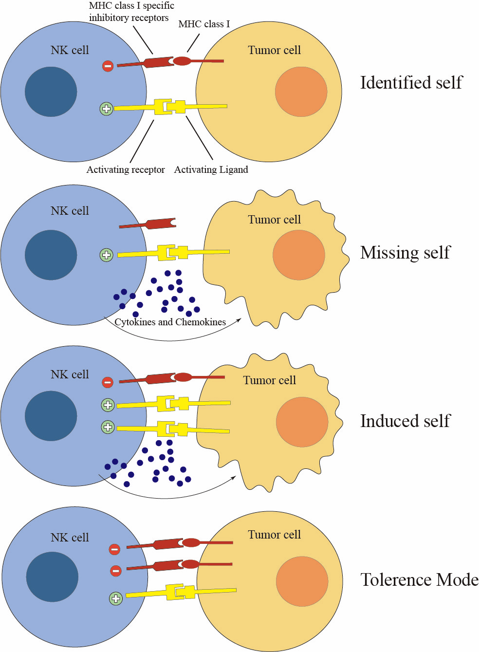
The Killer cell immunoglobulin-like receptor (KIR) family is one of the main groups of receptors that recognize MHC-I to maintain the tolerance phenotype of NK cells. The KIR family includes KIR2DL1, KIR2DL2/3, KIR3DL1, and KIR3DL2. NKG2A, also known as CD159a, and leukocyte immunoglobulin-like Receptor-1 (LIR-1) are other HLA-I receptors, which appear on different subsets of NK cells and also participate in the “self” identification mechanisms of NK cells (17). Interestingly, although there is diversity in HLA-I receptors on NK cells, these receptors use a common downstream signal pathway for inhibition. KIRs, NKG2A, and LIR-1 all contain cytoplasmic immunoreceptor tyrosine-based inhibition motifs (ITIMs). Phosphatase Src homology 2 domain-containing phosphatase 1 (SHP-1) is recruited around ITIMs in response to suppressive stimuli (18, 19). Other ITIM-containing NK inhibitory receptors do not recognize MHC-I. Killer Cell Lectin-like Receptor G1 (KLRG1) is the receptor of N-cadherin and E-cadherin (20, 21). KLRB1 binds the lectin-like molecule LLT1 and LLT1 homologs in humans and mice, respectively (22, 23). IRp60 recognizes phosphatidylserine (PS) and phosphatidylethanolamine (PE) (24). The ligands of sialic acid-binding Siglec-7 (CD238) and Siglec-9 (CD239) are sialoglycans. PD-1 can also be found on NK cells, which can inhibit NK cell activation by binding to PD-L1. TIGIT recognizes CD115 and CD112 (25). Unlike the inhibitory receptors, activation receptors induce diverse signaling cascades (Figure 2).
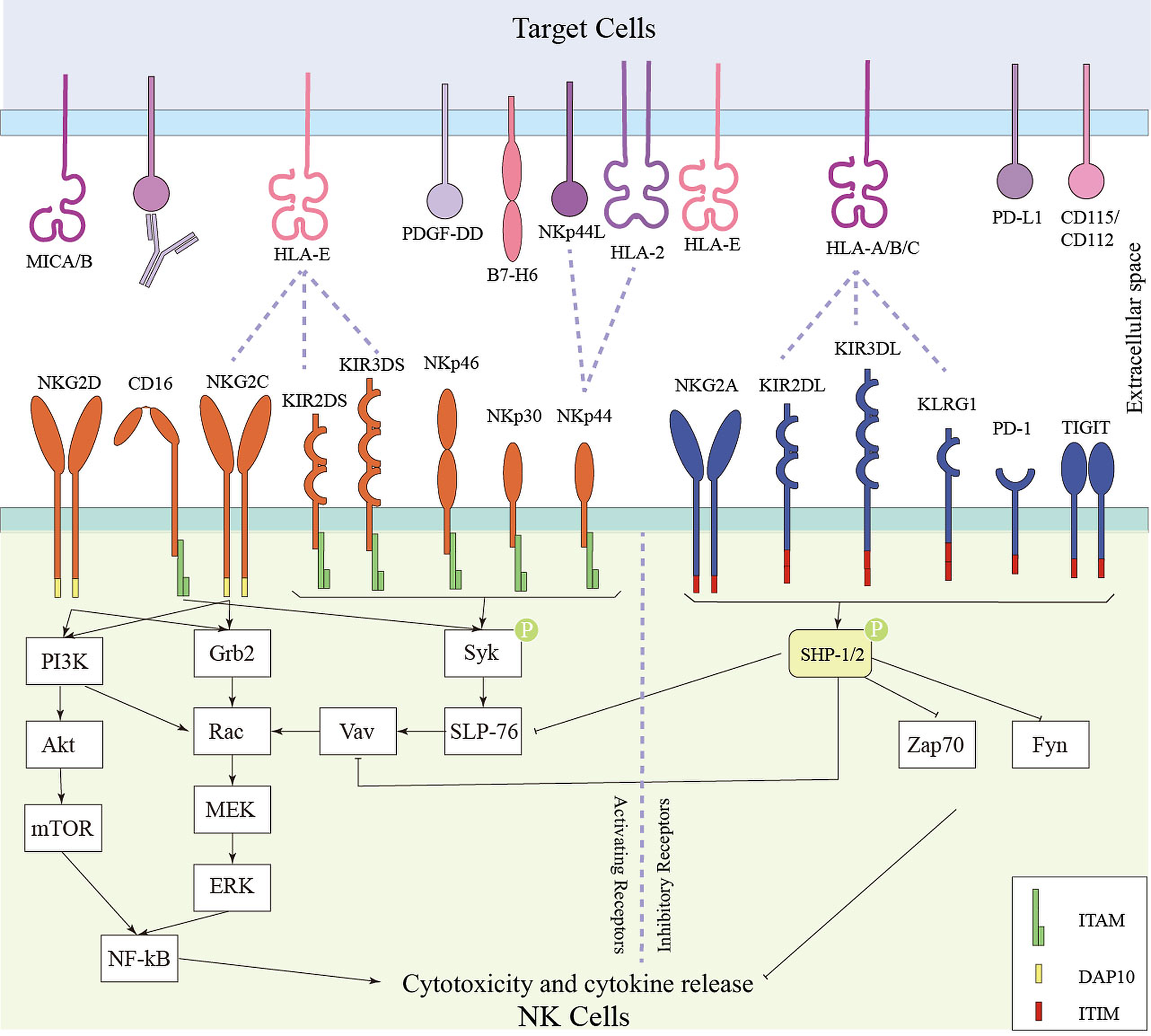
Groups of activation receptors contain an immunoreceptor tyrosine-based activation motif (ITAM). Among those receptors, KIR2DS, KIR3DS, NKG2C (CD159c) are closely related to MHC-I receptor counterparts, while CD16, NKp30 (CD337), and NKp46 (CD335) are not MHC-I receptor-related. CD16, NKp30, and NKp46 are expressed on most resting NK cells (26). After recognizing the Fc region of immunoglobulin, CD16 induces phosphorylation of ITAMs on the gamma domain of the high-affinity IgE receptor (FcεRIγ) and the CD3ζ chain, thereby triggering Antibody-dependent cell-mediated cytotoxicity (ADCC) (27, 28). It is an important tumor elimination approach in multiple monoclonal antibodies (mAb) in clinical practice, such as cetuximab, trastuzumab, and rituximab (29).
NKp30, NKp44, and NKp46 are classified as natural cytotoxicity receptors (NCRs), which are uniquely expressed by NK cells. NKp46 is highly conserved in mammals, but its ligands expressed on normal or tumor cells have yet to be identified (30, 31). The platelet-derived growth factor DD (PDGF-DD), secreted by many tumor types, is a widely studied ligand of NKp40. NKp30 will be discussed in detail in the next section. The best-known NK activation receptor lacking ITAM motifs is NKG2D (32, 33). Its ligands, MHC-I chain-related gene A (MICA), MHC-I chain-related gene B (MICB), and UL16-binding protein (ULBP), activate NK cells by phosphorylating the NKG2D-associated adapter protein DAP10, thereby stimulating the Phosphoinositide 3-kinase (PI3K) signaling cascade (34, 35). Tumor cells usually achieve immune escape by downregulating NK-cell activation signals, thereby shifting the NK cell from an activated to an inhibitory state.
Collectively, NK-cell activation is governed by the dynamic integration of stimulatory and inhibitory signals, creating a framework in which tumor-associated ligands such as B7-H6 can critically influence antitumor immune responses.
3. B7-H6 is a tumor-specific NK activation ligand
B7-H6 (NCR3LG1), a 51-kDa transmembrane protein with an IgV-IgC-like structure, was first identified in 2009 as an activation ligand for NK cells recognized by NKp30 (36, 37). The IgV-like domain has two glycosylation sites, and the IgC-like domain has four (38). B7-H6 contains an immunoreceptor tyrosine-based inhibition motif, an Src homology 2-binding domain, and an SH3-binding motif in the intracellular site, supporting unique intracellular functions (39, 40). B7-H6 is a tumor-specific marker that is undetectable in normal tissues at the mRNA level. But it is widely expressed in tumors, such as lymphoma, leukemia, ovarian carcinoma, brain cancer, breast cancer, and renal cell carcinoma, at both the mRNA level and protein level (41). Such tumor specialties make B7-H6 a potential therapeutic target for anti-tumor therapy. Expression of B7-H6 is closely related to histone deacetylases (HDACs).
The expression of B7-H6 was significantly induced or reduced by HDAC knockdown or HDAC3 overexpression, respectively (42). In addition to the membrane-binding type, B7-H6 also exists in soluble form. Matta et al. identified a soluble form of B7-H6 (sB7-H6), which could also associate with exosomes, is strictly expressed by activated monocytes and dendritic cells under inflammatory conditions, but can hardly be detected on the cell membrane of healthy donor PBMC (37, 43). Another type of sB7-H6 is generated by A disintegrin and metalloprotease (ADAM)-mediated protein shedding (Figure 3).
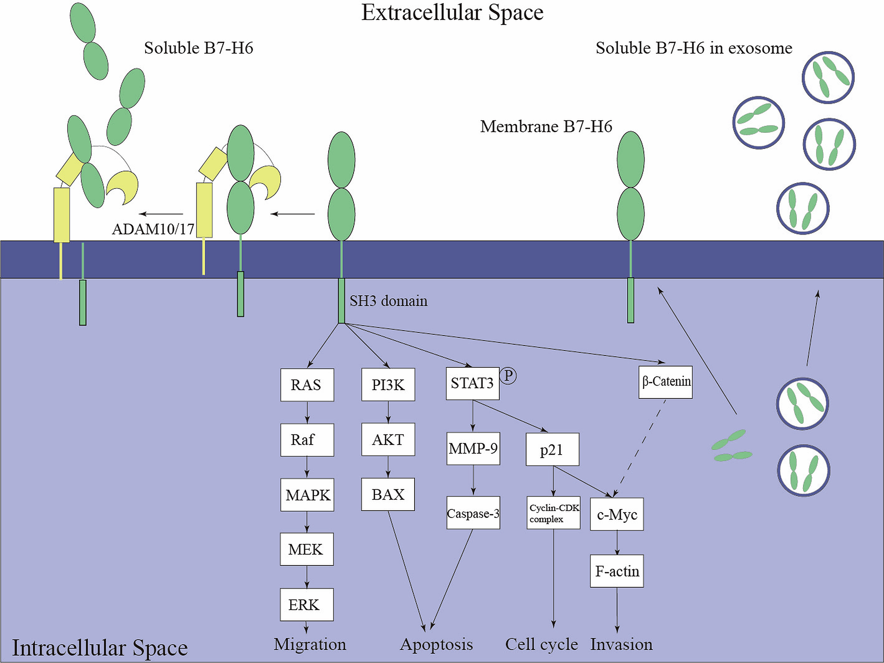
More specifically, ADAM-10 and ADAM-17 cleave the extracellular domain of B7-H6, potentially resulting in immune escape in melanoma (44). The ADAM inhibitor could significantly increase the membrane level of B7-H6 in the B7-H6 overexpression cell line. The shed-off part of B7-H6 was about 30 kDa, corresponding to the full-length extracellular domain. Furthermore, ADAM-10 and ADAM-17, as sheddases located on the extracellular membrane, are activated and cleave the target protein in the extracellular region. Moreover, two types of sB7-H6 were found in the peripheral blood of pregnant women (45). The function of those sB7-H6 in the pregnancy process remains ambiguous.
The existence of both membrane-bound and soluble forms of B7-H6 highlights the biological complexity of this molecule. Whereas membrane-associated B7-H6 primarily functions as a ligand for NKp30-mediated immune recognition, soluble B7-H6 may exert distinct immunological and tumor-promoting effects. The relative abundance, origin, and biological activity of these different B7-H6 species are likely to influence both tumor progression and therapeutic responsiveness. Consequently, understanding the mechanisms governing B7-H6 expression, shedding, and extracellular release has become an important area of investigation in cancer immunology.
Beyond epigenetic regulation, cellular stress pathways, inflammatory signaling, and oncogenic transformation have been proposed to contribute to the induction of B7-H6, although the underlying mechanisms remain incompletely defined. Although the regulatory mechanisms governing B7-H6 expression remain incompletely understood, current evidence suggests that B7-H6 expression reflects a state of cellular stress associated with malignant transformation rather than normal tissue homeostasis. The highly restricted expression pattern of B7-H6, together with its inducibility under pathological conditions, provides a strong rationale for its development as a selective immunotherapeutic target.
4. Regulation of B7-H6 expression in cancer
Although B7-H6 is widely recognized as a tumor-associated ligand for the activating NK-cell receptor NKp30, the mechanisms governing its expression remain incompletely understood. Unlike many immune regulatory molecules that are constitutively expressed in normal tissues, B7-H6 expression is largely absent under physiological conditions and becomes induced in response to pathological stress (37). This restricted expression pattern suggests that B7-H6 functions as a stress-associated molecule that signals cellular transformation and tissue damage to the immune system.
Current evidence indicates that B7-H6 expression is regulated at multiple levels, including epigenetic, transcriptional, and post-translational mechanisms. Among the best-characterized regulators are histone deacetylases (HDACs), which participate in chromatin remodeling and gene transcription. Experimental studies demonstrated that HDAC inhibitors can modulate B7-H6 expression and alter tumor-cell recognition by NK cells, whereas HDAC3 overexpression suppresses B7-H6 levels (46,47). These findings suggest that epigenetic mechanisms play an important role in controlling the availability of B7-H6 on the tumor cell surface. These observations suggest that epigenetic therapies may increase tumor susceptibility to NK-cell-mediated immune surveillance by enhancing B7-H6 expression.
In addition to epigenetic regulation, inflammatory and stress-associated signals have been proposed to contribute to B7-H6 induction, although the regulatory mechanisms remain incompletely defined (48). Chronic inflammation, a hallmark of many malignancies, is characterized by persistent cytokine production, oxidative stress, and tissue remodeling. These processes may create a microenvironment favorable for B7-H6 expression, thereby linking inflammatory signaling to NK-cell recognition of transformed cells.
Cellular stress responses associated with malignant transformation may also contribute to B7-H6 upregulation. Tumor cells are frequently exposed to genomic instability, oncogene activation, metabolic stress, hypoxia, and DNA damage, all of which activate stress-response pathways that alter surface ligand expression. Similar to other stress-induced NK-cell ligands, B7-H6 may function as a molecular indicator of cellular distress (37), allowing NK cells to identify and eliminate potentially dangerous cells before malignant progression occurs. However, the specific signaling pathways connecting these stress responses to B7-H6 transcription remain largely undefined and warrant further investigation.
Beyond transcriptional regulation, post-translational mechanisms also influence the biological availability of B7-H6. A major regulatory process involves ADAM10- and ADAM17-mediated proteolytic shedding, which releases the extracellular domain of B7-H6 into the tumor microenvironment and circulation (49). This process reduces membrane-associated B7-H6 while generating soluble B7-H6 species that may possess distinct immunological functions. Consequently, B7-H6 expression is not solely determined by transcriptional activity but also by dynamic regulation of protein stability and surface retention.
Collectively, current evidence suggests that B7-H6 expression is controlled by a complex network of epigenetic regulation, inflammatory signaling, cellular stress responses, and post-translational processing. A deeper understanding of these regulatory mechanisms will not only provide insight into the biological role of B7-H6 during tumor development but may also reveal new opportunities to enhance the efficacy of B7-H6-targeted immunotherapies by increasing target expression or preventing its loss through proteolytic shedding. The diverse mechanisms regulating B7-H6 expression suggest that this molecule is more than a passive marker of malignant transformation. Accumulating evidence suggests that B7-H6 may actively contribute to tumor progression through both immune-dependent and tumor-intrinsic mechanisms, as discussed below.
5. Biological roles of B7-H6 in cancer
Although B7-H6 was initially characterized as an immune recognition ligand, accumulating evidence indicates that it also participates directly in tumor cell biology. Emerging studies suggest that B7-H6 contributes to several hallmarks of cancer, including sustained proliferation, resistance to apoptosis, enhanced migratory capacity, and metastatic dissemination. These observations have shifted the perception of B7-H6 from a passive immune marker to an active regulator of malignant behavior. The expression patterns, biological functions, and clinical associations of B7-H6 across different malignancies are summarized in Table 1.
Table 1. Molecular and clinical significance of B7-H6 across human malignancies.
| Tumor types | Expression | Biological functions | Prognostic association | Proposed Mechanism | Ref |
|---|---|---|---|---|---|
| Hepatocellular Carcinoma (HCC) | Upregulated | Promotes proliferation, migration, and invasion | Conflicting; associated with both aggressive behavior and improved survival in different cohorts | STAT3 activation; MMP9 induction | 51, 56, 60 |
| Breast Cancer | Upregulated | Promotes tumor progression and survival | Worse overall survival | PI3K/Akt signaling; apoptosis regulation | 40,54 |
| Ovarian Cancer | Upregulated | Associated with metastasis and immune escape | Poor survival and disease progression | NKp30-suppression via soluble B7-H6 | 58 |
| Glioma/Medulloblastoma | Upregulated | Enhances migration and invasion | Not established | c-MYC activation; PI3K/Akt; ERK/MAPK; MMP2/9 | 53,57 |
| Non-Hodgkin Lymphoma (NHL) | Upregulated | Promotes proliferation, migration, and invasion | Limited clinical data | Ras/MEK/ERK signaling | 52 |
| Non-small cell lung cancer (NSCLC) | Variable | Associated with tumor differentiation | No significant association with survival | Not clearly defined | 59 |
| Cervical Cancer | Upregulated | Regulates proliferation and migration | Unknown | NKp30-dependent signaling interactions | 50 |
Abbreviations: HCC, hepatocellular carcinoma; NSCLC, non-small cell lung cancer; PI3K, phosphoinositide 3-kinase; Akt, protein kinase B; ERK, extracellular signal-regulated kinase; MMP, matrix metalloproteinase; STAT3, signal transducer and activator of transcription 3.
In addition to acting as the ligand for NKp30, B7-H6 also has intracellular regulatory functions. (Figure 3) When soluble NKp30 was co-cultured with the B7-H6-positive cervical cancer-derived cell lines, SiLA and HeLa, the proliferation and migration were significantly retarded (50). Knockdown of B7-H6 significantly attenuated tumor proliferation, migration, and invasion in the HepG2 and SMMC-7721 cell lines (51). Moreover, B7-H6 promotes cell proliferation, migration, and invasion via the Ras/MEK/ERK Pathway. Overexpression of B7-H6 in Jurkat and Raji cell lines significantly increased MEK and ERK phosphorylation (52). However, the MEK1/2 inhibitor can only partially reverse the cell migration and invasion. This reminds us that B7-H6 regulates tumor migration, proliferation, and invasion through multiple pathways. These pathways may function independently. Accumulating evidence revealed the relationship between B7-H6 and the PI3K/AKT pathway. Knockdown of B7-H6 in glioma stem-like cells decreased the expression of c-Myc and inactivated the PI3K/Akt and ERK/MAPK signaling pathways. RNMT (RNA guanine-7-methyltransferase) played a key role in c-Myc expression and was also downregulated by B7-H6 inhibition. C-Myc was known to regulate the PI3K/Akt and ERK signaling pathways. Moreover, enhanced c-Myc expression induced by B7-H6 also reorganizes F-actin, thereby improving the migration and invasion of Daoy cells (53). Furthermore, cell cycle regulatory proteins, such as cyclin D1, cyclin-dependent kinases 4 (CDK4) and CDK6, and phospho-Rb expression were significantly decreased upon B7-H6 inhibition (51). Knockdown of B7-H6 inhibits cell proliferation and promotes apoptosis, while it upregulates the expression of β‑catenin, caspase‑3, and Bax in MDA-MB-231 cells (54). Those proteins are also regulated by the PI3K/AKT and Ras/MEK/ERK pathways. Therefore, in addition to the Ras/MEK/ERK pathway, B7-H6 may regulate cell proliferation and invasion via the c-Myc/PI3K/Akt pathway.
Further studies have found that B7-H6 activates the STAT3 pathway (42). Cellular stress responses may also contribute to B7-H6 upregulation. In particular, therapeutic stress has been shown to enhance tumor sensitivity to NK-cell cytolysis by upregulating B7-H6 (55). B7-H6 promotes proliferation and invasion of HCC cells by inducing MMP-9 expression and STAT3 activation (56). The expression levels of MMP-2 and MMP-9 decreased upon B7-H6 knockdown in glioma cells (57). That research revealed that the pro-tumorigenesis function of B7-H6 may be mediated by the STAT3 pathway, promoting anti-apoptosis and pro-proliferation in cancer cells, but this function appears to depend on pathological conditions. In clinical practice, B7-H6 is closely associated with patient prognosis and survival. In a study on a cohort of 1100 breast cancer patients, the upregulation of B7-H6 at the mRNA level is significantly associated with worse overall survival (40). In ovarian cancer, high-level expression of B7-H6 in tumor tissues is correlated with a worse survival rate, tumor metastasis, and progression (58). However, in astrocytoma, gastric carcinoma, and non-small cell lung cancer (NSCLC), this protein expression does not confer prognostic significance (51). But in NSCLC, B7-H6-positive carcinomas were significantly correlated with the degree of differentiation but not with the three-year survival rate (59). In a study of hepatocellular carcinoma (HCC), high B7-H6 levels are associated with smaller tumor size and better survival (60). Therefore, the biological consequences of B7-H6 expression appear to be context-dependent and may vary with tumor type and microenvironmental conditions. Collectively, these findings suggest that B7-H6 may serve as a context-dependent indicator of tumor progression and clinical outcome. The prognostic significance of B7-H6 appears to be highly context-dependent. Differences in tumor lineage, immune cell infiltration, quantification methods, and the relative contributions of membrane-bound versus soluble B7-H6 may account for the inconsistent clinical associations reported across malignancies. These observations suggest that B7-H6 should not be viewed as a universal prognostic biomarker, but rather as a context-specific indicator whose biological and clinical relevance may vary according to tumor type and microenvironmental conditions.
Collectively, these findings suggest that B7-H6 functions as more than an NK-cell ligand. Through engagement of Ras/MEK/ERK, PI3K/Akt, c-Myc, and STAT3 signaling networks, B7-H6 appears capable of promoting multiple hallmarks of cancer, including sustained proliferation, resistance to apoptosis, and invasive behavior. These observations support an emerging model in which B7-H6 serves both immunological and tumor-intrinsic functions. This dual functionality places B7-H6 at a unique intersection between tumor immunology and cancer cell biology. Traditionally, B7-H6 has been viewed primarily as a danger-associated ligand that facilitates NK-cell recognition and elimination of transformed cells through NKp30 activation. However, accumulating evidence indicates that B7-H6 also exerts direct tumor-intrinsic effects by regulating signaling pathways involved in proliferation, survival, migration, and invasion. Moreover, proteolytic shedding and the generation of soluble B7-H6 may enable tumors to attenuate NK-cell surveillance while preserving or even enhancing protumorigenic functions. Consequently, B7-H6 may act as a molecular bridge linking immune surveillance, immune escape, and malignant progression. This integrated model offers a potential explanation for the context-dependent clinical associations observed across tumor types and highlights why therapeutic targeting of B7-H6 may simultaneously influence both tumor cells and the antitumor immune response.
The role of B7-H6 in tumor immune escape has gradually emerged through a series of experimental and clinical studies. The shedding of B7-H6 was considered one of the mechanisms by which tumor cells escape immune surveillance. The inhibition of ADAM-mediated B7-H6 shedding significantly increased NK-cell cytotoxicity (44). Furthermore, a high level of sB7-H6 was detected in peritoneal/ascitic fluid from ovarian cancer patients, which was associated with low NKp30 expression on NK-cells (58). In high-risk neuroblastoma, sB7-H6 in serum inhibited NK-cell functions when co-cultured with NK-cells in vitro (61). In hemodialysis patients, elevated serum sB7-H6 levels are associated with a higher risk of infection, and circulating sB7-H6 levels increase in patients with infectious complications (62). Therefore, B7-H6 may be related to inflammatory responses. However, there are 2 forms of B7-H6, and those studies haven’t explicitly stated the functional form of sB7-H6 in sera. Therefore, we can’t confirm the immunosuppressive function of cell-shed sB7-H6 in cancer progression. On the other hand, a recent study demonstrated that human recombinant sB7-H6 with an Fc tag has high NKp30-binding capacity and can induce both TNF-α and IFN-γ production (63). Whether cell-shed sB7-H6 can also induce NK-cell-mediated cytokine production and cytotoxicity remains to be experimentally verified. The biological significance of soluble B7-H6 (sB7-H6) remains one of the major unresolved questions surrounding the B7-H6/NKp30 axis. A prevailing hypothesis proposes that sB7-H6 functions as a decoy ligand that chronically engages NKp30, leading to receptor downregulation and reduced NK-cell responsiveness. This model is supported by studies demonstrating an inverse relationship between circulating sB7-H6 levels and NKp30 expression in cancer patients.
However, accumulating evidence suggests that the effects of sB7-H6 may be more complex than simple receptor blockade. Recombinant sB7-H6 has been shown to retain NKp30-binding activity and stimulate cytokine production under certain experimental conditions, raising the possibility that soluble B7-H6 may exert context-dependent immunomodulatory effects. These apparently conflicting findings may reflect differences in molecular origin, post-translational modification, concentration, or mode of release. For example, ADAM-mediated shedding, exosome-associated secretion, and inflammation-induced soluble isoforms may possess distinct biological properties. Clarifying these differences will be critical for understanding how B7-H6 contributes to immune escape and for optimizing future B7-H6-targeted therapeutic strategies. Collectively, current evidence suggests that soluble B7-H6 may function as both a biomarker of disease progression and a regulator of antitumor immunity, although its precise biological role remains to be fully elucidated.
6. Current anti-B7-H6 antibody therapy
The tumor-restricted expression pattern of B7-H6 has spurred the development of diverse therapeutic platforms to exploit the B7-H6/NKp30 axis. Unlike broadly expressed immune targets that raise concerns about on-target, off-tumor toxicity, B7-H6 offers an opportunity to selectively target malignant cells while simultaneously enhancing immune effector functions. Consequently, multiple antibody-based, cellular, and immune-engaging strategies have been developed to leverage B7-H6's unique biological properties. A diverse range of B7-H6-targeted therapeutic platforms has been developed, including bispecific engagers, immunoligands, CAR-based cellular therapies, and cytokine-enhanced immune approaches. The major strategies currently under investigation are summarized in Table 2.
Table 2. Current B7-H6-targeted therapeutic strategies.
| Strategy | Format | Mechanism | Outcome | Ref |
|---|---|---|---|---|
| NK-cell engager | B7-H6/NKp30 immunoligand | NK activation | Enhanced cytotoxicity | 64 |
| HER2-B7-H6 immunoligand | Fusion protein | HER2 targeting + NK activation | Improved ADCC | 65 |
| B7-H6/CD3 BiTE | T-cell engager | T-cell recruitment | Tumor elimination | 66 |
| TZ47 CAR-T | CAR-T | Direct targeting | In vivo efficacy | 67 |
| Humanized anti-B7-H6 antibody/scFv design | CAR-T | Reduced immunogenicity | Improved translation | 68 |
| Human scFv CAR-T | CAR-T | Alternative epitope | Strong efficacy | 69 |
| IL-15-enhanced BiTE | BiTE + cytokine | T/NK activation | Superior efficacy | 71 |
Abbreviations: ADCC, antibody-dependent cellular cytotoxicity; BiTE, bispecific T-cell engager; CAR, chimeric antigen receptor; IL-15, interleukin-15; NK, natural killer.
Pekar et al. generated a bispecific IgG-like NK-cell engager that fused the high-affinity N-terminal IgV domain of B7-H6 with antigen-binding fragments (Fabs) derived from cetuximab, an EGFR monoclonal antibody (64). The high-affinity N-terminal IgV domain of B7-H6 was obtained by affinity maturation. This bispecific IgG-like NK-cell engager increased NK-cell cytotoxicity and the release of IFN-γ and TNF-α compared with human IgG Fc (HFC), thereby promoting ADCC. A HER2/B7-H6 fusion antibody is also constructed to trigger NK-cell killing of HER2-positive breast cancer cells (65). The B7-H6:HER2-scFv shows good efficacy in enhancing NK-cell cytotoxicity and in improving ADCC when combined with trastuzumab and cetuximab 61. Several different bispecific T-cell engagers (BiTEs) were also generated. Wu et al. fused the anti-B7-H6 scFv with a CD3ε-specified scFv to lead T cells to mediate cell killing and cytokine release against B7-H6-positive tumor cells (66). The elimination effect was observed in both murine melanoma and ovarian cancer models. In vivo, perforin and IFN-γ levels increased following B7-H6/CD3 BiTE treatment, resulting in improved survival (66). They also constructed a B7-H6-specific CAR-T cell based on TZ47, which showed good tumor-elimination efficacy in the in vivo ovarian cancer model (67). Since TZ47 is a mouse IgG, Choi et al. humanized it to reduce its immunogenicity (68). Later in 2017, Hua et al. published a new human B7-H6-targeting scFv that binds a different epitope than TZ47 and generated a new CAR-T based on this antibody. This scFv can bind to both soluble and membrane-bound B7-H6, and the CAR-T also showed sufficient anti-cancer activity (69). BCL-2 overexpression, B7-H6-targeting CAR-T also showed promising therapeutic outcomes both in vitro and in vivo, alone and in combination with venetoclax (a BH3-mimetic small-molecule drug targeting BCL-2) against small-cell lung cancer (70). A recent study constructed BiTEs combined with the IL-15/IL-15Ra sushi fusion protein against B7-H6-positive cell lines. The cytotoxicity reached 90% in vitro, and tumor size was reduced by 79% in vivo. However, the BiTEs with the IL-15/IL-15Ra sushi fusion protein showed higher toxicity at high concentrations in vivo (71).
Taken together, these studies demonstrate that B7-H6 can be successfully targeted through multiple therapeutic modalities, including NK-cell engagers, bispecific antibodies, CAR-T cells, and cytokine-enhanced immune platforms. Although most evidence remains preclinical, the breadth of successful approaches highlights B7-H6's versatility as an immunotherapeutic target and provides a strong rationale for continued clinical development.
7. Clinical development status of B7-H6-targeted therapies
Despite the growing body of preclinical evidence supporting B7-H6 as an attractive immunotherapeutic target, clinical translation remains in its early stages. To date, most B7-H6-directed approaches have been evaluated primarily in cell-line systems and xenograft models (66, 67), with only limited progression into human clinical studies. The most advanced clinical program is represented by a B7-H6/CD3 bispecific T-cell engager evaluated in a phase I clinical trial (ClinicalTrials.gov identifier: NCT04752215). Although study completion and status updates have been reported, efficacy and detailed safety outcomes have not yet been publicly disclosed, making it difficult to assess the true clinical potential of B7-H6-targeted therapy.
The lack of clinical data highlights a broader challenge frequently encountered during immunotherapy development. While xenograft models have consistently demonstrated potent antitumor activity of B7-H6-targeting antibodies, BiTEs, and CAR-based cellular therapies, these models incompletely recapitulate the complexity of the human tumor microenvironment. Human tumors exhibit substantial heterogeneity in antigen expression, immune-cell infiltration, stromal composition, and immunosuppressive signaling, all of which may significantly influence therapeutic responses. Furthermore, xenograft systems often fail to accurately model soluble B7-H6 dynamics, antigen shedding, and long-term immune adaptation, factors that may become critical determinants of efficacy in patients.
The clinical development of B7-H6-targeted therapies can also be viewed in the context of other NK-cell-directed immunotherapeutic strategies. Therapeutic agents targeting NKG2D ligands, NKp46, CD16, NKG2A, and TIGIT have already entered clinical development and, in some cases, demonstrated encouraging early clinical activity (30). Compared with these targets, B7-H6 has the distinct advantage of being highly restricted in normal tissues or largely restricted to malignant and stressed cells, potentially reducing the risk of on-target, off-tumor toxicity. At the same time, uncertainties regarding expression heterogeneity, the biology of soluble B7-H6, and inducible expression under inflammatory conditions remain important translational considerations (47). Consequently, future clinical studies will be essential not only to establish efficacy but also to define optimal patient-selection strategies, combination regimens, and biomarkers to maximize the therapeutic potential of the B7-H6/NKp30 axis.
8. Discussion
Beyond demonstrating clinical efficacy, several biological and translational challenges must be addressed before B7-H6-targeted therapies can be successfully integrated into routine oncology practice. These include uncertainties regarding target heterogeneity, the biological consequences of soluble B7-H6, the mechanisms of therapeutic resistance, and the identification of predictive biomarkers to guide patient selection.
Currently, most B7-H6-targeting therapies focus on T-cell activation. However, a recent study has disclosed that B7-H6 expression is significantly increased on T cells, especially on CAR-T cells, in response to activation. B7-H6-mediated NKp30-dependent NK-cell activation resulted in depletion of activated T cells and limited the antitumor activity of CAR-T cells (72). Although there are no experimental results demonstrating T-cell depletion after T cells are activated by B7-H6-targeting T-cell engagers, there is a theoretical risk of T-cell elimination by NK cells, which could compromise the therapeutic efficacy of these engagers. Therefore, the development of B7-H6 target therapy based on activating NK cells might be more efficient. Furthermore, shedding B7-H6 is known as an immune escape strategy for cancer cells. For this reason, immunotherapy may lose efficacy. Therefore, B7-H6-targeting treatment combined with shedding inhibition may yield better outcomes. Another important consideration is the heterogeneous expression of B7-H6 across tumor types and even within individual tumors. Antigen heterogeneity may facilitate immune escape through selective pressure during treatment, potentially limiting the durability of therapeutic responses. Future studies should therefore investigate strategies to overcome antigen-loss variants, including dual-targeting approaches and combination immunotherapies.
What's more, there is a limited number of studies that have explored the drug-combination potential of B7-H6-targeting therapy. A study has shown that a B7-H6/CD3 T-cell engager can more effectively suppress tumor growth in combination with an anti-PD-1 drug. The combination of anti-PD-1 and B7-H6 reduced the effective concentration of both drugs (73). In addition to anti-PD-1, many other drugs are worth testing in combination with anti-B7-H6 antibodies. This direction needs more attempts. Also, B7-H6 is a highly tumor-specific surface marker. Therefore, B7-H6 is potentially a target for a tumor-specific drug delivery system, but this remains untested. B7-H6 retains considerable therapeutic potential. The development of robust predictive biomarkers also remains a major unmet need. While B7-H6 expression is frequently used as a selection criterion, it remains unclear whether expression level alone adequately predicts therapeutic responsiveness. Integrating tumor B7-H6 expression with measures of NK-cell infiltration, NKp30 expression, soluble B7-H6 concentration, and immune microenvironment characteristics may provide a more comprehensive framework for patient stratification.
B7-H6 occupies a unique position at the intersection of tumor immunology and cancer cell biology. Beyond functioning as a ligand for NKp30-mediated immune recognition, accumulating evidence suggests that B7-H6 actively contributes to tumor progression by regulating proliferative, survival, and invasive signaling networks. This dual role distinguishes B7-H6 from many conventional immunotherapeutic targets and creates opportunities for therapeutic strategies that can simultaneously enhance antitumor immunity and disrupt tumor-intrinsic oncogenic programs.
Despite substantial progress, several fundamental questions remain unanswered. The upstream signaling pathways that induce B7-H6 expression remain incompletely characterized; the biological functions of distinct soluble B7-H6 species remain controversial; and the mechanisms by which B7-H6 activates intracellular oncogenic pathways are poorly understood. Emerging evidence suggests that membrane-bound and soluble B7-H6 may exert distinct biological functions, yet their relative contributions to tumor progression and immune regulation remain unclear. Notably, soluble NKp30 has been reported to inhibit tumor growth, whereas inhibition of ADAM-mediated B7-H6 shedding can suppress tumor cell proliferation, highlighting the complex and potentially divergent roles of different B7-H6 forms. Whether the protumorigenic activities attributed to B7-H6 are mediated primarily by membrane-associated or soluble species remains unresolved. Clarifying the molecular mechanisms governing B7-H6 signaling, shedding, and functional diversification will be essential for refining therapeutic strategies, identifying predictive biomarkers, and determining which patient populations are most likely to benefit from B7-H6-targeted interventions.
Collectively, current evidence positions B7-H6 as a unique immuno-oncologic target that integrates immune surveillance, immune escape, and tumor-intrinsic signaling. Continued investigation into the regulation, biological functions, and therapeutic exploitation of the B7-H6/NKp30 axis will likely facilitate the development of next-generation immunotherapies that simultaneously enhance antitumor immunity and disrupt malignant progression.
References
1. Barrow AD, Martin CJ, Colonna M. The natural cytotoxicity receptors in health and disease. Front Immunol. 2019;10:909. https://doi.org/10.3389/fimmu.2019.00909
2. Laskowski TJ, Biederstädt A, Rezvani K. Natural killer cells in antitumour adoptive cell immunotherapy. Nat Rev Cancer. 2022;22(10):557–575. https://doi.org/10.1038/s41568-022-00491-0
3. Dhatchinamoorthy K, Colbert JD, Rock KL. Cancer immune evasion through loss of MHC class I antigen presentation. Front Immunol. 2021;12:636568. https://doi.org/10.3389/fimmu.2021.636568
4. Correia AL, Guimaraes JC, Auf der Maur P, De Silva D, Trefny MP, Okamoto R, et al. Hepatic stellate cells suppress NK cell-sustained breast cancer dormancy. Nature. 2021;594(7864):566–571. https://doi.org/10.1038/s41586-021-03614-z
5. Liu X, Song J, Zhang H, Liu X, Zuo F, Zhao Y, et al. Immune checkpoint HLA-E:CD94-NKG2A mediates evasion of circulating tumor cells from NK cell surveillance. Cancer Cell. 2023;41(2):272–287.e9. https://doi.org/10.1016/j.ccell.2023.01.001
6. Simonetta F, Alvarez M, Negrin RS. Natural killer cells in graft-versus-host-disease after allogeneic hematopoietic cell transplantation. Front Immunol. 2017;8:465. https://doi.org/10.3389/fimmu.2017.00465
7. Huang S, Xing F, Dai Y, Zhang Z, Zhou G, Yang S, et al. Navigating chimeric antigen receptor-engineered natural killer cells as drug carriers via three-dimensional mapping of the tumor microenvironment. J Control Release. 2023;362:524–535. https://doi.org/10.1016/j.jconrel.2023.09.007
8. Poli A, Michel T, Thérésine M, Andrès E, Hentges F, Zimmer J. CD56bright natural killer (NK) cells: an important NK cell subset. Immunology. 2009;126(4):458–465. https://doi.org/10.1111/j.1365-2567.2008.03027.x
9. Seymour F, Cavenagh JD, Mathews J, Gribben JG. NK cells CD56bright and CD56dim subset cytokine loss and exhaustion is associated with impaired survival in myeloma. Blood Adv. 2022;6(17):5152–5159. https://doi.org/10.1182/bloodadvances.2022007905
10. Weber S, Menees KB, Park J, Agin-Liebes J, Lin CC, Alcalay RN, et al. Distinctive CD56dim NK subset profiles and increased NKG2D expression in blood NK cells of Parkinson's disease patients. NPJ Parkinsons Dis. 2024;10(1):36. https://doi.org/10.1038/s41531-024-00652-y
11. Myers JA, Miller JS. Exploring the NK cell platform for cancer immunotherapy. Nat Rev Clin Oncol. 2021;18(2):85–100. https://doi.org/10.1038/s41571-020-0426-7
12. Albertsson PA, Basse PH, Hokland M, Goldfarb RH, Nagelkerke JF, Nannmark U, et al. NK cells and the tumour microenvironment: implications for NK-cell function and anti-tumour activity. Trends Immunol. 2003;24(11):603–609. https://doi.org/10.1016/j.it.2003.09.007
13. Mandal R, Şenbabaoğlu Y, Desrichard A, Havel JJ, Dalin MG, Riaz N, et al. The head and neck cancer immune landscape and its immunotherapeutic implications. JCI Insight. 2016;1(17):e89829. https://doi.org/10.1172/jci.insight.89829
14. Ljunggren HG, Kärre K. In search of the ‘missing self’: MHC molecules and NK cell recognition. Immunol Today. 1990;11(7):237–244. https://doi.org/10.1016/0167-5699(90)90097-s
15. Chen S, Zhu H, Jounaidi Y. Comprehensive snapshots of natural killer cell functions, signaling, molecular mechanisms and clinical utilization. Signal Transduct Target Ther. 2024;9(1):302. https://doi.org/10.1038/s41392-024-02005-w
16. Letafati A, Salahi Ardekani O, Naderisemiromi M, Norouzi M, Shafiei M, Nik S, et al. Unraveling the dynamic mechanisms of natural killer cells in viral infections: insights and implications. Virol J. 2024;21(1):18. https://doi.org/10.1186/s12985-024-02287-0
17. Sivori S, Della Chiesa M, Carlomagno S, Quatrini L, Munari E, Vacca P, et al. Inhibitory receptors and checkpoints in human NK cells, implications for the immunotherapy of cancer. Front Immunol. 2020;11:2156. https://doi.org/10.3389/fimmu.2020.02156
18. Bryceson YT, March ME, Ljunggren HG, Long EO. Activation, coactivation, and costimulation of resting human natural killer cells. Immunol Rev. 2006;214:73–91. https://doi.org/10.1111/j.1600-065x.2006.00457.x
19. Getahun A, Cambier JC. Of ITIMs, ITAMs, and ITAMis: revisiting immunoglobulin Fc receptor signaling. Immunol Rev. 2015;268(1):66–73. https://doi.org/10.1111/imr.12336
20. Butcher S, Arney KL, Cook GP. MAFA-L, an ITIM-containing receptor encoded by the human NK cell gene complex and expressed by basophils and NK cells. Eur J Immunol. 1998;28(11):3755–3762. https://doi.org/10.1002/(sici)1521-4141(199811)28:11<3755::AID-IMMU3755>3.0.CO;2-3
21. Zhang Y, Chen S, Tang X, Peng Y, Jiang T, Zhang X, et al. The role of KLRG1: a novel biomarker and new therapeutic target. Cell Commun Signal. 2024;22(1):337. https://doi.org/10.1186/s12964-024-01714-7
22. Aldemir H, Prod’homme V, Dumaurier MJ, Retiere C, Poupon G, Cazareth J, et al. Cutting edge: lectin-like transcript 1 is a ligand for the CD161 receptor. J Immunol. 2005;175(12):7791–7795. https://doi.org/10.4049/jimmunol.175.12.7791
23. Mathewson ND, Ashenberg O, Tirosh I, Gritsch S, Perez EM, Marx S, et al. Inhibitory CD161 receptor identified in glioma-infiltrating T cells by single-cell analysis. Cell. 2021;184(5):1281–1298.e26. https://doi.org/10.1016/j.cell.2021.01.022
24. Cantoni C, Bottino C, Augugliaro R, Morelli L, Marcenaro E, Castriconi R, et al. Molecular and functional characterization of IRp60, a member of the immunoglobulin superfamily that functions as an inhibitory receptor in human NK cells. Eur J Immunol. 1999;29(10):3148–3159. https://doi.org/10.1002/(SICI)1521-4141(199910)29:10<3148::AID-IMMU3148>3.0.CO;2-L
25. Mace EM. Human natural killer cells: form, function, and development. J Allergy Clin Immunol. 2023;151(2):371–385. https://doi.org/10.1016/j.jaci.2022.09.022
26. Demaria O, Gauthier L, Debroas G, Vivier E. Natural killer cell engagers in cancer immunotherapy: Next generation of immuno-oncology treatments. Eur J Immunol. 2021;51(8):1934–1942. https://doi.org/10.1002/eji.202048953
27. Perussia B, Trinchieri G, Jackson A, Warner NL, Faust J, Rumpold H, et al. The Fc receptor for IgG on human natural killer cells: phenotypic, functional, and comparative studies with monoclonal antibodies. J Immunol. 1984;133(1):180–189. https://doi.org/10.4049/jimmunol.133.1.180
28. Zhou G, Zhang Y, Gu Y, Cao B, Fan X, Yu D, et al. Enhanced antibody-mediated cellular cytotoxicity of germline-like anti-HER2 antibodies through a point mutation in complementarity-determining regions. J Immunol. 2025;214(7):1849–1859. https://doi.org/10.1093/jimmun/vkaf063
29. Bachanova V, Sarhan D, DeFor TE, Cooley S, Panoskaltsis-Mortari A, Blazar BR, et al. Haploidentical natural killer cells induce remissions in non-Hodgkin lymphoma patients with low levels of immune-suppressor cells. Cancer Immunol Immunother. 2018;67(3):483–494. https://doi.org/10.1007/s00262-017-2100-1
30. Gauthier L, Morel A, Anceriz N, Rossi B, Blanchard-Alvarez A, Grondin G, et al. Multifunctional natural killer cell engagers targeting NKp46 trigger protective tumor immunity. Cell. 2019;177(7):1701–1713.e16. https://doi.org/10.1016/j.cell.2019.04.041
31. Qian S, Zhou Y, Jin Z, Li X, Tian Y, Chen F, et al. Advancements in the study of the immune molecule NKp46 in immune system-related diseases. Clin Rev Allergy Immunol. 2024;67(1-3):96–110. https://doi.org/10.1007/s12016-024-09010-5
32. Cerwenka A, Lanier LL. NKG2D ligands: unconventional MHC class I-like molecules exploited by viruses and cancer. Tissue Antigens. 2003;61(5):335–343. https://doi.org/10.1034/j.1399-0039.2003.00070.x
33. Tang J, Lu Y, Chen M, Wu Q, Li Y, Qin Y, et al. The metabolic regulation of the NKG2D-positive NK and T cells and their role in disease progression. Biomolecules. 2025;15(11):1506. https://doi.org/10.3390/biom15111506
34. Segovis CM, Schoon RA, Dick CJ, Nacusi LP, Leibson PJ, Billadeau DD. PI3K Links NKG2D signaling to a CrkL pathway involved in natural killer cell adhesion, polarity, and granule secretion. J Immunol. 2009;182(11):6933–6942. https://doi.org/10.4049/jimmunol.0803840
35. Boneva E, Shivarov V, Ivanova M. A concise review of the role of the NKG2D receptor and its ligands in cancer. Immuno. 2025;5(1):9. https://doi.org/10.3390/immuno5010009
36. Joyce MG, Paul T, Zhuravleva MA, Jaw J, Colonna M, Sun PD. Crystal structure of human natural cytotoxicity receptor NKp30 and identification of its ligand binding site. Proc Natl Acad Sci U S A. 2011;108(15):6223–6228. https://doi.org/10.1073/pnas.1100622108
37. Brandt CS, Baratin M, Yi EC, Kennedy J, Gao Z, Fox B, et al. The B7 family member B7-H6 is a tumor cell ligand for the activating natural killer cell receptor NKp30 in humans. J Exp Med. 2009;206(7):1495–1503. https://doi.org/10.1084/jem.20090681
38. Li Y, Wang Q, Mariuzza RA. Structure of the human activating natural cytotoxicity receptor NKp30 bound to its tumor cell ligand B7-H6. J Exp Med. 2011;208(4):703–714. https://doi.org/10.1084/jem.20102548
39. Chen Y, Mo J, Jia X, He Y. The B7 family member B7-H6: a new bane of tumor. Pathol Oncol Res. 2018;24(4):717–721. https://doi.org/10.1007/s12253-017-0357-5
40. Xu Z, Shen J, Wang MH, Yi T, Yu Y, Zhu Y, et al. Comprehensive molecular profiling of the B7 family of immune-regulatory ligands in breast cancer. Oncoimmunology. 2016;5(8):e1207841. https://doi.org/10.1080/2162402X.2016.1207841
41. Ni L, Dong C. New B7 family checkpoints in human cancers. Mol Cancer Ther. 2017;16(7):1203–1211. https://doi.org/10.1158/1535-7163.MCT-16-0761
42. Hu Y, Zeng T, Xiao Z, Hu Q, Li Y, Tan X, et al. Immunological role and underlying mechanisms of B7-H6 in tumorigenesis. Clin Chim Acta. 2020;502:191–198. https://doi.org/10.1016/j.cca.2019.12.030
43. Matta J, Baratin M, Chiche L, Forel JM, Cognet C, Thomas G, et al. Induction of B7-H6, a ligand for the natural killer cell-activating receptor NKp30, in inflammatory conditions. Blood. 2013;122(3):394–404. https://doi.org/10.1182/blood-2013-01-481705
44. Schlecker E, Fiegler N, Arnold A, Altevogt P, Rose-John S, Moldenhauer G, et al. Metalloprotease-mediated tumor cell shedding of B7-H6, the ligand of the natural killer cell-activating receptor NKp30. Cancer Res. 2014;74(13):3429–3440. https://doi.org/10.1158/0008-5472.CAN-13-3017
45. Gutierrez-Franco J, Hernandez-Gutierrez R, Bueno-Topete MR, Haramati J, Navarro-Hernandez RE, Escarra-Senmarti M, et al. Characterization of B7H6, an endogenous ligand for the NK cell activating receptor NKp30, reveals the identity of two different soluble isoforms during normal human pregnancy. Immunobiology. 2018;223(1):57–63. https://doi.org/10.1016/j.imbio.2017.10.012
46. Fiegler N, Textor S, Arnold A, Rolle A, Köppel J, Witzens-Harig M, et al. Downregulation of the activating NKp30 ligand B7-H6 by HDAC inhibitors impairs tumor cell recognition by NK cells. Blood. 2013;122(5):684–693. https://doi.org/10.1182/blood-2013-02-482513
47. Lee S, Kim JH, Jang IH, Jo S, Lee SY, Oh SC, et al. Harnessing B7-H6 for anticancer immunotherapy: expression, pathways, and therapeutic strategies. Int J Mol Sci. 2024;25(19):10326. https://doi.org/10.3390/ijms251910326
48. Mohammadi A, Najafi S, Amini M, Mansoori B, Baghbanzadeh A, Hoheisel JD, et al. The potential of B7-H6 as a therapeutic target in cancer immunotherapy. Life Sci. 2022;304:120709. https://doi.org/10.1016/j.lfs.2022.120709
49. Pesce S, Tabellini G, Cantoni C, Patrizi O, Coltrini D, Rampinelli F, et al. B7-H6-mediated downregulation of NKp30 in NK cells contributes to ovarian carcinoma immune escape. Oncoimmunology. 2015;4(4):e1001224. https://doi.org/10.1080/2162402X.2014.1001224
50. Banu N, Riera-Leal A, Haramati J, Ortiz-Lazareno PC, Panikar SS, Bastidas-Ramirez BE, et al. B7-H6, an immunoligand for the natural killer cell activating receptor NKp30, reveals inhibitory effects on cell proliferation and migration, but not apoptosis, in cervical cancer derived-cell lines. BMC Cancer. 2020;20(1):1083. https://doi.org/10.1186/s12885-020-07608-4
51. Chen L, Feng J, Xu B, Zhou Y, Zheng X, Wu C, et al. B7-H6 expression in human hepatocellular carcinoma and its clinical significance. Cancer Cell Int. 2018;18:126. https://doi.org/10.1186/s12935-018-0627-7
52. Yang S, Yuan L, Wang Y, Zhu M, Wang J, Ke X. B7-H6 promotes cell proliferation, migration and invasion of non-Hodgkin lymphoma via Ras/MEK/ERK pathway based on quantitative phosphoproteomics data. Onco Targets Ther. 2020;13:5795–5805. https://doi.org/10.2147/OTT.S257512
53. Fan J, Wang Y, Liang X, Peng Y, Li S, Li X, et al. B7-H6 enhances F-actin rearrangement by targeting c-MYC activation to promote medulloblastoma migration and invasion. Med Oncol. 2023;40(3):85. https://doi.org/10.1007/s12032-023-01947-5
54. Zhang B, Sun J, Yao X, Li J, Tu Y, Yao F, et al. Knockdown of B7H6 inhibits tumor progression in triple-negative breast cancer. Oncol Lett. 2018;16(1):91–96. https://doi.org/10.3892/ol.2018.8689
55. Cao G, Wang J, Zheng X, Wei H, Tian Z, Sun R. Tumor therapeutics work as stress inducers to enhance tumor sensitivity to natural killer (NK) cell cytolysis by up-regulating NKp30 ligand B7-H6. J Biol Chem. 2015;290(50):29964–29973. https://doi.org/10.1074/jbc.M115.674010
56. Li YM, Liu ZY, Li ZC, Wang JC, Yu JM, Yang HJ, et al. Alterations of the immunologic co-stimulator B7 and TNFR families correlate with hepatocellular carcinoma prognosis and metastasis by inactivating STAT3. Int J Mol Sci. 2019;20(1):156. https://doi.org/10.3390/ijms20010156
57. Che F, Xie X, Wang L, Su Q, Jia F, Ye Y, et al. B7-H6 expression is induced by lipopolysaccharide and facilitates cancer invasion and metastasis in human gliomas. Int Immunopharmacol. 2018;59:318–327. https://doi.org/10.1016/j.intimp.2018.03.020
58. Zhou Y, Xu Y, Chen L, Xu B, Wu C, Jiang J. B7-H6 expression correlates with cancer progression and patient’s survival in human ovarian cancer. Int J Clin Exp Pathol. 2015;8(8):9428–9433. PMID: 26464699
59. Zhang X, Zhang G, Qin Y, Bai R, Huang J. B7-H6 expression in non-small cell lung cancers. Int J Clin Exp Pathol. 2014;7(10):6936–6942. PMID: 25400778
60. Qiu H, Gao S, Sun Z, Wang J. Dual role of B7-H6 as a novel prognostic marker in hepatocellular carcinoma. APMIS. 2021;129(3):105–117. https://doi.org/10.1111/apm.13099
61. Semeraro M, Rusakiewicz S, Minard-Colin V, Delahaye NF, Enot D, Vély F, et al. Clinical impact of the NKp30/B7-H6 axis in high-risk neuroblastoma patients. Sci Transl Med. 2015;7(283):283ra55. https://doi.org/10.1126/scitranslmed.aaa2327
62. Jin Y, Wang X, Kong W, Li X. Association between elevated serum soluble B7-H6 and infection in hemodialysis patients. Clin Exp Nephrol. 2025;29(10):1476–1483. https://doi.org/10.1007/s10157-025-02705-9
63. Phillips M, Romeo F, Bitsaktsis C, Sabatino D. B7H6-derived peptides trigger TNF-α-dependent immunostimulatory activity of lymphocytic NK92-MI cells. Peptide Science. 2016;106(5):658–672. https://doi.org/10.1002/bip.22879
64. Pekar L, Klausz K, Busch M, Valldorf B, Kolmar H, Wesch D, et al. Affinity maturation of B7-H6 translates into enhanced NK cell-mediated tumor cell lysis and improved proinflammatory cytokine release of bispecific immunoligands via NKp30 engagement. J Immunol. 2021;206(1):225–236. https://doi.org/10.4049/jimmunol.2001004
65. Peipp M, Derer S, Lohse S, Staudinger M, Klausz K, Valerius T, et al. HER2-specific immunoligands engaging NKp30 or NKp80 trigger NK-cell-mediated lysis of tumor cells and enhance antibody-dependent cell-mediated cytotoxicity. Oncotarget. 2015;6(31):32075–32088. https://doi.org/10.18632/oncotarget.5135
66. Wu MR, Zhang T, Gacerez AT, Coupet TA, DeMars LR, Sentman CL. B7H6-specific bispecific T cell engagers lead to tumor elimination and host antitumor immunity. J Immunol. 2015;194(11):5305–5311. https://doi.org/10.4049/jimmunol.1402517
67. Wu MR, Zhang T, DeMars LR, Sentman CL. B7H6-specific chimeric antigen receptors lead to tumor elimination and host antitumor immunity. Gene Ther. 2015;22(8):675–684. https://doi.org/10.1038/gt.2015.29
68. Choi Y, Hua C, Sentman CL, Ackerman ME, Bailey-Kellogg C. Antibody humanization by structure-based computational protein design. MAbs. 2015;7(6):1045–1057. https://doi.org/10.1080/19420862.2015.1076600
69. Hua CK, Gacerez AT, Sentman CL, Ackerman ME. Development of unique cytotoxic chimeric antigen receptors based on human scFv targeting B7H6. Protein Eng Des Sel. 2017;30(10):713–721. https://doi.org/10.1093/protein/gzx051
70. Zhang H, Xia L, Xuzhang W, Li Z, Zhang J, Li F, et al. BCL-2 mutant B7H6-CAR-T cells synergized with venetoclax for treating small cell lung cancer. J Immunother Cancer. 2025;13(5):e010073. https://doi.org/10.1136/jitc-2024-010073
71. Ma X, He H, Zhu Y, Zuo D, Wang F, Feng M, et al. Dual T/NK cell engagement via B7-H6-targeted bispecific antibodies and IL-15 eradicates chemo-resistant solid tumors. Front Immunol. 2025;16:1625813. https://doi.org/10.3389/fimmu.2025.1625813
72. Kilian M, Friedrich MJ, Lu KH-N, Vonhören D, Jansky S, Michel J, et al. The immunoglobulin superfamily ligand B7H6 subjects T cell responses to NK cell surveillance. Sci Immunol. 2024;9(95):eadj7970. https://doi.org/10.1126/sciimmunol.adj7970
73. Zhang W, Auguste A, Liao X, Walterskirchen C, Bauer K, Lin YH, et al. A novel B7-H6-targeted IgG-like T cell-engaging antibody for the treatment of gastrointestinal tumors. Clin Cancer Res. 2022;28(23):5190–5201. https://doi.org/10.1158/1078-0432.CCR-22-2108
Declarations
Funding Statement
This work is financially supported by the Science and Technology Development Fund of Macau (FDCT/0150/2025/AFJ, FDCT/0010/2023/AKP, FDCT/0009/2023/RIC and 0065/2025/ITP1), the Ministry of Education Frontiers Science Center for Precision Oncology, University of Macau (SP2026-00002-FSCPO, MYRG-GRG2024-00172-FHS).
Competing Interest
The authors declare no financial or personal relationships with other individuals or organizations that could inappropriately influence or bias the content of this work. All authors have read the final version of the manuscript and confirm that there are no competing interests.
Consent for Publication
Consent for publication: All authors have approved the final version of the manuscript.
Use of Artificial Intelligence Disclosure
This article was written by human contributors. Artificial intelligence-based tools were used to improve grammar, language, and readability without affecting the article's scientific content, data interpretation, or conclusions. The authors reviewed and verified all content to ensure its accuracy and integrity.
Data Availability Statement
“No datasets were generated or analyzed in the current study.”
Ethics approval and consent to participate
Not applicable, as this study did not involve the conduct of research.
Authors’ affiliations
1. Cancer Centre, Department of Biomedical Sciences, Faculty of Health Sciences, University of Macau, Macau SAR, 999078, China
2. MoE Frontiers Science Center for Precision Oncology, University of Macau, Taipa, Macau 999078, China
CRediT authorship contribution statement
Ziyi Yang: Writing – review & editing. Qi Zhao: Conceptualization, Funding acquisition, Writing – review & editing. All authors contributed to the work and approved the final version of the manuscript.