The Tumor Microenvironment in Non-Small Cell Lung Cancer Brain Metastases: Composition, Dynamics, and Therapeutic Implications
1 Medical Clinic A, Hematology, Oncology, Hemostaseology, Pneumology, University Hospital Muenster, Muenster, Germany
2 Medical Oncology Unit, University Hospital of Parma, Parma, Italy.
3 National Center for Cancer Immune Therapy (CCIT-DK), Herlev Hospital, Copenhagen, Denmark.
4 DKFZ Hector Cancer Institute & Department of Personalized Oncology, University Medicine Mannheim, Mannheim, Germany.
5 Liquid Biopsy and Single Cell Analysis Group (A420), German Cancer Research Center & Deutsches Zentrum für Lungenforschung (DZL-TLRC), Heidelberg, Germany.
6 Department of Medical Oncology and Early Drug Development, Gustave Roussy, Villejuif, France; Paris-Saclay University, INSERM U981, Paris, France
7 Department of Internal Medicine V, Hematology & Oncology, Medical University of Innsbruck, Innsbruck, Austria.
8 West German Cancer Center, University Hospital Muenster, 48149 Muenster, Germany.
9 Department of Pulmonary Diseases, GROW-Research Institute for Oncology and Reproduction, Maastricht University Medical Center, Maastricht, Netherlands.
Correspondence: Marcel Kemper (marcel.kemper@ukmuenster.de)
Received: April 20, 2026
Accepted: June 22, 2026
Published: August 12, 2026
© 2026 The Author(s). Published by GCINC Press, Spokane, Washington, United States. Open Access licensed under a Creative Commons Attribution 4.0 International License (CC BY 4.0), which permits unrestricted use, distribution, and reproduction in any medium, provided the original author(s) and source are credited. To view a copy of this license, visit: Creative Commons Attribution 4.0 International License (CC BY 4.0)
Abstract
Brain metastases (BM) are a common and clinically significant complication of non-small cell lung cancer (NSCLC), representing a major contributor to its high mortality. Among all metastatic sites, involvement of the central nervous system (CNS) is associated with a particularly profound deterioration in patients' quality of life. The incidence of BM varies considerably across different histological subtypes and molecularly defined groups of NSCLC. The development of BM is a multistep process involving primary tumor invasion, hematogenous dissemination, blood-brain barrier (BBB) transmigration, and successful colonization of the brain. This process is strongly shaped by reciprocal interactions between the tumor cells and the unique brain tumor microenvironment (TME). Brain endothelial cells, pericytes, astrocytes, microglia, neurons, and recruited peripheral immune and stromal cells collectively regulate BBB permeability, immune evasion, extracellular matrix remodeling, and metastatic outgrowth. Compared with primary lung tumors, BM displays a distinct immune landscape characterized by reduced lymphocyte infiltration, enrichment of immunosuppressive myeloid populations, impaired antigen presentation, and extensive crosstalk with CNS-resident cells. These features contribute to therapeutic resistance and help explain the heterogeneous intracranial efficacy of systemic treatments. While immune checkpoint inhibitors and chemotherapy combinations provide benefit in selected patients, the most pronounced intracranial responses are observed with CNS-penetrant targeted therapies in molecularly defined subsets. Emerging strategies aim to directly target the metastatic niche, including myeloid cells, tumor vasculature, immune checkpoints, and cellular immunotherapies. A deeper understanding of the brain metastatic ecosystem may enable the development of more effective, biology-driven therapeutic approaches for NSCLC BM.
Keywords
Non-small cell lung cancer; brain metastases; tumor microenvironment; central nervous system; immune evasion; blood-brain barrier; intracranial efficacy
1. Introduction
On a global scale, lung cancer continues to be the primary cause of cancer-related mortality (1). Large epidemiological datasets consistently show that this substantial disease burden is largely driven by late-stage diagnosis, as many patients present only after systemic dissemination has occurred (2). Despite major advances in molecular stratification and targeted therapies, metastatic progression remains the primary determinant of patient mortality. Brain metastases (BM) represent one of the most clinically challenging and significant sites of metastatic disease, with lung cancer accounting for approximately 40–50% of primary tumors in affected patients (3). Although the incidence and prevalence of BM are generally higher in small cell lung cancer (SCLC), this review focuses on BM in non-small cell lung cancer (NSCLC). In NSCLC, the age-adjusted incidence of synchronous BM at initial diagnosis is estimated at 4.60 per 100,000 population across all disease stages, with a higher incidence among males (5.18 per 100,000) and black individuals (5.62 per 100,000) (4). Patients with adenocarcinoma histology exhibit a greater propensity to develop central nervous system (CNS) involvement than those with squamous cell carcinoma (5). A systematic review and meta-analysis reported a pooled annual incidence of metachronous BM of 11% among patients with advanced or metastatic NSCLC who were free of BM at diagnosis (6). The incidence of BM also varied across molecular subgroups, with pooled annual incidences of 16% in epidermal growth factor receptor (EGFR)-mutant tumors, 12-17% in anaplastic lymphoma kinase (ALK)-rearranged disease, 10% in Kirsten rat sarcoma viral oncogene homolog (KRAS)-mutant NSCLC, 13% in c-ros oncogene 1 receptor tyrosine kinase (ROS1)-positive tumors, and 12% in REarranged during Transfection (RET)-rearranged tumors (6). However, accurately estimating the true incidence and prevalence of BM remains challenging, as routine brain imaging at diagnosis and during follow-up is not consistently performed across all disease stages.
Consequently, these figures likely underestimate the actual burden of intracranial disease. This notion is supported by data from recent phase III trials, such as MARIPOSA and FLAURA2, that reported baseline BM in approximately 40% of patients with newly diagnosed stage IV EGFR-mutant NSCLC (7, 8). These observations indicate that CNS involvement represents a major clinical challenge across multiple oncogenic subgroups. However, differences in imaging practices, surveillance intensity, and patient selection likely influence reported incidence estimates and should be considered when interpreting cross-study comparisons. Therapeutic approaches for NSCLC BM therefore vary widely and may include CNS-penetrant targeted therapies, chemotherapy, immune checkpoint inhibitors (ICI), stereotactic radiosurgery, whole-brain radiotherapy (WBRT), and neurosurgical resection. While next-generation targeted agents have substantially improved intracranial response rates in oncogene-driven NSCLC, durable disease control remains limited for many patients, particularly outside defined molecular subgroups. To improve the intracranial efficacy of current and future therapies, a more comprehensive understanding of BM development is required. BM formation is a highly complex, multistep process.
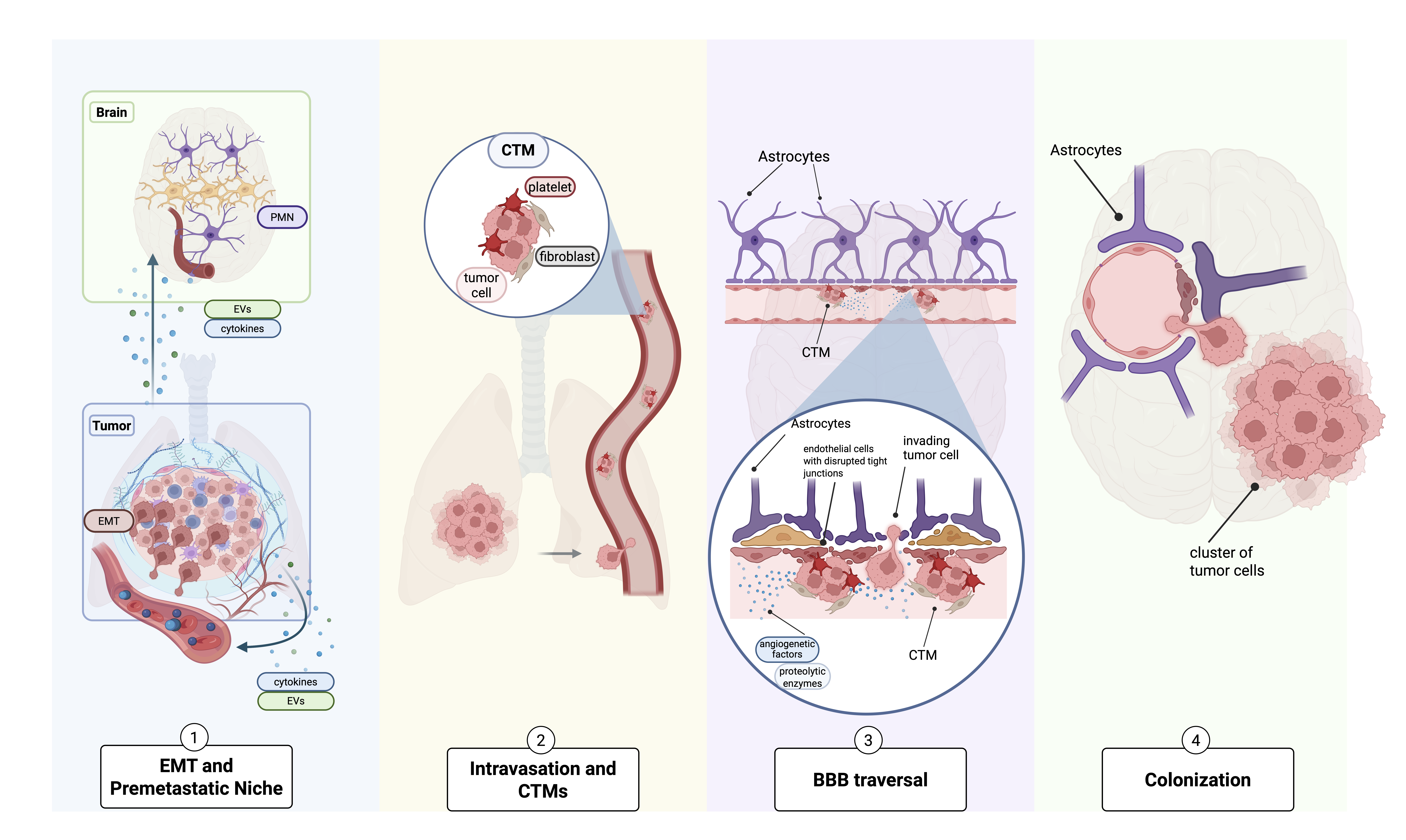
It involves primary tumor cell invasion into the vasculature, hematogenous dissemination, survival in circulation, and successful extravasation of circulating tumor cells (CTCs) across the blood-brain barrier (BBB). Once within the CNS, disseminated tumor cells encounter a highly specialized microenvironment characterized by unique metabolic constraints, immune privilege, and tightly regulated vascular architecture (9, 10). To successfully colonize the brain, tumor cells must adapt to and actively remodel this niche, engaging in dynamic crosstalk with CNS-resident cell populations, including astrocytes, microglia, macrophages, endothelial cells, and neurons. The distinct tumor microenvironment (TME) of BM critically shapes therapeutic response. The BBB limits drug penetration, reducing effective intracranial concentrations of many systemic agents (9, 10). In addition, astrocyte-mediated chemoprotection (11, 12), microglial polarization and immune modulation (13–15), and metabolic reprogramming within the CNS niche collectively contribute to treatment resistance and disease persistence. These interactions underscore that BM are not merely anatomical extensions of systemic disease but rather represent biologically distinct ecosystems. Given the substantial clinical burden of BM in NSCLC, including neurological morbidity, profound reductions in quality of life, and persistently poor prognosis, a comprehensive understanding of the brain TME is urgently needed. In this review, we synthesize current knowledge on the molecular determinants and biological mechanisms underlying BM in NSCLC. Particular emphasis is placed on the cellular and molecular composition of the brain TME and its functional interactions with CNS-resident immune and stromal cells. In addition, we summarize intracranial efficacy data for current treatment strategies and discuss recent advances in novel therapeutic approaches targeting BM in NSCLC. By integrating these insights, we aim to identify potential therapeutic vulnerabilities and outline future strategies to improve clinical outcomes for patients with NSCLC BM (Figure 1).
2. Materials and Methods
This narrative review provides a comprehensive synthesis of current knowledge on the TME of BM in NSCLC. We conducted a literature search using PubMed, Web of Science, and Google Scholar, covering publications from January 2000 to April 2026. The search strategy used keyword combinations, including NSCLC, lung cancer, brain metastases, tumor microenvironment, pre-metastatic niche, microglia, astrocytes, immunotherapy, and intracranial efficacy. To ensure a high-quality evidence base, we prioritized original peer-reviewed articles, translational studies, and reviews, primarily in English, that provided significant mechanistic insights. Conference abstracts were included when they provided relevant updates on emerging biomarkers or clinical outcomes. Particular attention was given to studies employing modern molecular and spatial profiling approaches, including single-cell RNA sequencing, spatial transcriptomics, proteomics, and multiplex immunohistochemistry, as well as comparative studies addressing the distinct features of the BM-TME versus primary lung tumors. The selected literature was screened for scientific rigor and clinical relevance, and key findings were synthesized to provide an integrated overview of current knowledge regarding cellular composition, immune interactions, and microenvironmental adaptations that characterize NSCLC.
3. The Brain Metastatic Cascade in NSCLC
The development of BM in NSCLC is a complex, tightly regulated, multistep process. This phenomenon is conceptually explained by Paget’s classical “seed-and-soil” hypothesis, which proposes that successful metastatic colonization depends on the compatibility between disseminating tumor cells (the “seed”) and the microenvironment of the target organ (the “soil”) (16). According to this concept, metastatic outgrowth occurs only when tumor cells encounter a permissive microenvironment that supports their survival and proliferation. Based on this framework, the metastatic spread of NSCLC to the brain can be broadly divided into four major stages (Figure 1) (17). Although the brain metastatic cascade is commonly presented as a sequential process, individual steps frequently overlap, and their relative importance likely varies according to tumor genotype, treatment history, and the evolving brain microenvironment.
The first step involves detaching tumor cells from the primary lung tumor and establishing a premetastatic niche (PMN). Interactions between tumor cells and the local TME induce epithelial-mesenchymal transition (EMT), a process characterized by cytoskeletal reorganization and changes in cell adhesion molecules that confer increased motility and invasiveness in cancer cells (18) (Figure 1). Importantly, the primary tumor can influence distant organs even before tumor cell dissemination occurs. Through the systemic release of soluble mediators and extracellular vesicles (EVs), lung tumors can modify immune responses and remodel the brain microenvironment, thereby creating a permissive PMN that facilitates future metastatic colonization (19) (Figure 1). In this context, inflammatory cytokines, including interleukin (IL)-6, tumor necrosis factor-α (TNF-α), and transforming growth factor-β (TGF-β), help shape the TME and regulate tumor cell plasticity, including the induction of EMT (20, 21). TGF-β additionally promotes angiogenesis by stimulating the expression of vascular endothelial growth factor (VEGF) (20).
Furthermore, tumor-derived EVs have been implicated in directing organ-specific metastasis, as integrin molecules expressed on EV surfaces can influence organotropism and guide metastatic spread to specific tissues, including the brain (22, 23). Recent evidence from breast cancer models suggests that tumor-free pre-metastatic organs can release EVs that are taken up by tumor cells and promote organ-specific metastatic colonization. These findings indicate that EV-mediated organotropism may be driven not only by tumor-derived EVs but also by EVs originating from the PMN itself (24). However, this concept has not yet been demonstrated in NSCLC.
The second stage is marked by tumor cell entry into the bloodstream (intravasation). In circulation, cancer cells may aggregate into circulating tumor microemboli (CTM), multicellular clusters that enhance tumor cell survival under the mechanical stress of blood flow (25) (Figure 1). The presence of stromal and platelet components within CTM provides additional protection against anoikis and apoptotic signals, thereby increasing the likelihood that these clusters will attach within the cerebral microvasculature and initiate metastatic seeding (26). Recent evidence further suggests that these CTC clusters are not only critical mediators of metastatic dissemination but may also play a role in late-stage disease progression and cancer-related mortality. Dynamic increases in CTC counts and cluster size have been observed immediately prior to death, accompanied by tumor emboli formation and infiltration of large vessels. This process can contribute to vascular obstruction and systemic failure, highlighting the broader pathophysiological relevance of CTM beyond metastatic seeding and underscoring their potential role as both prognostic biomarkers and therapeutic targets in advanced cancer (27). However, while metastatic CTC clusters have been well described in breast cancer, comparable functional evidence in NSCLC remains limited, and current NSCLC data mainly support substantial CTC heterogeneity rather than well-defined BM-initiating CTC populations.
The third step involves traversal of the BBB, a critical and often rate-limiting stage in the metastatic cascade. NSCLC cells, together with associated stromal elements, secrete angiogenic factors and proteolytic enzymes that degrade components of the extracellular matrix (ECM) and disrupt endothelial tight junctions (TJ). This process increases vascular permeability and facilitates the transendothelial migration of tumor cells into the brain parenchyma (28) (Figure 1). Recent work further highlights the active role of the endothelium as a regulatory interface in this process. Endothelial cells do not merely represent a passive barrier but actively determine tumor cell fate through angiocrine signaling. Endothelial-derived Wnt signaling has been shown to influence whether disseminated tumor cells undergo extravasation and enter a latent state or remain intravascular and proliferate. This response is partly predetermined by the epigenetic state of tumor cells: hypomethylated cells exhibit increased Wnt responsiveness and are more likely to extravasate and adopt a latent phenotype, whereas more highly methylated cells show reduced Wnt activity and preferentially proliferate within the vasculature.
These findings underscore the evidence that endothelial-tumor cell interactions, together with tumor-intrinsic epigenetic programming, critically shape early metastatic seeding and colonization of the brain (29). Finally, after extravasation, disseminated tumor cells adapt to the unique microenvironment of the CNS and begin to colonize the brain tissue. Interactions with resident brain cells, including astrocytes, microglia, and other stromal components, drive reciprocal signaling processes that promote tumor cell survival, immune modulation, and proliferation (Table 1). These dynamic tumor-stroma interactions ultimately lead to the establishment and expansion of clinically detectable BM (30).
Table 1. Cellular Components of the Brain Metastasis Tumor Microenvironment and Their Functional Roles.
| Cell type | Main role in BM-TME | Key functions in NSCLC BM | Mechanistic links to therapy resistance |
|---|---|---|---|
| Endothelial cells | Core component of the BBB. | Tumor cell adhesion and extravasation (ALCAM, E-selectin/CD15); tight junction disruption and BBB leakiness (TNF-α, TGF-β, VEGFA, ANGPT2, MMPs, ADAM9). | Restrict effector T-cell trafficking across the BBB through vascular dysfunction and reduced adhesion molecule expression, promoting immune exclusion and reduced sensitivity to PD-1/PD-L1 blockade. |
| Pericytes | Stabilize cerebral vessels and BBB. | Maintain vascular integrity (PDGFRβ); regulate vascular remodeling and tumor invasion (TGF-β, Desmin); disrupt endothelial-pericyte interactions (PGE2, N-cadherin/Cx43). | Pericyte dysfunction promotes BBB instability, immune-cell exclusion, and may limit ICI efficacy. |
| Astrocytes | Central regulators of neuroinflammation and BBB function. | Premetastatic niche formation (CXCL1, IL-15, VEGF); BBB disruption and ECM remodeling (MMPs, CXCL10/CXCR3, IL-6/CCL2); immune evasion and tumor support (PI3K/AKT, STAT3, PD-L1, VEGF-A); early anti-tumor activity (PA, TRPA1/FGFR2). | Promote chemotherapy resistance through Cx43-mediated gap junctions and calcium buffering; reduce ICI efficacy through STAT3/IL-11/PD-L1 signaling and CD8⁺ T-cell apoptosis. |
| Microglia | Brain-resident immune surveillance cells. | T-cell suppression (PGE2, FasL, VISTA, PD-L1); ECM remodeling and metastatic colonization (TGF-β/MMPs, IL-6/JAK2/STAT3, Wnt/β-catenin, CXCL16/CXCR6). | Pro-tumor polarization, impaired antigen presentation, and PD-L1/VISTA expression suppress T-cell activation and may reduce ICI efficacy. |
| Neurons | Functional cells of the CNS. | Neurotransmitter-mediated tumor support (GABA); brain adaptation (Reelin); tumor motility and invasion (SNPH). | Potential contribution to immunosuppression and therapy resistance remains investigational in NSCLC BM. |
| MDMs / TAMs | Major recruited myeloid population. | M1/M2 polarization; T-cell suppression (IL-10, TGF-β); tumor promotion (NF-κB, JAK/STAT, NOTCH); angiogenesis, ECM remodeling, and BBB disruption (VEGF, MMP-9, CXCL12). | M2 polarization suppresses CD8⁺ T-cell function, promotes Treg differentiation, and maintains an immunosuppressive myeloid niche associated with ICI resistance. |
| TANs | Innate immune cells with pro-metastatic functions. | Premetastatic niche formation and NET release (IL-8, G-CSF, S100A8/A9); N2-mediated T-cell suppression (PD-L1, ARG1, TGF-β); angiogenesis and invasion (VEGFA, MMP9, PI3K/AKT). | PD-L1⁺/ARG1⁺ TANs suppress cytotoxic T-cell responses, while NETs reinforce immune exclusion and metastatic persistence. |
| CAFs | Activated stromal fibroblasts. | ECM remodeling; EMT and invasion (IL-6/TGF-β, MET/HGF, JAK/STAT3); immune suppression and stromal remodeling. | Promote chemotherapy and targeted therapy resistance, T-cell exclusion, and impaired drug penetration through ECM remodeling and IL-6/CX3CL1 signaling. |
| CD8+ T cells | Main cytotoxic lymphocyte population. | Antitumor cytotoxicity regulated by PD-1, TIM-3, TIGIT, LAG-3, CTLA-4, and IFN-γ. | Low infiltration and exhausted checkpoint-high phenotypes are associated with reduced ICI responsiveness. |
| CD4+ T cells / Tregs | Adaptive immune regulation. | Immune suppression through IL-10 and TGF-β secretion. | Treg enrichment suppresses cytotoxic immunity and may reduce responses to ICIs and adoptive cell therapies. |
| B cells / plasma cells | Humoral immune compartment. | Support tertiary lymphoid structure formation and adaptive immune activation. | TLS-rich tumors may predict improved ICI responsiveness, whereas TLS deficiency may reflect impaired local immune priming. |
| NK cells | Innate cytotoxic lymphocytes. | Antitumor cytotoxicity mediated by NKG2D, FasL, and CX3CL1. | Reduced abundance and functional impairment may limit innate antitumor immunity; direct effects on therapeutic resistance remain incompletely defined. |
Abbreviations: BBB, blood-brain barrier; BM, brain metastases; BM-TME, brain metastasis tumor microenvironment; CAFs, cancer-associated fibroblasts; CNS, central nervous system; ECM, extracellular matrix; EMT, epithelial–mesenchymal transition; IL, interleukin; NK, natural killer; PMN, pre-metastatic niche; TAMs, tumor-associated macrophages; TANs, tumor-associated neutrophils; TGF-β, transforming growth factor beta.
4. The cellular components of the TME in the brain
CNS-resident cells
Brain endothelial cells – key components of the BBB
Brain endothelial cells line the cerebral vasculature and represent the principal cellular component of the BBB, a highly specialized structure that restricts both immune cell infiltration and tumor cell entry into the CNS (Figure 2).
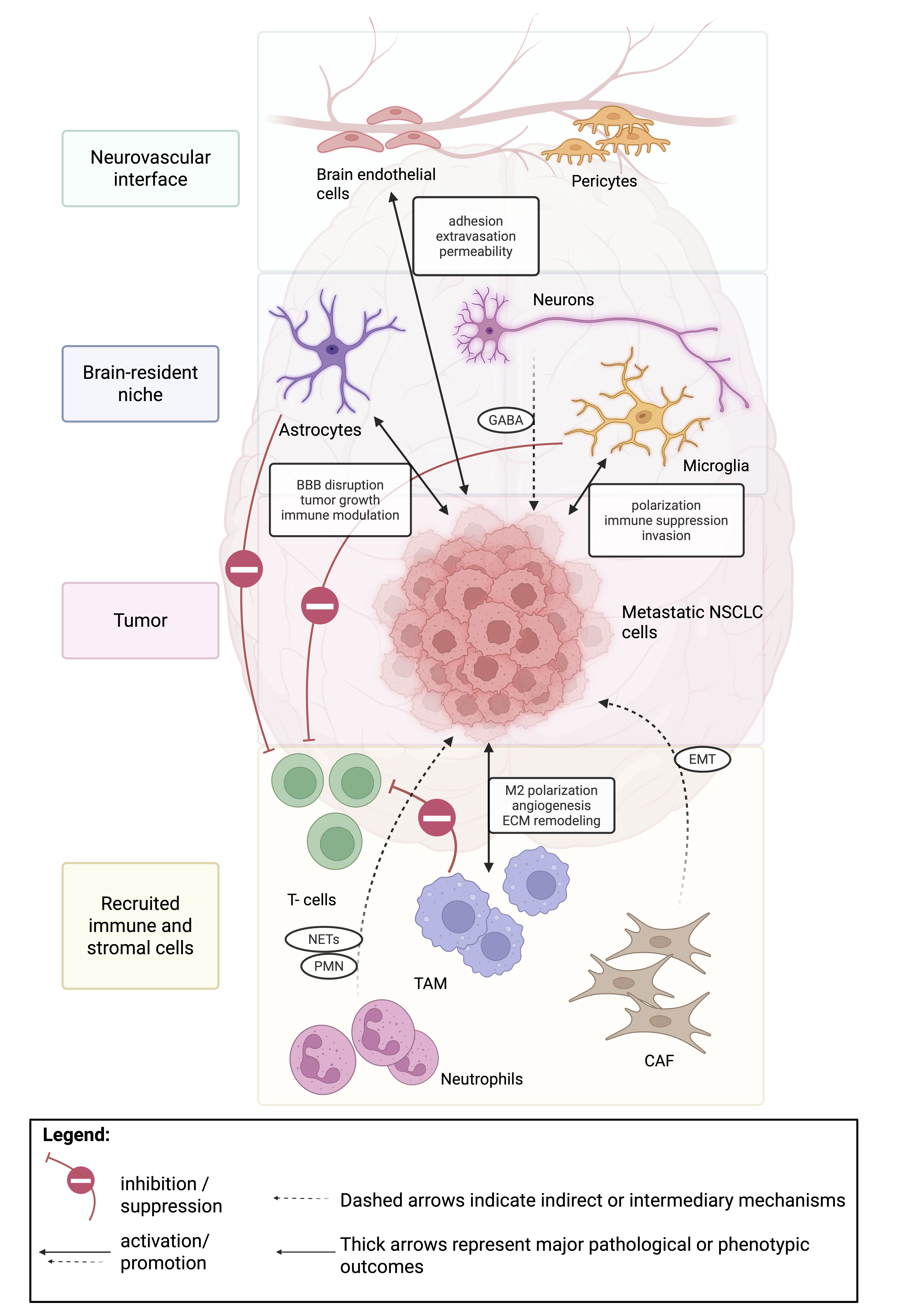
Consequently, they serve as key regulators of BM development in NSCLC. Successful metastatic dissemination requires CTCs to adhere to and penetrate this endothelial barrier. NSCLC cells express adhesion molecules such as activated leukocyte cell adhesion molecule (ALCAM/CD166), which facilitate binding to endothelial cells and promote subsequent extravasation (31). Tumor-derived factors, including VEGF and TNF-α, contribute to degradation of the endothelial glycocalyx, leading to the exposure of E-selectin (32). E-selectin interacts with CD15 on tumor cells, thereby promoting vascular arrest and facilitating metastatic seeding (33–35). In parallel, disruption of TJ integrity increases BBB permeability. TNF-α further enhances TGF-β-induced endothelial-to-mesenchymal transition (EndMT), resulting in downregulation of TJ proteins and increased vascular permeability, which facilitates tumor cell transmigration across the BBB (36, 37). Additional tumor-intrinsic mechanisms also contribute to BBB disruption. NSCLC-derived TGF-β induces the release of exosomal long noncoding RNA (lnc)-MMP-2, which suppresses microRNA (miR)-1207-5p and weakens TJ integrity (38). Moreover, NSCLC-associated aldo-keto reductase family 1 member B10 (AKR1B10) expression activates mitogen-activated protein kinase (MAPK) signaling, leading to upregulation of matrix metalloproteinases (MMPs) 2 and 9, which further degrade TJ components. Activation of several signaling pathways also increases VEGFA production (39), while proteins such as a disintegrin and metalloprotease domain 9 (ADAM9) regulate endothelial expression of VEGFA and angiopoietin-2 (ANGPT2) (40).
Collectively, these mechanisms support a central role for endothelial dysfunction in metastatic colonization. Nevertheless, the relative contributions of individual pathways in human NSCLC BM remain incompletely defined because much of the mechanistic evidence derives from experimental models (Figure 2). In NSCLC BM, the cerebral vasculature is transcriptionally reprogrammed toward a glioblastoma-like, immunosuppressive phenotype that upregulates endothelial checkpoints such as CD276/B7-H3 (alongside CD200, Galectin-9 (LGALS9) and Tryptophan 2,3-dioxygenase (TDO2)) and downregulates the intercellular adhesion molecules 1 and 2 (ICAM1, ICAM2) as well as the vascular cell adhesion molecule-1 (VCAM1) required for effector T cell infiltration (41). Therefore, this vascular immune barrier likely contributes to resistance to immune checkpoint blockade by enforcing a T cell-excluded phenotype and restricting cytotoxic infiltration into intracranial lesions, a process also observed in SCLC (42).
Pericytes – regulate vascular stability and BBB permeability
Pericytes are specialized mural cells that envelop endothelial cells and are essential for microvascular stability, vessel maturation, and maintenance of BBB integrity (Figure 2). In the context of NSCLC BM, pericytes play a dual and highly dynamic role in regulating vascular homeostasis and metastatic progression. Experimental data suggest that intact pericyte coverage can suppress NSCLC BM in vitro (43), underscoring their protective function. Conversely, a reduction in platelet-derived growth factor receptor ß (PDGFRβ)-positive pericytes has been associated with increased BBB permeability (44). Loss of pericyte coverage destabilizes endothelial TJ, promotes endothelial hyperplasia, and contributes to abnormal angiogenesis (45), thereby facilitating tumor cell extravasation. Tumor-derived signals further reprogram pericytes. For instance, TGF-β induces a transition toward a myofibroblast-like phenotype, characterized by excessive ECM deposition, including collagen and fibronectin, which leads to vascular remodeling and enhanced tumor invasion (46). Alterations in pericyte subpopulations also influence vascular function: enrichment of desmin-positive pericytes correlates with increased vascular leakiness, highlighting pericyte heterogeneity as a determinant of barrier integrity and a potential therapeutic target (47). Consistently, tumor-associated pericytes exhibit structural and phenotypic abnormalities, including altered marker expression, loose endothelial attachment, and abnormal integration into endothelial sprouts, collectively contributing to disorganized tumor vasculature, impaired barrier function, and progressive tumor growth (48). Inflammatory mediators further exacerbate pericyte dysfunction. Prostaglandin E2 (PGE2), which is elevated in cancer and neurological disorders, disrupts pericyte-endothelial interactions by downregulating N-cadherin and Connexin-43 (Cx43) via prostaglandin receptors EP1- and EP4-dependent signaling pathways and suppressing R-Ras expression, thereby promoting a migratory, weakly adherent pericyte phenotype and reducing pericyte coverage in vivo (49). Reduced N-cadherin expression and its aberrant regulation on metastatic cells further weaken endothelial-pericyte contacts, facilitating transendothelial migration and brain colonization (50).
Spatial transcriptomic data further support a role for perivascular stromal remodeling in NSCLC BM. In a NanoString GeoMx digital spatial profiling study of paired primary lung tumors, BM, and tumor-adjacent brain microenvironment from 44 NSCLC patients, Zhang et al. identified extensive remodeling of the brain TME toward an immunosuppressive and fibrogenic niche. Fibrosis was observed in a substantial fraction of the BM, and the authors noted enrichment of pericytes and CAFs in the tumor brain microenvironment compared with control brain tissue, along with increased expression of fibrogenic regulators such as PDGFRβ, C-X-C motif chemokine receptor 4 (CXCR4), and TGF-Β1. These spatial data do not define a pericyte-specific mechanism in NSCLC BM but support the concept that PDGFRβ-positive perivascular stromal cells may contribute to ECM remodeling, angiogenesis, and vascular niche adaptation in the brain metastatic microenvironment (30).
Collectively, current evidence suggests that pericytes contribute substantially to vascular remodeling and BBB integrity. However, their precise functional roles in NSCLC BM remain incompletely resolved because direct mechanistic studies are relatively limited (51). Whether endothelial immune reprogramming represents a driver of immune escape or a secondary adaptation to the metastatic niche remains an important unresolved question.
Astrocytes – major regulators of neuroinflammation, BBB integrity, and chemoprotection
Astrocytes are the most abundant glial cells in the CNS and play essential roles in maintaining neuronal homeostasis, regulating synaptic activity, and preserving BBB integrity (52). In the context of brain-metastatic NSCLC, astrocytes serve as central regulators of neuroinflammation and vascular stability while engaging in complex, dynamic interactions with tumor cells (53). These interactions are highly context-dependent and evolve throughout metastatic progression (Figure 2). Importantly, many astrocyte-associated signaling pathways are also activated during physiological CNS injury and repair, complicating efforts to therapeutically target these mechanisms without disrupting normal brain homeostasis.
Astrocytes in PMN Formation
Even before tumor cells colonize the brain, astrocytes contribute to the formation of the PMN. Tumor-derived EVs can reprogram astrocytes and disrupt BBB integrity. For example, EV-associated miR-181c promotes BBB breakdown by inducing abnormal actin cytoskeletal organization in astrocytes through downregulation of its target gene, 3-phosphoinositide-dependent protein kinase-1 (PDPK1) (54). In addition, exosomes derived from H1299 NSCLC cells induce astrocyte apoptosis by upregulating apoptosis-related proteins, including mitogen-activated protein 2 kinase 1 (MAP2K1), tubulin alpha 1c (TUBA1C), RELA proto-oncogene (RELA), and caspase 6 (CASP6). These tumor-derived vesicles also stimulate astrocytes to release a broad panel of pro-inflammatory and immunomodulatory cytokines, such as C-X-C motif chemokine ligand 1 (CXCL1), interferon gamma (IFN-γ), IL-3, IL-5, IL-15, leukemia inhibitory factor (LIF), macrophage colony-stimulating factor (M-CSF), platelet-derived growth factor (PDGF), nerve growth factor (NGF), and VEGF, which promote the recruitment of immunosuppressive immune cells and create a permissive microenvironment for metastatic seeding (55).
Astrocytes and BBB Disruption
Under physiological conditions, astrocytes support BBB integrity by secreting factors that stabilize endothelial TJ proteins (56), but during metastatic progression, they undergo pathological reprogramming and actively facilitate tumor cell invasion across the BBB. Reactive astrocytes promote ECM remodeling through downregulation of tissue inhibitor of metalloproteinases 2 (TIMP-2) and upregulation of astrocyte elevated gene 1 (AEG-1), thereby enhancing matrix degradation and weakening endothelial TJ, a process further amplified by tumor-intrinsic AEG-1 expression (57). Astrocytes also secrete C-X-C motif chemokine ligand 10 (CXCL10), which binds to C-X-C motif chemokine receptor 3 (CXCR3) on CTCs, directing their migration toward the brain. Concurrently, tumor cells form invadopodia that release pro-MMP-2 and -9, which are activated by urokinase-type plasminogen activator (uPA) and plasmin, thereby degrading ECM components and facilitating BBB disruption (58). Astrocytic sphingosine-1-phosphate receptor 3 (S1P3) signaling further induces the expression of IL-6 and CC chemokine ligand 2 (CCL2), promoting recruitment of myeloid cells and weakening endothelial adhesion (59). Together, these processes position astrocytes as critical drivers of BBB breakdown and tumor cell extravasation.
Early Anti-tumor Functions of Astrocytes
Despite these pro-metastatic effects, astrocytes can initially exert anti-tumor activity. Reactive astrocytes secrete plasminogen activators (PA), which induce apoptosis in invading tumor cells (60). However, NSCLC cells can evade this defense by producing anti-PA serpins that inhibit plasmin-mediated cytotoxicity (53). Astrocytes may also suppress metastatic progression through the exosomal transfer of miR-142-3p, which downregulates transient receptor potential ankyrin 1 (TRPA1) in tumor cells, disrupts the TRPA1-fibroblast growth factor receptor 2 (FGFR2) axis, and limits metastatic growth (61).
Pro-tumorigenic Astrocyte Reprogramming
During metastatic evolution, astrocytes frequently shift toward a pro-tumorigenic phenotype. Experimental models show that NSCLC BM are surrounded by glial fibrillary acidic protein (GFAP)-positive activated astrocytes. Tumor-derived factors such as macrophage migration inhibitory factor (MIF), IL-8, and plasminogen activator inhibitor 1 (PAI-1) stimulate astrocytes to produce inflammatory cytokines including IL-6, TNF-α, and IL-1β, which promote tumor cell proliferation. Spatial transcriptomic profiling of human BM demonstrated that astrocytes within the tumor-brain microenvironment undergo profound transcriptional reprogramming. Compared with normal brain tissue, brain metastasis-associated astrocytes showed loss of mature astrocyte markers including solute carrier family 1 member (SLC1A2), Aldolase C (ALDOC) and γ-aminobutyric acid type A receptor subunit alpha 2 (GABRA2), while simultaneously acquiring reactive astrocyte programs characterized by inflammatory and signal transducer and activator of transcription 3 (STAT3)-associated signaling, including increased oncostatin M receptor (OSMR) and thrombospondin 1 (THBS1) expression (30, 62). These findings indicate that astrocytes are not merely activated but are reprogrammed toward a distinct reactive state that supports remodeling of the metastatic niche and may facilitate tumor adaptation to the CNS environment (30). Upregulation of IL-6 receptor components on cancer cells further strengthens this reciprocal signaling loop. Similar astrocyte activation patterns have been observed in human BM samples, highlighting the clinical relevance of tumor–astrocyte crosstalk (63). NSCLC cells further reprogram astrocytes by inducing endothelin-1 (ET-1) expression, activating phosphoinositide 3-kinase (PI3K)/ protein kinase B (AKT) and MAPK signaling pathways, and promoting protocadherin-7 (PCDH7)-mediated Cx43 gap junction formation (12, 64). These gap junctions allow the transfer of cyclic guanosine monophosphate-adenosine monophosphate (cGAMP) from tumor cells to astrocytes, activating stimulator of interferon genes (STING)/ signal transducer and activator of transcription 3 (STAT3) signaling that supports metastatic growth (64).
Recent multimodal single-cell and spatial transcriptomic analyses of treatment-naïve NSCLC BM identified neural-like transcriptional programs that were strongly enriched in metastatic tumor cells compared with primary lung tumors. Spatial mapping localized these neural-like tumor populations to CNS-specific niches enriched for resident glial cells. These findings suggest that astrocyte-rich microenvironments may actively support the acquisition and maintenance of neural-like tumor states, thereby facilitating metastatic adaptation and persistence within the brain. Such observations extend the concept of astrocyte–tumor crosstalk beyond inflammatory signaling and indicate that astrocytes may directly contribute to the transcriptional reprogramming of metastatic NSCLC cells within the brain niche (65).
In addition, astrocyte-derived exosomal miR-19a suppresses phosphatase and tensin homolog (PTEN) in tumor cells, leading to increased CCL2 secretion and recruitment of ionized calcium-binding adapter molecule1 (IBA1)-positive myeloid cells (66). This astrocyte-mediated reprogramming has direct therapeutic implications, particularly for chemotherapy resistance. Astrocytes can protect metastatic tumor cells from cytotoxic agents through gap-junction-dependent signaling and induction of survival programs, resulting in reduced apoptosis and increased expression of pro-survival and stress-response genes (67). In experimental models of BM, astrocyte contact has been associated with resistance to chemotherapeutic agents through mechanisms involving Cx43-mediated intercellular communication, intracellular calcium sequestration, and upregulation of genes such as glutathione S-transferase alpha 5 (GSTA5), Bcl-2-like protein 1 (BCL2L1), and TWIST1. Therefore, the same astrocyte–tumor crosstalk that supports metastatic outgrowth may also create a non-genetic, microenvironment-mediated state of drug tolerance within the brain metastatic niche.
Astrocyte Activation and Immune Modulation
The Janus kinase (JAK)/STAT signaling pathway, particularly STAT3 activation, plays a central role in astrocyte-mediated tumor support. STAT3⁺ reactive astrocytes acquire immunosuppressive properties through upregulation of programmed death-ligand 1 (PD-L1), VEGF-A, lipocalin-2, and TIMP-1 (62). Furthermore, STAT3⁺ astrocytes produce MIF, which suppresses microglial M1 polarization, thereby promoting brain tumorigenesis (68). Consequently, STAT3⁺ astrocytes represent a key cellular population within the brain metastatic niche (62). Reactive astrocytes also exhibit increased secretion of IL-11. In vitro, IL-11 induces PD-L1 expression in metastatic tumor cells and promotes apoptosis of CD8⁺ T cells by activating both EGFR signaling and the canonical IL-11Rα/glycoprotein (gp)130 pathway. In vivo, pharmacologic inhibition of gp130 and EGFR signaling suppresses BM growth and prolongs survival in murine models, underscoring this axis as a promising therapeutic target (69). Their role in immune suppression in the TME has been shown to diminish the potential effect of ICIs in BM (70). Taken together, astrocytes exhibit a context-dependent dual role in NSCLC BM. While they initially exert anti-tumor effects that limit metastatic colonization, tumor-derived signals progressively reprogram astrocytes into pro-tumorigenic and immunosuppressive cells that promote BBB disruption, tumor cell survival, and metastatic growth. Astrocyte-tumor cell interactions represent a critical determinant of disease progression, therapeutic response, and prognosis in NSCLC BM (71).
Microglia – brain-resident macrophage-like immune cells involved in immune surveillance and polarization (pro- vs. anti-tumor phenotypes)
Microglia are brain-resident immune cells derived from yolk sac macrophages that play essential roles in CNS homeostasis, immune surveillance, and tissue repair (72–74). Within the TME of BM, microglia exhibit highly plastic, context-dependent functions that contribute to both tumor suppression and progression (75). Transcriptomic analyses have demonstrated that microglia possess gene expression profiles distinct from those of peripheral monocytes and macrophages (76). It remains debated whether their activation states can be fully categorized into the classical macrophage M1 (“pro-inflammatory”) and M2 (“anti-inflammatory”) phenotypes (77–79), although microglia clearly undergo dynamic functional polarization during metastatic colonization of the brain (80). In BM, tumor-associated microglia are frequently enriched at the invasive margins of metastatic lesions, where they interact closely with infiltrating tumor cells (81). Tumor-derived signals can induce PGE2 production in microglia (82), which exerts immunosuppressive effects by inhibiting T cell activation and reducing TNF-α expression (83). Additional immune evasion mechanisms include the upregulation of Fas ligand (FasL) (84) and reduced expression of major histocompatibility complex class II (MHC II) molecules (85), both of which further attenuate anti-tumor immune responses.
Beyond immune modulation, microglia also contribute to ECM remodeling and tumor invasion. Tumor-associated microglia can promote invasive tumor growth by promoting TGF-β1-dependent MMP-9 induction (86) and by secreting IL-6 (87). Interactions between microglia and other components of the neurovascular unit further shape the metastatic niche. At the BBB, pericyte-derived cytokines can stimulate microglial activation, thereby promoting tissue remodeling and facilitating tumor invasion (88). Conversely, M2-like microglia may induce PDGFRβ expression in tumor cells, thereby enhancing pericyte recruitment and vascular remodeling within metastatic lesions (89). Several signaling pathways have been implicated in driving microglial polarization toward a pro-tumorigenic phenotype, including Wnt/β-catenin signaling (90) and the CXCL16-CXCR6 axis (91).
In addition, emerging evidence indicates that EVs represent an important mechanism of tumor-microglia communication (15, 92). For example, co-culture studies with NSCLC cell lines identified IL-6 as a key factor inducing M2-like microglial polarization via activation of the JAK2/STAT3 signaling pathway, thereby facilitating brain colonization (13, 93). Microglia can also directly suppress adaptive immune responses. Upregulation of immune checkpoint molecules such as V-domain Ig suppressor of T cell activation (VISTA) and PD-L1 via activation of the PI3K/AKT pathway has been shown to inhibit T cell function within the TME and can limit the efficacy of ICIs in NSCLC BM (94, 95).
Consistent with these experimental findings, spatial transcriptomic profiling of human NSCLC BM revealed substantial reprogramming of the monocyte-derived macrophage (MDM)-microglia axis within the tumor-brain microenvironment. BM-associated myeloid signatures were enriched in highly fibrotic metastatic niches, supporting a role for resident and recruited myeloid populations in establishing an immunosuppressive metastatic niche (30). These findings support a multifaceted role for microglia in NSCLC BM. However, considerable uncertainty remains regarding the extent to which experimentally defined polarization states accurately reflect the diverse activation programs observed in human disease (96). Current classifications based on M1/M2 phenotypes likely oversimplify the functional diversity of microglia, emphasizing the need for single-cell and spatial approaches to define clinically relevant microglial states.
Neurons - provide metabolic support and form functional synapse-like interactions with tumor cells
Neurons are the principal functional cells of the brain, responsible for transmitting and integrating electrical and chemical signals that regulate cognition, sensation, and motor control. Although neurons play a central role in brain physiology, their contribution to the establishment and progression of BM remain incompletely understood, and current evidence is still limited. Within the metastatic niche, interactions between tumor cells and the neuronal microenvironment appear to influence tumor growth and adaptation. Activated astrocytes and microglia in the TME can impair neuronal function (97), while neuronal activity, soluble mediators, and neurotransmitters have been shown to promote tumor cell survival and proliferation in glioma (98–100). Recent data indicate that metastatic cancer cells actively adapt to the neuronal milieu. For example, breast cancer cells can acquire a γ-aminobutyric acid (GABA)-related phenotype, allowing them to better integrate into the brain microenvironment (101). Co-culture experiments demonstrate that direct neuronal contact induces the expression of neurotransmitter receptors and synaptic signaling mediators in breast and NSCLC cells. In breast cancer models, tumor cells transition from autocrine signaling to paracrine dependence on neuron-derived GABA, establishing a GABA-responsive phenotype that facilitates early adaptation to the CNS. Moreover, neuronal interaction can epigenetically reactivate Reelin expression in tumor cells, further supporting their acclimatization to the brain microenvironment (102).
Although recent evidence suggests that SCLC cells may hijack synaptic signaling pathways to promote tumor progression within the brain (103), data supporting a comparable role for tumor-neuron crosstalk in NSCLC remain limited. However, recent multimodal single-cell and spatial transcriptomic analyses of treatment-naïve NSCLC BM identified neural-like transcriptional programs that were strongly enriched in metastatic tumor cells compared with primary lung tumors. Spatial mapping further localized these neural-like tumor populations to CNS-specific niches, suggesting acquisition of neural-like states may represent an important mechanism supporting metastatic adaptation and persistence within the brain (65). Nevertheless, initial findings indicate that neuronal interactions could contribute to tumor adaptation and metastatic growth in the brain. In line with this concept, recent studies demonstrate that tumor cells, including NSCLC, exploit neuronal programs of mitochondrial trafficking to enhance metastatic capacity. Reduced expression of the neuronal regulator syntaphilin (SNPH) facilitates mitochondrial redistribution to the cell periphery, thereby promoting tumor cell motility and invasion, and is associated with increased metastatic potential and poorer clinical outcomes in lung adenocarcinoma (104).
These observations suggest that NSCLC cells may adopt neuron-like mechanisms to optimize energy distribution and support metastatic dissemination within the brain microenvironment, warranting further investigation into their biological and therapeutic relevance. There is also growing evidence that neurons and neuronal injury may contribute to anti-PD-1 resistance, as recently shown in peripheral nerves, where cancer-induced nerve injury drives a chronic inflammatory response that skews the tumor microenvironment toward an immunosuppressive state (105, 106).
Recruited peripheral immune and stromal cells
Monocyte-derived macrophages (MDMs) / Tumor-associated macrophages (TAMs)
MDMs and TAMs represent major immune cell populations within the brain TME and are closely linked to tumor progression in NSCLC BM. TAMs exhibit pronounced functional plasticity and can adopt distinct activation states during metastatic development. In early stages, M1-like macrophages exert anti-tumor functions through antigen presentation, immune surveillance, and secretion of pro-inflammatory mediators such as nitric oxide (NO), TNF-α, and IL-6 (107). However, in established BM, the macrophage compartment is predominantly composed of M2-like TAMs, which promote immune evasion and tumor growth (107, 108). These cells suppress cytotoxic T cell responses by impairing antigen presentation and secreting anti-inflammatory cytokines such as IL-10 and TGF-β, thereby promoting regulatory T cell differentiation and T cell exhaustion (108, 109). In addition, IL-10 can directly enhance NSCLC metastatic potential by activating the JAK/STAT, nuclear factor of kappa light-chain enhancer of B cells (NF-κB), and NOTCH signaling pathways (110). Tumor cells actively contribute to this polarization process: NSCLC-derived IL-17 recruits macrophages to the PMN and induces PGE2-mediated M2 polarization (111), while EGFR-mutant tumors release exosomal SRY-box transcription factor 2-overlapping transcript (SOX2-OT), which modulates suppressor of mothers against decapentaplegic homolog (SMAD) signaling and further promotes M2 differentiation (112).
Beyond immune regulation, TAMs play important roles in ECM remodeling, vascular remodeling, and BBB disruption. M2-polarized TAMs increase arginase-1 (ARG-1) activity, promoting the synthesis of ornithine and polyamines that facilitate tissue remodeling and tumor invasion (113). They also secrete MMP-9, which degrades ECM components and TJ proteins (114). Inflammatory signaling pathways, including NF-κB and Wnt, activated in response to tissue damage, further amplify cytokine production and immune cell recruitment, thereby exacerbating BBB permeability (115). Moreover, TAM-derived factors, such as VEGF, PDGF, insulin-like growth factor (IGF), and CXCL12, promote angiogenesis, recruit additional myeloid cells, and contribute to vascular destabilization within metastatic lesions (108, 116).
Bone marrow-derived TAMs, which are often enriched in the tumor core, display particularly strong immunosuppressive gene expression profiles (81, 117). Through positive feedback interactions within the TME, M2-like TAMs reinforce immunosuppression by promoting regulatory T cell differentiation and sustaining an immune-permissive niche 108. Recent spatial transcriptomic analyses of human NSCLC BM support these observations by identifying myeloid-rich metastatic niches characterized by enrichment of M2-like macrophages, increased TGF-β1 expression, and ECM remodeling. Furthermore, BM-associated myeloid signatures were particularly enriched in highly fibrotic tumor-brain microenvironments, suggesting an association between macrophage polarization and the establishment of an immunosuppressive and pro-fibrotic metastatic niche (30, 118). In addition, CD74⁺ TAMs can interact with macrophage MIF secreted by STAT3⁺ reactive astrocytes, inducing midkine (MDK) expression and thereby supporting NSCLC proliferation (119).
These findings highlight TAMs as central regulators of immune suppression, ECM remodeling, and vascular niche formation in the NSCLC BM. These mechanisms also directly contribute to immunotherapy resistance. In particular, M2-like TAM polarization can limit the efficacy of ICIs by reducing antigen presentation, suppressing cytotoxic CD8+ T-cell function, promoting Treg differentiation via IL-10 and TGF-β, and maintaining a myeloid-dominated, T-cell-excluded niche (120, 121). An immunosuppressive vascular niche formed by mesenchymal-like endothelial cells promotes osteopontin-dependent macrophage polarization through a transcriptional program mediated by TWIST1 and special AT-rich sequence-binding protein 1 (SATB1). In glioblastoma models, genetic or pharmacological inhibition of TWIST1 increased T-cell infiltration and sensitized tumors to chimeric antigen receptor T-cell (CAR-T) therapy (122). Therapeutic strategies aimed at macrophage depletion or reprogramming have shown encouraging preclinical activity; however, their clinical efficacy may depend on preserving essential tissue-repair functions while selectively targeting immunosuppressive macrophage subsets.
Tumor-associated Neutrophils (TANs)
Neutrophils represent a major component of the innate immune system. They are predominantly found circulating in the bloodstream, where they can interact with CTCs and promote their survival during hematogenous dissemination (123, 124). In addition to this systemic role, TANs are frequently detected within the TME of parenchymal BM (125). Their recruitment to metastatic lesions is largely driven by tumor-derived chemokines such as IL-8 and granulocyte colony-stimulating factor (G-CSF). These infiltrating neutrophils also often display increased resistance to reactive oxygen species (ROS) (126). Recent single-cell and spatial transcriptomic analyses of human BM have shown that neutrophils are enriched in NSCLC BM and contribute to multiple aspects of metastatic progression (30, 127–129). Within the metastatic niche, TANs predominantly exhibit an immunosuppressive N2 phenotype, characterized by elevated expression of PD-L1 and ARG-1, activation of TNF-α signaling, and increased expression of infiltration and transmigration markers, including integrin A3 (ITGA3), CD177, CD15, and CD11b (126, 130). The polarization of N1 neutrophils toward pro-tumorigenic N2 neutrophils is promoted by tumor-derived TGF-β signaling and by interactions between cancer cells and cancer-associated fibroblasts (CAFs) (131, 132). Functionally, N2-polarized TANs suppress anti-tumor immunity by impairing effector T cell responses, for example, through programmed death protein 1 (PD-1)/PD-L1 interactions with CD8⁺ T cells (126).
Single-cell transcriptomic studies have demonstrated that TANs in NSCLC exhibit substantial heterogeneity that extends beyond the traditional N1/N2 paradigm, comprising multiple, functionally distinct subpopulations shaped by tumor- and microenvironmental cues (133). These diverse TAN states, including mature-primed, interferon-responsive, hybrid (antigen-presenting), immunomodulatory, and pro-angiogenic/pro-metastatic subsets, each with unique transcriptional and functional profiles (133). Among these, pro-angiogenic TANs represent a particularly relevant subset, characterized by the expression of factors including VEGFA, MMP9, and prokineticin-2 (PROK2). Neutrophils constitute a major source of VEGFA within the NSCLC TME and thereby directly contribute to the angiogenic switch. Functionally, these cells, which are more concentrated in tumor tissue compared to normal lung tissue, display features of chronically activated or exhausted neutrophils (133). Overall, recent high-resolution single-cell and spatial transcriptomic studies suggest that TAN populations can exert diverse and sometimes opposing functions, ranging from antitumor immunity to immunosuppression, angiogenesis, and metastatic progression. Their marked plasticity and the existence of multiple transcriptionally distinct functional states highlight both the limitations of the traditional N1/N2 classification model and the need for subtype-specific therapeutic strategies (129, 133–135).
Beyond that, neutrophils have been implicated in the PMN formation, in part through astrocyte-driven neuroinflammatory processes (136), and they can promote angiogenesis within metastatic lesions (135). A key mediator of neutrophil recruitment is the S100A8/A9 protein complex (calprotectin), which induces chemotactic attraction of neutrophils and promotes the formation of neutrophil extracellular traps (NETs) (137–139). TANs themselves can produce S100A8/A9, creating a positive feedback loop that amplifies neutrophil recruitment to the metastatic site (126). In addition, S100A8/A9 signaling enhances tumor growth and invasion by activating oncogenic pathways including NF-κB, PI3K/AKT, mechanistic target of rapamycin (mTOR), and MAPK (140). Elevated neutrophil infiltration has also been shown to have prognostic implications: a higher neutrophil-to-lymphocyte ratio (NLR) in BM has been associated with poorer survival in patients with NSCLC (141, 142).
Collectively, these mechanisms nominate several candidate therapeutic vulnerabilities, including disruption of neutrophil recruitment via CXCR1/2 or IL-8 blockade, interference with TGF-β-driven N2 polarization, and targeting of the S100A8/A9-NET axis (e.g., peptidyl arginine deiminase 4 (PAD4) inhibition). However, evidence in NSCLC BM specifically remains largely preclinical, and the extent to which these strategies translate across the blood-brain barrier and the distinct neuroinflammatory niche is still unknown. Whether these transcriptionally distinct neutrophil states can be therapeutically manipulated without compromising host defense remains an important translational challenge.
Cancer-associated fibroblasts (CAFs)
CAFs are activated stromal fibroblasts within the TME that interact extensively with tumor cells and other stromal components. Through remodeling of the ECM, secretion of growth factors and cytokines, and modulation of immune responses, CAFs contribute to tumor progression, immune suppression, and therapy resistance in many cancers, including NSCLC (143). Increasing evidence suggests that CAF populations are highly heterogeneous and may influence organ-specific metastatic dissemination. Single-cell sequencing analyses of primary tumors from patients with advanced NSCLC have identified three major CAF subtypes: myofibroblastic CAFs (myCAFs), inflammatory CAFs (iCAFs), and antigen-presenting CAFs (apCAFs) (144, 145). Notably, a relatively small single-cell RNA-sequencing study of 14 treatment-naïve patients with advanced NSCLC observed differences in the relative abundance of CAF subtypes by metastatic pattern. Specifically, iCAFs were enriched in tumors from patients with BM, whereas apCAFs were more prevalent in tumors from patients with bone metastases.
Ligand-receptor analyses further suggested that iCAFs may interact with tumor cells through the MET-HGF axis. At the same time, apCAFs were linked to secreted phosphoprotein 1 (SPP1)-CD44/SPP1-prostaglandin E receptor 4 (PTGER4) signaling, highlighting potential mechanisms by which CAF heterogeneity may contribute to organ-specific metastatic progression (146). In vivo experiments further demonstrated that co-injection of BM-associated CAFs (BM-CAFs) with NSCLC cells accelerates tumor growth. These effects are linked to the induction of EMT and the acquisition of cancer stem cell-like properties through IL-26-mediated activation of the JAK/STAT3 pathway and chemokine C-X3-C motif ligand 1 (CX3CL1)-driven activation of the JAK/STAT3 and AKT/mTOR signaling pathways. Inhibition of IL-26 or CX3CL1 signaling reversed these pro-tumorigenic effects, highlighting these pathways as potential therapeutic targets (147).
EMT induction represents one of the central mechanisms by which CAFs facilitate metastatic spread. CAF-derived exosomal miR-210, for example, has been shown to activate the PTEN/PI3K/AKT signaling pathway (148), while CAF-secreted IL-6 promotes EMT and tumor progression by enhancing TGF-β signaling (149). CAFs also contribute to ECM remodeling by secreting MMPs, which facilitate tumor invasion and metastatic dissemination (150). Beyond their functional roles in tumor progression, CAF abundance and activity have also been associated with clinical outcomes and impaired responses to immunotherapy (151), suggesting their potential utility as prognostic biomarkers in NSCLC (152–154). ECM remodeling may also contribute to therapeutic resistance through both physical and signaling-mediated mechanisms. Dense or aberrantly remodeled ECM can increase tissue stiffness, alter interstitial pressure, and create perivascular or stromal niches that limit homogeneous drug distribution within metastatic lesions (155, 156).
These mechanisms may impair responses not only to immunotherapy by reinforcing T-cell exclusion and exhaustion, but also to chemotherapy and targeted therapies by reducing effective drug exposure and sustaining downstream survival signaling despite cytotoxic or oncogene-directed treatment. However, direct evidence in NSCLC BM remains limited. Moreover, CAF-targeted therapies must account for the marked functional heterogeneity of fibroblast populations, as indiscriminate depletion may also eliminate stromal subsets with tumor-restraining properties.
Lymphocytes – T cells and Natural killer (NK) cells
Lymphocytes, particularly T cells, are among the most abundant immune cell populations in the brain TME (130, 157). In NSCLC BM, tumor-infiltrating T lymphocytes (TILs) typically display an activated yet functionally exhausted phenotype, characterized by the expression of immune checkpoint molecules such as PD-1, T cell immunoglobulin and mucin domain-containing protein 3 (TIM-3), cytotoxic T-lymphocyte-associated protein 4 (CTLA-4), T cell immunoreceptor with Ig and ITIM domains (TIGIT), and lymphocyte activation gene 3 (LAG-3) (157–159). Spatial analyses have revealed functional heterogeneity within the CD8⁺ T cell compartment. Memory-like CD8⁺ T cells are predominantly located at the peripheral stromal regions of metastatic lesions and resemble circulating CD8⁺ T cells, suggesting a reactive immune state. CD8⁺ T cells within the tumor core, on the other hand, are often more tumor-specific but exhibit pronounced exhaustion with high checkpoint expression and minimal overlap with circulating or bystander CD8⁺ T cells (158). The density of CD8⁺ TILs has been clinically correlated with the extent of brain edema and is associated with improved overall survival (OS) in patients with BM (160, 161). In comparison, CD4⁺ T cells in human BM frequently display a hyporesponsive or anergic phenotype (130), and malignant brain tumors show a higher frequency of regulatory T cells (Tregs) than benign brain lesions (162). Tregs contribute to an immunosuppressive TME by secreting anti-inflammatory cytokines such as IL-10 and TGF-β, thereby limiting anti-tumor immune responses (163, 164).
NK cells represent another lymphocyte population with potential anti-tumor activity, although they are relatively rare within the BM TME. CD56⁺ NK cells are recruited to the brain via CX3CL1 signaling (165) but typically constitute only approximately 2.5% of infiltrating leukocytes in BM (166). Single-cell and spatial transcriptomic studies have consistently described an immunosuppressive In line with these findings, GeoMx digital spatial profiling of paired NSCLC primary tumors and BM demonstrated reduced T-cell and B-cell abundance, impaired cytotoxic lymphocyte signatures, and reduced antigen-presentation programs in the brain metastatic TME compared with the primary lung TME (30). Recent multimodal single-cell, TCR sequencing, and spatial analyses of treatment-naïve NSCLC BM refined this view by showing that BM are depleted of CD8⁺ T cells across activated, differentiating, effector, and resident memory states compared with primary tumors. In contrast, suppressive Tregs, naïve T cells, and NK cells are relatively enriched. This was supported by multiplexed immunofluorescence, which confirmed significantly lower CD8⁺ TIL infiltration in BM than in primary tumors, and by TCR-seq, which showed limited clonal expansion, consistent with impaired CD8⁺ T-cell infiltration, differentiation, and function in NSCLC BM (65).
Collectively, these studies support the presence of an immunosuppressive BM microenvironment characterized by depletion of activated and effector CD8⁺ T-cell populations, enrichment of regulatory T cells, impaired antigen-presentation programs, and tumor-intrinsic immune evasion mechanisms including MHC class I downregulation, chromosomal instability-associated alterations in cGAMP synthase (cGAS)/STING signaling, and the emergence of neural-like transcriptional tumor states (30, 65). In addition, metabolic and immune regulatory pathways can further impair NK-cell activity. For instance, indoleamine 2,3-dioxygenase 1 (IDO1) suppresses NK-cell–mediated cytotoxicity against NSCLC cells by downregulating natural killer group 2D (NKG2D) ligands via ADAM10 activity (167). Reduced expression of NK-cell ligands has also been linked to immune evasion by inducing slow-cycling states and stem-cell-like phenotypes in NSCLC cells (168). Furthermore, neutrophils within the metastatic niche can negatively modulate NK-cell anti-tumor responses, thereby reinforcing an immunosuppressive TME (169). Despite their limited abundance in BM, NK cells remain of considerable interest as potential therapeutic targets, and ongoing research aims to better understand their function and therapeutic exploitation in the BM setting (166, 170).
5. Comparative Features of the Brain Tumor Microenvironment
Histology-specific differences in the TME between squamous and non-squamous NSCLC
Substantial histology-specific differences exist in the TME between squamous and non-squamous NSCLC, which have important implications for therapeutic decision-making, particularly regarding the use of immunotherapy versus combination approaches. Early immunohistochemical analyses demonstrated that squamous tumors exhibit higher intratumoral CD8⁺ T-cell infiltration compared with non-squamous NSCLC, suggesting a more inflamed phenotype. However, CD8⁺ T-cell density, rather than PD-L1 expression, emerged as the key prognostic factor across both histologies, with PD-L1 showing inconsistent associations with survival and even an inverse relationship with T-cell infiltration (171). These findings highlight that the functional immune contexture, rather than single biomarkers, is critical in determining treatment response. More recent single-cell transcriptomic analyses have further refined this concept by revealing that non-squamous NSCLC, particularly lung adenocarcinoma, is characterized by a more coordinated and functionally active TME, including enhanced antigen presentation, increased dendritic cell activity, and stronger cytotoxic T- and NK-cell responses.
In contrast, squamous tumors display a more stromal-dominated and immunosuppressive TME, marked by increased numbers of CAFs, extensive ECM remodeling, and activation of immunosuppressive signaling pathways such as Wnt and thrombospondin (THBS). Importantly, these stromal features appear to impair immune cell-tumor interactions, resulting in functional immune exclusion despite the presence of TILs (172). Adding to this complexity, immunogenomic analyses of squamous NSCLC have identified distinct subtypes ranging from immune-active to immune-suppressed phenotypes. While immunity-high tumors exhibit increased cytotoxic activity, INF-γ signaling, and checkpoint expression, suggesting potential sensitivity to ICIs, immunity-low tumors are characterized by impaired T-cell function and immune evasion mechanisms, including 𝑁6-methyladenosine (m6A)-related pathways, which may contribute to resistance to immunotherapy. Notably, even within immune-infiltrated tumors, clinical outcomes vary, underscoring the importance of TME composition and functional immune states rather than immune cell presence alone (173). These histology-specific differences in the TME have direct therapeutic implications. The more immune-permissive and antigen-presenting environment observed in non-squamous NSCLC may partly explain the efficacy of ICI monotherapy in selected patients, particularly those with high PD-L1 expression. In contrast, the stromal barriers and immunosuppressive signaling characteristic of squamous tumors support the use of combination strategies, such as chemo-immunotherapy or dual checkpoint inhibition, to overcome resistance mechanisms and enhance treatment efficacy.
Although these data are largely derived from primary tumors, they are highly relevant to the setting of BM, where the TME is intrinsically immunosuppressive and further constrained by the BBB. TME features such as ECM remodeling, impaired antigen presentation, and dysfunctional T-cell activity are likely amplified in the CNS, potentially exacerbating immune exclusion. Consequently, tumors with a squamous-like, stromal-dominated phenotype may be particularly dependent on combination strategies to achieve intracranial control. In contrast, tumors with a more immune-active profile may retain sensitivity to immunotherapy-based approaches. It must be emphasized, however, that these findings are derived primarily from tumor samples. Extrapolating these models to the BM niche presents intrinsic limitations, dictated by the anatomical compartmentalization of the CNS and the restrictive nature of the BBB. In addition, while stromal features observed at the primary site may also be present in the metastatic niche, the functional roles of resident microglia and astrocytes in modulating therapeutic resistance within the BM remain partially characterized. Therefore, distinguishing between primary immune phenotypes and intracranial-specific TME adaptations necessitates further validation through spatial profiling and models based on matched primary-metastasis pairs. Overall, these findings emphasize that histology-specific TME architecture should be considered in treatment selection and may be especially relevant for optimizing therapeutic strategies in NSCLC patients with BM.
Differences in the TME of brain metastases versus primary lung tumors
The TME of NSCLC BM differs substantially from that of primary lung tumors. These differences arise from both the immune-specialized environment of the CNS and the evolutionary processes associated with metastatic dissemination. Comparative transcriptomic, spatial, and immunohistochemical analyses consistently show that BM exhibit reduced lymphocyte infiltration, enrichment of immunosuppressive myeloid populations, and extensive interactions with brain-resident cells such as microglia and astrocytes. Together, these features establish a distinct metastatic niche that facilitates immune evasion and tumor progression.
Altered immune cell composition
One of the most consistent differences between primary NSCLC tumors and BM is the composition of infiltrating immune cells. Primary tumors are typically enriched in T lymphocytes, whereas the BM show reduced adaptive immune infiltration and greater dominance of myeloid populations. Single-cell RNA sequencing analyses comparing lung adenocarcinoma primary tumors and matched BM demonstrated that T cells are the predominant immune population in primary lesions. In contrast, microglia are the major immune component in BM (174). Consistently, multiple studies report significantly lower lymphocyte infiltration in the BM than in primary tumors. Paired tumor analyses have shown marked reductions in CD8⁺ T cell density, T cell richness, and overall lymphocyte infiltration within BM. Gene expression profiling further revealed suppression of pathways involved in dendritic cell maturation, Th1 responses, and leukocyte extravasation, along with reduced expression of adhesion molecules such as VCAM1, which may limit lymphocyte recruitment to the CNS (175). Multiplex immunofluorescence studies confirmed significantly lower densities of both CD4⁺ and CD8⁺ T cells in the tumor and stromal compartments of BM (176). Nevertheless, higher T cell densities within BM correlate with improved patient survival, indicating that intracranial immune responses remain clinically relevant despite the overall immunosuppressive environment.
Myeloid and stromal remodeling in the brain metastatic niche
In parallel with reduced T cell infiltration, BM commonly shows enrichment of immunosuppressive myeloid populations. Myeloid cells exhibit substantial functional heterogeneity and plasticity within the TME, with phenotypes that are dynamically shaped by tumor-intrinsic factors, spatial context, and temporal cues (177). In this setting, TAMs with an M2-like phenotype are more abundant in the BM, and higher densities of CD204⁺ macrophages have been observed in metastatic lesions than in primary tumors (175, 176). In addition, the brain TME introduces unique resident immune populations, particularly microglia, which interact extensively with tumor and stromal cells through signaling pathways such as delta-like ligand 4 (DLL4)/NOTCH4 and VEGF-mediated interactions. In addition, metastatic lesions demonstrated downregulation of multiple human leukocyte antigen (HLA) genes, indicating impaired antigen presentation and suggesting a mechanism by which tumor cells evade immune recognition within the CNS niche (174). Spatial transcriptomic analyses further revealed infiltration of neutrophils, macrophages, and CAFs in intracranial lesions, accompanied by ECM remodeling and activation of TGF-β-related signaling pathways. These changes contribute to a fibrotic and immunosuppressive environment that promotes tumor cell survival and metastatic colonization. Brain-resident cells, including microglia and astrocytes, undergo functional reprogramming within metastatic lesions and adopt tumor-supportive phenotypes that promote immune suppression and tumor growth (30, 118).
Immune checkpoint regulation and T cell repertoire
Immune checkpoint signaling and adaptive immune responses also differ between primary lung tumors and BM. PD-L1 expression frequently shows spatial heterogeneity between primary tumors and metastatic lesions, with discordant expression observed in a substantial proportion of matched tumor pairs (178). While some studies report increased PD-L1 expression in metastatic lesions, others report comparable expression levels between primary tumors and BM, with considerable interpatient variability (179). Additional checkpoint molecules, including B7-H3, B7-H4, and IDO1, also show heterogeneous expression patterns between primary and metastatic tumors (180). Notably, high B7-H4 expression and increased CD68⁺ TAM infiltration in BM have been associated with poorer OS. The adaptive immune repertoire is likewise altered in BM. T cell receptor sequencing studies demonstrate reduced clonal richness within BM, indicating contraction of the T cell repertoire in intracranial lesions (181). Overlap of T cell clones between primary tumors and BM is often limited, suggesting spatial heterogeneity in antitumor immune responses. However, dominant T cell clones may occasionally be shared between sites, implying recognition of common tumor antigens but impaired immune expansion within the CNS microenvironment (175). Recent multi-omic studies identified subsets of BM, particularly from NSCLC, that harbor clonally expanded CXCL13⁺CD39⁺ CD8⁺ T cells with transcriptional signatures of tumor reactivity and exhaustion. These potentially tumor-reactive T cells are associated with interferon signaling, antigen-presentation programs, and T cell-recruiting chemokines such as CXCL9-CXCL11, suggesting that a subset of BM may retain immunologically active microenvironments (182).
Evolutionary and tumor-intrinsic adaptations
In addition to immune remodeling, genomic and evolutionary differences contribute to the distinct microenvironment of BM. Phylogenetic analyses indicate that metastatic clones often diverge early from the primary tumor lineage and subsequently evolve independently. While key driver mutations such as EGFR, KRAS, or TP53 are generally conserved between primary tumors and metastases (183–187), BM frequently acquires additional genomic alterations, including copy-number changes affecting MYC, Yes-associated protein 1 (YAP1), and Cyclin-Dependent Kinase Inhibitor 2A/B (CDKN2A/B) (184–186, 188). These alterations are associated with increased chromosomal instability and may enhance metastatic fitness within the brain (188). Spatial and single-cell transcriptomic analyses further demonstrate that metastatic tumor cells often adopt neural-like transcriptional programs and engage in extensive interactions with brain-resident stromal and immune cells, highlighting the strong influence of the CNS microenvironment on metastatic tumor biology (65).
NSCLC BM develops within a highly specialized microenvironment characterized by reduced lymphocyte infiltration, enrichment of immunosuppressive myeloid populations, impaired antigen presentation, and extensive interactions with brain-resident cells. These features distinguish BM from primary lung tumors and create a metastatic niche that promotes immune escape and tumor progression. Understanding these TME differences is essential for the development of effective therapeutic strategies, as the unique biology of the BM niche may influence responses to immunotherapy and targeted treatments.
Taken together, these brain-specific microenvironmental features provide mechanistic links between the BM-TME and therapeutic resistance. Astrocyte-mediated tumor support may promote resistance to cytotoxic chemotherapy by inducing pro-survival programs, reducing apoptosis, and protecting metastatic cells through gap-junction-dependent signaling. Myeloid polarization, particularly the enrichment of M2-like TAMs and immunosuppressive microglia, can limit the efficacy of immune checkpoint inhibitors by impairing antigen presentation, suppressing cytotoxic T-cell activity, promoting Treg differentiation, and maintaining an immune-excluded phenotype. In parallel, ECM remodeling by astrocytes, TAMs, CAFs, and pericyte-like stromal cells may reinforce resistance by facilitating invasion, increasing tissue stiffness, altering local drug distribution, and creating stromal niches that support tumor persistence. Thus, the BM-TME should be viewed as a permissive niche for metastatic colonization rather than an active determinant of resistance to cancer treatment.
6. Therapeutic Implications
Systemic treatment of NSCLC BM has evolved substantially with the introduction of immunotherapy and CNS-penetrant targeted therapies. Nevertheless, intracranial efficacy varies considerably between treatment strategies and is strongly influenced by the biological characteristics of the TME.
Immunotherapy Strategies without Chemotherapy
Chemotherapy-free immunotherapy has demonstrated clinically meaningful but heterogeneous intracranial activity in NSCLC patients with BM (Tables 2 and 3). Since most clinical trials have excluded patients with untreated or active BM, high-quality evidence on the intracranial activity of ICIs remains limited. In NSCLC patients with pretreated or stable BM, results of phase III trials and real-world data indicate clinical benefit with pembrolizumab (189), atezolizumab (190, 191), nivolumab (192), and cemiplimab (193), as well as with the combination of nivolumab plus ipilimumab (194), particularly in tumors with high PD-L1 expression (>50%). However, the true magnitude of intracranial efficacy is difficult to determine because of confounding by prior local therapies and inherent limitations of the trial design. Prospective studies specifically including patients with untreated BM are therefore required to define intracranial activity more accurately. Among ICI monotherapies, the strongest prospective evidence for intracranial activity derives from pembrolizumab. In a phase II study of NSCLC patients (both squamous and non-squamous) with untreated or progressive BM (n = 42), intracranial responses were largely restricted to tumors with PD-L1 expression ≥1%, whereas minimal activity was observed in PD-L1-negative disease (195). However, only about half of the patients were naïve to prior local CNS-directed therapy, whereas the remaining patients had unequivocal progression after radiotherapy, limiting the interpretability of the results.
Overall, intracranial efficacy was modest, with a median CNS progression-free survival (PFS) of 2.3 months, although approximately 33% of the patients remained progression-free in the brain at one year. Discordant intracranial and systemic responses were observed in a subset of patients, indicating compartment-specific differences in treatment activity. Median OS was 9.9 months, with a two-year OS rate of 34%. Notably, eight patients (19%) did not receive any local CNS therapy before, during, or after trial participation due to sustained intracranial responses to pembrolizumab. However, no significant association was observed between prior local therapy and intracranial response. While overall response rates (ORR) were low, a subset of patients achieved durable intracranial benefit, suggesting clinically meaningful activity in selected individuals.
Nevertheless, given the small sample size and heterogeneity in prior treatments, these findings must be interpreted with caution (195). Prospective evidence for dual immunotherapy is limited to the single-arm phase III CheckMate-817 trial, in which nivolumab plus ipilimumab achieved a median OS of 12.8 months and a three-year OS rate of 21% in patients with untreated BM and with squamous or non-squamous NSCLC (196). However, intracranial efficacy could not be adequately assessed due to data-collection limitations.
Immunotherapy Combined with Chemotherapy
Combination strategies integrating ICIs with chemotherapy have demonstrated more consistent intracranial activity than chemotherapy-free regimens. In pooled analyses of KEYNOTE-021, KEYNOTE-189, and KEYNOTE-407, pembrolizumab plus platinum-based chemotherapy significantly improved outcomes compared with chemotherapy alone in patients with previously treated or untreated disease with a stable baseline BM (Table 2). This table summarizes prospective clinical trials evaluating immunotherapy-based treatment strategies in patients with untreated or progressive NSCLC brain metastases. Reported outcomes include overall response rate (ORR), intracranial response rate (ICR/icORR), disease control rate (DCR), complete response (CR), partial response (PR), progression-free survival (PFS), central nervous system progression-free survival (CNS-PFS or IC-PFS), and overall survival (OS). Median values are indicated as mPFS, mOS, or mIC-PFS where applicable. Survival rates are provided at specified time points (e.g., 1-year, 2-year, or 3-year OS/PFS rates).
Table 2. Clinical outcomes of immune checkpoint inhibitor-based regimens in patients with non-small cell lung cancer (NSCLC) and untreated brain metastases (BM).
| Immunotherapy Regimen | Key Trial / Study | Population (n) | Outcomes | Reference |
|---|---|---|---|---|
| Pembrolizumab | Phase II | Untreated or progressive NSCLC BM (squamous + non-squamous) (n = 42) | In patients with PD-L1 ≥1%: ORR 18.9%; intracranial response rate (ICR) 29.4%; CNS-PFS 2.3 months; 1-year CNS-PFS rate 33%; mPFS 1.9 months; mOS 9.9 months; 2-year OS rate 34% | (195, 197) |
| Nivolumab + Ipilimumab | CheckMate 817 Phase III | Untreated NSCLC BM (squamous + non-squamous) (n = 49) | mOS 12.8 months; 3-year OS rate 21%; mPFS 2.8 months; 3-year PFS rate 14.2% | (196) |
| Atezolizumab + Carboplatin/Pemetrexed | ATEZO-BRAIN Phase II | Untreated non-squamous NSCLC BM (n=40) | Intracranial RR 40%; ORR 47.5%; median CNS-PFS 6.9 months; mOS 13.6 months; 2-year OS rate 30.5%; mPFS 8.9 months | (198) |
| Camrelizumab + Carboplatin/Pemetrexed | CAP-BRAIN Phase II | Untreated non-squamous NSCLC BM (n=45) | Intracranial ORR (icORR) 50%; DCR 87.5%; CR 12.5%; PR 40%; mIC-PFS 7.6 months; mOS 21 months; 12-month OS rate 71.2%; mPFS 7.4 months | (199) |
| Nivolumab/Ipilimumab + platinum-based chemotherapy | NIVIPI-Brain Phase II | Untreated squamous + non-squamous NSCLC BM (n = 66) | Intracranial ORR 41.5%; intracranial DCR 41-46%; mIC-PFS 4.9 months; mOS 10.6 months; 2-year OS rate 29.4% | (200) |
| Nivolumab/Ipilimumab + Carboplatin/Nab-Paclitaxel + Bevacizumab |
BreakB5 Phase II |
Untreated non-squamous NSCLC BM (n = 35) | CNS clinical benefit rate of 42.9%, Intracranial ORR 54%, extracranial ORR 43%, median CNS PFS 6.0 months, and median OS 15.6 months, 2-year OS rate 40.3% | (201) |
Abbreviations: BM, brain metastases; CNS, central nervous system; CR, complete response; DCR, disease control rate; IC, intracranial; ICR, intracranial response; mOS, median overall survival; mPFS, median progression-free survival; NSCLC, non-small cell lung cancer; ORR, overall response rate; OS, overall survival; PD-L1, programmed death-ligand 1; PFS, progression-free survival; PR, partial response.
Patients with previously treated BM were clinically stable for two or more weeks, had no evidence of new or enlarging BM, and had no steroid use at least three days before dosing. Patients with known untreated asymptomatic BM required regular imaging of the brain. In this analysis, the extracranial ORR was 39.0% versus 19.7%, median PFS 6.9 versus 4.1 months, and median OS 18.8 versus 7.6 months (202). However, there are no data on intracranial responses in these studies, as brain lesions were considered nontarget.
Prospective trials specifically evaluating patients with BM have further supported these findings. In the single-arm, phase II ATEZO-BRAIN trial, atezolizumab combined with carboplatin and pemetrexed was investigated in patients with stage IV non-squamous NSCLC without EGFR or ALK alterations and with untreated BM (198). Patients were required to be neurologically asymptomatic at baseline; however, anticonvulsants and dexamethasone ≤4 mg daily were permitted. Of the 40 patients included, 55% received baseline dexamethasone and 50% had PD-L1-positive tumors. The combination demonstrated clinically meaningful intracranial activity, with an intracranial response rate of 40% (including complete and partial responses) and few discordant responses between intracranial and systemic disease. Intracranial disease control was durable, with a median CNS PFS of 6.9 months, comparable to systemic PFS (8.9 months). Notably, intracranial efficacy appeared independent of PD-L1 expression and baseline corticosteroid use, suggesting activity across patient subgroups. Median OS was 13.6 months, with a 2-year OS rate of 30.5%. These results support the use of this chemoimmunotherapy (CIT) regimen as an effective treatment option with relevant intracranial activity in patients with untreated BM.
Similarly, the single-arm phase II CAP-BRAIN trial evaluated camrelizumab plus carboplatin and pemetrexed in treatment-naïve patients with stage IV non-squamous NSCLC without EGFR or ALK alterations and with baseline BM (199). Patients were required to be asymptomatic or to have neurological symptoms controlled with dexamethasone (≤10 mg/day) and mannitol. Brain radiotherapy was not allowed during study treatment. Of the 45 enrolled patients, 28.9% presented with symptomatic BM, and seven patients required corticosteroids plus mannitol at baseline. The regimen demonstrated substantial intracranial activity, with a confirmed intracranial ORR of approximately 50% and a disease control rate (DCR) of 87.5%, with complete responses in 12.5% and partial responses in 40% of patients. Responses were observed irrespective of symptom status, with comparable intracranial response rates in symptomatic and asymptomatic patients. Intracranial responses occurred rapidly (median time to response 1.5 months) and were durable, with a median intracranial PFS of 7.6 months, similar to systemic PFS (7.4 months). Median OS reached 21.0 months, with a 12-month OS rate of 71.2%. Outcomes were particularly favorable in patients achieving intracranial responses (median OS not reached (NR), 95% confidence interval (CI):19.7-NR) compared to those with stable disease (median OS 12.3 months, 95% CI: 5.0-NR). These findings further support CIT as an effective strategy with robust intracranial efficacy in NSCLC patients with untreated BM.
The phase II NIVIPI-Brain trial evaluated the efficacy and safety of nivolumab plus ipilimumab in combination with platinum-based chemotherapy in patients with advanced NSCLC (both non-squamous and squamous) without EGFR, ALK, or ROS1 alterations and with synchronous BM. A total of 66 patients were enrolled, of whom 65 received treatment (39 in cohort A with asymptomatic BM and no corticosteroids, and 26 in cohort B with oligosymptomatic BM controlled with dexamethasone ≤4 mg/day) (200). The median age was 64 years, and most patients were male (73%), never-smokers (89%), and had adenocarcinoma (77%). Systemic and intracranial ORR were both 41.5% (systemic: 27 partial responses; intracranial: six complete responses and 21 partial responses), indicating comparable activity across compartments. Intracranial DCR was modest, with 41% in cohort A and 46% in cohort B. The median intracranial PFS was 4.9 months, shorter than systemic PFS (6.7 months), and six-month intracranial PFS rates remained below the predefined target of 50%. With a median follow-up of 25 months, the median OS was 10.6 months, and the 2-year OS rate was 29.4%. Overall, despite demonstrating some level of intracranial activity, the combination of nivolumab/ipilimumab with chemotherapy did not meet the expected efficacy thresholds in this patient population, and the specific contribution of ipilimumab remains difficult to assess in the absence of a comparator arm.
The major prospective clinical trials evaluating immune checkpoint inhibitor–based regimens in NSCLC patients with brain metastases are summarized in Table 3. This table summarizes pooled analyses and phase III clinical trials evaluating immune checkpoint inhibitor (ICI)-based regimens versus chemotherapy in patients with non-small cell lung cancer (NSCLC) and brain metastases (BM). Included populations comprise patients with treated, stable, or asymptomatic BM, as well as selected cohorts with symptomatic or previously irradiated lesions. Outcomes focus on intracranial efficacy and survival, including overall response rate (ORR), intracranial response rate (ICR), progression-free survival (PFS), intracranial PFS, and overall survival (OS). Median values are reported as mPFS and mOS, and long-term survival outcomes are provided where available (e.g., 5-year OS or intracranial PFS rates). Hazard ratios (HR) with corresponding confidence intervals (CI) are included when reported. Comparisons are presented between ICI-based regimens and chemotherapy (CTX), highlighting the added benefit of immunotherapy, alone or in combination with chemotherapy, across PD-L1 expression subgroups and histologies (squamous and non-squamous).
Table 3. Efficacy of immune checkpoint inhibitor-based therapies compared with chemotherapy in NSCLC patients with treated brain metastases.
| Immunotherapy Regimen | Key Trial / Study | Population (n) | Outcomes | Reference |
|---|---|---|---|---|
| Pembrolizumab + chemotherapy vs. chemotherapy | KEYNOTE-021, -189, -407 | Treated (KN-021, -189, -407) or untreated (KN-189, -407) stable BM; non-squamous (021+189), squamous (407); (n=171) | mOS: 18.8 vs 7.6 months; mPFS: 6.9 vs 4.1 months; ORR: 39.0% vs 19.7% | (202) |
| Pembrolizumab vs. chemotherapy | KEYNOTE-001, -010, -024, -042 | Treated stable BM (squamous/non-squamous), PD-L1 ≥1% (n=293) | PD-L1 ≥1%: mOS 13.4 vs 10.3 months; PD-L1 ≥50%: mOS 19.7 vs 9.7 months | (189) |
| Atezolizumab vs. docetaxel |
OAK Phase III |
Treated asymptomatic BM (squamous/non-squamous) (n=61 vs 62) | ORR: 13.7% vs 11.8%; ICR: NR vs NR; mOS: 16.0 vs 11.9 months; reduced risk of new symptomatic BM | (190) |
| Nivolumab | EVIDENS, ENLARGE Retrospective registry (France, Germany, Canada) | Baseline BM (n=477) | mOS: 9.7 months (with BM) vs 11.9 months (without BM) | (192) |
| Cemiplimab vs chemotherapy | EMPOWER-Lung 1 Phase III | Symptomatic, radiotherapy-treated BM (squamous/non-squamous), PD-L1 ≥50% (n=69) | mOS: 52.4 vs 20.7 months; mPFS: 12.5 vs 5.3 months | (193, 203) |
| Nivolumab + Ipilimumab vs chemotherapy |
CheckMate 227 Phase III |
Treated NSCLC BM (n=68 vs 66) | mOS: 17.4 vs 13.7 months; 5-year OS: 20% vs 6%; 5-year intracranial PFS: 16% vs 6% | (194) |
| Nivolumab + Ipilimumab + chemotherapy vs chemotherapy |
CheckMate 9LA Phase III |
Treated squamous + non-squamous NSCLC BM (n=51 vs 50) | mOS: 20.0 vs 6.8 months (HR 0.44); 5-year OS: 20% vs 6%; intracranial PFS improved (HR 0.40); 5-year intracranial PFS: 9% | (204–206) |
| Atezolizumab + Bevacizumab + Carboplatin + Paclitaxel (ABCP) vs Bevacizumab + Carboplatin + Paclitaxel (BCP) |
IMpower150 Phase III |
Treated stable non-squamous NSCLC BM; EGFR- and ALK-positive NSCLC allowed | Trend toward delayed development of new BM (HR 0.68) with ABCP versus BCP, suggesting potential CNS-protective effect of combination therapy | (207) |
Abbreviations: BM, brain metastases; CI, confidence interval; CTX, chemotherapy; HR, hazard ratio; ICI, immune checkpoint inhibitor; ICR, intracranial response rate; mOS, median overall survival; mPFS, median progression-free survival; NSCLC, non-small cell lung cancer; ORR, overall response rate; OS, overall survival; PD-L1, programmed death-ligand 1; PFS, progression-free survival; sq, squamous; non-sq, non-squamous; NR, not reported.
The only phase III data in this setting come from the CheckMate 9LA trial, which included only patients with pretreated BM. Eligible patients had stage IV or recurrent squamous or non-squamous NSCLC without sensitizing EGFR or ALK alterations (205). Patients with BM were required to be asymptomatic for at least two weeks prior to treatment initiation and to have adequately treated lesions; corticosteroids were permitted if the doses were stable or decreasing (≤10 mg prednisone equivalent) for at least two weeks before therapy. With a minimum follow-up of 57.3 months, nivolumab plus ipilimumab combined with chemotherapy demonstrated a sustained OS benefit compared with chemotherapy alone, with a median OS of 15.8 versus 11.0 months (Hazard Ratio (HR) 0.73; 95% CI 0.62–0.85) and five-year OS rates of 18% versus 11% in the overall population. Importantly, exploratory analyses showed a pronounced benefit in patients with baseline BM, with a median OS of 20.0 versus 6.8 months (HR 0.44) and five-year OS rates of 20% versus 6%. In patients without BM, median OS was 15.6 versus 12.1 months, with corresponding five-year OS rates of 18% versus 11%. PFS outcomes also improved, with systemic PFS favoring the combination in both subgroups (five-year rates: 12% vs. 0% in patients with BM and 9% vs. 5% in those without BM). Intracranial PFS was significantly prolonged (HR 0.40), with a five-year intracranial PFS rate of 9% in the combination arm. The incidence of new BM during treatment was low and comparable between treatment arms (204). Overall, these results indicate that nivolumab/ipilimumab plus chemotherapy provides durable systemic and intracranial benefit, particularly in patients with pretreated BM.
Anti-angiogenic Therapy
Antiangiogenic agents have emerged as a promising therapeutic strategy in NSCLC patients with BM, as they can modulate tumor vascular permeability, reduce peritumoral edema, and potentially enhance intracranial treatment efficacy. This therapeutic rationale is supported by biological insights indicating that metastatic outgrowth in the brain critically depends on tumor-vascular interactions, including perivascular niche formation, vessel co-option, and angiogenesis, thereby rendering the tumor vasculature an attractive target (208). The first evidence came from the IMpower150 trial, in which intracranial activity was assessed indirectly, as patients with untreated BM were excluded. The atezolizumab-bevacizumab-chemotherapy regimen (ABCP) showed a trend toward delayed development of new BM compared with BCP (HR 0.68), and bevacizumab-containing regimens were associated with lower rates of CNS progression overall (207) (Table 3). These findings suggested a potential role in preventing or delaying intracranial disease, although they remain exploratory. Therefore, prospective trials specifically including patients with untreated BM were needed. The phase II BRAIN trial was a prospective, multicenter study evaluating bevacizumab-based therapy in patients with asymptomatic, untreated BM from non-squamous NSCLC (209). In the first-line cohort, bevacizumab plus carboplatin and paclitaxel showed promising intracranial activity, with response rates in BM comparable to those in extracranial disease and encouraging survival outcomes (median OS ~16 months), exceeding historical expectations for this population. The regimen also achieved meaningful disease control, allowing delay of WBRT in many patients, with an acceptable safety profile and low rates of intracranial hemorrhage. Overall, the trial provided the first prospective evidence that bevacizumab-containing systemic therapy can be both effective and safe in untreated BM, supporting its potential role as an alternative to immediate radiotherapy.
The phase III BAP BRAIN trial evaluated bevacizumab plus pemetrexed-platinum chemotherapy versus chemotherapy alone in treatment-naïve patients with non-squamous NSCLC and asymptomatic BM or with controlled symptoms of intracranial hypertension after dehydration therapy with dexamethasone (< 10mg/day or equivalent) and/or mannitol (210). Radiotherapy to the brain was prohibited when patients were on study treatment. The addition of bevacizumab significantly improved intracranial outcomes, with a median intracranial PFS of 11.1 versus 7.4 months (HR 0.49), and markedly higher intracranial response rates (69.6% vs. 32.4%). Systemic efficacy was also enhanced, including longer PFS (8.8 vs. 5.2 months) and higher ORR. Although OS showed a numerical improvement (28.1 vs. 18.5 months), this did not reach statistical significance. Importantly, bevacizumab also led to a substantial reduction of cerebral edema and had a manageable safety profile without increased risk of severe intracranial hemorrhage. Overall, the study provided strong randomized evidence supporting bevacizumab-based chemotherapy as an effective first-line option for patients with NSCLC and BM. The phase II Break-B5 trial evaluated a combination of nivolumab, ipilimumab, platinum-based chemotherapy, and bevacizumab in treatment-naïve NSCLC patients with untreated BM that was asymptomatic or symptomatic with no urgent need for local therapy. Corticosteroids at any dose were allowed for the first six weeks. An interim analysis was presented at the European Society for Medical Oncology (ESMO) annual meeting in 2025 (201). In this challenging population, the regimen demonstrated promising intracranial activity, with a CNS clinical benefit rate of 42.9% and an intracranial ORR of 54%, exceeding the extracranial response rate (43%). Intracranial disease control was moderate, with a median CNS PFS of 6.0 months; OS reached 15.6 months, with a two-year OS rate of 40.3%. Importantly, the inclusion of bevacizumab allowed a steroid-sparing approach while maintaining a manageable safety profile. Overall, the study suggests that this multimodal regimen may be an effective strategy for patients with active BM, including those typically excluded from clinical trials.
Targeted Therapies in Oncogenic NSCLC
Targeted therapies have demonstrated substantial intracranial efficacy in molecularly defined subsets of NSCLC patients with BM (Table 4). BM occur frequently in tumors harboring oncogenic driver alterations, including EGFR mutations, ALK rearrangements, ROS1 fusions, RET fusions, MET exon 14 skipping mutations, KRAS G12C mutations, and NTRK fusions.
In EGFR-mutant NSCLC, the third-generation EGFR inhibitor osimertinib demonstrated superior efficacy over earlier-generation tyrosine kinase inhibitors (TKIs) in the FLAURA trial, including improved CNS PFS (211). Building on these results, the phase III FLAURA2 trial established the efficacy of combining osimertinib with platinum-pemetrexed chemotherapy in patients with asymptomatic or stable BM. The combination significantly enhanced intracranial disease control compared with osimertinib monotherapy, primarily by delaying CNS progression (HR 0.58) and reducing the 24-month incidence of CNS progression (9% vs. 23%). While CNS ORR were similarly high in both arms (~70%), the combination achieved higher CR rates (59% vs. 43%) and greater depth of intracranial tumor shrinkage, indicating more durable CNS control (212). These intracranial benefits translated into improved survival outcomes: in patients with baseline BM, median OS was 40.9 versus 29.7 months (HR 0.72), with 36-month OS rates of 57% versus 40% for combination therapy and monotherapy, respectively (213).
Collectively, these findings support adding chemotherapy to osimertinib to further optimize both intracranial and overall outcomes in EGFR-mutant NSCLC. Similarly, the combination of the third-generation EGFR TKI lazertinib with the bispecific EGFR/MET antibody amivantamab has shown clinical benefit in patients with BM in the MARIPOSA trial, which included individuals with asymptomatic or stable CNS involvement 7. In patients with BM, the combination improved OS compared with lazertinib monotherapy (median OS 18.3 vs. 13.0 months; HR 0.69). However, dedicated intracranial efficacy data have not yet been fully reported, limiting conclusions regarding its specific activity within the CNS.
Next-generation ALK inhibitors have similarly demonstrated strong intracranial activity. The phase III ALEX study demonstrated that the second-generation ALK TKI alectinib provides markedly superior intracranial efficacy compared with crizotinib in treatment-naïve ALK-positive NSCLC. Among 303 patients (40% with baseline BM), alectinib significantly prolonged PFS (HR 0.47; median 34.8 vs. 10.9 months) and reduced the risk of CNS progression (HR ~0.16). The 12-month cumulative incidence of CNS progression was substantially lower with alectinib (16% vs. 58% in patients with baseline BM). Intracranial response rates were higher with alectinib (78.6% vs. 40.0% without prior radiotherapy), with notably increased complete response rates (up to 61.5% vs. 10.8%) (214). This improved CNS control translated into a trend toward prolonged OS: median OS was 63.4 vs. 30.9 months in patients with baseline BM (HR 0.68) and 94.0 vs. 69.8 months in those without BM (HR 0.87). Overall, alectinib provides more durable and consistent control of systemic and intracranial disease, irrespective of baseline CNS status. In a post hoc analysis from the phase III CROWN study, the third-generation ALK TKI lorlatinib demonstrated outstanding intracranial efficacy compared with crizotinib in treatment-naïve ALK-positive NSCLC. In patients with baseline BM, intracranial ORR was markedly higher with lorlatinib (66% vs. 20%), including CR in 61% vs. 15%. Among patients with measurable BM, CR rates reached 71% with lorlatinib versus 8% with crizotinib. Lorlatinib also profoundly reduced CNS progression, with a 12-month cumulative incidence rate of only 7% compared with 72% for crizotinib (HR 0.07), and even in patients without baseline BM, the rate was 1% vs. 18% (HR 0.05) (215). These results highlight both strong intracranial response induction and effective CNS disease prevention with lorlatinib.
Additional targeted agents with CNS activity include taletrectinib, repotrectinib and zidesamtinib for ROS1-rearranged NSCLC (216–218), selpercatinib and pralsetinib for RET-fused tumors (219–221), capmatinib and tepotinib for MET exon 14 skipping mutations (222, 223), and larotrectinib and repotrectinib for NTRK-fusion cancers (224, 225). The strong intracranial efficacy of these agents is largely attributed to improved BBB penetration and pharmacokinetic properties, enabling sustained drug exposure within the CNS. In addition, oncogenic signaling pathways such as EGFR and ALK can influence immune signaling, cytokine production, and tumor-stroma interactions, suggesting that targeted therapies may also modulate the intracranial TME. However, resistance inevitably develops through secondary mutations, bypass signaling pathways, or adaptive changes within the TME, highlighting the need for optimized treatment sequencing and rational combination strategies. Table 4 summarizes key phase I-III clinical trials evaluating targeted therapies in patients with oncogene-driven non-small cell lung cancer (NSCLC), including EGFR-mutant, ALK-rearranged, ROS1-positive, RET fusion–positive, and NTRK fusion–positive disease. The included populations predominantly comprise patients with asymptomatic, treated, or stable brain metastases (BM), with some studies also including untreated but clinically stable CNS disease. Reported intracranial outcomes include intracranial overall response rate (IC ORR), intracranial complete response (CR), duration of intracranial response (IC DOR), intracranial progression-free survival (IC-PFS), and time to CNS progression. Where not explicitly reported, CNS-specific endpoints are derived from subgroup or exploratory analyses. Systemic efficacy outcomes include overall response rate (ORR), duration of response (DOR), progression-free survival (PFS), and overall survival (OS). Median values are indicated as mPFS, mOS, and mDOR where applicable. Across trials, next-generation tyrosine kinase inhibitors (TKIs) demonstrate substantial intracranial activity, with high intracranial response rates, durable CNS control, and reduced risk of CNS progression compared with earlier-generation therapies or chemotherapy. Combination approaches (e.g., EGFR TKI plus chemotherapy or bispecific antibody combinations) further enhance systemic and intracranial disease control in selected populations.
Table 4. Intracranial and systemic efficacy of targeted therapies in oncogene-driven NSCLC with brain metastases.
| Treatment | Key Trial / Study | Population (n) | Intracranial Outcomes | Systemic Outcomes | Refs |
|---|---|---|---|---|---|
| Osimertinib vs standard EGFR-TKI |
FLAURA Phase III |
Untreated EGFR-mutant NSCLC (Ex19del/L858R); asymptomatic or stable treated BM (n=53 vs 63) | CNS subgroup: 18-mo PFS 58% vs 40%; CNS ORR not reported | ORR 80% vs 76%; PFS 18.9 vs 10.2 mo; OS 38.6 vs 31.8 mo; DOR 17.2 vs 8.5 mo | (211, 226) |
| Osimertinib + platinum/pemetrexed vs osimertinib |
FLAURA2 Phase III |
Untreated EGFR-mutant NSCLC (Ex19del/L858R); asymptomatic or stable treated BM (n=116 vs 110) | Delayed CNS progression (HR 0.58); 24-mo CNS progression 9% vs 23%; IC CR 59% vs 43%; BM subgroup OS 40.9 vs 29.7 mo | ORR 83% vs 76%; PFS 25.5 vs 16.7 mo; DOR 24.0 vs 15.3 mo; OS 47.5 vs 37.6 mo | (212, 213) |
| Amivantamab + lazertinib vs osimertinib |
MARIPOSA Phase III |
Untreated EGFR-mutant NSCLC (Ex19del/L858R); asymptomatic or stable treated BM (n=178 vs 172) | BM subgroup PFS 18.3 vs 13.0 mo; mandatory serial brain MRI; IC ORR 78% vs 77% | ORR 86% vs 85%; median PFS 23.7 vs 16.6 mo; median OS not reached vs 36.7 mo in later OS update | (7, 227) |
| Alectinib vs crizotinib |
ALEX Phase III |
Untreated ALK-positive NSCLC; asymptomatic or stable treated BM (n=64 vs 58) | IC ORR 79% vs 40%; 12-mo CNS progression 16% vs 58%; BM subgroup OS 63.4 vs 30.9 mo | ORR 82.9% vs 75.5%; PFS 34.8 vs 10.9 mo; OS 81.1 vs 54.2 mo; 7-year OS 48.6% vs 38.2% | (214, 228) |
| Brigatinib vs crizotinib |
ALTA-1L Phase III |
Untreated ALK-positive NSCLC; asymptomatic or stable treated BM (n=43 vs 47) | IC ORR 78% vs 26% | ORR 74% vs 62%; 2-year PFS 48% vs 15%; 2-year OS 76% vs 74% | (229, 230) |
| Lorlatinib vs crizotinib |
CROWN Phase III |
Untreated ALK-positive NSCLC; treated or untreated asymptomatic BM (n=38 vs 40) | IC ORR 82% vs 23%; IC CR 71% vs 8%; 5-year intracranial progression-free rate 92% vs 21% | PFS NR vs 9.1 mo; 5-year PFS 60% vs 8%; OS immature | (215, 231, 232) |
| Entrectinib |
ALKA-372-001, STARTRK-1, STARTRK-2 Phase I/II |
ROS1-positive NSCLC; asymptomatic or controlled BM (n=56) | IC ORR 55% | ORR 77%; PFS 19.0 mo; DOR 24.6 mo; OS NR | (233) |
| Repotrectinib |
TRIDENT-1 Phase I/II |
ROS1-positive NSCLC; asymptomatic treated/untreated BM (n=43) | IC ORR 89% (TKI-naïve) vs 38% (pretreated); 12-mo IC DOR 83% vs 60%; 12-mo IC PFS 91% vs 82% | ORR 79% vs 38%; PFS 35.7 vs 9.0 mo; DOR 34.1 vs 14.8 mo; ROS1 G2032R ORR 59% | (217) |
| Taletrectinib |
TRUST-I/ TRUST-II Phase I/II |
ROS1-positive NSCLC; asymptomatic or stable treated BM (n=92) | IC ORR 76.5% vs 65.6%; IC DOR 14.7 vs 11.9 mo | ORR 88.8% vs 55.8%; PFS 45.6 vs 9.7 mo; DOR 44.2 vs 16.6 mo | (216) |
| Zidesamtinib |
ARROS-1 Phase I/II |
ROS1-positive NSCLC; asymptomatic or stable treated BM (49% with BM) | IC ORR 83% (TKI-naïve), 48% (all pretreated), 58% (1 prior ROS1 TKI); 12-mo IC DOR 71–82% | ORR 89%, 44%, and 51%; 12-mo DOR 96%, 78%, and 93% | (218) |
| Pralsetinib |
ARROW Phase I/II |
ROS1-positive NSCLC (n=281) asymptomatic or previously treated stable BM allowed (n=97) | IC ORR 53–73%; DOR 14.8 mo | ORR 78% vs 63%; PFS 12.1 vs 16.4 mo; OS 50.1 vs 39.7 mo | (220, 221) |
| Selpercatinib vs chemotherapy ± pembrolizumab |
LIBRETTO-431 Phase III |
Untreated RET fusion-positive NSCLC; asymptomatic or stable treated BM (n=42) | IC ORR 82% vs 58%; 12-mo CNS progression 6% vs 29% | ORR 84% vs 65%; DOR 24.2 vs 11.5 mo; PFS 24.8 vs 11.2 mo; OS immature | (234) |
| Capmatinib |
GEOMETRY Phase II |
ORR 84% vs 65%; DOR 24.2 vs 11.5 mo; PFS 24.8 vs 11.2 mo; OS immature | IC ORR 57% | ORR 68% (treatment-naïve) vs 44% (pretreated) | (222) |
| Tepotinib |
VISION Phase II |
MET exon 14–mutant NSCLC; asymptomatic or stable treated BM (n=11) | IC ORR 55%; DOR 9.5 mo; PFS 10.9 mo | ORR 46%; DOR 11.1 mo; PFS 8.5 mo; OS 17.1 mo | (223) |
| Repotrectinib |
TRIDENT-1 Phase I/II |
NTRK fusion-positive solid tumors (TKI-naïve, n=51; TKI-pretreated, n=69), including NSCLC (n=27 and n=17, respectively); asymptomatic treated or untreated BM (n=10 and n=16, respectively) | IC ORR 67% in TKI-naïve and pretreated patients | NSCLC ORR 63% vs 53%; PFS 30.3 vs 7.4 mo; OS immature | (225) |
| Larotrectinib | Pooled NTRK analysis of three phase I/II trials | NTRK fusion-positive tumors including NSCLC; asymptomatic or stable treated BM (n=13) | NTRK fusion-positive tumors including NSCLC; asymptomatic or stable treated BM (n=13) | ORR 79%; DOR 35.2 mo; PFS 28.3 mo; OS 44.4 mo; 12-mo PFS 67%; OS 88% | (235) |
Abbreviations: BM, brain metastases; CNS, central nervous system; CR, complete response; DOR, duration of response; EGFR, epidermal growth factor receptor; IC, intracranial; IC ORR, intracranial overall response rate; IC-PFS, intracranial progression-free survival; NSCLC, non-small cell lung cancer; ORR, overall response rate; OS, overall survival; PFS, progression-free survival; RET, rearranged during transfection; ROS1, c-ros oncogene 1; TKI, tyrosine kinase inhibitor.
Novel Approaches
Despite advances in immunotherapy, chemoimmunotherapy, and CNS-penetrant targeted therapies, intracranial disease control in NSCLC remains limited by the highly specialized and immunosuppressive BM TME. Consequently, several emerging strategies aim to directly target cellular and vascular components of the metastatic niche beyond conventional PD-1/PD-L1 blockade.
Targeting TAMs
One promising approach involves targeting TAMs, including resident microglia and infiltrating macrophages, which constitute a dominant immune population in the BM TME. Recent transcriptomic analyses identified interleukin-1 receptors 1 and 2 (IL1R1/IL1R2) and triggering receptor expressed on myeloid cells 2 (TREM2) as upregulated pathways in microglia and macrophages within BM, highlighting potential therapeutic targets for modulating the myeloid compartment (236). Preclinical studies further demonstrated that TREM2 blockade can reprogram TAMs and enhance immunotherapy responses, providing a rationale for combining myeloid-directed therapies with ICIs (237, 238).
Targeting B7-H3
Another emerging therapeutic strategy targets the BBB and tumor vasculature. The immune checkpoint molecule B7-H3 (CD276) has been shown to be significantly upregulated in the vasculature of BM, and its blockade prolonged survival in preclinical models (239). Given that B7-H3 suppresses T-cell proliferation, vascular targeting may not only disrupt tumor-associated angiogenesis but also enhance immune cell infiltration and alleviate immune suppression within the CNS metastatic niche. Consistent with this rationale, early clinical data support the intracranial activity of B7-H3-directed therapies. In a phase I exploratory analysis, the antibody-drug conjugate (ADC) YL201 demonstrated encouraging efficacy in pretreated patients with SCLC and NSCLC and stable (asymptomatic and without steroids) BM (treated or untreated) (n = 29). Median intracranial PFS was 6.2 months, with an intracranial response rate of 30.8% and a DCR of 100% among patients with measurable brain lesions. Notably, responses were higher (57.1%) in patients without recent prior CNS radiotherapy. Treatment was well tolerated, with <1% experiencing grade ≥3 CNS adverse events (240). These findings highlight B7-H3-targeted approaches as a promising avenue for improving intracranial disease control, with additional agents such as HS-20093 currently under clinical investigation in NSCLC (241).
Cellular Therapies and Bispecific Antibodies
T cell-based immunotherapies, including chimeric antigen receptor (CAR) T cells, T cell receptor-engineered T cells (TCR-T), and bispecific T cell engagers (BiTEs), are increasingly being explored to overcome the limited immune infiltration characteristic of BM (242). Preclinical data suggest that CAR-T cells directed against tumor-associated antigens such as Epithelial Cell Adhesion Molecule (EpCAM), CD133, or B7-H3 may enhance CNS trafficking and exert meaningful intracranial antitumor activity (243–245). Beyond cellular therapies, bispecific antibody approaches have also emerged as a promising strategy to improve intracranial disease control. In an exploratory analysis of the phase I/II AK112-201/202 trials, the PD-1/VEGF bispecific antibody ivonescimab demonstrated encouraging intracranial activity in treatment-naïve non-oncogenic NSCLC patients with asymptomatic BM (n = 35). Only patients with asymptomatic BM at baseline were eligible. Patients with symptomatic BM who received prior radiotherapy were not evaluable for response assessment. Intracranial responses were observed in 34% of patients, including complete responses, particularly with CIT combinations, and median intracranial PFS reached 19.3 months without evidence of intracranial hemorrhage, indicating a favorable safety profile (246). Consistent with these findings, phase III data further support the intracranial efficacy of ivonescimab in pretreated EGFR-mutated NSCLC after progression on third-generation EGFR-TKI. In the HARMONi study, ivonescimab combined with chemotherapy significantly improved intracranial PFS compared with chemotherapy alone, both in patients with baseline BM (10.1 vs. 6.5 months; HR 0.53) and in the overall population (15.7 vs. 11.6 months; HR 0.72), while also reducing the cumulative risk of CNS progression.
These benefits were observed across subgroups and were accompanied by a manageable safety profile (247); however, confirmation in a full peer-reviewed publication is warranted. In the context of NSCLC BM, the dual-targeting mechanism of ivonescimab is particularly compelling, given the highly immunosuppressive and angiogenic brain TME. By dual PD-1/VEGF blockade, ivonescimab may restore anti-tumor immunity while normalizing the aberrant tumor vasculature. This could enhance immune cell trafficking into the CNS, counteract VEGF-induced immunosuppression, and improve therapeutic efficacy in a compartment that is typically resistant to conventional immunotherapy (248). Other bispecific immunotherapy strategies are also under active investigation. For example, the PD-1/CTLA-4 bispecific antibody cadonilimab is currently being evaluated in combination with bevacizumab and chemotherapy in patients with advanced non-squamous NSCLC and untreated BM (NCT05812534), including those with controlled or minimally symptomatic disease while receiving low-dose corticosteroids (dexamethasone ≤ 4mg once daily).
ADCs
ADCs targeting tumor-associated surface antigens such as human epidermal growth factor receptor 2 (HER2) and 3 (HER3), as well as trophoblast cell surface antigen 2 (TROP2) are increasingly being investigated for the treatment of BM in NSCLC. Among these agents, trastuzumab deruxtecan (T-DXd) has demonstrated notable intracranial efficacy. In a large real-world cohort of patients with HER2-mutant NSCLC, T-DXd achieved an intracranial ORR of 74.1% in patients with measurable BM, including complete responses (249). Similarly, patritumab deruxtecan (HER3-DXd) showed promising intracranial activity in the phase II TUXEDO-3 trial, with an intracranial ORR of 30% in patients with active BM, including both untreated and progressing lesions, and a manageable safety profile with no treatment-related deaths (250). Targeting TROP2, datopotamab deruxtecan (Dato-DXd) also demonstrated clinically meaningful CNS activity in a retrospective analysis of the phase III TROPION-Lung01 trial. In patients with untreated measurable BM, Dato-DXd achieved an intracranial ORR of 37.5% and improved intracranial PFS, whereas no intracranial responses were observed with docetaxel (251). In addition to these agents, ongoing clinical trials are exploring further ADC-based strategies. For example, a current study is evaluating the combination of sacituzumab govitecan (anti-TROP2) and bevacizumab in patients with non-squamous NSCLC and progressive BM following chemotherapy and immunotherapy (NCT06401824).
7. Discussion and Future Perspectives
The above-named emerging strategies highlight a shift toward TME-directed therapies targeting myeloid cells, regulatory immune pathways, tumor vasculature, and immune cell trafficking. However, most of these approaches remain investigational, and dedicated CNS-focused clinical trials are needed to determine their therapeutic potential in NSCLC BM. Similarly, vaccine-based strategies are being explored to enhance CNS-directed antitumor immunity, including dendritic cell vaccines employing mRNA-encoded tumor antigens (NCT02808416) and intrathecal administration of autologous dendritic cells (NCT03638765). Rational combination approaches targeting complementary resistance pathways may further improve outcomes. The phase II HUDSON trial, for example, investigated the PD-L1 inhibitor durvalumab in combination with agents targeting DNA damage response and immune evasion, including ceralasertib (ataxia telangiectasia and Rad3-related (ATR) inhibitor), olaparib (poly (ADP-ribose) polymerase 1 (PARP) inhibitor), danvatirsen (STAT3 antisense oligonucleotide), and oleclumab (anti-CD73 antibody) (252). Although not specifically designed for BM, such combinatorial strategies may be particularly relevant for overcoming the immunosuppressive BM TME.
Nevertheless, the further clinical development of several of these combinations was discontinued due to limited efficacy observed in clinical studies. Therapeutic efficacy in BM is also constrained by limited drug delivery across the BBB. Recent evidence suggests that integrating immunotherapy with local modalities such as radiotherapy or focused ultrasound (FUS) may help overcome these barriers, with radiotherapy promoting immune priming and FUS enhancing penetration of drugs and immune cells into the CNS (253). This concept is currently being evaluated in the ongoing randomized LIMITLESS trial (NCT05317858), which assesses Magnetic Resonance (MR)-guided microbubble-mediated BBB opening combined with standard systemic therapy versus standard therapy alone in NSCLC patients with BM (254).
Beyond their therapeutic relevance, several BM TME-associated features may also serve as candidate biomarkers for patient stratification and clinical trial selection in NSCLC BM. For example, enrichment of TREM2-positive or IL1R1/IL1R2-expressing myeloid populations could potentially identify patients more likely to benefit from macrophage-directed or combinatorial immunotherapeutic approaches. Similarly, vascular- and immune-related markers, such as B7-H3 or VEGF expression, may help select patients for vascular-targeting or bispecific antibody strategies, including PD-1/VEGF blockade. The degree of immune cell infiltration, microglial activation states, and BBB disruption may further serve as biologically relevant indicators of intracranial treatment sensitivity and drug-delivery potential.
Collectively, these observations support a shift toward biomarker-driven, TME-directed, and multimodal therapeutic strategies in NSCLC BM. However, the prospective integration of transcriptomic, spatial, and circulating biomarker analyses into dedicated CNS-focused clinical trials will be essential for refining patient selection and assessing clinical benefit. However, biomarker reproducibility across independent cohorts remains limited, reflecting differences in tissue sampling, spatial heterogeneity, analytical platforms, and treatment exposure.
BM remain a major cause of morbidity and mortality in patients with NSCLC and represent a biologically distinct manifestation of systemic disease. Their development is driven by a complex metastatic cascade that requires tumor cell dissemination, traversal of the BBB, and successful adaptation to the highly specialized TME of the brain. Growing evidence indicates that the TME of BM differs substantially from that of primary lung tumors, characterized by reduced lymphocyte infiltration, enrichment of immunosuppressive myeloid populations, and extensive interactions with brain-resident cells, such as astrocytes and microglia. These features contribute to immune evasion, metastatic outgrowth, and heterogeneous responses to systemic therapies. While advances in BBB-penetrant targeted therapies and immunotherapy-based strategies have improved outcomes in selected patient populations, durable intracranial disease control remains limited for many patients. Continued integration of mechanistic studies with prospective clinical investigations, spatial multi-omic profiling, and biomarker-guided therapeutic development will likely be required to translate these biological insights into clinically meaningful improvements for patients with NSCLC brain metastases. Future research integrating multi-omic and spatial technologies with translational and clinical studies may help identify novel, therapeutically exploitable vulnerabilities and enable the design of rational combination approaches to improve outcomes for patients with NSCLC BM.
References
1. Bray F, Laversanne M, Sung H, Ferlay J, Siegel RL, Soerjomataram I, et al. Global cancer statistics 2022: GLOBOCAN estimates of incidence and mortality worldwide for 36 cancers in 185 countries. CA Cancer J Clin. 2024;74(3):229–263. https://doi.org/10.3322/caac.21834
2. Ganti AK, Klein AB, Cotarla I, Seal B, Chou E. Update of incidence, prevalence, survival, and initial treatment in patients with non-small cell lung cancer in the US. JAMA Oncol. 2021;7(12):1824–1832. https://doi.org/10.1001/jamaoncol.2021.4932
3. Berghoff AS, Schur S, Füreder LM, Gatterbauer B, Dieckmann K, Widhalm G, et al. Descriptive statistical analysis of a real life cohort of 2419 patients with brain metastases of solid cancers. ESMO Open. 2016;1(2):e000024. https://doi.org/10.1136/esmoopen-2015-000024
4. Zhang C, Zhang H, Jia W. Trends of incidence and survival of brain metastases from lung cancer. World Neurosurg. 2026;205:124636. https://doi.org/10.1016/j.wneu.2025.124636
5. Miccio JA, Tian Z, Mahase SS, Lin C, Choi S, Zacharia BE, et al. Estimating the risk of brain metastasis for patients newly diagnosed with cancer. Commun Med (Lond). 2024;4(1):27. https://doi.org/10.1038/s43856-024-00445-7
6. Gillespie CS, Mustafa MA, Richardson GE, Alam AM, Lee KS, Hughes DM, et al. Genomic alterations and the incidence of brain metastases in advanced and metastatic NSCLC: a systematic review and meta-analysis. J Thorac Oncol. 2023;18(12):1703–1713. https://doi.org/10.1016/j.jtho.2023.06.017
7. Cho BC, Lu S, Felip E, Spira AI, Girard N, Lee JS, et al. Amivantamab plus lazertinib in previously untreated EGFR-mutated advanced NSCLC. N Engl J Med. 2024;391(16):1486–1498. https://doi.org/10.1056/NEJMoa2403614
8. Planchard D, Jänne PA, Cheng Y, Yang JC-H, Yanagitani N, Kim SW, et al. Osimertinib with or without chemotherapy in EGFR-mutated advanced NSCLC. N Engl J Med. 2023;389(21):1935–1948. https://doi.org/10.1056/NEJMoa2306434
9. Achrol AS, Rennert RC, Anders C, Soffietti R, Ahluwalia MS, Nayak L, et al. Brain metastases. Nat Rev Dis Primers. 2019;5(1):5. https://doi.org/10.1038/s41572-018-0055-y
10. Chafe SC, Mobilio D, Zhai K, Kieliszek AM, Venugopal C, Singh SK. Emerging paradigms in the study of brain metastases. Sci Signal. 2026;19(932):eadj9726. https://doi.org/10.1126/scisignal.adj9726
11. Placone AL, Quiñones-Hinojosa A, Searson PC. The role of astrocytes in the progression of brain cancer: complicating the picture of the tumor microenvironment. Tumour Biol. 2016;37(1):61–69. https://doi.org/10.1007/s13277-015-4242-0
12. Kim SW, Choi HJ, Lee H-J, He J, Wu Q, Langley RR, et al. Role of the endothelin axis in astrocyte- and endothelial cell-mediated chemoprotection of cancer cells. Neuro Oncol. 2014;16(12):1585–1598. https://doi.org/10.1093/neuonc/nou128
13. Jin Y, Kang Y, Wang M, Wu B, Su B, Yin H, et al. Targeting polarized phenotype of microglia via IL6/JAK2/STAT3 signaling to reduce NSCLC brain metastasis. Signal Transduct Target Ther. 2022;7(1):52. https://doi.org/10.1038/s41392-022-00872-9
14. Xia Z, Liu W, Guo D, Chen L, Zhao M, Zhao Y, et al. circADAMTS12 inhibits the antitumor activity of microglia to enable metastatic colonization in the brain. Cancer Res. 2025;85(24):4977–4994. https://doi.org/10.1158/0008-5472.CAN-25-0738
15. Xu W, Patel N, Deng Y, Ding S, Wang T, Zhang H. Extracellular vesicle-derived LINC00482 induces microglial M2 polarization to facilitate brain metastasis of NSCLC. Cancer Lett. 2023;561:216146. https://doi.org/10.1016/j.canlet.2023.216146
16. Paget S. The distribution of secondary growths in cancer of the breast. Lancet. 1889;133(3421):571–573. https://doi.org/10.1016/S0140-6736(00)49915-0
17. Kim MS, Lee J, Lee JE, An JH, Heo JY, Yeo MK. Brain metastasis from non-small cell lung cancer: crosstalk between cancer cells and tumor microenvironment components. Exp Mol Med. 2025;57(12):2749–2762. https://doi.org/10.1038/s12276-025-01604-z
18. Gavert N, Ben-Ze’ev A. Epithelial-mesenchymal transition and the invasive potential of tumors. Trends Mol Med. 2008;14(5):199–209. https://doi.org/10.1016/j.molmed.2008.03.004
19. Peinado H, Zhang H, Matei IR, Costa-Silva B, Hoshino A, Rodrigues G, et al. Pre-metastatic niches: organ-specific homes for metastases. Nat Rev Cancer. 2017;17(5):302–317. https://doi.org/10.1038/nrc.2017.6
20. Hao Y, Baker D, Ten Dijke P. TGF-β-mediated epithelial-mesenchymal transition and cancer metastasis. Int J Mol Sci. 2019;20(11):2767. https://doi.org/10.3390/ijms20112767
21. Thuya WL, Cao Y, Ho PC-L, Wong AL-A, Wang L, Zhou J, et al. Insights into IL-6/JAK/STAT3 signaling in the tumor microenvironment: implications for cancer therapy. Cytokine Growth Factor Rev. 2025;85:26–42. https://doi.org/10.1016/j.cytogfr.2025.01.003
22. Zhao L, Ma X, Yu J. Exosomes and organ-specific metastasis. Mol Ther Methods Clin Dev. 2021;22:133–147. https://doi.org/10.1016/j.omtm.2021.05.016
23. Hoshino A, Costa-Silva B, Shen T-L, Rodrigues G, Hashimoto A, Tesic Mark M, et al. Tumour exosome integrins determine organotropic metastasis. Nature. 2015;527(7578):329–335. https://doi.org/10.1038/nature15756
24. Cheytan T, Schneider M, Würth R, Schwerd-Kleine P, Gutjahr E, Thewes V, et al. Small extracellular vesicles and particles (sEVPs) derived from tumor-free pre-metastatic organs promote breast cancer metastasis and support organotropism. Mol Cancer. 2025;24(1):72. https://doi.org/10.1186/s12943-025-02235-8
25. Donato C, Kunz L, Castro-Giner F, Paasinen-Sohns A, Strittmatter K, Szczerba BM, et al. Hypoxia triggers the intravasation of clustered circulating tumor cells. Cell Rep. 2020;32(10):108105. https://doi.org/10.1016/j.celrep.2020.108105
26. Lambert AW, Pattabiraman DR, Weinberg RA. Emerging biological principles of metastasis. Cell. 2017;168(4):670–691. https://doi.org/10.1016/j.cell.2016.11.037
27. Newcomer K, Bifolco A, Reeves S, Pontecorvi G, Asawa S, Sznurkowska MK, et al. Macrovascular tumor infiltration and circulating tumor cell cluster dynamics in patients with cancer approaching the end of life. Nat Med. 2025;31(12):4140–4149. https://doi.org/10.1038/s41591-025-03966-3
28. Frezzetti D, Gallo M, Roma C, D'Alessio A, Maiello MR, Bevilacqua S, et al. Vascular endothelial growth factor A regulates the secretion of different angiogenic factors in lung cancer cells. J Cell Physiol. 2016;231(7):1514–1521. https://doi.org/10.1002/jcp.25243
29. Jakab M, Lee KH, Uvarovskii A, Ovchinnikova S, Kulkarni SR, Jakab S, et al. Lung endothelium exploits susceptible tumor cell states to instruct metastatic latency. Nat Cancer. 2024;5(5):716–730. https://doi.org/10.1038/s43018-023-00716-7
30. Zhang Q, Abdo R, Iosef C, Kaneko T, Cecchini M, Han VK, et al. The spatial transcriptomic landscape of non-small cell lung cancer brain metastasis. Nat Commun. 2022;13(1):5983. https://doi.org/10.1038/s41467-022-33365-y
31. Münsterberg J, Loreth D, Brylka L, Werner S, Karbanova J, Gandrass M, et al. ALCAM contributes to brain metastasis formation in non-small-cell lung cancer through interaction with the vascular endothelium. Neuro Oncol. 2020;22(7):955–966. https://doi.org/10.1093/neuonc/noaa028
32. Rai S, Nejadhamzeeigilani Z, Gutowski NJ, Whatmore JL. Loss of the endothelial glycocalyx is associated with increased E-selectin mediated adhesion of lung tumour cells to the brain microvascular endothelium. J Exp Clin Cancer Res. 2015;34:105. https://doi.org/10.1186/s13046-015-0223-9
33. Jassam SA, Maherally Z, Smith JR, Ashkan K, Roncaroli F, Fillmore HL, et al. TNF-α enhancement of CD62E mediates adhesion of non-small cell lung cancer cells to brain endothelium via CD15 in lung-brain metastasis. Neuro Oncol. 2016;18(5):679–690. https://doi.org/10.1093/neuonc/nov248
34. Jassam SA, Maherally Z, Smith JR, Ashkan K, Roncaroli F, Fillmore HL, et al. CD15s/CD62E interaction mediates the adhesion of non-small cell lung cancer cells on brain endothelial cells: implications for cerebral metastasis. Int J Mol Sci. 2017;18(7):1474. https://doi.org/10.3390/ijms18071474
35. Jassam SA, Maherally Z, Ashkan K, Pilkington GJ, Fillmore HL. Fucosyltransferase 4 and 7 mediates adhesion of non-small cell lung cancer cells to brain-derived endothelial cells and results in modification of the blood-brain-barrier: in vitro investigation of CD15 and CD15s in lung-to-brain metastasis. J Neurooncol. 2019;143(3):405–415. https://doi.org/10.1007/s11060-019-03188-x
36. Yoshimatsu Y, Wakabayashi I, Kimuro S, Takahashi N, Takahashi K, Kobayashi M, et al. TNF-α enhances TGF-β-induced endothelial-to-mesenchymal transition via TGF-β signal augmentation. Cancer Sci. 2020;111(7):2385–2399. https://doi.org/10.1111/cas.14455
37. Ciccone V, Filippelli A, Bacchella C, Monzani E, Morbidelli L. The nitric oxide donor (Zn(PipNONO)Cl) exhibits antitumor activity through inhibition of epithelial and endothelial mesenchymal transitions. Cancers (Basel). 2022;14(17):4240. https://doi.org/10.3390/cancers14174240
38. Wu D, Deng S, Li L, Liu T, Zhang T, Li J, et al. TGF-β1-mediated exosomal lnc-MMP2-2 increases blood-brain barrier permeability via the miRNA-1207-5p/EPB41L5 axis to promote non-small cell lung cancer brain metastasis. Cell Death Dis. 2021;12(8):721. https://doi.org/10.1038/s41419-021-04004-z
39. Liu W, Song J, Du X, Zhou Y, Li Y, Li R, et al. AKR1B10 (Aldo-keto reductase family 1 B10) promotes brain metastasis of lung cancer cells in a multi-organ microfluidic chip model. Acta Biomater. 2019;91:195–208. https://doi.org/10.1016/j.actbio.2019.04.053
40. Lin CY, Cho CF, Bai ST, Liu JP, Kuo TT, Wang LJ, et al. ADAM9 promotes lung cancer progression through vascular remodeling by VEGFA, ANGPT2, and PLAT. Sci Rep. 2017;7(1):15108. https://doi.org/10.1038/s41598-017-15159-1
41. Amersfoort J, Eelen G, Carmeliet P. Immunomodulation by endothelial cells — partnering up with the immune system? Nat Rev Immunol. 2022;22(9):576–588. https://doi.org/10.1038/s41577-022-00694-4
42. Wang Y, Zhong A, Wang B, Zhai X, Lei C, Liang Z, et al. A blood-brain barrier-like vascular gate limits immunotherapy efficacy in neuroendocrine cancers. Cell. 2026;189(13):3922–3939.e20. https://doi.org/10.1016/j.cell.2026.04.017
43. Fujimoto T, Nakagawa S, Morofuji Y, Watanabe D, Ujifuku K, Horie N, et al. Pericytes suppress brain metastasis from lung cancer in vitro. Cell Mol Neurobiol. 2020;40(1):113–121. https://doi.org/10.1007/s10571-019-00725-0
44. Armulik A, Genové G, Mäe M, Nisancioglu MH, Wallgard E, Niaudet C, et al. Pericytes regulate the blood-brain barrier. Nature. 2010;468(7323):557–561. https://doi.org/10.1038/nature09522
45. Hellström M, Gerhardt H, Kalén M, Li X, Eriksson U, Wolburg H, et al. Lack of pericytes leads to endothelial hyperplasia and abnormal vascular morphogenesis. J Cell Biol. 2001;153(3):543–553. https://doi.org/10.1083/jcb.153.3.543
46. Zhao Z, Zhang Y, Zhang C, Zhang J, Luo X, Qiu Q, et al. TGF-β promotes pericyte-myofibroblast transition in subretinal fibrosis through the Smad2/3 and Akt/mTOR pathways. Exp Mol Med. 2022;54(5):673–684. https://doi.org/10.1038/s12276-022-00778-0
47. Lyle LT, Lockman PR, Adkins CE, Mohammad AS, Sechrest E, Hua E, et al. Alterations in pericyte subpopulations are associated with elevated blood-tumor barrier permeability in experimental brain metastasis of breast cancer. Clin Cancer Res. 2016;22(21):5287–5299. https://doi.org/10.1158/1078-0432.CCR-15-1836
48. Morikawa S, Baluk P, Kaidoh T, Haskell A, Jain RK, McDonald DM. Abnormalities in pericytes on blood vessels and endothelial sprouts in tumors. Am J Pathol. 2002;160(3):985–1000. https://doi.org/10.1016/S0002-9440(10)64920-6
49. Perrot CY, Herrera JL, Fournier-Goss AE, Komatsu M. Prostaglandin E2 breaks down pericyte–endothelial cell interaction via EP1 and EP4-dependent downregulation of pericyte N-cadherin, connexin-43, and R-Ras. Sci Rep. 2020;10(1):11186. https://doi.org/10.1038/s41598-020-68019-w
50. Xu S, Zhang H, Tian Y. Pericytes in hematogenous metastasis: mechanistic insights and therapeutic approaches. Cell Oncol. 2025;48(4):921–941. https://doi.org/10.1007/s13402-025-01073-6
51. Bejarano L, Kauzlaric A, Lamprou E, Lourenco J, Fournier N, Ballabio M, et al. Interrogation of endothelial and mural cells in brain metastasis reveals key immune-regulatory mechanisms. Cancer Cell. 2024;42(3):378–395.e10. https://doi.org/10.1016/j.ccell.2023.12.018
52. Linnerbauer M, Wheeler MA, Quintana FJ. Astrocyte crosstalk in CNS inflammation. Neuron. 2020;108(4):608–622. https://doi.org/10.1016/j.neuron.2020.08.012
53. Valiente M, Obenauf AC, Jin X, Chen Q, Zhang XH, Lee DJ, et al. Serpins promote cancer cell survival and vascular co-option in brain metastasis. Cell. 2014;156(5):1002–1016. https://doi.org/10.1016/j.cell.2014.01.040
54. Tominaga N, Kosaka N, Ono M, Katsuda T, Yoshioka Y, Tamura K, et al. Brain metastatic cancer cells release microRNA-181c-containing extracellular vesicles capable of destructing blood-brain barrier. Nat Commun. 2015;6:6716. https://doi.org/10.1038/ncomms7716
55. Ye L, Wu Y, Zhou J, Xie M, Zhang Z, Su C. Influence of exosomes on astrocytes in the pre-metastatic niche of lung cancer brain metastases. Biol Proced Online. 2023;25(1):5. https://doi.org/10.1186/s12575-023-00192-4
56. Manu DR, Slevin M, Barcutean L, Forro T, Boghitoiu T, Balasa R. Astrocyte involvement in blood-brain barrier function: a critical update highlighting novel, complex, neurovascular interactions. Int J Mol Sci. 2023;24(24):17146. https://doi.org/10.3390/ijms242417146
57. Sun S, Ke Z, Wang F, Li S, Chen W, Han A, et al. Overexpression of astrocyte-elevated gene-1 is closely correlated with poor prognosis in human non-small cell lung cancer and mediates its metastasis through up-regulation of matrix metalloproteinase-9 expression. Hum Pathol. 2012;43(7):1051–1060. https://doi.org/10.1016/j.humpath.2011.08.017
58. Zhou T, Yan Y, Lin M, Zeng C, Mao X, Zhu Y, et al. Astrocyte involvement in brain metastasis: from biological mechanisms to therapeutic strategies. Cancer Metastasis Rev. 2025;44(3):60. https://doi.org/10.1007/s10555-025-10276-0
59. Gril B, Paranjape AN, Woditschka S, Hua E, Dolan EL, Hanson J, et al. Reactive astrocytic S1P3 signaling modulates the blood-tumor barrier in brain metastases. Nat Commun. 2018;9(1):2705. https://doi.org/10.1038/s41467-018-05030-w
60. Wasilewski D, Priego N, Fustero-Torre C, Valiente M. Reactive astrocytes in brain metastasis. Front Oncol. 2017;7:298. https://doi.org/10.3389/fonc.2017.00298
61. Berrout J, Kyriakopoulou E, Moparthi L, Hogea AS, Berrout L, Ivan C, et al. TRPA1-FGFR2 binding event is a regulatory oncogenic driver modulated by miRNA-142–3p. Nat Commun. 2017;8(1):947. https://doi.org/10.1038/s41467-017-00983-w
62. Priego N, Zhu L, Monteiro C, Mulders M, Wasilewski D, Bindeman W, et al. STAT3 labels a subpopulation of reactive astrocytes required for brain metastasis. Nat Med. 2018;24(7):1024–1035. https://doi.org/10.1038/s41591-018-0044-4
63. Seike T, Fujita K, Yamakawa Y, Kido MA, Takiguchi S, Teramoto N, et al. Interaction between lung cancer cells and astrocytes via specific inflammatory cytokines in the microenvironment of brain metastasis. Clin Exp Metastasis. 2011;28(1):13–25. https://doi.org/10.1007/s10585-010-9354-8
64. Chen Q, Boire A, Jin X, Valiente M, Er EE, Lopez-Soto A, et al. Carcinoma-astrocyte gap junctions promote brain metastasis by cGAMP transfer. Nature. 2016;533(7604):493–498. https://doi.org/10.1038/nature18268
65. Tagore S, Caprio L, Amin AD, Bestak K, Luthria K, D'Souza E, et al. Single-cell and spatial genomic landscape of non-small cell lung cancer brain metastases. Nat Med. 2025;31(4):1351–1363. https://doi.org/10.1038/s41591-025-03530-z
66. Zhang L, Zhang S, Yao J, Lowery FJ, Zhang Q, Huang WC, et al. Microenvironment-induced PTEN loss by exosomal microRNA primes brain metastasis outgrowth. Nature. 2015;527(7576):100–104. https://doi.org/10.1038/nature15376
67. Lin Q, Balasubramanian K, Fan D, Kim SJ, Guo L, Wang H, et al. Reactive astrocytes protect melanoma cells from chemotherapy by sequestering intracellular calcium through gap junction communication channels. Neoplasia. 2010;12(9):748–754. https://doi.org/10.1593/neo.10602
68. Ghoochani A, Schwarz MA, Yakubov E, Engelhorn T, Doerfler A, Buchfelder M, et al. MIF-CD74 signaling impedes microglial M1 polarization and facilitates brain tumorigenesis. Oncogene. 2016;35(48):6246–6261. https://doi.org/10.1038/onc.2016.160
69. Tang M, Xu M, Wang J, Liu Y, Liang K, Jin Y, et al. Brain metastasis from EGFR-mutated non-small cell lung cancer: secretion of IL11 from astrocytes up-regulates PDL1 and promotes immune escape. Adv Sci (Weinh). 2024;11(26):e2306348. https://doi.org/10.1002/advs.202306348
70. Faust Akl C, Quintana FJ. Astrocyte-driven immunosuppression in the brain tumor microenvironment. Nat Immunol. 2026;27(5):912–922. https://doi.org/10.1038/s41590-026-02488-5
71. Dai W, Zhu H, Chen G, Gu H, Gu Y, Sun X, et al. Orchestration of the crosstalk between astrocytes and cancer cells affects the treatment and prognosis of lung cancer sufferers with brain metastasis. J Thorac Dis. 2016;8(11):E1450–E1454. https://doi.org/10.21037/jtd.2016.11.11
72. Nimmerjahn A, Kirchhoff F, Helmchen F. Resting microglial cells are highly dynamic surveillants of brain parenchyma in vivo. Science. 2005;308(5726):1314–1318. https://doi.org/10.1126/science.1110647
73. Davalos D, Grutzendler J, Yang G, Kim JV, Zuo Y, Jung S, et al. ATP mediates rapid microglial response to local brain injury in vivo. Nat Neurosci. 2005;8(6):752–758. https://doi.org/10.1038/nn1472
74. Mayer MG, Fischer T. Microglia at the blood brain barrier in health and disease. Front Cell Neurosci. 2024;18:1360195. https://doi.org/10.3389/fncel.2024.1360195
75. Cole AP, Hoffmeyer E, Lakshmana Chetty S, Cruz-Cruz J, Hamrick F, Youssef O, et al. Microglia in the brain tumor microenvironment. Adv Exp Med Biol. 2020;1273:197–208. https://doi.org/10.1007/978-3-030-49270-0_11
76. Haage V, Semtner M, Oliveira Vidal R, Perez Hernandez D, Pong WW, Chen Z, et al. Comprehensive gene expression meta-analysis identifies signature genes that distinguish microglia from peripheral monocytes/macrophages in health and glioma. Acta Neuropathol Commun. 2019;7(1):20. https://doi.org/10.1186/s40478-019-0665-y
77. Wes PD, Holtman IR, Boddeke EWGM, Möller T, Eggen BJL. Next generation transcriptomics and genomics elucidate biological complexity of microglia in health and disease. Glia. 2016;64(2):197–213. https://doi.org/10.1002/glia.22866
78. Ransohoff RM. A polarizing question: do M1 and M2 microglia exist? Nat Neurosci. 2016;19(8):987–991. https://doi.org/10.1038/nn.4338
79. Yuan A, Hsiao YJ, Chen HY, Chen HW, Ho CC, Chen YY, et al. Opposite effects of M1 and M2 macrophage subtypes on lung cancer progression. Sci Rep. 2015;5:14273. https://doi.org/10.1038/srep14273
80. He BP, Wang JJ, Zhang X, Wu Y, Wang M, Bay BH, Chang AYC. Differential reactions of microglia to brain metastasis of lung cancer. Mol Med. 2006;12(7–8):161–170. https://doi.org/10.2119/2006-00033.He
81. Darmanis S, Sloan SA, Croote D, Mignardi M, Chernikova S, Samghababi P, et al. Single-cell RNA-seq analysis of infiltrating neoplastic cells at the migrating front of human glioblastoma. Cell Rep. 2017;21(5):1399–1410. https://doi.org/10.1016/j.celrep.2017.10.030
82. Nakano Y, Kuroda E, Kito T, Uematsu S, Akira S, Yokota A, et al. Induction of prostaglandin E2 synthesis and microsomal prostaglandin E synthase-1 expression in murine microglia by glioma-derived soluble factors. J Neurosurg. 2008;108(2):311–319. https://doi.org/10.3171/JNS/2008/108/2/0311
83. Hao C, Beguinot F, Condorelli G, Trencia A, Van Meir EG, Yong VW, et al. Induction and intracellular regulation of tumor necrosis factor-related apoptosis-inducing ligand (TRAIL) mediated apoptosis in human malignant glioma cells. Cancer Res. 2001;61(3):1162–1170. PMID: 11221847
84. Badie B, Schartner J, Prabakaran S, Paul J, Vorpahl J. Expression of Fas ligand by microglia: possible role in glioma immune evasion. J Neuroimmunol. 2001;120(1–2):19–24. https://doi.org/10.1016/S0165-5728(01)00361-7
85. Klemm F, Maas RR, Bowman RL, Kornete M, Soukup K, Nassiri S, et al. Interrogation of the microenvironmental landscape in brain tumors reveals disease-specific alterations of immune cells. Cell. 2020;181(7):1643–1660.e17. https://doi.org/10.1016/j.cell.2020.05.007
86. Ye XZ, Xu SL, Xin YH, Yu SC, Ping YF, Chen L, et al. Tumor-associated microglia/macrophages enhance the invasion of glioma stem-like cells via TGF-β1 signaling pathway. J Immunol. 2012;189(1):444–453. https://doi.org/10.4049/jimmunol.1103248
87. Zhang J, Sarkar S, Cua R, Zhou Y, Hader W, Yong VW. A dialog between glioma and microglia that promotes tumor invasiveness through the CCL2/CCR2/interleukin-6 axis. Carcinogenesis. 2012;33(2):312–319. https://doi.org/10.1093/carcin/bgr289
88. Matsumoto J, Takata F, Machida T, Takahashi H, Soejima Y, Funakoshi M, et al. Tumor necrosis factor-α-stimulated brain pericytes possess a unique cytokine and chemokine release profile and enhance microglial activation. Neurosci Lett. 2014;578:133–138. https://doi.org/10.1016/j.neulet.2014.06.052
89. Wallmann T, Zhang XM, Wallerius M, Bolin S, Joly AL, Sobocki C, et al. Microglia induce PDGFRB expression in glioma cells to enhance their migratory capacity. iScience. 2018;9:71–83. https://doi.org/10.1016/j.isci.2018.10.011
90. Matias D, Dubois LG, Pontes B, Rosário L, Ferrer VP, Balça-Silva J, et al. GBM-derived Wnt3a induces M2-like phenotype in microglial cells through Wnt/β-catenin signaling. Mol Neurobiol. 2019;56(2):1517–1530. https://doi.org/10.1007/s12035-018-1150-5
91. Lepore F, D'Alessandro G, Antonangeli F, Santoro A, Esposito V, Limatola C, et al. CXCL16/CXCR6 axis drives microglia/macrophages phenotype in physiological conditions and plays a crucial role in glioma. Front Immunol. 2018;9:2750. https://doi.org/10.3389/fimmu.2018.02750
92. Abels ER, Maas SLN, Nieland L, Wei Z, Cheah PS, Tai E, et al. Glioblastoma-associated microglia reprogramming is mediated by functional transfer of extracellular miR-21. Cell Rep. 2019;28(12):3105–3119.e7. https://doi.org/10.1016/j.celrep.2019.08.036
93. Doron H, Pukrop T, Erez N. A blazing landscape: neuroinflammation shapes brain metastasis. Cancer Res. 2019;79(3):423–436. https://doi.org/10.1158/0008-5472.CAN-18-1805
94. Saponaro C, Cianciulli A, Calvello R, Dragone T, Iacobazzi F, Panaro MA. The PI3K/Akt pathway is required for LPS activation of microglial cells. Immunopharmacol Immunotoxicol. 2012;34(5):858–865. https://doi.org/10.3109/08923973.2012.665461
95. Cianciulli A, Porro C, Calvello R, Trotta T, Lofrumento DD, Panaro MA. Microglia mediated neuroinflammation: focus on PI3K modulation. Biomolecules. 2020;10(1):137. https://doi.org/10.3390/biom10010137
96. Vinnakota JM, Adams RC, Athanassopoulos D, Schmidt D, Biavasco F, Zähringer A, et al. Anti-PD-1 cancer immunotherapy induces central nervous system immune-related adverse events by microglia activation. Sci Transl Med. 2024;16(751):eadj9672. https://doi.org/10.1126/scitranslmed.adj9672
97. Liddelow SA, Guttenplan KA, Clarke LE, Bennett FC, Bohlen CJ, Schirmer L, et al. Neurotoxic reactive astrocytes are induced by activated microglia. Nature. 2017;541(7638):481–487. https://doi.org/10.1038/nature21029
98. Gillespie S, Monje M. An active role for neurons in glioma progression: making sense of Scherer’s structures. Neuro Oncol. 2018;20(10):1292–1299. https://doi.org/10.1093/neuonc/noy083
99. Venkatesh HS, Johung TB, Caretti V, Noll A, Tang Y, Nagaraja S, et al. Neuronal activity promotes glioma growth through neuroligin-3 secretion. Cell. 2015;161(4):803–816. https://doi.org/10.1016/j.cell.2015.04.012
100. Venkatesh H, Monje M. Neuronal activity in ontogeny and oncology. Trends Cancer. 2017;3(2):89–112. https://doi.org/10.1016/j.trecan.2016.12.008
101. Neman J, Termini J, Wilczynski S, Vaidehi N, Choy C, Kowolik CM, et al. Human breast cancer metastases to the brain display GABAergic properties in the neural niche. Proc Natl Acad Sci U S A. 2014;111(3):984–989. https://doi.org/10.1073/pnas.1322098111
102. Deshpande K, Martirosian V, Nakamura BN, Iyer M, Julian A, Eisenbarth R, et al. Neuronal exposure induces neurotransmitter signaling and synaptic mediators in tumors early in brain metastasis. Neuro Oncol. 2022;24(6):914–924. https://doi.org/10.1093/neuonc/noab290
103. Sakthivelu V, Schmitt A, Odenthal F, Ndoci K, Touet M, Shaib AH, et al. Functional synapses between neurons and small cell lung cancer. Nature. 2025;646(8087):1243–1253. https://doi.org/10.1038/s41586-025-09434-9
104. Caino MC, Seo JH, Aguinaldo A, Wait E, Bryant KG, Kossenkov AV, et al. A neuronal network of mitochondrial dynamics regulates metastasis. Nat Commun. 2016;7:13730. https://doi.org/10.1038/ncomms13730
105. Pu T, Sun J, Ren G, Li H. Neuro-immune crosstalk in cancer: mechanisms and therapeutic implications. Signal Transduct Target Ther. 2025;10(1):176. https://doi.org/10.1038/s41392-025-02241-8
106. Baruch EN, Gleber-Netto FO, Nagarajan P, Rao X, Akhter S, Eichwald T, et al. Cancer-induced nerve injury promotes resistance to anti-PD-1 therapy. Nature. 2025;646(8084):462–473. https://doi.org/10.1038/s41586-025-09370-8
107. Shapouri-Moghaddam A, Mohammadian S, Vazini H, Taghadosi M, Esmaeili SA, Mardani F, et al. Macrophage plasticity, polarization, and function in health and disease. J Cell Physiol. 2018;233(9):6425–6440. https://doi.org/10.1002/jcp.26429
108. Wang S, Wang J, Chen Z, Luo J, Guo W, Sun L. Targeting M2-like tumor-associated macrophages is a potential therapeutic approach to overcome antitumor drug resistance. NPJ Precis Oncol. 2024;8(1):31. https://doi.org/10.1038/s41698-024-00522-z
109. Jiang Y, Li Y, Zhu B. T-cell exhaustion in the tumor microenvironment. Cell Death Dis. 2015;6(6):e1792. https://doi.org/10.1038/cddis.2015.162
110. Yang L, Dong Y, Li Y, Wang D, Liu S, Wang D, et al. IL-10 derived from M2 macrophage promotes cancer stemness via JAK1/STAT1/NF-κB/Notch1 pathway in non-small cell lung cancer. Int J Cancer. 2019;145(4):1099–1110. https://doi.org/10.1002/ijc.32151
111. Liu L, Ge D, Ma L, Mei J, Liu S, Zhang Q, et al. Interleukin-17 and prostaglandin E2 are involved in formation of an M2 macrophage-dominant microenvironment in lung cancer. J Thorac Oncol. 2012;7(7):1091–1100. https://doi.org/10.1097/JTO.0b013e3182542752
112. Zhou D, Xia Z, Xie M, Gao Y, Yu Q, He B. Exosomal long non-coding RNA SOX2 overlapping transcript enhances the resistance to EGFR-TKIs in non-small cell lung cancer cell line H1975. Hum Cell. 2021;34(5):1478–1489. https://doi.org/10.1007/s13577-021-00572-6
113. Rath M, Müller I, Kropf P, Closs EI, Munder M. Metabolism via arginase or nitric oxide synthase: two competing arginine pathways in macrophages. Front Immunol. 2014;5:532. https://doi.org/10.3389/fimmu.2014.00532
114. Wang R, Zhang J, Chen S, Lu M, Luo X, Yao S, et al. Tumor-associated macrophages provide a suitable microenvironment for non-small lung cancer invasion and progression. Lung Cancer. 2011;74(2):188–196. https://doi.org/10.1016/j.lungcan.2011.04.009
115. Chuang HN, van Rossum D, Sieger D, Siam L, Klemm F, Bleckmann A, et al. Carcinoma cells misuse the host tissue damage response to invade the brain. Glia. 2013;61(8):1331–1346. https://doi.org/10.1002/glia.22518
116. Wang Y, Chang T, Wu T, Xu W, Dou G, Wang Y, et al. M2 macrophages promote vasculogenesis during retinal neovascularization by regulating bone marrow-derived cells via SDF-1/VEGF. Cell Tissue Res. 2020;380(3):469–486. https://doi.org/10.1007/s00441-019-03166-9
117. Bowman RL, Klemm F, Akkari L, Pyonteck SM, Sevenich L, Quail DF, et al. Macrophage ontogeny underlies differences in tumor-specific education in brain malignancies. Cell Rep. 2016;17(9):2445–2459. https://doi.org/10.1016/j.celrep.2016.10.052
118. Kim N, Kim HK, Lee K, Hong Y, Cho JH, Choi JW, et al. Single-cell RNA sequencing demonstrates the molecular and cellular reprogramming of metastatic lung adenocarcinoma. Nat Commun. 2020;11(1):2285. https://doi.org/10.1038/s41467-020-16164-1
119. Shin DH, Jo JY, Kim SH, Choi M, Han C, Choi BK, et al. Midkine is a potential therapeutic target of tumorigenesis, angiogenesis, and metastasis in non-small cell lung cancer. Cancers (Basel). 2020;12(9):2402. https://doi.org/10.3390/cancers12092402
120. Molgora M, Esaulova E, Vermi W, Hou J, Chen Y, Luo J, et al. TREM2 modulation remodels the tumor myeloid landscape enhancing anti-PD-1 immunotherapy. Cell. 2020;182(4):886–900.e17. https://doi.org/10.1016/j.cell.2020.07.013
121. Zhang H, Liu Z, Wen H, Guo Y, Xu F, Zhu Q, et al. Immunosuppressive TREM2(+) macrophages are associated with undesirable prognosis and responses to anti-PD-1 immunotherapy in non-small cell lung cancer. Cancer Immunol Immunother. 2022;71(10):2511–2522. https://doi.org/10.1007/s00262-022-03173-w
122. Yang F, Akhtar MN, Zhang D, El-Mayta R, Shin J, Dorsey JF, et al. An immunosuppressive vascular niche drives macrophage polarization and immunotherapy resistance in glioblastoma. Sci Adv. 2024;10(9):eadj4678. https://doi.org/10.1126/sciadv.adj4678
123. Szczerba BM, Castro-Giner F, Vetter M, Krol I, Gkountela S, Landin J, et al. Neutrophils escort circulating tumour cells to enable cell cycle progression. Nature. 2019;566(7745):553–557. https://doi.org/10.1038/s41586-019-0915-y
124. Hu C, Long L, Lou J, Leng M, Yang Q, Xu X, Zhou X. CTC-neutrophil interaction: a key driver and therapeutic target of cancer metastasis. Biomed Pharmacother. 2024;180:117474. https://doi.org/10.1016/j.biopha.2024.117474
125. Schreurs LD, Vom Stein AF, Jünger ST, Timmer M, Noh KW, Buettner R, et al. The immune landscape in brain metastasis. Neuro Oncol. 2025;27(1):50–62. https://doi.org/10.1093/neuonc/noae219
126. Maas RR, Soukup K, Fournier N, Massara M, Galland S, Kornete M, et al. The local microenvironment drives activation of neutrophils in human brain tumors. Cell. 2023;186(21):4546–4566.e27. https://doi.org/10.1016/j.cell.2023.08.043
127. Li M, Hou X, Sai K, Wu L, Chen J, Zhang B, et al. Immune suppressive microenvironment in brain metastatic non-small cell lung cancer: comprehensive immune microenvironment profiling of brain metastases versus paired primary lung tumors (GASTO 1060). Oncoimmunology. 2022;11(1):2059874. https://doi.org/10.1080/2162402X.2022.2059874
128. Stankovic B, Bjørhovde HAK, Skarshaug R, Aamodt H, Frafjord A, Müller E, et al. Immune cell composition in human non-small cell lung cancer. Front Immunol. 2019;9:3101. https://doi.org/10.3389/fimmu.2018.03101
129. Gonzalez H, Mei W, Robles I, Hagerling C, Allen BM, Okholm TLH, et al. Cellular architecture of human brain metastases. Cell. 2022;185(4):729–745.e20. https://doi.org/10.1016/j.cell.2021.12.043
130. Lin H, Liu C, Hu A, Zhang D, Yang H. Understanding the immunosuppressive microenvironment of glioma: mechanistic insights and clinical perspectives. J Hematol Oncol. 2024;17:31. https://doi.org/10.1186/s13045-024-01544-7
131. Tsai YM, Wu KL, Liu YW, Chang WA, Huang YC, Chang CY, et al. Cooperation between cancer and fibroblasts in vascular mimicry and N2-type neutrophil recruitment via Notch2-Jagged1 interaction in lung cancer. Front Oncol. 2021;11:696931. https://doi.org/10.3389/fonc.2021.696931
132. Fridlender ZG, Sun J, Kim S, Kapoor V, Cheng G, Ling L, et al. Polarization of tumor-associated neutrophil phenotype by TGF-beta: "N1" versus "N2" TAN. Cancer Cell. 2009;16(3):183–194. https://doi.org/10.1016/j.ccr.2009.06.017
133. Salcher S, Sturm G, Horvath L, Untergasser G, Kuempers C, Fotakis G, et al. High-resolution single-cell atlas reveals diversity and plasticity of tissue-resident neutrophils in non-small cell lung cancer. Cancer Cell. 2022;40(12):1503–1520.e8. https://doi.org/10.1016/j.ccell.2022.10.008
134. Horvath L, Puschmann C, Scheiber A, Martowicz A, Sturm G, Trajanoski Z, et al. Beyond binary: bridging neutrophil diversity to new therapeutic approaches in NSCLC. Trends Cancer. 2024;10(5):457–474. https://doi.org/10.1016/j.trecan.2024.01.010
135. Sun HF, Li LD, Lao IW, Li X, Xu BJ, Cao YQ, et al. Single-cell RNA sequencing reveals cellular and molecular reprograming landscape of gliomas and lung cancer brain metastases. Clin Transl Med. 2022;12(11):e1101. https://doi.org/10.1002/ctm2.1101
136. Adler O, Zait Y, Cohen N, Blazquez R, Doron H, Monteran L, et al. Reciprocal interactions between innate immune cells and astrocytes facilitate neuroinflammation and brain metastasis via lipocalin-2. Nat Cancer. 2023;4(3):401–418. https://doi.org/10.1038/s43018-023-00519-w
137. Cools-Lartigue J, Spicer J, McDonald B, Gowing S, Chow S, Giannias B, et al. Neutrophil extracellular traps sequester circulating tumor cells and promote metastasis. J Clin Invest. 2013;123(8):3446–3458. https://doi.org/10.1172/JCI67484
138. Liu Y, Kosaka A, Ikeura M, Kohanbash G, Fellows-Mayle W, Snyder LA, et al. Premetastatic soil and prevention of breast cancer brain metastasis. Neuro Oncol. 2013;15(7):891–903. https://doi.org/10.1093/neuonc/not031
139. Sprenkeler EGG, Zandstra J, van Kleef ND, Goetschalckx I, Verstegen B, Aarts CEMA, et al. S100A8/A9 is a marker for the release of neutrophil extracellular traps and induces neutrophil activation. Cells. 2022;11(2):236. https://doi.org/10.3390/cells11020236
140. Zhou H, Zhao C, Shao R, Xu Y, Zhao W. The functions and regulatory pathways of S100A8/A9 and its receptors in cancers. Front Pharmacol. 2023;14:1187741. https://doi.org/10.3389/fphar.2023.1187741
141. Cho A, Kranawetter B, Untersteiner H, Khalaveh F, Dorfer C, Rössler K, et al. Neutrophil-to-lymphocyte ratio is superior to other leukocyte-based ratios as a prognostic predictor in non-small cell lung cancer patients with radiosurgically treated brain metastases under immunotherapy or targeted therapy. World Neurosurg. 2021;151:e324–e331. https://doi.org/10.1016/j.wneu.2021.04.033
142. Mitsuya K, Nakasu Y, Kurakane T, Hayashi N, Harada H, Nozaki K. Elevated preoperative neutrophil-to-lymphocyte ratio as a predictor of worse survival after resection in patients with brain metastasis. J Neurosurg. 2017;127(2):433–437. https://doi.org/10.3171/2016.8.JNS16899
143. Mao X, Xu J, Wang W, Liang C, Hua J, Liu J, et al. Crosstalk between cancer-associated fibroblasts and immune cells in the tumor microenvironment: new findings and future perspectives. Mol Cancer. 2021;20(1):131. https://doi.org/10.1186/s12943-021-01428-1
144. Elyada E, Bolisetty M, Laise P, Flynn WF, Courtois ET, Burkhart RA, et al. Cross-species single-cell analysis of pancreatic ductal adenocarcinoma reveals antigen-presenting cancer-associated fibroblasts. Cancer Discov. 2019;9(8):1102–1123. https://doi.org/10.1158/2159-8290.CD-19-0094
145. Lambrechts D, Wauters E, Boeckx B, Aibar S, Nittner D, Burton O, et al. Phenotype molding of stromal cells in the lung tumor microenvironment. Nat Med. 2018;24(8):1277–1289. https://doi.org/10.1038/s41591-018-0096-5
146. Xu K, Wang H, Zou YX, Zhang HH, Wang YN, Ren XR, et al. Distinct fibroblast subpopulations associated with bone, brain or intrapulmonary metastasis in advanced non-small-cell lung cancer. Clin Transl Med. 2024;14(3):e1605. https://doi.org/10.1002/ctm2.1605
147. Salam SMA, Jahan E, Ahn EJ, Kim SS, Kim YJ, Park SJ, et al. Brain metastasis-associated cancer fibroblasts drive tumor progression and therapeutic resistance through IL26 and CX3CL1 signaling in non-small-cell lung cancer. Exp Hematol Oncol. 2025;14(1):120. https://doi.org/10.1186/s40164-025-00713-9
148. Yang F, Yan Y, Yang Y, Hong X, Wang M, Yang Z, et al. MiR-210 in exosomes derived from CAFs promotes non-small cell lung cancer migration and invasion through PTEN/PI3K/AKT pathway. Cell Signal. 2020;73:109675. https://doi.org/10.1016/j.cellsig.2020.109675
149. Abulaiti A, Shintani Y, Funaki S, Nakagiri T, Inoue M, Sawabata N, et al. Interaction between non-small-cell lung cancer cells and fibroblasts via enhancement of TGF-β signaling by IL-6. Lung Cancer. 2013;82(2):204–213. https://doi.org/10.1016/j.lungcan.2013.08.008
150. Merchant N, Nagaraju GP, Rajitha B, Lammata S, Jella KK, Buchwald ZS, et al. Matrix metalloproteinases: their functional role in lung cancer. Carcinogenesis. 2017;38(8):766–780. https://doi.org/10.1093/carcin/bgx063
151. Zarodniuk M, Steele A, Lu X, Li J, Datta M. CNS tumor stroma transcriptomics identify perivascular fibroblasts as predictors of immunotherapy resistance in glioblastoma patients. NPJ Genom Med. 2023;8(1):35. https://doi.org/10.1038/s41525-023-00381-w
152. Bremnes RM, Dønnem T, Al-Saad S, Al-Shibli K, Andersen S, Sirera R, et al. The role of tumor stroma in cancer progression and prognosis: emphasis on carcinoma-associated fibroblasts and non-small cell lung cancer. J Thorac Oncol. 2011;6(1):209–217. https://doi.org/10.1097/JTO.0b013e3181f8a1bd
153. Mathieson L, Koppensteiner L, Dorward DA, O’Connor RA, Akram AR. Cancer-associated fibroblasts expressing fibroblast activation protein and podoplanin in non-small cell lung cancer predict poor clinical outcome. Br J Cancer. 2024;130(11):1758–1769. https://doi.org/10.1038/s41416-024-02671-1
154. Schulze AB, Schmidt LH, Heitkötter B, Huss S, Mohr M, Marra A, et al. Prognostic impact of CD34 and SMA in cancer-associated fibroblasts in stage I-III NSCLC. Thorac Cancer. 2020;11(1):120–129. https://doi.org/10.1111/1759-7714.13248
155. Prakash J, Shaked Y. The interplay between extracellular matrix remodeling and cancer therapeutics. Cancer Discov. 2024;14(8):1375–1388. https://doi.org/10.1158/2159-8290.CD-24-0002
156. Yuan Z, Li Y, Zhang S, Wang X, Dou H, Yu X, et al. Extracellular matrix remodeling in tumor progression and immune escape: from mechanisms to treatments. Mol Cancer. 2023;22(1):48. https://doi.org/10.1186/s12943-023-01744-8
157. Friebel E, Kapolou K, Unger S, Gonzalo Núñez N, Utz S, Rushing EJ, et al. Single-cell mapping of human brain cancer reveals tumor-specific instruction of tissue-invading leukocytes. Cell. 2020;181(7):1626–1642.e20. https://doi.org/10.1016/j.cell.2020.04.055
158. Sudmeier LJ, Hoang KB, Nduom EK, Wieland A, Neill SG, Schniederjan MJ, et al. Distinct phenotypic states and spatial distribution of CD8+ T cell clonotypes in human brain metastases. Cell Rep Med. 2022;3(5):100620. https://doi.org/10.1016/j.xcrm.2022.100620
159. Lu BY, Gupta R, Aguirre-Ducler A, Gianino N, Wyatt H, Ribeiro M, et al. Spatially resolved analysis of the T cell immune contexture in lung cancer-associated brain metastases. J Immunother Cancer. 2021;9(10):e002684. https://doi.org/10.1136/jitc-2021-002684
160. Berghoff AS, Fuchs E, Ricken G, Mlecnik B, Bindea G, Spanberger T, et al. Density of tumor-infiltrating lymphocytes correlates with extent of brain edema and overall survival time in patients with brain metastases. Oncoimmunology. 2016;5(1):e1057388. https://doi.org/10.1080/2162402X.2015.1057388
161. Berghoff AS, Ricken G, Wilhelm D, Rajky O, Widhalm G, Dieckmann K, et al. Tumor infiltrating lymphocytes and PD-L1 expression in brain metastases of small cell lung cancer (SCLC). J Neurooncol. 2016;130(1):19–29. https://doi.org/10.1007/s11060-016-2216-8
162. Jacobs JFM, Idema AJ, Bol KF, Nierkens S, Grauer OM, Wesseling P, et al. Regulatory T cells and the PD-L1/PD-1 pathway mediate immune suppression in malignant human brain tumors. Neuro Oncol. 2009;11(4):394–402. https://doi.org/10.1215/15228517-2008-104
163. Tanaka A, Sakaguchi S. Regulatory T cells in cancer immunotherapy. Cell Res. 2017;27(1):109–118. https://doi.org/10.1038/cr.2016.151
164. Ooi YC, Tran P, Ung N, Thill K, Trang A, Fong BM, et al. The role of regulatory T-cells in glioma immunology. Clin Neurol Neurosurg. 2014;119:125–132. https://doi.org/10.1016/j.clineuro.2013.12.004
165. Ren F, Zhao Q, Huang L, Zheng Y, Li L, He Q, et al. The R132H mutation in IDH1 promotes the recruitment of NK cells through CX3CL1/CX3CR1 chemotaxis and is correlated with a better prognosis in gliomas. Immunol Cell Biol. 2019;97(5):457–469. https://doi.org/10.1111/imcb.12225
166. Balatsoukas A, Rossignoli F, Shah K. NK cells in the brain: implications for brain tumor development and therapy. Trends Mol Med. 2022;28(3):194–209. https://doi.org/10.1016/j.molmed.2021.12.008
167. Fang X, Guo L, Xing Z, Shi L, Liang H, Li A, et al. IDO1 can impair NK cells function against non-small cell lung cancer by downregulation of NKG2D ligand via ADAM10. Pharmacol Res. 2022;177:106132. https://doi.org/10.1016/j.phrs.2022.106132
168. Malladi S, Macalinao DG, Jin X, He L, Basnet H, Zou Y, et al. Metastatic latency and immune evasion through autocrine inhibition of WNT. Cell. 2016;165(1):45–60. https://doi.org/10.1016/j.cell.2016.02.025
169. Palano MT, Gallazzi M, Cucchiara M, De Lerma Barbaro A, Gallo D, Bassani B, et al. Neutrophil and natural killer cell interactions in cancers: dangerous liaisons instructing immunosuppression and angiogenesis. Vaccines (Basel). 2021;9(12):1488. https://doi.org/10.3390/vaccines9121488
170. McLaughlin Q, Sojka DK, Kennedy K, Piersma SJ, Sethakorn N. Unleashing NK cells for cancer immunotherapy in lung cancer: biologic challenges and clinical advances. J Exp Clin Cancer Res. 2025;44(1):251. https://doi.org/10.1186/s13046-025-03503-7
171. Meng X, Gao Y, Yang L, Jing H, Teng F, Huang Z, et al. Immune microenvironment differences between squamous and non-squamous non-small-cell lung cancer and their influence on the prognosis. Clin Lung Cancer. 2019;20(1):48–58. https://doi.org/10.1016/j.cllc.2018.09.012
172. Fang X, Li D, Wan S, Hu J, Zhang P, Jie D, et al. Insights into the heterogeneity of the tumor microenvironment in lung adenocarcinoma and squamous carcinoma through single-cell transcriptomic analysis: implications for distinct immunotherapy outcomes. J Gene Med. 2024;26(6):e3694. https://doi.org/10.1002/jgm.3694
173. Fu D, Zhang B, Zhang Y, Feng J, Jiang H. Immunogenomic classification of lung squamous cell carcinoma characterizes tumor immune microenvironment and predicts cancer therapy. Genes Dis. 2023;10(6):2274–2277. https://doi.org/10.1016/j.gendis.2023.01.022
174. Souza VGP, Telkar N, Lam WL, Reis PP. Comprehensive analysis of lung adenocarcinoma and brain metastasis through integrated single-cell transcriptomics. Int J Mol Sci. 2024;25(7):3779. https://doi.org/10.3390/ijms25073779
175. Kudo Y, Haymaker C, Zhang J, Reuben A, Duose DY, Fujimoto J, et al. Suppressed immune microenvironment and repertoire in brain metastases from patients with resected non-small-cell lung cancer. Ann Oncol. 2019;30(9):1521–1530. https://doi.org/10.1093/annonc/mdz207
176. Ikarashi D, Okimoto T, Shukuya T, Onagi H, Hayashi T, Sinicropi-Yao SL, et al. Comparison of tumor microenvironments between primary tumors and brain metastases in patients with NSCLC. JTO Clin Res Rep. 2021;2(10):100230. https://doi.org/10.1016/j.jtocrr.2021.100230
177. Nogales-Pons M, Munárriz-Paños M, Aceña-Gonzalo T, Casanova-Acebes M. Myeloid diversity in tumors: shaped by genes, location, and time. J Exp Med. 2026;223(4):e20252267. https://doi.org/10.1084/jem.20252267
178. Zhou J, Gong Z, Jia Q, Wu Y, Yang ZZ, Zhu B. Programmed death ligand 1 expression and CD8+ tumor-infiltrating lymphocyte density differences between paired primary and brain metastatic lesions in non-small cell lung cancer. Biochem Biophys Res Commun. 2018;498(4):751–757. https://doi.org/10.1016/j.bbrc.2018.03.053
179. Moutafi MK, Tao W, Huang R, Haberberger J, Alexander B, Ramkissoon S, et al. Comparison of programmed death-ligand 1 protein expression between primary and metastatic lesions in patients with lung cancer. J Immunother Cancer. 2021;9(4):e002230. https://doi.org/10.1136/jitc-2020-002230
180. Liu JS, Cai YX, He YZ, Xu J, Tian SF, Li ZQ. Spatial and temporal heterogeneity of tumor immune microenvironment between primary tumor and brain metastases in NSCLC. BMC Cancer. 2024;24(1):123. https://doi.org/10.1186/s12885-024-11875-w
181. Mansfield AS, Ren H, Sutor S, Sarangi V, Nair A, Davila J, et al. Contraction of T cell richness in lung cancer brain metastases. Sci Rep. 2018;8(1):2171. https://doi.org/10.1038/s41598-018-20622-8
182. Wischnewski V, Maas RR, Guerrero Aruffo P, Soukup K, Galletti G, Kornete M, et al. Phenotypic diversity of T cells in human primary and metastatic brain tumors revealed by multiomic interrogation. Nat Cancer. 2023;4(6):908–924. https://doi.org/10.1038/s43018-023-00566-3
183. Jiang T, Yan Y, Zhou K, Su C, Ren S, Li N, et al. Characterization of evolution trajectory and immune profiling of brain metastasis in lung adenocarcinoma. NPJ Precis Oncol. 2021;5(1):6. https://doi.org/10.1038/s41698-021-00151-w
184. Shih DJH, Nayyar N, Bihun I, Dagogo-Jack I, Gill CM, Aquilanti E, et al. Genomic characterization of human brain metastases identifies drivers of metastatic lung adenocarcinoma. Nat Genet. 2020;52(4):371–377. https://doi.org/10.1038/s41588-020-0592-7
185. Vassella E, Kashani E, Zens P, Kündig A, Fung C, Scherz A, et al. Mutational profiles of primary pulmonary adenocarcinoma and paired brain metastases disclose the importance of KRAS mutations. Eur J Cancer. 2021;159:227–236. https://doi.org/10.1016/j.ejca.2021.10.006
186. Lengel HB, Mastrogiacomo B, Connolly JG, Tan KS, Liu Y, Fick CN, et al. Genomic mapping of metastatic organotropism in lung adenocarcinoma. Cancer Cell. 2023;41(5):970–985.e3. https://doi.org/10.1016/j.ccell.2023.03.018
187. Li L, Liu Z, Han R, Li L, Wang M, Huang D, et al. Genetic heterogeneity between paired primary and brain metastases in lung adenocarcinoma. Clin Med Insights Oncol. 2020;14:1179554920947335. https://doi.org/10.1177/1179554920947335
188. Nicoś M, Harbers L, Patrucco E, Kramer-Drauberg M, Zhang X, Voena C, et al. Genomic profiling identifies putative pathogenic alterations in NSCLC brain metastases. JTO Clin Res Rep. 2022;3(12):100435. https://doi.org/10.1016/j.jtocrr.2022.100435
189. Mansfield AS, Herbst RS, de Castro G Jr, Hui R, Peled N, Kim DW, et al. Outcomes with pembrolizumab monotherapy in patients with programmed death-ligand 1-positive NSCLC with brain metastases: pooled analysis of KEYNOTE-001, 010, 024, and 042. JTO Clin Res Rep. 2021;2(8):100205. https://doi.org/10.1016/j.jtocrr.2021.100205
190. Gadgeel SM, Lukas RV, Goldschmidt J, Conkling P, Park K, Cortinovis D, et al. Atezolizumab in patients with advanced non-small cell lung cancer and history of asymptomatic, treated brain metastases: exploratory analyses of the phase III OAK study. Lung Cancer. 2019;128:105–112. https://doi.org/10.1016/j.lungcan.2018.12.017
191. Rittmeyer A, Barlesi F, Waterkamp D, Park K, Ciardiello F, von Pawel J, et al. Atezolizumab versus docetaxel in patients with previously treated non-small-cell lung cancer (OAK): a phase 3, open-label, multicentre randomised controlled trial. Lancet. 2017;389(10066):255–265. https://doi.org/10.1016/S0140-6736(16)32517-X
192. Debieuvre D, Juergens RA, Asselain B, Audigier-Valette C, Auliac JB, Barlesi F, et al. Two-year survival with nivolumab in previously treated advanced non-small-cell lung cancer: a real-world pooled analysis of patients from France, Germany, and Canada. Lung Cancer. 2021;157:40–47. https://doi.org/10.1016/j.lungcan.2021.04.022
193. Kilickap S, Özgüroğlu M, Sezer A, Gümüş M, Bondarenko I, Gogishvili M, et al. Cemiplimab monotherapy as first-line treatment of patients with brain metastases from advanced non-small cell lung cancer with programmed cell death-ligand 1 ≥50. Cancer. 2025;131(10):e35864. https://doi.org/10.1002/cncr.35864
194. Reck M, Ciuleanu TE, Lee JS, Schenker M, Zurawski B, Kim SW, et al. Systemic and intracranial outcomes with first-line nivolumab plus ipilimumab in patients with metastatic NSCLC and baseline brain metastases from CheckMate 227 Part 1. J Thorac Oncol. 2023;18(8):1055–1069. https://doi.org/10.1016/j.jtho.2023.04.021
195. Goldberg SB, Schalper KA, Gettinger SN, Mahajan A, Herbst RS, Chiang AC, et al. Pembrolizumab for management of patients with NSCLC and brain metastases: long-term results and biomarker analysis from a non-randomised, open-label, phase 2 trial. Lancet Oncol. 2020;21(5):655–663. https://doi.org/10.1016/S1470-2045(20)30111-X
196. Ready NE, Audigier-Valette C, Goldman JW, Felip E, Ciuleanu TE, García Campelo MR, et al. First-line nivolumab plus ipilimumab for metastatic non-small cell lung cancer, including patients with ECOG performance status 2 and other special populations: CheckMate 817. J Immunother Cancer. 2023;11(2):e006127. https://doi.org/10.1136/jitc-2022-006127
197. Goldberg SB, Gettinger SN, Mahajan A, Chiang AC, Herbst RS, Sznol M, et al. Pembrolizumab for patients with melanoma or non-small-cell lung cancer and untreated brain metastases: early analysis of a non-randomized, open-label, phase 2 trial. Lancet Oncol. 2016;17(7):976–983. https://doi.org/10.1016/S1470-2045(16)30053-5
198. Nadal E, Rodríguez-Abreu D, Simó M, Massutí B, Juan O, Huidobro G, et al. Phase II trial of atezolizumab combined with carboplatin and pemetrexed for patients with advanced nonsquamous non-small-cell lung cancer with untreated brain metastases (Atezo-Brain, GECP17/05). J Clin Oncol. 2023;41(28):4478–4485. https://doi.org/10.1200/JCO.22.02561
199. Hou X, Zhou C, Wu G, Lin W, Xie Z, Zhang H, et al. Efficacy, safety, and health-related quality of life with camrelizumab plus pemetrexed and carboplatin as first-line treatment for advanced nonsquamous NSCLC with brain metastases (CAP-BRAIN): a multicenter, open-label, single-arm, phase 2 study. J Thorac Oncol. 2023;18(6):769–779. https://doi.org/10.1016/j.jtho.2023.01.083
200. Nadal E, Cantero A, Paz-Ares L, Ortega AL, Barba A, Iranzo P, et al. NIVIPI-Brain: phase 2 study of nivolumab + ipilimumab combined with chemotherapy in NSCLC with synchronous brain metastases. J Thorac Oncol. 2025;20(10 Suppl 1):S19–S20. https://doi.org/10.1016/j.jtho.2025.09.044
201. Heudobler D, Schulz C, Griesinger F, Hoffmann J, Illerhaus G, Knott M, et al. Nivolumab, ipilimumab and bevacizumab together with 2 cycles of induction chemotherapy in patients with non-squamous NSCLC and untreated brain metastases (CA209-7WF/Break B5-BM-NSCLC/AIO-TRK-0220/ass). Ann Oncol. 2025;36(Suppl 2):S453–S454. https://doi.org/10.1016/j.annonc.2025.08.1233
202. Powell SF, Rodríguez-Abreu D, Langer CJ, Tafreshi A, Paz-Ares L, Kopp HG, et al. Outcomes with pembrolizumab plus platinum-based chemotherapy for patients with NSCLC and stable brain metastases: pooled analysis of KEYNOTE-021, -189, and -407. J Thorac Oncol. 2021;16(11):1883–1892. https://doi.org/10.1016/j.jtho.2021.06.020
203. Sezer A, Kilickap S, Gümüş M, Bondarenko I, Özgüroğlu M, Gogishvili M, et al. Cemiplimab monotherapy for first-line treatment of advanced non-small-cell lung cancer with PD-L1 of at least 50%: a multicentre, open-label, global, phase 3, randomized, controlled trial (EMPOWER-Lung 1). Lancet. 2021;397(10274):592–604. https://doi.org/10.1016/S0140-6736(21)00228-2
204. Reck M, Ciuleanu TE, Schenker M, Bordenave S, Cobo M, Juan-Vidal O, et al. Five-year outcomes with first-line nivolumab plus ipilimumab with 2 cycles of chemotherapy versus 4 cycles of chemotherapy alone in patients with metastatic non-small cell lung cancer in the randomized CheckMate 9LA trial. Eur J Cancer. 2024;211:114296. https://doi.org/10.1016/j.ejca.2024.114296
205. Paz-Ares LG, Ciuleanu TE, Cobo M, Bennouna J, Schenker M, Cheng Y, et al. First-line nivolumab plus ipilimumab with chemotherapy versus chemotherapy alone for metastatic NSCLC in CheckMate 9LA: 3-year clinical update and outcomes in patients with brain metastases or select somatic mutations. J Thorac Oncol. 2023;18(2):204–222. https://doi.org/10.1016/j.jtho.2022.10.014
206. Carbone D, Ciuleanu T, Cobo M, Schenker M, Zurawski B, Menezes J, et al. OA09.01 First-line nivolumab + ipilimumab + chemo in patients with advanced NSCLC and brain metastases: results from CheckMate 9LA. J Thorac Oncol. 2021;16(10):S862. https://doi.org/10.1016/j.jtho.2021.08.061
207. Nogami N, Barlesi F, Socinski MA, Reck M, Thomas CA, Cappuzzo F, et al. IMpower150 final exploratory analyses for atezolizumab plus bevacizumab and chemotherapy in key NSCLC patient subgroups with EGFR mutations or metastases in the liver or brain. J Thorac Oncol. 2022;17(2):309–323. https://doi.org/10.1016/j.jtho.2021.09.014
208. Kienast Y, von Baumgarten L, Fuhrmann M, Klinkert WEF, Goldbrunner R, Herms J, et al. Real-time imaging reveals the single steps of brain metastasis formation. Nat Med. 2010;16(1):116–122. https://doi.org/10.1038/nm.2072
209. Besse B, Le Moulec S, Mazières J, Senellart H, Barlesi F, Chouaid C, et al. Bevacizumab in patients with nonsquamous non-small cell lung cancer and asymptomatic, untreated brain metastases (BRAIN): a nonrandomized, phase II study. Clin Cancer Res. 2015;21(8):1896–1903. https://doi.org/10.1158/1078-0432.CCR-14-2082
210. Li M, Pan Y, Jiang G, Wang Q, Wu G, Feng W, et al. Bevacizumab plus chemotherapy versus chemotherapy in untreated advanced non-squamous non-small-cell lung cancer patients with brain metastases (BAP BRAIN): an open-label, randomized, multicenter, phase III study. ESMO Open. 2025;10(12):105908. https://doi.org/10.1016/j.esmoop.2025.105908
211. Soria JC, Ohe Y, Vansteenkiste J, Reungwetwattana T, Chewaskulyong B, Lee KH, et al. Osimertinib in untreated EGFR-mutated advanced non-small-cell lung cancer. N Engl J Med. 2018;378(2):113–125. https://doi.org/10.1056/NEJMoa1713137
212. Jänne PA, Planchard D, Kobayashi K, Cheng Y, Lee CK, Valdiviezo N, et al. CNS efficacy of osimertinib with or without chemotherapy in epidermal growth factor receptor-mutated advanced non-small-cell lung cancer. J Clin Oncol. 2024;42(7):808–820. https://doi.org/10.1200/JCO.23.02219
213. Jänne PA, Planchard D, Kobayashi K, Yang JCH, Liu Y, Valdiviezo N, et al. Survival with osimertinib plus chemotherapy in EGFR-mutated advanced NSCLC. N Engl J Med. 2026;394(1):27–38. https://doi.org/10.1056/NEJMoa2510308
214. Gadgeel S, Peters S, Mok T, Shaw AT, Kim DW, Ou SI, et al. Alectinib versus crizotinib in treatment-naive anaplastic lymphoma kinase-positive (ALK+) non-small-cell lung cancer: CNS efficacy results from the ALEX study. Ann Oncol. 2018;29(11):2214–2222. https://doi.org/10.1093/annonc/mdy405
215. Solomon BJ, Bauer TM, Ou SHI, Liu G, Hayashi H, Bearz A, et al. Post hoc analysis of lorlatinib intracranial efficacy and safety in patients with ALK-positive advanced non-small-cell lung cancer from the phase III CROWN study. J Clin Oncol. 2022;40(31):3593–3602. https://doi.org/10.1200/JCO.21.02278
216. Pérol M, Li W, Pennell NA, Liu G, Ohe Y, De Braud F, et al. Taletrectinib in ROS1+ non-small cell lung cancer: TRUST. J Clin Oncol. 2025;43(16):1920–1929. https://doi.org/10.1200/JCO-25-00275
217. Drilon A, Camidge DR, Lin JJ, Kim SW, Solomon BJ, Dziadziuszko R, et al. Repotrectinib in ROS1 fusion-positive non-small-cell lung cancer. N Engl J Med. 2024;390(2):118–131. https://doi.org/10.1056/NEJMoa2302299
218. Drilon AE, Cho BC, Lin JJ, Solomon BJ, Lin CC, de Langen AJ, et al. PL02.15 Pivotal ARROS-1 efficacy and safety data: zidesamtinib in TKI pre-treated patients with advanced/metastatic ROS1+ NSCLC. J Thorac Oncol. 2025;20(10):S3. https://doi.org/10.1016/j.jtho.2025.09.018
219. Drilon A, Oxnard GR, Tan DSW, Loong HHF, Johnson M, Gainor J, et al. Efficacy of selpercatinib in RET fusion-positive non-small-cell lung cancer. N Engl J Med. 2020;383(9):813–824. https://doi.org/10.1056/NEJMoa2005653
220. Griesinger F, Curigliano G, Thomas M, Subbiah V, Baik CS, Tan DSW, et al. Safety and efficacy of pralsetinib in RET fusion-positive non-small-cell lung cancer including as first-line therapy: update from the ARROW trial. Ann Oncol. 2022;33(11):1168–1178. https://doi.org/10.1016/j.annonc.2022.08.002
221. Besse B, Subbiah V, Curigliano G, Bowles DW, Doebele RC, Mansfield AS, et al. Final efficacy and safety data from the phase I/II ARROW study of pralsetinib in patients with advanced RET fusion-positive non-small cell lung cancer. J Clin Oncol. 2026;44(13):1190–1197. https://doi.org/10.1200/JCO-25-01489
222. Wolf J, Hochmair M, Han JY, Reguart N, Souquet PJ, Smit EFS, et al. Capmatinib in MET exon 14-mutated non-small-cell lung cancer: final results from the open-label, phase 2 GEOMETRY mono-1 trial. Lancet Oncol. 2024;25(10):1357–1370. https://doi.org/10.1016/S1470-2045(24)00441-8
223. Paik PK, Felip E, Veillon R, Sakai H, Cortot AB, Garassino MC, et al. Tepotinib in non-small-cell lung cancer with MET exon 14 skipping mutations. N Engl J Med. 2020;383(10):931–943. https://doi.org/10.1056/NEJMoa2004407
224. Drilon A, Tan DSW, Lassen UN, Leyvraz S, Liu Y, Patel JD, et al. Efficacy and safety of larotrectinib in patients with tropomyosin receptor kinase fusion-positive lung cancers. JCO Precis Oncol. 2022;6:e2100418. https://doi.org/10.1200/PO.21.00418
225. Besse B, Lin JJ, Bazhenova L, Goto K, de Langen AJ, Kim DW, et al. Repotrectinib in NTRK fusion-positive advanced solid tumors: a phase 1/2 trial. Nat Med. 2026;32(2):682–689. https://doi.org/10.1038/s41591-025-04079-7
226. Ramalingam SS, Vansteenkiste J, Planchard D, Cho BC, Gray JE, Ohe Y, et al. Overall survival with osimertinib in untreated, EGFR-mutated advanced NSCLC. N Engl J Med. 2020;382(1):41–50. https://doi.org/10.1056/NEJMoa1913662
227. Yang JCH, Lu S, Hayashi H, Felip E, Spira AI, Girard N, et al. Overall survival with amivantamab-lazertinib in EGFR-mutated advanced NSCLC. N Engl J Med. 2025;393(17):1681–1693. https://doi.org/10.1056/NEJMoa2503001
228. Peters S, Camidge R, Dziadziuszko R, Gadgeel S, Shaw AT, Kim DW, et al. Alectinib versus crizotinib in previously untreated ALK-positive advanced non-small cell lung cancer: final overall survival analysis of the phase III ALEX study. Ann Oncol. 2026;37(1):92–103. https://doi.org/10.1016/j.annonc.2025.09.018
229. Camidge DR, Kim HR, Ahn MJ, Yang JCH, Han JY, Hochmair MJ, et al. Brigatinib versus crizotinib in ALK inhibitor-naive advanced ALK-positive NSCLC: final results of phase 3 ALTA-1L trial. J Thorac Oncol. 2021;16(12):2091–2108. https://doi.org/10.1016/j.jtho.2021.07.035
230. Camidge DR, Kim HR, Ahn MJ, Yang JCH, Han JY, Lee JS, et al. Brigatinib versus crizotinib in ALK-positive non-small-cell lung cancer. N Engl J Med. 2018;379(21):2027–2039. https://doi.org/10.1056/NEJMoa1810171
231. Shaw AT, Bauer TM, de Marinis F, Felip E, Goto Y, Liu G, et al. First-line lorlatinib or crizotinib in advanced ALK-positive lung cancer. N Engl J Med. 2020;383(21):2018–2029. https://doi.org/10.1056/NEJMoa2027187
232. Solomon BJ, Liu G, Felip E, Mok TSK, Soo RA, Mazieres J, et al. Lorlatinib versus crizotinib in patients with advanced ALK-positive non-small cell lung cancer: 5-year outcomes from the phase III CROWN study. J Clin Oncol. 2024;42(29):3400–3409. https://doi.org/10.1200/JCO.24.00581
233. Dziadziuszko R, Krebs MG, De Braud F, Siena S, Drilon A, Doebele RC, et al. Updated integrated analysis of the efficacy and safety of entrectinib in locally advanced or metastatic ROS1 fusion-positive non-small-cell lung cancer. J Clin Oncol. 2021;39(11):1253–1263. https://doi.org/10.1200/JCO.20.03025
234. Zhou C, Solomon B, Loong HH, Park K, Pérol M, Arriola E, et al. First-line selpercatinib or chemotherapy and pembrolizumab in RET fusion-positive NSCLC. N Engl J Med. 2023;389(20):1839–1850. https://doi.org/10.1056/NEJMoa2309457
235. Hong DS, DuBois SG, Kummar S, Farago AF, Albert CM, Rohrberg KS, et al. Larotrectinib in patients with TRK fusion-positive solid tumours: a pooled analysis of three phase 1/2 clinical trials. Lancet Oncol. 2020;21(4):531–540. https://doi.org/10.1016/S1470-2045(19)30856-3
236. Álvarez-Prado ÁF, Maas RR, Soukup K, Klemm F, Kornete M, Krebs FS, et al. Immunogenomic analysis of human brain metastases reveals diverse immune landscapes across genetically distinct tumors. Cell Rep Med. 2023;4(1):100900. https://doi.org/10.1016/j.xcrm.2022.100900
237. Tartey S, Neale G, Vogel P, Malireddi RKS, Kanneganti TD. A MyD88/IL1R axis regulates PD-1 expression on tumor-associated macrophages and sustains their immunosuppressive function in melanoma. Cancer Res. 2021;81(9):2358–2372. https://doi.org/10.1158/0008-5472.CAN-20-3510
238. Katzenelenbogen Y, Sheban F, Yalin A, Yofe I, Svetlichnyy D, Jaitin DA, et al. Coupled scRNA-seq and intracellular protein activity reveal an immunosuppressive role of TREM2 in cancer. Cell. 2020;182(4):872–885.e19. https://doi.org/10.1016/j.cell.2020.06.032
239. Seaman S, Zhu Z, Saha S, Zhang XM, Yang MY, Hilton MB, et al. Eradication of tumors through simultaneous ablation of CD276/B7-H3-positive tumor cells and tumor vasculature. Cancer Cell. 2017;31(4):501–515.e8. https://doi.org/10.1016/j.ccell.2017.03.005
240. Ma Y, Yang Y, Huang Y, Fang W, Xue J, Meng X, et al. A B7H3-targeting antibody-drug conjugate in advanced solid tumors: a phase 1/1b trial. Nat Med. 2025;31(6):1949–1957. https://doi.org/10.1038/s41591-025-03600-2
241. Duan J, Sun Y, Wang Q, Xing L, Wu L, Han L, et al. HS-20093, a B7-H3-targeted antibody-drug conjugate in lung cancer: results from the ARTEMIS-001 phase 1a/b trial. Cancer Cell. 2026;44(4):846–857.e3. https://doi.org/10.1016/j.ccell.2026.02.006
242. Paisana E, Cascão R, Alvoeiro M, Félix F, Martins G, Guerreiro C, et al. Immunotherapy in lung cancer brain metastases. NPJ Precis Onc. 2025;9(1):130. https://doi.org/10.1038/s41698-025-00901-0
243. Xu T, Karschnia P, Cadilha BL, Dede S, Lorenz M, Seewaldt N, et al. In vivo dynamics and anti-tumor effects of EpCAM-directed CAR T-cells against brain metastases from lung cancer. Oncoimmunology. 2023;12(1):2163781. https://doi.org/10.1080/2162402X.2022.2163781
244. Li H, Harrison EB, Li H, Hirabayashi K, Chen J, Li QX, et al. Targeting brain lesions of non-small cell lung cancer by enhancing CCL2-mediated CAR-T cell migration. Nat Commun. 2022;13(1):2154. https://doi.org/10.1038/s41467-022-29647-0
245. Kieliszek AM, Mobilio D, Upreti D, Bloemberg D, Escudero L, Kwiecien JM, et al. Intratumoral delivery of chimeric antigen receptor T cells targeting CD133 effectively treats brain metastases. Clin Cancer Res. 2024;30(3):554–563. https://doi.org/10.1158/1078-0432.CCR-23-1735
246. Zhang L, Zhou C, Fang WF, Du Y, Zhao Y, Chen J, et al. 174P Intracranial (IC) activity of ivonescimab (ivo) alone or in combination with platinum doublet chemotherapy (PC) in patients (Pts) with advanced non-small cell lung cancer (aNSCLC) and brain metastases (BMs). ESMO Open. 2024;9(Suppl 3):102749. https://doi.org/10.1016/j.esmoop.2024.102749
247. Le X, Passaro A, Laskin J, Rodriguez-Abreu D, Calles Blanco A, Bazhenova L, et al. Intracranial efficacy of ivonescimab plus chemotherapy in epidermal growth factor receptor tyrosine kinase inhibitor-resistant, EGFR-mutated non-small cell lung cancer in the HARMONi study. ESMO Open. 2026;11(Suppl 3):106322. https://doi.org/10.1016/j.esmoop.2026.106322
248. Shi F, Yang L, Gao Y, Hou Y, Lv Q, Cao F, et al. Unlocking new horizons in oncology: ivonescimab’s dual-target approach to anti-VEGF/PD-1(L1) therapy. Front Immunol. 2025;16:1599181. https://doi.org/10.3389/fimmu.2025.1599181
249. Illini O, Hund AC, Kopp HG, Moskovitz M, Mountzios G, Brueckl WM, et al. Systemic and intracranial efficacy and safety of trastuzumab deruxtecan in patients with HER2-mutant non-small cell lung cancer across treatment lines: evidence from the TRACER/HERTras real-world cohort. J Thorac Oncol. 2026;21(7):103651. https://doi.org/10.1016/j.jtho.2026.103651
250. Fuereder T, Garde-Noguera J, García-Mosquera JJ, Ruiz-Borrego M, Valero M, Llombart-Cussac A, et al. Patritumab deruxtecan (HER3-DXd) in patients with active brain metastases of non-small-cell lung cancer (TUXEDO-3): a multicentre, single-arm, phase 2 trial. Lancet Oncol. 2025;26(11):1454–1466. https://doi.org/10.1016/S1470-2045(25)00465-6
251. Pons-Tostivint E, Okamoto I, Ahn MJ, Girard N, Cornelissen R, Hong MH, et al. OA10.01 intracranial efficacy of datopotamab deruxtecan (Dato-DXd) in patients with advanced/metastatic NSCLC in TROPION-Lung01. J Thorac Oncol. 2025;20(10):S30–S31. https://doi.org/10.1016/j.jtho.2025.09.061
252. Besse B, Pons-Tostivint E, Park K, Hartl S, Forde PM, Hochmair MJ, et al. Biomarker-directed targeted therapy plus durvalumab in advanced non-small-cell lung cancer: a phase 2 umbrella trial. Nat Med. 2024;30(3):716–729. https://doi.org/10.1038/s41591-024-02808-y
253. Boffelli L, Fimiani C, Núñez NG, Kienzler JC. Synergy of radiotherapy, focused ultrasound, and immunotherapy in the treatment of brain metastases. J Neurooncol. 2026;176(1):124. https://doi.org/10.1007/s11060-025-05379-1
254. Ahluwalia MS, Ozair A, Sahni KS, Sanai N, Sahgal A, McDermott MW, et al. A multicenter, randomized, controlled, pivotal trial of microbubble-enhanced transcranial focused ultrasound for patients with NSCLC brain metastases (LIMITLESS). J Clin Oncol. 2025;43(16_suppl). https://doi.org/10.1200/JCO.2025.43.16_suppl.TPS2089
Declarations
Funding Statement
This work received no external funding.
Competing Interest
M.K. received honoraria or grants unrelated to this manuscript from Accord Healthcare, Arttempi, AstraZeneca, BeOne, Boehringer Ingelheim, Daiichi Sankyo, Deciphera, FOMF, Gilead, Johnson and Johnson, Novartis, Onko Update, Pierre Fabre, Roche, and Takeda.
L.R. received honoraria or travel grants not related to this manuscript from Accord Healthcare, Arttempi, AstraZeneca, BeOne, and Daiichi Sankyo.
A.L. received speaker’s fees from AstraZeneca, BMS, MSD, Regeneron, Sanofi, and Takeda. A.L. has been on the advisory board of Amgen, AstraZeneca, BeOne, Novartis, and Sanofi. A.L. has received a consultant’s fee from Amgen. A.L. has attended editorial activities sponsored by Eli Lilly, Novartis, and Roche. A.L. has received travel support from Daiichi Sankyo, MSD, Novartis, Roche, and Takeda.
Y.G. received honoraria for lectures and travel grants from Regeneron, Daiichi-Sankyo, Novartis, Roche, Pierre-Fabre, and Bristol-Myers Squibb, consulting fees from Need AI and Oncobites/Medistream, and serves as Social Media Editor for JNCI journals.
M.J. received speakers’ and /or advisory board honoraria from Roche, Amgen, AstraZeneca, Novartis, Takeda, MSD, Daiichi Sanchyo and travel support from AstraZeneca and Takeda. MJ is part of the Daiichi Sankyo Endeavour Lung Program (2025-2027).
M.A. received research funding from Amgen, AstraZeneca, Owkin, and Lifen; honoraria for advisory board service from Elipsses, Summit Therapeutics, and Johnson & Johnson.
A.B. received honoraria, consulting fees, or travel grants unrelated to this manuscript from Amgen, Arrtempi, AstraZeneca, Bayer, Boehringer Ingelheim, BMS, Daiichi Sankyo, Janssen, Lilly, Merck, MSD, Novartis, Onkowissen.de, Onko Update, Pfizer, RG GmbH, Roche, Sanofi, Takeda.
A.P. received honoraria, consulting fees, or grants unrelated to this manuscript from Amgen, AstraZeneca, Daiichi Sankyo, Johnson & Johnson, Merck, MSD, Pharma Mar, and Roche.
L.H. received honoraria or grants not related to this manuscript from AbbVie, Amgen, Anhearth, AstraZeneca, Bayer, BeOne, Benecke, Blueprint, Boehringer Ingelheim, BMS, Daiichi Sankyo, Gilead, GSK, High5Oncology, Janssen, Lilly, Medimix, Medtalks, Merck, Mirati, Novartis, Pfizer, Pierre Fabre, Revolution Medicines, Roche Genentech, Sanofi, Summit Therapeutics, Takeda, VJOncology.
Consent for Publication
Consent for publication: All authors have approved the final version of the manuscript.
Use of Artificial Intelligence Disclosure
This article was written by human contributors. Artificial intelligence-based tools were used to improve grammar, language, and readability without affecting the article's scientific content, data interpretation, or conclusions. The authors reviewed and verified all content to ensure its accuracy and integrity.
Data Availability Statement
“No datasets were generated or analyzed in the current study.”
Ethics approval and consent to participate
Not applicable, as this study did not involve the conduct of research.
Authors’ affiliations
1. Medical Clinic A, Hematology, Oncology, Hemostaseology, Pneumology, University Hospital Muenster, Muenster, Germany
2. Medical Oncology Unit, University Hospital of Parma, Parma, Italy.
3. National Center for Cancer Immune Therapy (CCIT-DK), Herlev Hospital, Copenhagen, Denmark.
4. DKFZ Hector Cancer Institute & Department of Personalized Oncology, University Medicine Mannheim, Mannheim, Germany.
5. Liquid Biopsy and Single Cell Analysis Group (A420), German Cancer Research Center & Deutsches Zentrum für Lungenforschung (DZL-TLRC), Heidelberg, Germany.
6. Department of Medical Oncology and Early Drug Development, Gustave Roussy, Villejuif, France; Paris-Saclay University, INSERM U981, Paris, France
7. Department of Internal Medicine V, Hematology & Oncology, Medical University of Innsbruck, Innsbruck, Austria.
8. West German Cancer Center, University Hospital Muenster, 48149 Muenster, Germany.
9. Department of Pulmonary Diseases, GROW-Research Institute for Oncology and Reproduction, Maastricht University Medical Center, Maastricht, Netherlands.
CRediT authorship contribution statement
MK: Conceptualization, Methodology, Project administration, Writing – original draft, Writing – review & editing. LR: Visualization, Writing – original draft, Writing – review & editing. AL: Writing – review & editing. YG: Writing – review & editing. MJ: Writing – review & editing. MA: Writing – review & editing. AP: Supervision, Writing – review & editing. AB: Supervision, Writing – review & editing. LH: Supervision, Writing – review & editing.
ORCID ID
Marcel Kemper: https://orcid.org/0000-0001-8906-5297