From Cold to Constrained: Why MSS Colorectal Cancer Resists Immunotherapy
1 Department of Pathology and Laboratory Medicine, University of Kansas Medical Center, Kansas City, USA
Correspondence: Christophe Nicot (cnicot@kumc.edu)
Received: April 16, 2026
Accepted: April 24, 2026
Published: May 12, 2026
© 2026 The Author(s). Published by GCINC Press, Spokane, Washington, United States. Open Access licensed under a Creative Commons Attribution 4.0 International License (CC BY 4.0), which permits unrestricted use, distribution, and reproduction in any medium, provided the original author(s) and source are credited. To view a copy of this license, visit: Creative Commons Attribution 4.0 International License (CC BY 4.0)
Immune checkpoint blockade (ICB) has reshaped the treatment of multiple malignancies. However, its success in colorectal cancer (CRC) has been largely restricted to the minority of tumors exhibiting microsatellite instability-high (MSI-H) status. In contrast, microsatellite-stable (MSS) colorectal cancer remains largely refractory (1, 2). For nearly a decade, this resistance has been framed in deceptively simple terms: MSS tumors are “immune cold,” lack sufficient neoantigens, and therefore fail to respond to checkpoint inhibition. While conceptually convenient, this explanation has proven clinically insufficient. MSS CRC is not immunologically barren; rather, it is immunologically constrained. This commentary examines and contextualizes the accompanying review by Mi et al. on immune resistance in MSS colorectal cancer (3). Mi and colleagues present a timely and integrative reframing of MSS CRC immune failure, arguing that resistance to ICB is driven not by immune absence per se, but by a coordinated network of metabolic, microbial, and microenvironmental constraints that actively suppress immune competence. By integrating insights from tumor immunology, immunometabolism, and microbiome research, the authors propose a tripartite model in which host metabolism, gut dysbiosis, and immune suppression form a self-reinforcing ecosystem that limits the efficacy of checkpoint blockade alone. This reframing clarifies a long-standing misconception and directly informs therapeutic strategy. The persistence of the “cold tumor” paradigm may itself have delayed progress. By attributing failure primarily to the absence of immunogenicity, the field has often underprioritized reversible ecosystem-level barriers such as nutrient deprivation, stromal exclusion, and microbial conditioning. Reframing MSS CRC as constrained rather than inert, therefore, carries not only biological significance but therapeutic urgency.
Beyond “cold” tumors: immune presence without immune function
A central contribution of this review is its explicit challenge to the binary “hot versus cold” tumor paradigm. Accumulating evidence indicates that many MSS CRCs harbor immune infiltrates, including CD8⁺ T cells, myeloid populations, and tertiary lymphoid structures, yet remain functionally unresponsive to ICB (4–6). This paradox underscores a key insight emphasized by Mi et al.: immune presence does not equate to immune competence (7, 8). This is not a failure of immunogenicity; it is a failure of context. The review carefully dissects how chronic antigen exposure, inhibitory receptor signaling, and metabolic insufficiency converge to drive T-cell dysfunction. Effector lymphocytes operating in glucose-depleted, hypoxic, lactate-rich environments cannot sustain cytotoxic programs, even when checkpoint signaling is relieved (9–11). In this context, checkpoint blockade in MSS CRC often releases the brake on an immune system that lacks metabolic capacity. This metabolic framing provides a mechanistic explanation for the repeated failure of ICB monotherapy despite demonstrable immune infiltration. It reframes immune resistance in MSS CRC as a problem of functional suppression rather than immune absence, with direct implications for the design and evaluation of immunotherapeutic strategies.
Metabolism as an upstream immune checkpoint
Another major strength of the review lies in its positioning of tumor metabolism as a dominant upstream regulator of immune function. Aerobic glycolysis, hypoxia-induced HIF-1α signaling, amino acid depletion, and lipid metabolic rewiring collectively generate a tumor microenvironment (TME) that selectively favors regulatory T cells and suppressive myeloid populations over cytotoxic effectors. These metabolic constraints are not passive byproducts of tumor growth; they are actively enforced by oncogenic signaling pathways commonly operative in MSS CRC. By integrating immunometabolic principles, the authors highlight a frequently overlooked reality: immune checkpoint pathways are metabolically contingent. PD-1 signaling suppresses glycolysis and promotes fatty-acid oxidation, while hypoxia and nutrient scarcity independently inhibit mTOR- and MYC-dependent immune activation programs. As a result, metabolic stress and checkpoint signaling function as a “double brake” on antitumor immunity (12). This observation argues that metabolic reconditioning is not merely complementary to immunotherapy but may represent a prerequisite for its success in MSS CRC. These insights suggest that restoring metabolic permissiveness may be a necessary first step before checkpoint inhibition can exert meaningful clinical benefit. Checkpoint blockade cannot rescue an immune system that is metabolically incapacitated. In this framework, metabolic insufficiency is not a secondary barrier to immunity—it is a primary determinant of therapeutic failure.
The microbiome: conditioning immune tone rather than triggering immunity
Equally compelling is the review’s nuanced treatment of the gut microbiome. Rather than portraying microbial modulation as a universal immunotherapy sensitizer, Mi et al. emphasize biological context and mechanism. Dysbiosis in CRC promotes epithelial barrier disruption, chronic inflammation, and the production of immunomodulatory metabolites, including secondary bile acids, trimethylamine-N-oxide (TMAO), and kynurenines (13). These signals collectively bias immune tone toward tolerance and suppression, reinforcing myeloid dominance and regulatory T-cell expansion. The review avoids a reductive “beneficial versus harmful bacteria” narrative. Instead, it frames the microbiome as a metabolic and inflammatory conditioning layer that interacts bidirectionally with both tumor metabolism and immune state. This framing helps explain why microbiome-targeted interventions have shown striking effects in some settings yet yielded inconsistent outcomes in CRC (14–16). The implication is clear: microbiome modulation alone is unlikely to restore immune function unless dominant metabolic and stromal constraints are addressed concurrently.
Axis dominance as a framework for stratification
Perhaps the most forward-looking contribution of the review by Mi and colleagues is the proposal that MSS CRC should be viewed not as a single immune-refractory entity, but as a collection of axis-dominant suppression states. Metabolically stress-dominant, microbiome-driven, stromal-exclusion, and immune-intrinsic checkpoint-dominant phenotypes impose distinct bottlenecks on immune activation (2). This framework has direct implications for trial design, arguing against empiric “add-on” approaches in favor of biomarker-guided ecosystem reconditioning. Metabolic inhibitors, microbiome modulation, stromal reprogramming, or dual-checkpoint blockade may be effective, but only when aligned with the dominant suppressive constraint in a given tumor. More broadly, it advances a decision-theoretic paradigm in which immune resistance is diagnosed before treatment and provides a unifying lens for aligning therapeutic strategies with the functional bottleneck governing immune failure in individual tumors (Figure 1).
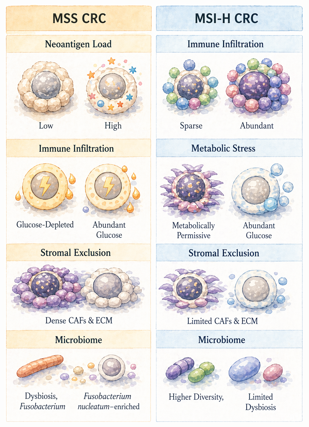
Unlike consensus molecular subtypes or conventional immune-phenotype classifications, the axis-dominance model prioritizes identification of the actionable suppressive constraint governing immune failure in a given tumor (17). The failure of prior combination trials likely reflects axis mismatch rather than intrinsic futility (18, 19). Efforts to pair immunotherapy with metabolic or microenvironmental modulators illustrate this principle: PD-1 blockade combined with IDO inhibition failed to overcome immunosuppression when broader nutrient competition and hypoxia persisted (20). Similarly, stromal-targeting approaches, such as FAP-directed strategies, enhance immune infiltration only when stromal dominance is the primary constraint (21). Microbiome-focused interventions, including Akkermansia muciniphila enrichment, have likewise shown inconsistent efficacy in CRC despite success in other tumor types (14). Together, these observations indicate that therapeutic success depends on aligning interventions with the dominant axis of immune constraint rather than indiscriminate combinations; efficacy is conditional and diluted when only a subset of tumors depends on the targeted axis.
Toward ecosystem reconditioning as a therapeutic paradigm
Immune resistance in MSS CRC is a dynamic, multilayered state shaped by metabolic stress, hypoxia, suppressive metabolites, microbial signals, and stromal architecture. Releasing inhibitory receptors without addressing these upstream constraints is unlikely to produce a durable benefit (Figure 2).

The next generation of MSS CRC immunotherapy trials should not be judged solely by response rate. Early metabolic normalization, stromal remodeling, restoration of T-cell fitness, or microbiome reprogramming may represent meaningful intermediate endpoints that precede objective tumor regression. The translational implications of the axis-dominance framework are outlined in Table 1, which links dominant suppressive constraints in MSS colorectal cancer with candidate biomarkers and rational therapeutic strategies.
Table 1. Dominant immune-constraint axes in microsatellite-stable colorectal cancer: candidate biomarkers and rational therapeutic strategies.
| Dominant Axis | Representative Biomarkers | Functional Effect | Potential Therapeutic Strategy |
|---|---|---|---|
| Metabolic stress | HIF1α, LDHA, lactate signature | T-cell dysfunction | Metabolic inhibitor + PD-1 |
| Stromal exclusion | TGF-β, CAF/FAP signature | Immune exclusion | Stromal/TGF-β targeting |
| Microbiome dysbiosis | Microbial composition, bile acid profile | Tolerogenic inflammation | Microbiome modulation / FMT |
| Checkpoint dominant | PD-L1, exhausted T-cell markers | Adaptive inhibition | Dual checkpoint blockade |
| Myeloid suppression | MDSC/TAM signatures | Antigen suppression | CSF1R / myeloid targeting |
Abbreviations: HIF1α, hypoxia-inducible factor 1α; LDHA, lactate dehydrogenase A; CAF, cancer-associated fibroblasts; FAP, fibroblast activation protein; FMT, fecal microbiota transplantation; MDSC, myeloid-derived suppressor cells; TAM, tumor-associated macrophages; PD-1, programmed cell death protein 1.
Integration of spatial transcriptomics, metabolomics, and microbiome profiling will be essential to operationalize this framework (22, 23). Such approaches can reveal dominant suppressive axes within individual tumors and guide rational combination strategies. While challenges remain, including biomarker validation, therapeutic toxicity, and clinical feasibility, the ecosystem-reconditioning paradigm offers a coherent path beyond the stagnation that has characterized MSS CRC immunotherapy to date.
By reframing MSS CRC as constrained rather than cold, this review provides both conceptual clarity and practical direction. It invites the field to move beyond reductive explanations toward a more precise, systems-level understanding of immune resistance, one that may ultimately enable durable immunotherapeutic benefit for the majority of patients with colorectal cancer. The field now faces a clear mandate: diagnose the dominant axis of immune constraint in each tumor and intervene accordingly.
MSS colorectal cancer is not immune “cold” but functionally constrained by metabolic, microbial, and microenvironmental barriers. This reframes resistance as a systems-level problem, positioning metabolic and ecological constraints as primary determinants of response. Progress will depend not on intensifying checkpoint blockade, but on diagnosing and dismantling the dominant constraint in each tumor. This shift from immune activation to ecosystem reconditioning represents a necessary evolution for achieving durable immunotherapy responses.
References
1. Le DT, Uram JN, Wang H, Bartlett BR, Kemberling H, Eyring AD, et al. PD-1 blockade in mismatch-repair deficiency. N Engl J Med. 2015;372(26):2509–2520. https://doi.org/10.1056/nejmoa1500596
2. Ganesh K, Stadler ZK, Cercek A, Shia J, Segal NH, Diaz LA Jr, et al. Immunotherapy in colorectal cancer: challenges and opportunities. Nat Rev Gastroenterol Hepatol. 2019;16(6):361–375. https://doi.org/10.1038/s41575-019-0126-x
3. Mi Y, Lin Z, Zhang Q, Li N, Zhang Z, Zhou H. Microbiota-Immune Crosstalk in Colorectal Cancer: Mechanisms, Metabolism, and Therapeutic Opportunities. Cancer Biome Target Ther. 2026;1(2):224–251. https://doi.org/10.66505/cbtt.v1i2.39
4. Mlecnik B, Bindea G, Angell HK, Maby P, Angelova M, Tougeron D, et al. Integrative immune analysis in colorectal cancer. Immunity. 2016;44(4):912–924. https://doi.org/10.1016/j.immuni.2016.02.025
5. Pelka K, Hofree M, Chen JH, Sarkizova S, Pirl JD, Jorgji V, et al. Spatially organized multicellular immune hubs in human colorectal cancer. Cell. 2021;184(18):4734–4752.e20. https://doi.org/10.1016/j.cell.2021.08.003
6. Keren L, Bosse M, Marquez D, Angoshtari R, Jain S, Varma S, et al. A structured tumor-immune microenvironment in triple negative breast cancer revealed by multiplexed ion beam imaging. Cell. 2018;174(6):1373–1387.e19. https://doi.org/10.1016/j.cell.2018.08.039
7. Thommen DS, Schumacher TN. T cell exhaustion in cancer. Nat Rev Immunol. 2018;18(6):409–421. https://doi.org/10.1038/s41577-018-0013-1
8. Zhang L, Yu X, Zheng L, Zhang Y, Li Y, Fang Q, et al. Lineage tracking reveals dynamic relationships of T cells in colorectal cancer. Nature. 2020;564(7735):268–272. https://doi.org/10.1038/s41586-018-0694-x
9. Ho PC, Bihuniak JD, Macintyre AN, Staron M, Liu X, Amezquita R, et al. Phosphoenolpyruvate is a metabolic checkpoint of anti-tumor T cell responses. Cell. 2015;162(6):1217–1228. https://doi.org/10.1016/j.cell.2015.08.012
10. Scharping NE, Menk AV, Moreci RS, Whetstone RD, Dadey RE, Watkins SC, et al. The tumor microenvironment represses T cell mitochondrial biogenesis to drive intratumoral T cell metabolic failure. Nat Immunol. 2021;22(3):361–374. https://doi.org/10.1038/s41590-020-00846-5
11. Sharma P, Hu-Lieskovan S, Wargo JA, Ribas A. Primary, adaptive, and acquired resistance to cancer immunotherapy. Cell. 2017;168(4):707–723. https://doi.org/10.1016/j.cell.2017.01.017
12. Buck MD, Sowell RT, Kaech SM, Pearce EL. Metabolic instruction of immunity. Cell. 2017;169(4):570–586. https://doi.org/10.1016/j.cell.2017.04.004
13. Wong SH, Yu J. Gut microbiota in colorectal cancer: mechanisms of action and clinical applications. Nat Rev Gastroenterol Hepatol. 2019;16(11):690–704. https://doi.org/10.1038/s41575-019-0209-8
14. Routy B, Le Chatelier E, Derosa L, Duong CPM, Alou MT, Daillère R, et al. Gut microbiome influences efficacy of PD-1-based immunotherapy against epithelial tumors. Science. 2018;359(6371):91–97. https://doi.org/10.1126/science.aan3706
15. Lee SH, Griffiths JR. How and why are cancers acidic? Carbonic anhydrase IX and the homeostatic control of tumour extracellular pH. Cancers (Basel). 2020;12(6):1616. https://doi.org/10.3390/cancers12061616
16. Reinfeld BI, Madden MZ, Wolf MM, Chytil A, Bader JE, Patterson AR, et al. Cell-programmed nutrient partitioning in the tumour microenvironment. Nature. 2021;593(7858):282–288. https://doi.org/10.1038/s41586-021-03442-1
17. Guinney J, Dienstmann R, Wang X, de Reyniès A, Schlicker A, Soneson C, et al. The consensus molecular subtypes of colorectal cancer. Nat Med. 2015;21(11):1350–1356. https://doi.org/10.1038/nm.3967
18. Llosa NJ, Cruise M, Tam A, Wicks EC, Hechenbleikner EM, Taube JM, et al. The vigorous immune microenvironment of microsatellite instable colon cancer is balanced by multiple counter-inhibitory checkpoints. Cancer Discov. 2015;5(1):43–51. https://doi.org/10.1158/2159-8290.CD-14-0863
19. Overman MJ, McDermott R, Leach JL, Lonardi S, Lenz HJ, Morse MA, et al. Nivolumab in patients with metastatic DNA mismatch repair-deficient or microsatellite instability-high colorectal cancer (CheckMate 142): an open-label, multicentre, phase 2 study. Lancet Oncol. 2017;18(9):1182–1191. https://doi.org/10.1016/S1470-2045(17)30422-9
20. Long GV, Dummer R, Hamid O, Gajewski TF, Caglevic C, Dalle S, et al. Epacadostat plus pembrolizumab versus pembrolizumab alone in advanced melanoma (ECHO 301/KEYNOTE 252): a phase 3 trial. Lancet Oncol. 2019;20(8):1083–1097. https://doi.org/10.1016/S1470-2045(19)30274-8
21. Kraman M, Bambrough PJ, Arnold JN, Roberts EW, Magiera L, Jones JO, et al. Suppression of antitumor immunity by stromal cells expressing fibroblast activation protein α. Science. 2010;330(6005):827–830. https://doi.org/10.1126/science.1195300
22. Schürch CM, Bhate SS, Barlow GL, Phillips DJ, Noti L, Zlobec I, et al. Coordinated cellular neighborhoods orchestrate antitumoral immunity at the colorectal cancer invasive front. Cell. 2020;182(5):1341–1359.e19. https://doi.org/10.1016/j.cell.2020.07.005
23. Gopalakrishnan V, Spencer CN, Nezi L, Reuben A, Andrews MC, Karpinets TV, et al. Gut microbiome modulates response to anti-PD-1 immunotherapy in melanoma patients. Science. 2018;359(6371):97–103. https://doi.org/10.1126/science.aan4236
Declarations
Funding Statement: This work was supported by a grant award from the Kansas University Cancer Center (KUCC) National Cancer Institute Cancer Center Support Grant P30CA168524.
Competing Interest: The author declares no financial or personal relationships with other individuals or organizations that could inappropriately influence or bias the content of this work. The author has read the final version of the manuscript and confirms that there are no competing interests.
Consent for publication: The author has approved the final version of the manuscript.
Use of Artificial Intelligence Disclosure: This article was written by human contributors. Artificial intelligence-based tools were used to improve grammar, language, and readability without affecting the article's scientific content, data interpretation, or conclusions. The author reviewed and verified all content to ensure its accuracy and integrity.
Data Availability Statement: “No datasets were generated or analyzed in the current study.”
Ethics approval and consent to participate: Not applicable, as this study did not involve the conduct of research.
Authors’ affiliations
1. Department of Pathology and Laboratory Medicine, University of Kansas Medical Center, Kansas City, USA
CRediT authorship contribution statement: Christophe Nicot: Writing - review & editing, Funding acquisition.
ORCID ID
Christophe Nicot: https://orcid.org/0000-0001-5927-2869
Note: This article was commissioned. The content was reviewed by the editorial team for accuracy, relevance, and adherence to journal standards.