Targeting Immune Cell Metabolism for Cancer Immunotherapy
1 Key Laboratory of Medical Molecular Virology (MOE/NHC/CAMS) and Shanghai Institute of Infectious Disease and Biosecurity, School of Basic Medical Sciences, Fudan University, Shanghai 200032, China
2 Shanghai Engineering Research Center for Synthetic Immunology, Shanghai 200032, China
Correspondence: Yan-ling Wu (yanlingwu@fudan.edu.cn); Tian-lei Ying (tlying@fudan.edu.cn)
Received: April 5, 2026
Accepted: June 22, 2026
Published: August 12, 2026
© 2026 The Author(s). Published by GCINC Press, Spokane, Washington, United States. Open Access licensed under a Creative Commons Attribution 4.0 International License (CC BY 4.0), which permits unrestricted use, distribution, and reproduction in any medium, provided the original author(s) and source are credited. To view a copy of this license, visit: Creative Commons Attribution 4.0 International License (CC BY 4.0)
Abstract
Metabolic reprogramming is a fundamental regulator of immune cell fate and function in the tumor microenvironment (TME), though the relative contributions of individual metabolic pathways vary across immune cell subsets, tumor types, and disease stages. Competition for nutrients, hypoxia, and the accumulation of immunosuppressive metabolites reshape the TME's metabolic landscape, driving functional exhaustion of antitumor immune cells while promoting the persistence of immunosuppressive populations. These metabolic adaptations have emerged as critical determinants of immune evasion, therapeutic resistance, and responses to cancer immunotherapy. In this review, we examine immune cell metabolism from four complementary perspectives: the physiological metabolic programs of major immune cell populations and their remodeling within the TME; the functional consequences of metabolic reprogramming for antitumor immunity; the principal metabolic drivers, including hypoxia, nutrient competition, and suppressive metabolites; and current therapeutic strategies targeting key immunometabolic pathways, with particular emphasis on glutamine, fatty acid, and lactate metabolism. We further discuss emerging metabolic modulators, their interactions with immune checkpoint blockade, and the opportunities and limitations of combining metabolic intervention with established cancer therapies. Finally, we highlight major translational challenges, including metabolic heterogeneity, limited clinical validation, biomarker identification, and the need for standardized metabolomic and single-cell approaches to enable precision immunometabolism. A more comprehensive understanding of immune cell metabolic regulation may facilitate the development of biomarker-guided therapeutic strategies that selectively restore antitumor immunity while minimizing systemic toxicity, thereby advancing the next generation of personalized cancer immunotherapies.
Keywords
Immunometabolism; tumor microenvironment; metabolic reprogramming; tumor-infiltrating immune cells; cancer immunotherapy; immune cell metabolism; fatty acid metabolism
1. Introduction
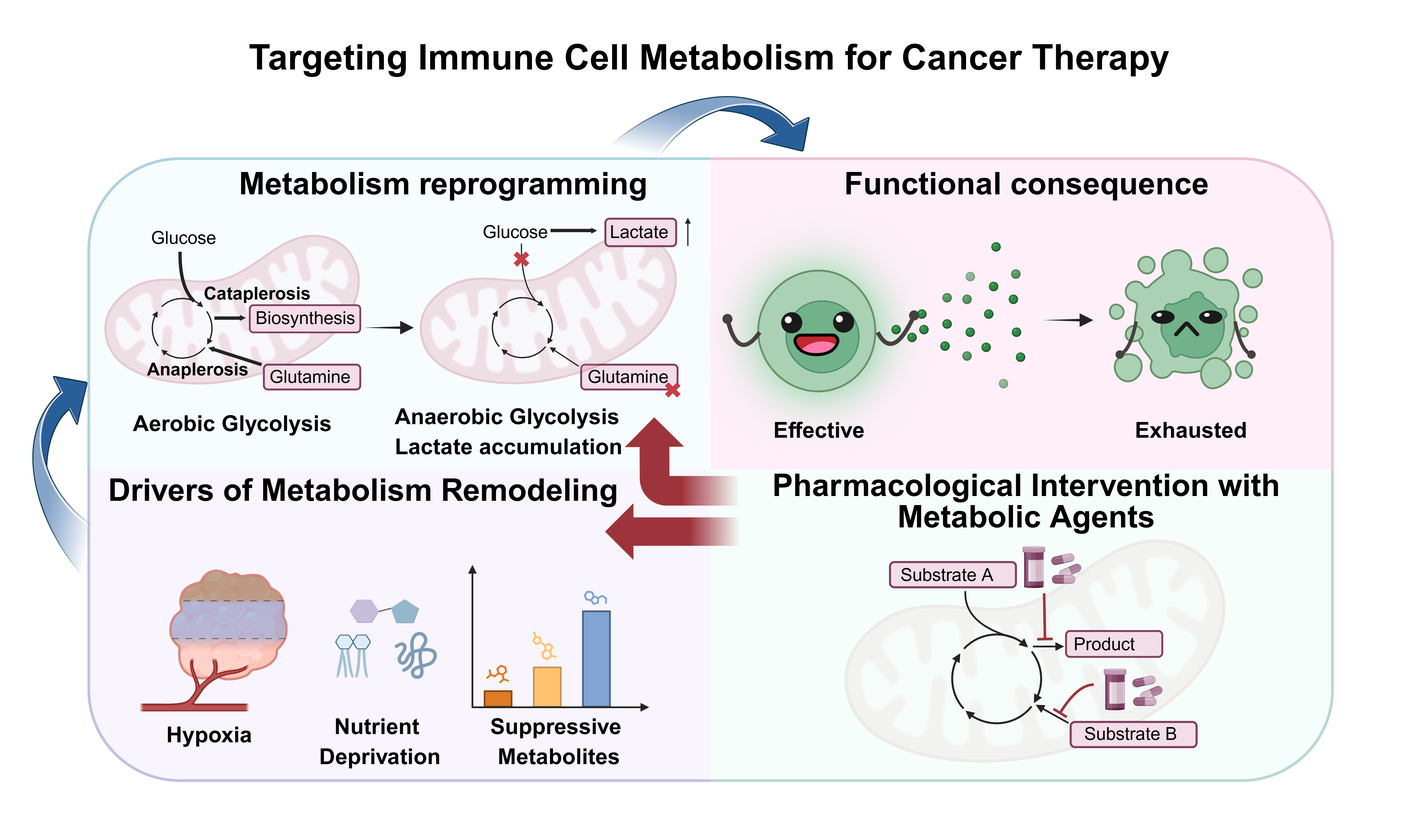
Since the discovery of the Warburg effect in the early twentieth century, tumor metabolism has remained a major focus of cancer research. Multiple metabolism-targeted strategies have entered clinical development; however, clinical efficacy has generally been more modest than anticipated owing to metabolic plasticity, pathway redundancy, and dose-limiting toxicities (1). However, most of these approaches were designed to disrupt tumor cell metabolism, and their clinical benefit has been limited by tumor heterogeneity, metabolic plasticity, and the complexity of the tumor microenvironment (TME) (2-4). Beyond malignant cells, non-malignant components of the TME, including tumor-infiltrating lymphocytes (TILs) and stromal cells, also undergo profound metabolic reprogramming (5). Importantly, activated immune cells and tumor cells share several core metabolic pathways, particularly glycolysis, raising the risk that metabolism-targeting therapies may produce on-target effects that inadvertently impair antitumor immunity (6). These limitations have shifted attention from tumor cell metabolism to the metabolic states of immune cells within the TME. The metabolic state of immune cells is tightly coupled to their differentiation and effector function. Resting T cells primarily rely on oxidative phosphorylation (OXPHOS), whereas activated T cells increase glycolysis and glutamine metabolism to support proliferation, biosynthesis, and cytotoxic activity (7). By contrast, during tumor progression, tumor-derived signals such as colony-stimulating factor 1 (CSF1) promote the recruitment of tumor-associated macrophages (TAMs) that preferentially engage in fatty acid oxidation (FAO), thereby reinforcing an M2-like state (8). Metabolic reprogramming should therefore be viewed not merely as a consequence of immune activation but as a critical regulatory layer that shapes immune cell fate and function in the TME. Targeting immune cell metabolism has therefore emerged as a promising complementary therapeutic strategy, although its clinical success will likely depend on selective modulation of immune rather than tumor metabolism. In this review, we examine immune cell metabolism in the TME from four perspectives: the physiological metabolic states of immune cells and how they shift in the TME, the functional consequences of these changes, the metabolic drivers of immune cell reprogramming, and therapeutic strategies that target major metabolic pathways, with particular emphasis on glutamine, fatty acid, and lactate metabolism (Figure 1).
2. Metabolism of tumor-infiltrating immune cells
We focus on seven immune cell types that collectively represent the major innate and adaptive compartments of the tumor immune microenvironment, including antigen-presenting, effector, suppressive, and humoral populations. Together, these cell types capture the principal metabolic programs that shape immune activation, tolerance, and dysfunction in cancer.
Baseline Metabolic States of Immune Cells
Macrophages arise from embryonically seeded tissue-resident populations and from bone marrow-derived monocytes, with their development dependent on CSF1R signaling and tissue-specific niche cues. For instance, alveolar macrophages require a GM-CSF-induced PPARγ program (9). Under homeostatic conditions, macrophages mediate apoptotic cell clearance, tissue maintenance, and immune surveillance. Metabolically, they generally rely on an intact tricarboxylic acid (TCA) cycle and mitochondrial OXPHOS. In contrast, M1-like macrophages increase glycolysis and pentose phosphate pathway (PPP) activity and rewire the TCA cycle, whereas M2-like macrophages maintain higher OXPHOS, fatty acid oxidation, and glutamine metabolism (10). T cells develop from lymphoid progenitors through thymic selection and differentiate into CD4, CD8, and thymus-derived regulatory T cells (Treg) (11). Following antigenic stimulation, peripheral T cells further diversify into effector, memory, or induced Treg states (12). Resting naive and memory T cells primarily rely on OXPHOS and fatty acid utilization. Upon activation, effector T cells switch to aerobic glycolysis, accompanied by enhanced glutaminolysis and lipid synthesis, under the control of MYC, HIF-1α, and PI3K-AKT-mTORC1 signaling (13). By contrast, Treg cells preferentially use OXPHOS and fatty acid oxidation and can also exploit lactate to maintain suppressive function. Dendritic cells (DCs) include cDC1, cDC2, pDCs, Langerhans cells, and inflammatory monocyte-derived DCs, and develop through Flt3L-Flt3 signaling. Immature DCs primarily rely on OXPHOS and fatty acid oxidation and are specialized for antigen uptake. After activation, they rapidly increase glycolysis and lipid synthesis and rewire the TCA cycle to support maturation and antigen presentation (14).
Myeloid-derived suppressor cells (MDSCs), including the monocytic and polymorphonuclear subsets, expand in response to tumor- and inflammation-associated factors, including GM-CSF, IL-6, VEGF, and STAT3 signaling (15). They suppress T cell responses via Arginase 1 (ARG1), iNOS, and reactive oxygen species (ROS) while promoting fatty acid uptake and oxidation, lipid droplet accumulation, and elevated OXPHOS. Among granulocytes, neutrophils are metabolically distinctive because they rely predominantly on glycolysis even at rest and have abundant glycogen stores (16). Upon activation, glycolysis and PPP flux further increase to generate NADPH for ROS production and neutrophil extracellular trap formation, whereas mitochondrial OXPHOS contributes less (17). Resting B cells primarily use OXPHOS; activated B cells increase both glycolysis and mitochondrial metabolism; memory B cells revert to an OXPHOS-dominant state; and plasma cells depend on OXPHOS and glutamine metabolism to sustain antibody production (18). Likewise, resting NK cells primarily rely on OXPHOS, whereas activated NK cells coordinately enhance glycolysis and mitochondrial metabolism to support effector function (19). Overall, immune cell metabolism is not fixed but dynamically adjusted to developmental stage and functional demand. Most resting immune cells rely primarily on oxidative metabolism, whereas activation is generally accompanied by increased glycolysis and other anabolic pathways. Memory cells and immunosuppressive cell subsets tend to favor fatty acid oxidation and OXPHOS, whereas neutrophils are unusual in relying predominantly on glycolysis even when resting (20). Although these metabolic programs provide a useful conceptual framework, growing evidence indicates that immune cell phenotypes span metabolic continua rather than discrete binary states, particularly in heterogeneous human tumors.
Metabolic Reprogramming of Immune Cells Under Tumor Pressure
Cancer immunoediting is the dynamic interplay between the immune system and cancer cells, unfolding in three phases: elimination, equilibrium, and escape (21). In neoplastic tissues, cancer cells, stromal cells, and newly activated immune cells all exhibit metabolic features typical of proliferating cells, underscoring their shared metabolic demands. As tumor cells rapidly consume nutrients, they reshape the TME by limiting nutrient availability, inducing hypoxia, and generating immunosuppressive metabolites (22). Consequently, immune cells undergo adaptive metabolic reprogramming, although the magnitude and direction of these changes depend on nutrient availability, tumor genotype, tissue context, and immune composition (23) (Figure 2).
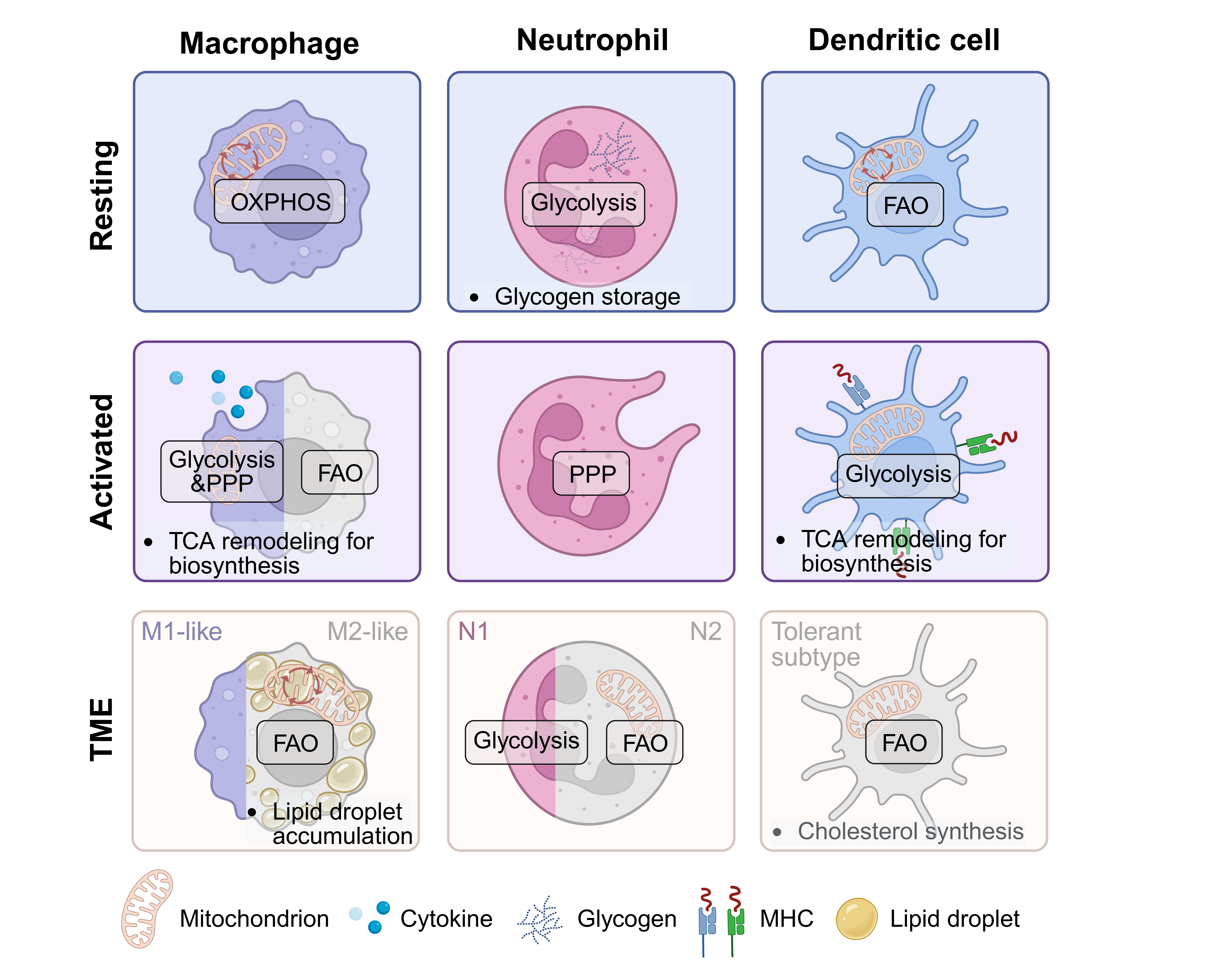
Within the TME, TAMs commonly adopt an M2-like phenotype. Because tumor cells consume large amounts of glucose, glycolysis in TAMs is often restricted, and their metabolism gradually shifts toward fatty acid oxidation (FAO). By upregulating lipid transporters such as CD36, TAMs increase fatty acid uptake, and tumor-derived lipids and apoptotic cell debris serve as exogenous substrates that support their immunosuppressive functions (24).
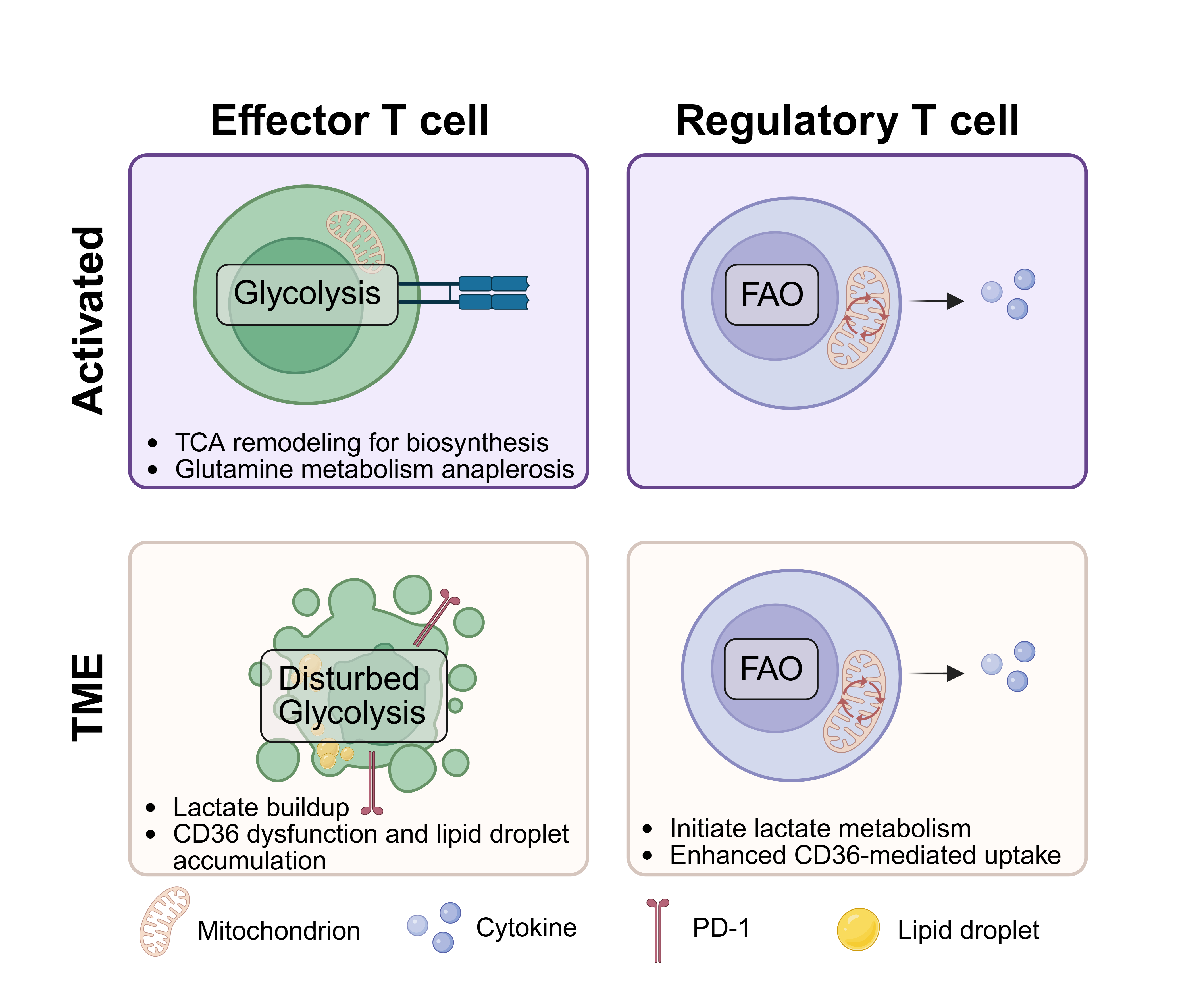
Effector T cells experience profound metabolic stress in the TME and progressively develop functional exhaustion (25). Competition for glucose limits GLUT1-mediated uptake, while lactate accumulation suppresses glycolysis by acidifying the microenvironment and disrupting metabolic homeostasis. In parallel, T cells aberrantly take up oxidized lipids via CD36, leading to lipid peroxide accumulation and induction of ferroptosis, which further reduces their abundance and function (26, 27). In addition, depletion of amino acids such as glutamine constrains T-cell proliferation and biosynthesis (28). In contrast, Treg cells exhibit greater metabolic adaptability within the TME. They are less dependent on glucose, can use lactate as an alternative carbon source, and increase fatty acid uptake via CD36, thereby relying on FAO and OXPHOS to sustain survival and suppressive activity (Figure 3) (29).
DCs in the TME shift from an immune-activating state toward an immune-tolerant state. Glucose deprivation and lactate accumulation suppress the glycolytic activation required for DC maturation, whereas tumor-derived lipids reprogram DC metabolism via SREBP2- and MerTK-mediated pathways, promoting dependence on OXPHOS and lipid metabolism and activating cholesterol biosynthesis, ultimately impairing antigen presentation (30).
By contrast, MDSCs undergo marked expansion in tumors and adopt a lipid-centered metabolic program characterized by increased fatty acid uptake via CD36 and FATP2, lipid and cholesterol accumulation, and enhanced FAO and OXPHOS, with glycolysis also increasing under certain conditions (31). Arginine depletion mediated by ARG1, together with iNOS activity, ROS production, and lipid peroxidation-related signaling, forms the metabolic basis for their T-cell suppressive activity (32).
Neutrophils rely on glycolysis and glycogen storage to support rapid responses (33). In the TME, however, glucose scarcity limits glycolysis and PPP activity, reducing NADPH production and attenuating ROS generation. Under these conditions, neutrophils undergo metabolic divergence: N1-like cells partially maintain glycolysis, whereas N2-like cells upregulate CD36 and FATP2 and become more dependent on FAO. This “glycolytic restriction plus FAO compensation” pattern is closely associated with protumor functions (34).
Metabolic studies of B cells in the TME remain limited. Existing evidence suggests that depletion of glucose and glutamine may impair germinal center responses and plasma cell antibody secretion, whereas the differentiation of regulatory B cells (Bregs) may depend on glycolysis (35). In addition, tertiary lymphoid structures (TLS) may provide local metabolic support for B cells, although their metabolic characteristics likely vary across tissues. NK cells also undergo substantial metabolic impairment in the TME. Competition for glucose suppresses the initiation of glycolysis; TGF-β inhibits mTORC1 activity, thereby blocking metabolic reprogramming; and hypoxia, together with oxidative stress, damages mitochondrial function, promotes ROS accumulation, and reduces metabolic flexibility (36). As a result, NK cells ultimately enter a state characterized by restricted glycolysis, impaired OXPHOS, and mitochondrial dysfunction, which provides an important metabolic basis for their functional exhaustion.
Taken together, metabolic reprogramming constitutes a central regulatory layer through which the tumor microenvironment integrates external cues and actively shapes immune cell functional states. Importantly, most mechanistic evidence derives from murine models or in vitro systems, whereas direct validation in human tumors remains limited.
3. Functional consequences of immune cell metabolic reprogramming
Cellular metabolism not only sustains immune-cell survival but also actively regulates differentiation and function; however, these relationships are bidirectional, as immune activation itself further reshapes metabolic programs. For example, promoting FAO or inhibiting glycolysis enhances the survival and memory formation of CD8+ T cells, whereas increased glycolysis drives their differentiation toward an effector phenotype, underscoring metabolism as a key regulator of immune cell fate (37, 38).
Lymphoid Cells
In the TME, CD8+ effector T cells progressively shift from potent cytotoxic cells to a functionally exhausted state under persistent antigen stimulation and metabolic stress. Glucose deprivation and lactate accumulation reduce the production of effector molecules such as granzymes, perforin, and IFN-γ, while inhibitory receptors, including PD-1, TIM-3, and LAG-3, remain highly expressed, ultimately impairing tumor cell clearance (39). In contrast, Tregs are metabolically advantaged in this hostile environment. By relying on fatty acid oxidation and lactate utilization, they maintain stable survival and suppressive activity, further inhibiting effector T cells through IL-10, TGF-β, and high CTLA-4 expression (40). Together, the exhaustion of CD8+ T cells and the reinforcement of Treg function drive antitumor immunity from active attack to functional failure, although the degree of T-cell exhaustion varies considerably across tumor types and therapeutic settings.
B cells exhibit marked functional duality in the TME. Under physiological conditions, B cells contribute to adaptive immunity through antibody production, antigen presentation, and cytokine secretion (5). In tumors, however, their functions diverge in opposing directions. On the one hand, B cells within tertiary lymphoid structures (TLS) can undergo clonal expansion and class switching, generate tumor-reactive antibodies, and promote tumor killing through antibody-dependent cellular cytotoxicity (ADCC), complement-dependent cytotoxicity (CDC), and antigen presentation, which explains why TLS are often associated with a favorable prognosis and better responses to immunotherapy (41, 42). On the other hand, regulatory B cells (Bregs) secrete IL-10, IL-35, and TGF-β, upregulate PD-L1, and recruit MDSCs and Tregs, thereby amplifying local immunosuppression and promoting tumor progression (43, 44). Thus, the net effect of B cells in cancer depends on the balance between TLS-associated antitumor B cells and immunosuppressive Bregs (45).
NK cells are key innate immune effectors capable of killing target cells without prior sensitization, and their IFN-γ production and cytotoxicity depend on coordinated enhancement of glycolysis and mitochondrial metabolism. In the TME, however, glucose deprivation and suppressive signals such as TGF-β and PGE2 impair glycolytic activation and reduce mTORC1 activity. At the same time, activating receptors are downregulated, whereas inhibitory receptors are upregulated. Hypoxia, together with oxidative stress, further impairs mitochondrial function, exacerbating NK-cell dysfunction (46). As a result, NK cells gradually shift from rapid innate killers to metabolically constrained, exhausted cells, becoming an important component of tumor immune evasion (47).
Myeloid Cells
Under physiological conditions, macrophages serve as guardians of tissue homeostasis by mediating phagocytosis, antigen presentation, and inflammatory regulation, and they can dynamically switch between pro-inflammatory and reparative states. In the TME, however, glucose deprivation, lactate accumulation, hypoxia, and signals such as CSF1, IL-10, and TGF-β drive macrophages toward an M2-like phenotype, converting them from immune defenders into tumor accomplices. TAMs exhibit reduced phagocytic and tumoricidal capacity and impaired antigen presentation, yet secrete high levels of IL-10, TGF-β, and VEGF, thereby suppressing CD8+ T cell function (48) and promoting angiogenesis and tissue remodeling. In addition, TAMs can induce T-cell exhaustion through PD-L1 expression (49) and recruit Tregs via chemokines such as CCL22 (50). Consequently, TAMs frequently function as central regulators of immunosuppression, although their phenotypic and metabolic heterogeneity suggests that not all macrophage subsets should be considered uniformly protumorigenic. Metabolic reprogramming also transforms DCs from initiators of immune responses into facilitators of immune tolerance. In tumors, DCs exhibit impaired antigen cross-presentation and are less able to prime naïve T cells (51). At the same time, they upregulate suppressive molecules such as PD-L1 and Indoleamine 2,3-dioxygenase (IDO) and gain the capacity to induce Treg differentiation, thereby establishing a local barrier to immune tolerance (52).
MDSCs do not normally exist as a stable, mature suppressive cell population, but in tumors they undergo abnormal expansion and become locked in an immunosuppressive state (53). They consume large amounts of arginine through ARG1 and iNOS (54), deplete tryptophan via IDO, and generate ROS and peroxynitrite, thereby directly suppressing T-cell activation and proliferation at the metabolic level. In parallel, they secrete IL-10 and TGF-β and upregulate PD-L1, further reinforcing the suppressive network (55). MDSCs can also promote angiogenesis via VEGF and contribute to the formation of the premetastatic niche, thereby serving as a critical link among metabolic reprogramming, immune evasion, and therapeutic resistance (56).
Neutrophils normally rely on glycolysis to drive respiratory burst, ROS production, and NET formation, functioning as rapid responders in innate immunity. In the TME, however, tumor-associated neutrophils can polarize into N1 and N2 states (57). N1 neutrophils retain some antitumor activity, whereas metabolic pressures within the TME preferentially promote N2 polarization. N2 neutrophils suppress antitumor immunity by depleting arginine via ARG1, secreting IL-10 and TGF-β, and releasing NETs, while also promoting angiogenesis via VEGF, thereby functioning as protumor effectors (58, 59). Because glucose deprivation imposes stronger selective pressure on glycolysis-dependent N1 cells, whereas FAO-biased N2 cells gain a survival advantage, N2 neutrophils often become dominant in tumors and establish a positive feedback loop that further reinforces the immunosuppressive microenvironment (60).
4. Remodeling of immune cell metabolism by key metabolites and microenvironmental cues
Hypoxia, nutrient competition, and suppressive metabolites are key drivers of immune cell reprogramming in the TME, although their relative contributions vary among tumor microenvironments.
Hypoxia and Imbalanced Nutrient Distribution
Hypoxia and altered nutrient distribution are widely recognized drivers of immune metabolic reprogramming; however, the severity of these constraints varies with vascular architecture, tumor growth kinetics, and tissue origin (61). This state does not arise solely from rapid tumor cell proliferation but from the combined effects of aberrant angiogenesis, insufficient perfusion, restricted oxygen diffusion, and the high metabolic burden of tumors (62). As a result, the TME commonly exhibits hypoxia, reduced nutrient availability, metabolic disorder, and immunosuppression, thereby shaping the metabolic niche in which immune cells reside. Hypoxia is not merely a reduction in oxygen supply but a broader constraint on cellular metabolism. As the terminal electron acceptor in mitochondrial OXPHOS, oxygen deficiency directly limits immune cells' ability to sustain efficient oxidative metabolism and, through HIF-associated programs, reshapes glucose utilization, lactate production, and stress adaptation. For effector T cells, NK cells, and mature DCs, this typically leads to restricted energy metabolism and impaired effector function; in contrast, Tregs, TAMs, and MDSCs are better able to undergo adaptive metabolic reprogramming under hypoxic conditions, thereby gaining survival and functional advantages within the TME (63).
Compared with the broad concept of “nutrient deprivation,” “imbalanced nutrient distribution” more accurately captures resource competition in tumors. Different cellular populations do not merely compete passively for the same substrates; rather, nutrient allocation appears to be at least partially programmed. Myeloid cells often show a greater capacity for glucose uptake, whereas tumor cells tend to dominate glutamine utilization (64). Thus, immune cells face not only generalized nutrient scarcity but also an unequal distribution of key substrates among different cell populations. For effector T cells and NK cells, reduced glucose availability suppresses glycolysis and its associated effector programs, whereas glutamine insufficiency further compromises proliferation, cytotoxicity, and antibody secretion. At the same time, intratumoral T cells often exhibit cholesterol deficiency, whereas tumor cells and immunosuppressive myeloid cells are enriched in cholesterol. Low cholesterol levels can inhibit T-cell proliferation and induce autophagy-related apoptosis, with particularly marked effects on cytotoxic T cells (31).
In addition to glucose and glutamine, local depletion of amino acids such as arginine and tryptophan is another important metabolic constraint. Tumor-associated myeloid cells can degrade arginine via ARG1, thereby reducing CD3ζ chain expression and weakening TCR signaling in T cells (65). Meanwhile, tumor cells and tolerogenic antigen-presenting cells can catabolize tryptophan via IDO/tryptophan 2,3-dioxygenase (TDO), leading to local tryptophan insufficiency and the accumulation of inhibitory downstream metabolites such as kynurenine, which further promote T cell anergy and Treg skewing. Overall, hypoxia and imbalanced nutrient distribution not only limit the acquisition of fuel and biosynthetic substrates by immune cells but also alter their redox state, signal transduction, and fate decisions, thereby laying the groundwork for the subsequent accumulation of suppressive metabolites and stress-associated products.
Accumulation of Suppressive Metabolites and Metabolic Stress Products
The persistent accumulation of suppressive metabolites and metabolic stress products is another major driver of immune cell metabolic reprogramming in the TME (66). Among these, the lactate/acidosis axis is a representative suppressive metabolic pathway (67). Under highly glycolytic conditions, tumor cells continuously convert pyruvate to lactate via lactate dehydrogenase, leading to the co-accumulation of lactate and protons in the local microenvironment. Some highly glycolytic immune cells, particularly certain myeloid populations, may also contribute to this lactate pool. Lactate/acidosis suppresses glycolysis, cytokine secretion, and cytotoxicity in effector T cells and NK cells, while Tregs take up and use lactate as a metabolic substrate, thereby conferring a metabolic adaptive advantage on Tregs (68). Tumor-derived lactate can also drive macrophages toward an M2-like phenotype. Recent studies further suggest that high lactate levels induce signaling changes, such as protein lactylation, thereby reshaping innate immune sensing and transcriptional programs (69).
Adenosine and kynurenine exemplify immunosuppressive signaling metabolites with well-defined origins. Adenosine is primarily generated from extracellular ATP released by stressed, hypoxic, or necrotic cells and is then sequentially converted through the ectoenzymatic cascade mediated by CD39 and CD73 (70). This process can involve tumor cells, Tregs, MDSCs, TAMs, and tumor-derived exosomes (71). Accumulated adenosine suppresses T-cell and NK-cell activation via A2A receptors (72) and impairs antigen presentation by DCs. Similarly, kynurenine is generated via tryptophan catabolism catalyzed by IDO/TDO, primarily in tumor cells and antigen-presenting cells, especially tolerogenic DCs (73). Its accumulation not only reflects local tryptophan depletion but also directly suppresses T-cell proliferation and promotes Treg differentiation, thereby converting “amino acid insufficiency” into an “immunosuppressive phenotype.” In addition, polyamines produced by TAMs through arginine metabolism can promote immune evasion (74).
Beyond soluble metabolites, metabolic stress products, particularly ROS and lipid peroxides, have defined sources and important immunological functions. ROS may arise from mitochondrial electron leakage in the highly metabolic state of tumor cells or from release by myeloid cells such as MDSCs and TAMs, thereby creating persistent oxidative stress in the local environment. Excessive ROS damage mitochondria in T and NK cells, disrupt redox homeostasis, and promote functional exhaustion (75). Lipid peroxides mainly originate from lipid-rich tumor niches, oxidized lipids released by dying or damaged tumor cells, and abnormal lipid uptake by immune cells. For example, CD8+ T cells can take up oxidized lipids through CD36 and undergo lipid peroxidation, resulting in functional impairment and a tendency toward ferroptosis (76). Excessive lipid accumulation in DC can also induce endoplasmic reticulum stress and impair antigen processing and presentation. In contrast, immunosuppressive populations such as Treg and TAM are generally better adapted to lipid-rich environments, thereby further amplifying this metabolic imbalance (77).
Therefore, the significance of suppressive metabolites and metabolic stress products lies in their traceable cellular origins, defined biosynthetic pathways, and specific immune targets. If hypoxia and imbalanced nutrient distribution form the background conditions for immune metabolic reprogramming, the accumulation of molecules with defined cellular origins, such as lactate/acidosis, adenosine, kynurenine, ROS, and oxidized lipids, translates this background pressure into direct mechanisms of immune dysfunction, phenotypic skewing, and weakened antitumor responses. Whether these metabolites primarily initiate immune dysfunction or reinforce pre-existing suppressive programs remains incompletely resolved and likely depends on tumor context.
Synergistic Amplification of Metabolic Suppression by Cytokine and Angiogenic Signaling
The metabolic pressures described above, cytokines, and angiogenesis-related signals in the TME act synergistically to further stabilize the immunosuppressive metabolic state. TGF-β is a representative factor linking immunosuppression to metabolic reprogramming. It not only directly inhibits activation programs in effector T cells and NK cells but also affects mTORC1-associated metabolic activity, weakens glycolysis-dependent effector responses, and promotes the maintenance of Treg and immunosuppressive macrophage phenotypes (78). In addition, CSF1 and GM-CSF help sustain the expansion, survival, and functional programs of TAMs and MDSCs, thereby indirectly reinforcing their dependence on lipid metabolism and their suppressive metabolic phenotypes. VEGF exacerbates hypoxia and imbalanced nutrient distribution by promoting aberrant angiogenesis, whereas IL-10 favors a tolerogenic state in antigen-presenting cells and macrophages, making the aforementioned metabolic pressures more likely to result in persistent immunosuppression (79).
Altogether, cytokines and angiogenic signals, by regulating metabolic enzyme expression, nutrient utilization patterns, and differentiation trajectories, cooperate with hypoxia, nutrient restriction, and the accumulation of suppressive metabolites to form an amplification loop of immune cell metabolic reprogramming.
5. Therapeutic modulators targeting immunometabolic pathways in the tumor microenvironment
Metabolic pathways represent attractive therapeutic targets; however, achieving sufficient selectivity to preserve physiological immune function while disrupting tumor-associated metabolic programs remains a major challenge. However, strategies that broadly target tumor-cell metabolism are often limited by systemic toxicity, pathway redundancy, and metabolic plasticity, whereas dysfunctional immune cells themselves require direct reprogramming (80).
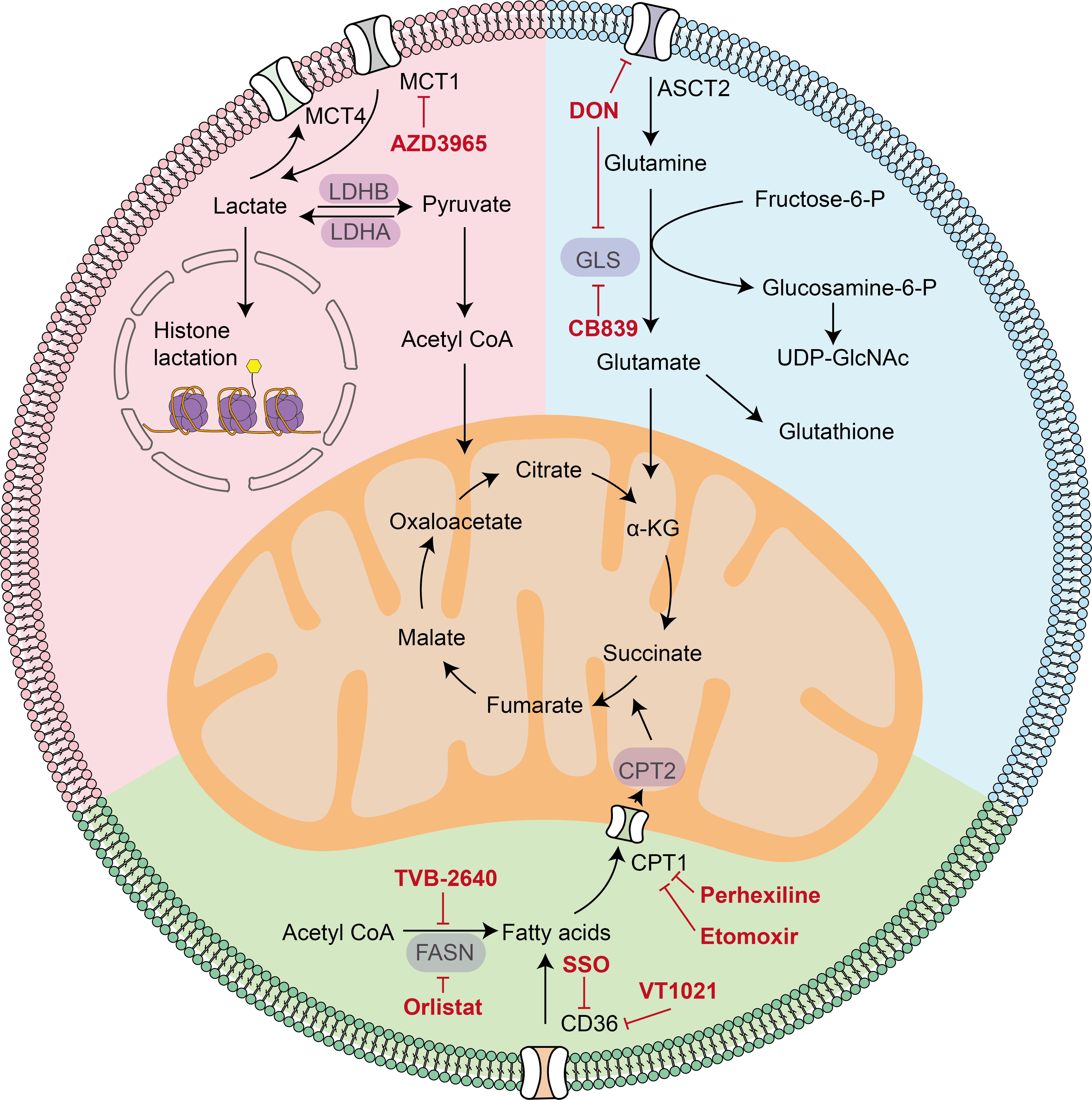
This section focuses on agents that directly modulate immune-cell metabolism within the TME, with an emphasis on three therapeutically relevant axes: lactate, glutamine, and fatty acid metabolism (Figure 4).
Representative therapeutic approaches targeting these metabolic pathways are summarized in Table 1 below.
Table 1. Metabolism regulators acting on immune cells
| Name | Targeting metabolism | Phase of clinical trials * | Effect on immune cells |
|---|---|---|---|
| CPI-613 | TCA cycle | Approved (Acute myeloid leukemia) | Inhibit PDH, restore PC and promote the killing of T cells |
| AZD3965 | Lactate metabolism | Phase I (Monotherapy for advanced cancer, NCT01791595) | Reduces the lactate/pyruvate ratio in tumor-infiltrating lymphocytes (TILs) and enhances immune activity |
| DON | Glutamine metabolism | Preclinical | Promote OXPHOS and pentose phosphate pathway of T cells; Conversion of MDSCs into pro-inflammatory immune cells |
| DRP-104 | Glutamine metabolism | Phase I/II (with durvalumab for advanced stage fibrolamellar carcinoma; NCT06027086) | Increase immune cell infiltration, improve CD8+ T cell activity, stimulate macrophages to switch to M1 type, and reverse T cell exhaustion |
| CB839 | Glutamine metabolism | Phase II ** (with Nivolumab for melanoma, ccRCC and NSCLC, NCT02771626; with Palbociclib for solid tumors; NCT03965845) | Enhances IFN-γ expression and effector function of CD8⁺ T cells |
| Etomoxir | FAO, OXPHOS | Preclinical | Inhibit the FAO of immune cells, and act on OXPHOS at high doses |
| Perhexiline | FAO | Preclinical | Promote the polarization of macrophages to M1 and inhibit M2; promote the secretion of IFN-γ and granzyme B by CD8+ T cells |
| VT1021 | FAO | Phase III (glioblastoma; NCT03970447) | Stimulate TSP-1 production and promote the conversion of M2 macrophages to M1 |
| SSO | Fatty acid synthesis | Preclinical | Enhance the secretion of CD8+ T cytokines TNF and IFN-γ |
| TVB-2640 | Fatty acid synthesis | Phase III (KRAS NSCLC; NCT03808558), (with Bevacizumab for recurrent GBM, NCT05118776) | Enhance MHC-1 expression in tumor cells |
| Orlistat | Fatty acid synthesis | Preclinical | Increases MHC-I expression, suppresses FABP1-mediated immunosuppressive signaling, and alleviates immune suppression |
Abbreviations: ccRCC, clear cell renal cell carcinoma; CD8, cluster of differentiation 8; FAO, fatty acid oxidation; FABP1, fatty acid-binding protein 1; GBM, glioblastoma; IFN-γ, interferon gamma; KRAS, Kirsten rat sarcoma viral oncogene homolog; M1, classically activated macrophage; M2, alternatively activated macrophage; MDSCs, myeloid-derived suppressor cells; MHC-I, major histocompatibility complex class I; NSCLC, non-small cell lung cancer; OXPHOS, oxidative phosphorylation; PC, pyruvate carboxylase; PDH, pyruvate dehydrogenase; TCA, tricarboxylic acid; TILs, tumor-infiltrating lymphocytes; TNF, tumor necrosis factor; TSP-1, thrombospondin-1. * Refers only to the clinical development stage for oncology indications. ** Here, the list just represents the representative Phase II trials
Targeting Lactate Metabolism
Lactate metabolism can be targeted at two main levels: lactate production and lactate transport. Lactate dehydrogenase (LDH) is an attractive target, but clinically available LDH inhibitors remain limited. Although liver-directed LDHA silencing has been achieved with Nedosiran in primary hyperoxaluria, this agent is not an oncology drug and does not directly address immune-cell metabolic reprogramming in tumors (81). Several LDH inhibitors, including Gen140, NCI-006, and Galloflavin, are in development and have shown antitumor activity in vitro and in vivo (82-84), but immune-cell-specific effects remain unclear. Thus, current immunometabolic interest has shifted more strongly toward monocarboxylate transporters (MCTs) and lactate-associated signaling.
Targeting Lactate Transport
Lactate and associated H+ ions enter immune cells through several mechanisms, with proton-dependent MCTs serving as the principal transporters (85). MCT1 is typically associated with lactate uptake in oxidative cells, whereas MCT4 is more commonly associated with lactate export in highly glycolytic cells. MCT1 mediates lactate-associated effects in macrophages, mast cells, and CD8+ T cells (86). Inhibition of MCT1 with AZD3965 has been reported to increase lymphocyte infiltration, reduce the lactate-to-pyruvate ratio, and enhance immune cell activity in the TME (87, 88). In phase I studies, AZD3965 showed acceptable tolerability, with mainly grade 1/2 adverse events, but dose-limiting ocular and metabolic toxicities were observed, and the efficacy of single-agent MCT1 inhibition remained limited (89).
Targeting Lactate-Associated Metabolic Signaling
Beyond transport, lactate can reshape intracellular metabolic signaling. Tumor-derived lactate alters the balance of pyruvate entry into the TCA cycle, shifting it from pyruvate carboxylase (PC)-dependent reactions to pyruvate dehydrogenase (PDH)-dependent reactions. Inhibiting PDH with CPI-613 restores PC-dependent anaplerosis and succinate-SUCNR1 signaling in CD8+ T cells, thereby rescuing T-cell cytotoxicity in lactate-rich environments (90). In parallel, Tregs can channel lactate-derived carbon into phosphoenolpyruvate (PEP), promoting their proliferation; accordingly, the PEP carboxykinase inhibitor 3MP significantly reduces Treg proliferation (91). These findings suggest that targeting lactate signaling may be as important as blocking lactate accumulation.
In summary, these agents intervene in lactate metabolism from upstream production to downstream signaling by inhibiting lactate production, blocking lactate transport (e.g., the MCT1 inhibitor AZD3965), or disrupting lactate‑driven signaling (e.g., the PDH inhibitor CPI-613 and the PEP carboxykinase inhibitor 3MP), thereby reprogramming immune cell function. Accordingly, future studies should determine whether metabolic biomarkers can identify patients most likely to benefit from lactate-targeted interventions.
Targeting Glutamine Metabolism
Glutamine enters cells primarily through the alanine-serine-cysteine transporter 2 (ASCT2) and is converted by glutaminase (GLS) into glutamate, which replenishes the TCA cycle and supports biosynthesis, energy production, hexosamine synthesis, and glutathione generation for redox homeostasis (92). Because glutamine metabolism is required by both tumor and immune cells, targeting strategies must account for their distinct metabolic dependencies and plasticity.
Glutamine Antagonists and Prodrug Strategies
Interest in glutamine antagonism dates to the 1950s, when glutamine analogs such as 6-diazo-5-oxo-L-norleucine (DON), Azotomycin, Azaserine, and Acivixin were shown to inhibit glutamine-dependent enzymes and suppress tumor growth (93-95). However, severe gastrointestinal toxicity limited clinical success (96). Prodrug strategies were therefore developed to improve tissue selectivity and reduce toxicity (97). In 2016, Barbara S. Slusher and colleagues reported that ester modification and amino-group protection could improve DON delivery, identifying the prodrug 5c, which showed greater plasma stability and cerebrospinal fluid distribution in non-human primates, although immune effects were not assessed (98).
To date, several glutamine analogs have been shown to enhance immune activity within the TME. A major advance came with JHU083. In 2019, Slusher and Jonathan D. Powell demonstrated that the oral DON prodrug JHU083 exerted potent antitumor effects in mice in a CD8+ T-cell-dependent manner; its active metabolite enhanced T-cell OXPHOS and pentose phosphate pathway activity while further suppressing tumor metabolism (99). JHU083 also reduced MDSCs and repolarized TAM toward a pro-inflammatory phenotype (100). These studies showed that glutamine antagonism can enhance antitumor immunity despite the glutamine dependence of activated T cells, because the net effect in tumors reflects different metabolic requirements among tumor cells, suppressive myeloid cells, Tregs, and effector CD8+ T cells (101, 102). DRP-104 (sirpiglenastat) is the first DON prodrug to enter clinical testing. Compared with JHU083, DRP-104 shows improved stability and bioavailability, with higher tumor/plasma and tumor/gastrointestinal exposure ratios (103). Preclinical studies indicate that DRP-104 enhances immune cell infiltration, increases CD8+ T cell activity, promotes macrophage polarization toward the M1 phenotype, and reverses T cell exhaustion (104, 105). Overall, prodrug strategies such as JHU083 and DRP-104 better illustrate the therapeutic potential of glutamine antagonism to simultaneously restrain tumor anabolism and reprogram suppressive immune states.
Glutaminase Inhibitors
GLS encodes two proteins, GLS1 and GLS2. GLS1 is highly expressed in many tumors and comprises the kidney-type (KGA) and glutaminase C (GAC) isoforms. BPTES, first identified in 2001, is a selective allosteric GLS inhibitor (106) that preferentially inhibits KGA (107, 108) and suppresses the growth of B-cell lymphoma, colon cancer, and ovarian cancer (109). Due to poor solubility, later studies focused on improving its oral bioavailability (110, 111).
This led to the development of CB-839 and IACS-6274. CB-839 (telaglenastat), developed by Calithera Biosciences, inhibits both KGA and GAC and exhibits slow, reversible pharmacodynamics (112). It has shown promising activity in triple-negative breast cancer, acute myeloid leukemia, and melanoma, with oral efficacy and acceptable safety (113, 114). In co-culture systems of TILs and patient-derived melanoma cells, CB-839 enhanced immune-cell cytotoxicity while reducing glutamine conversion to α-ketoglutarate in tumor cells. Preclinical data supported combining it with PD-1/CTLA-4 blockade, but subsequent phase I/II evaluation of telaglenastat plus nivolumab showed acceptable tolerability without a consistent signal of efficacy across tumor cohorts (115, 116). A phase II trial of CB-839 plus nivolumab was later terminated for disease progression and suboptimal efficacy (NCT02771626). IACS-6274 (IPN60090), developed at MD Anderson, has shown pharmacologic activity at substantially lower doses (117) and remains under active clinical evaluation, although its immune-cell-specific effects are still unclear (118). Thus, GLS inhibition remains mechanistically attractive, but its capacity to selectively enhance antitumor immunity in patients remains uncertain.
Targeting glutamine metabolism through two complementary approaches: glutamine antagonism with prodrugs (e.g., JHU083, DRP-104) and glutaminase inhibition (e.g., CB-839, IACS-6274). These agents intervene at the entry and conversion steps of glutamine utilization, respectively, thereby simultaneously suppressing tumor anabolism and reprogramming immune cell function. Prodrug strategies have consistently demonstrated antitumor immunity in preclinical models, whereas GLS inhibitors remain uncertain in clinical efficacy. Although glutamine antagonists have shown encouraging immunomodulatory activity in preclinical models, their therapeutic window and long-term effects on normal immune function remain incompletely characterized.
Targeting Fatty Acid Metabolism
Inhibiting Fatty Acid Oxidation
FAO depends on carnitine palmitoyltransferase 1 (CPT1), which catalyzes the rate-limiting step in transporting long-chain fatty acids into mitochondria. High CPT1 expression has been linked to protumor TAM polarization (119). Etomoxir, a CPT1 inhibitor, was investigated for non-cancer indications, but its clinical development was halted due to congestive heart failure and hepatotoxicity; in vivo, it is converted to etomoxir-CoA (120). In lung carcinoma and colon adenocarcinoma models, etomoxir inhibited FAO in MDSCs and reversed tumor-promoting effects by reducing their infiltration (121). However, interpretation of etomoxir studies requires caution. At commonly used concentrations, etomoxir exerts CPT1-independent effects, including inhibition of OXPHOS, electron transport chain function, adenine nucleotide translocator activity, mitochondrial swelling, and ROS production in macrophages and proliferating effector T cells (122-124). These off-target effects complicate conclusions about CPT1 as a therapeutic target.
Perhexiline, a CPT1/2 inhibitor used clinically to treat angina and arrhythmias, has also attracted interest in cancer research (125). Preclinical studies suggest that it promotes macrophage polarization toward the M1 phenotype, suppresses the immunosuppressive activity of M2 macrophages (126), and enhances CD8+ T cell cytotoxicity, including IFN-γ and granzyme B production (127). Nonetheless, its use in cancer immunotherapy remains preclinical.
Targeting Fatty Acid Uptake and CD36-Related Signaling
CD36 is a key fatty acid transporter that is highly expressed in many tumors and immune cells. In tumor-infiltrating CD8+ T cells, CD36 upregulation increases fatty acid and low-density lipoprotein (LDL) uptake, driving lipid peroxidation and ferroptosis, which suppress cytokine secretion and impair antitumor function (128, 129). By contrast, CD36 is selectively upregulated in Tregs, supporting their metabolic fitness in the TME (29). These findings make CD36 an attractive immunometabolic target (130).
Several CD36-targeted strategies are in development. Thrombospondin-1 (TSP-1), a major CD36 ligand, has inspired agents such as ABT510, a TSP-1 peptide analog active against glioma and ovarian cancer (131), and VT1021, a cyclic peptide that stimulates TSP-1 production and is in phase III testing for glioma (NCT03970447). TSP-1 can bind CD36 on macrophages, promoting conversion of M2 to M1 phenotypes, and can also engage CD47 to relieve immunosuppression (132). CD36 monoclonal antibodies, including CRF D2712/JC63.1 and FA6-152, remain largely preclinical but broadly inhibit oxidized LDL binding and show antitumor potential, including possible synergy with PD-1 blockade (29, 133). The tool compound sulfosuccinimidyl oleate (SSO), which binds lysine 164 of CD36, inhibits fatty acid uptake and intracellular calcium signaling (134); by blocking oxidized LDL uptake, SSO restores cytokine production, including IFNγ and TNF, in vitro (129).
Inhibiting Fatty Acid Synthesis
Immune metabolism may also be altered by targeting fatty acid synthesis. Fatty acid synthase (FASN) is central to lipogenesis in tumor cells. TVB-2640, an orally available FASN inhibitor, has entered clinical development and demonstrated anticancer activity. In combination with orlistat, it can increase MHC-I expression on tumor cells, potentially enhancing immune recognition (135). Orlistat may also alleviate immunosuppression in liver cancer by inhibiting TAM secretion of fatty acid-binding protein 1 (136). However, most studies remain preclinical, and the precise effects of fatty acid synthesis inhibition on immune cells are still unclear. For example, the FASN inhibitor C75 suppresses TAM secretion of TNF-α, IL-6, and IL-10, indicating broader effects on inflammatory programming (137). Accordingly, this therapeutic axis requires further mechanistic validation. From FAO and fatty acid uptake to de novo synthesis, lipid metabolism can be targeted at multiple levels. CPT1 inhibitors (etomoxir, perhexiline) and CD36-targeting agents (VT1021, antibodies, SSO) modulate immune polarization and cytotoxicity, whereas FASN inhibitors (TVB-2640, orlistat) may enhance immune recognition. However, off-target concerns and limited clinical validation for immune endpoints temper current conclusions. These observations provide a compelling rationale for combining immune checkpoint blockade with metabolic modulation, although optimal treatment sequencing, dosing, and patient selection remain unresolved.
Crosstalk Between Immune Checkpoint Blockade and Immune-Cell Metabolism
Immune checkpoint molecules such as PD-1 and CTLA-4 suppress immune activation and, when engaged by tumor or tumor-associated cells, contribute to impaired antitumor immunity. Increasing evidence indicates that checkpoint signaling is tightly linked to cellular metabolism; therefore, immune checkpoint blockade (ICB) can reshape immune-cell metabolic programs and may also influence metabolic competition within the TME (138, 139).
Although both PD-1 and CTLA-4 suppress activation-associated metabolism in effector T cells, they do so through distinct mechanisms (140). Both inhibit CD3/CD28-driven Akt activation and glucose metabolism, but PD-1 more specifically suppresses glycolysis and amino acid metabolism while promoting lipolysis and FAO, whereas CTLA-4 mainly restrains glycolytic reprogramming and maintains a more quiescent metabolic state (141). By contrast, co-stimulatory pathways enhance metabolic fitness: CD28 promotes mitochondrial elongation and spare respiratory capacity early after activation (142), while 4-1BB enhances mitochondrial function and biogenesis. Agonism of 4-1BB combined with PD-1 blockade improves tumor control in B16 melanoma (143, 144).
These observations provide a strong rationale for combining ICB with metabolic therapies. However, checkpoint and co-stimulatory signals regulate not only metabolism but also proliferation, differentiation, survival, and effector function (145). Consequently, their therapeutic effects are multifaceted and highly context-dependent.
6. Discussion
In recent years, immunometabolism has emerged as a key framework for understanding how metabolic programs regulate immune cell differentiation, activation, and effector functions (146). Growing evidence indicates that immune cells in the tumor microenvironment undergo profound metabolic reprogramming, which not only reflects adaptation to local stress but also actively shapes antitumor or immunosuppressive responses. Among the pathways discussed in this review, glutamine metabolism is particularly notable. The distinct dependence of tumor and immune cells on glutamine has led to the concept of “metabolic checkpoints,” highlighting the possibility that metabolic intervention may reprogram immunity rather than simply inhibit tumor growth (99). Despite rapid advances, much of the current evidence remains preclinical, and relatively few metabolic interventions have demonstrated durable clinical benefit when evaluated as monotherapy.
The therapeutic strategies summarized in this review target multiple layers of metabolic regulation, ranging from intracellular enzymes and nutrient transporters to cell-surface receptors and rational combination therapies. In some settings, multitarget agents such as DON appear to exert broader immunoregulatory effects than more selective inhibitors such as CB-839 (97). However, the activity of metabolic modulators varies substantially across tumor types, reflecting differences in metabolic wiring, nutrient availability, immune composition, and the plasticity of both malignant and non-malignant cells (143, 147). This context dependence suggests that future development should rely more on biomarker-guided, disease-specific strategies. In parallel, modulation of upstream signaling networks, particularly co-stimulatory pathways that sustain metabolic fitness, may further strengthen the rationale for combining metabolic interventions with immunotherapy (138). An additional challenge is the substantial metabolic heterogeneity observed both between tumor types and within individual tumors, suggesting that universal metabolic interventions are unlikely to be broadly effective.
Limitations
Despite considerable progress, several important barriers continue to limit the clinical translation of immunometabolic therapies (148). Metabolic heterogeneity remains a fundamental challenge. An analysis of 900 clinical datasets across seven cancer types found that only a limited number of metabolites, including acylcarnitines, lactate, taurine, and kynurenine, are consistently altered across tumors (149). Furthermore, metabolic dependencies vary substantially across tumor types, disease stages, and even within the same tumor, limiting the applicability of universal metabolic interventions. In addition, most mechanistic insights have been generated using murine models or in vitro systems, which do not fully recapitulate the metabolic complexity, spatial organization, and immune diversity of human cancers.
Clinical development has also highlighted important therapeutic limitations. Most metabolic modulators demonstrate greater efficacy when combined with immune checkpoint blockade, chemotherapy, or radiotherapy than as monotherapies (150, 151). Moreover, because many metabolic pathways are shared by malignant and normal immune cells, achieving sufficient therapeutic selectivity while minimizing on-target toxicity remains a major challenge. Collectively, these findings indicate that metabolic modulation alone is unlikely to provide durable benefit in most cancers and will likely require rational combination strategies alongside improved patient selection.
Remaining Challenges and Future Directions
Future progress will depend on a more precise understanding of immune-cell metabolism within individual tumor ecosystems. Integrating standardized metabolomic profiling with single-cell sequencing, spatial transcriptomics, and functional metabolic imaging will enable higher-resolution characterization of immune-cell metabolic states and their interactions within the tumor microenvironment. Such multidimensional approaches may facilitate the identification of robust predictive biomarkers for patient stratification, therapeutic monitoring, and the rational design of combination therapies.
Because metabolic dependencies vary considerably across tumor types, disease stages, and immune landscapes, biomarker-guided patient stratification will likely be essential for maximizing therapeutic benefit while minimizing unnecessary toxicity. Equally important will be the development of strategies that selectively reprogram dysfunctional immune cells while preserving normal immune homeostasis, thereby avoiding unintended impairment of protective immunity. Future advances should also emphasize standardized metabolomic methodologies and prospective clinical validation to improve reproducibility across studies and accelerate clinical translation.
Beyond oncology, immunometabolic principles may have broad therapeutic implications for autoimmune diseases, chronic inflammatory disorders, and transplantation, where immune metabolism similarly regulates immune activation and tolerance (152, 153). Taken together, current evidence supports immunometabolism as an important conceptual framework for cancer therapy; however, translating mechanistic discoveries into clinically effective and broadly applicable metabolic interventions will require rigorous prospective validation, biomarker-guided therapeutic strategies, and the multidisciplinary integration of systems biology with clinical investigation.
References
1. Xiao Y, Yu TJ, Xu Y, Ding R, Wang YP, Jiang YZ, et al. Emerging therapies in cancer metabolism. Cell Metab. 2023;35(8):1283–1303. https://doi.org/10.1016/j.cmet.2023.07.006
2. Chen D, Zhang Y, Wang W, Chen H, Ling T, Yang R, et al. Identification and characterization of robust hepatocellular carcinoma prognostic subtypes based on an integrative metabolite-protein interaction network. Adv Sci (Weinh). 2021;8(17):2100311. https://doi.org/10.1002/advs.202100311
3. Gentric G, Kieffer Y, Mieulet V, Goundiam O, Bonneau C, Nemati F, et al. PML-regulated mitochondrial metabolism enhances chemosensitivity in human ovarian cancers. Cell Metab. 2019;29(1):156–173.e10. https://doi.org/10.1016/j.cmet.2018.09.002
4. Missiaen R, Anderson NM, Kim LC, Nance B, Burrows M, Skuli N, et al. GCN2 inhibition sensitizes arginine-deprived hepatocellular carcinoma cells to senolytic treatment. Cell Metab. 2022;34(8):1151–1167.e7. https://doi.org/10.1016/j.cmet.2022.06.010
5. Mellman I, Chen DS, Powles T, Turley SJ. The cancer-immunity cycle: Indication, genotype, and immunotype. Immunity. 2023;56(10):2188–2205. https://doi.org/10.1016/j.immuni.2023.09.011
6. Leone RD, Powell JD. Metabolism of immune cells in cancer. Nat Rev Cancer. 2020;20(9):516–531. https://doi.org/10.1038/s41568-020-0273-y
7. Zhang L, Romero P. Metabolic control of CD8+ T-cell fate decisions and antitumor immunity. Trends Mol Med. 2018;24(1):30–48. https://doi.org/10.1016/j.molmed.2017.11.005
8. Nomura M, Liu J, Rovira II, Gonzalez-Hurtado E, Lee J, Wolfgang MJ, et al. Fatty acid oxidation in macrophage polarization. Nat Immunol. 2016;17(3):216–217. https://doi.org/10.1038/ni.3366
9. Xu J, Ding L, Mei J, Hu Y, Kong X, Dai S, et al. Dual roles and therapeutic targeting of tumor-associated macrophages in tumor microenvironments. Signal Transduct Target Ther. 2025;10(1):268. https://doi.org/10.1038/s41392-025-02325-5
10. Li M, Yang Y, Xiong L, Jiang P, Wang J, Li C. Metabolism, metabolites, and macrophages in cancer. J Hematol Oncol. 2023;16(1):80. https://doi.org/10.1186/s13045-023-01478-6
11. Savage PA, Klawon DEJ, Miller CH. Regulatory T cell development. Annu Rev Immunol. 2020;38:421–453. https://doi.org/10.1146/annurev-immunol-100219-020937
12. Koh C-H, Lee S, Kwak M, Kim B-S, Chung Y. CD8 T-cell subsets: heterogeneity, functions, and therapeutic potential. Exp Mol Med. 2023;55(11):2287–2299. https://doi.org/10.1038/s12276-023-01105-x
13. Chen Y, Xu Z, Sun H, Ouyang X, Han Y, Yu H, et al. Regulation of CD8+ T memory and exhaustion by the mTOR signals. Cell Mol Immunol. 2023;20(9):1023–1039. https://doi.org/10.1038/s41423-023-01064-3
14. Nobs SP, Kolodziejczyk AA, Adler L, Horesh N, Botscharnikow C, Herzog E, et al. Lung dendritic-cell metabolism underlies susceptibility to viral infection in diabetes. Nature. 2023;624(7992):645–652. https://doi.org/10.1038/s41586-023-06803-0
15. Cheng S, Li Z, Gao R, Xing B, Gao Y, Yang Y, et al. A pan-cancer single-cell transcriptional atlas of tumor infiltrating myeloid cells. Cell. 2021;184(3):792–809.e23. https://doi.org/10.1016/j.cell.2021.01.010
16. Zhang F, Xia Y, Su J, Quan F, Zhou H, Li Q, et al. Neutrophil diversity and function in health and disease. Signal Transduct Target Ther. 2024;9(1):343. https://doi.org/10.1038/s41392-024-02049-y
17. Willson JA, Arienti S, Sadiku P, Reyes L, Coelho P, Morrison T, et al. Neutrophil HIF-1α stabilization is augmented by mitochondrial ROS produced via the glycerol 3-phosphate shuttle. Blood. 2022;139(2):281–286. https://doi.org/10.1182/blood.2021011010
18. Martinis E, Tonon S, Colamatteo A, La Cava A, Matarese G, Pucillo CEM. B cell immunometabolism in health and disease. Nat Immunol. 2025;26(3):366–377. https://doi.org/10.1038/s41590-025-02102-0
19. O'Brien KL, Finlay DK. Immunometabolism and natural killer cell responses. Nat Rev Immunol. 2019;19(5):282–290. https://doi.org/10.1038/s41577-019-0139-2
20. Ran GH, Lin YQ, Tian L, Zhang T, Yan DM, Yu JH, et al. Natural killer cell homing and trafficking in tissues and tumors: from biology to application. Signal Transduct Target Ther. 2022;7(1):205. https://doi.org/10.1038/s41392-022-01058-z
21. Kao K-C, Vilbois S, Tsai C-H, Ho P-C. Metabolic communication in the tumour-immune microenvironment. Nat Cell Biol. 2022;24(11):1574–1583. https://doi.org/10.1038/s41556-022-01002-x
22. Andrejeva G, Rathmell JC. Similarities and distinctions of cancer and immune metabolism in inflammation and tumors. Cell Metab. 2017;26(1):49–70. https://doi.org/10.1016/j.cmet.2017.06.004
23. Binnewies M, Roberts EW, Kersten K, Chan V, Fearon DF, Merad M, et al. Understanding the tumor immune microenvironment (TIME) for effective therapy. Nat Med. 2018;24(5):541–550. https://doi.org/10.1038/s41591-018-0014-x
24. Yang P, Qin H, Li Y, Xiao A, Zheng E, Zeng H, et al. CD36-mediated metabolic crosstalk between tumor cells and macrophages affects liver metastasis. Nat Commun. 2022;13(1):5782. https://doi.org/10.1038/s41467-022-33349-y
25. Reina-Campos M, Scharping NE, Goldrath AW. CD8+ T cell metabolism in infection and cancer. Nat Rev Immunol. 2021;21(11):718–738. https://doi.org/10.1038/s41577-021-00537-8
26. Wang W, Green M, Choi JE, Gijón M, Kennedy PD, Johnson JK, et al. CD8+ T cells regulate tumour ferroptosis during cancer immunotherapy. Nature. 2019;569(7755):270–274. https://doi.org/10.1038/s41586-019-1170-y
27. Liao P, Wang W, Wang W, Kryczek I, Li X, Bian Y, et al. CD8(+) T cells and fatty acids orchestrate tumor ferroptosis and immunity via ACSL4. Cancer Cell. 2022;40(4):365–378.e6. https://doi.org/10.1016/j.ccell.2022.02.003
28. Jaccard A, Wyss T, Maldonado-Pérez N, Rath JA, Bevilacqua A, Peng J-J, et al. Reductive carboxylation epigenetically instructs T cell differentiation. Nature. 2023;621(7980):849–856. https://doi.org/10.1038/s41586-023-06546-y
29. Wang H, Franco F, Tsui Y-C, Xie X, Trefny MP, Zappasodi R, et al. CD36-mediated metabolic adaptation supports regulatory T cell survival and function in tumors. Nat Immunol. 2020;21(3):298–308. https://doi.org/10.1038/s41590-019-0589-5
30. Kelly B, O'Neill LAJ. Metabolic reprogramming in macrophages and dendritic cells in innate immunity. Cell Res. 2015;25(7):771–784. https://doi.org/10.1038/cr.2015.68
31. Yan C, Zheng L, Jiang S, Yang H, Guo J, Jiang L-Y, et al. Exhaustion-associated cholesterol deficiency dampens the cytotoxic arm of antitumor immunity. Cancer Cell. 2023;41(7):1276–1293.e11. https://doi.org/10.1016/j.ccell.2023.04.016
32. Zhao Z, Qin J, Qian Y, Huang C, Liu X, Wang N, et al. FFAR2 expressing myeloid-derived suppressor cells drive cancer immunoevasion. J Hematol Oncol. 2024;17(1):9. https://doi.org/10.1186/s13045-024-01529-6
33. Sadiku P, Willson JA, Ryan EM, Sammut D, Coelho P, Watts ER, et al. Neutrophils fuel effective immune responses through gluconeogenesis and glycogenesis. Cell Metab. 2021;33(2):411–423.e4. https://doi.org/10.1016/j.cmet.2020.11.016
34. Tang W, Luan Y, Yuan Q, Li A, Chen S, Menacherry S, et al. LDL receptor-related protein 5 selectively transports unesterified polyunsaturated fatty acids to intracellular compartments. Nat Commun. 2024;15(1):3068. https://doi.org/10.1038/s41467-024-47262-z
35. Wang X-Y, Wei Y, Hu B, Liao Y, Wang X, Wan W-H, et al. c-Myc-driven glycolysis polarizes functional regulatory B cells that trigger pathogenic inflammatory responses. Signal Transduct Target Ther. 2022;7(1):105. https://doi.org/10.1038/s41392-022-00948-6
36. Wu SY, Fu T, Jiang YZ, Shao ZM. Natural killer cells in cancer biology and therapy. Mol Cancer. 2020;19(1):120. https://doi.org/10.1186/s12943-020-01238-x
37. Franco F, Jaccard A, Romero P, Yu YR, Ho P-C. Metabolic and epigenetic regulation of T-cell exhaustion. Nat Metab. 2020;2(10):1001–1012. https://doi.org/10.1038/s42255-020-00280-9
38. Cao J, Liao S, Zeng F, Liao Q, Luo G, Zhou Y. Effects of altered glycolysis levels on CD8(+) T cell activation and function. Cell Death Dis. 2023;14(7):407. https://doi.org/10.1038/s41419-023-05937-3
39. Gebhardt T, Park SL, Parish IA. Stem-like exhausted and memory CD8+ T cells in cancer. Nat Rev Cancer. 2023;23(11):780–798. https://doi.org/10.1038/s41568-023-00615-0
40. Marangoni F, Zhakyp A, Corsini M, Geels SN, Carrizosa E, Thelen M, et al. Expansion of tumor-associated Treg cells upon disruption of a CTLA-4-dependent feedback loop. Cell. 2021;184(15):3998–4015.e19. https://doi.org/10.1016/j.cell.2021.05.027
41. Hu C, You W, Kong D, Huang Y, Lu J, Zhao M, et al. Tertiary lymphoid structure-associated B cells enhance CXCL13(+)CD103(+)CD8(+) tissue-resident memory T-cell response to programmed cell death protein 1 blockade in cancer immunotherapy. Gastroenterology. 2024;166(6):1069–1084. https://doi.org/10.1053/j.gastro.2023.10.022
42. Fridman WH, Meylan M, Pupier G, Calvez A, Hernandez I, Sautès-Fridman C. Tertiary lymphoid structures and B cells: an intratumoral immunity cycle. Immunity. 2023;56(10):2254–2269. https://doi.org/10.1016/j.immuni.2023.08.009
43. Khan AR, Hams E, Floudas A, Sparwasser T, Weaver CT, Fallon PG. PD-L1hi B cells are critical regulators of humoral immunity. Nat Commun. 2015;6(1):5997. https://doi.org/10.1038/ncomms6997
44. Wu H, Xia L, Jia D, Zou H, Jin G, Qian W, et al. PD-L1(+) regulatory B cells act as a T cell suppressor in a PD-L1-dependent manner in melanoma patients with bone metastasis. Mol Immunol. 2020;119:83–91. https://doi.org/10.1016/j.molimm.2020.01.008
45. Mauri C, Bosma A. Immune regulatory function of B cells. Annu Rev Immunol. 2012;30:221–241. https://doi.org/10.1146/annurev-immunol-020711-074934
46. Zhou Y, Cheng L, Liu L, Li X. NK cells are never alone: crosstalk and communication in tumour microenvironments. Mol Cancer. 2023;22(1):34. https://doi.org/10.1186/s12943-023-01737-7
47. Myers JA, Miller JS. Exploring the NK cell platform for cancer immunotherapy. Nat Rev Clin Oncol. 2021;18(2):85–100. https://doi.org/10.1038/s41571-020-0426-7
48. Kersten K, Hu KH, Combes AJ, Samad B, Harwin T, Ray A, et al. Spatiotemporal co-dependency between macrophages and exhausted CD8(+) T cells in cancer. Cancer Cell. 2022;40(6):624–638.e9. https://doi.org/10.1016/j.ccell.2022.05.004
49. Wen Z, Sun H, Zhang Z, Zheng Y, Zheng S, Bin J, et al. High baseline tumor burden-associated macrophages promote an immunosuppressive microenvironment and reduce the efficacy of immune checkpoint inhibitors through the IGFBP2-STAT3-PD-L1 pathway. Cancer Commun (Lond). 2023;43(5):562–581. https://doi.org/10.1002/cac2.12420
50. Carbó JM, León TE, Font-Díaz J, de la Rosa JV, Castrillo A, Picard FR, et al. Pharmacologic activation of LXR alters the expression profile of tumor-associated macrophages and the abundance of regulatory T cells in the tumor microenvironment. Cancer Res. 2021;81(4):968–985. https://doi.org/10.1158/0008-5472.CAN-19-3360
51. Shapir Itai Y, Barboy O, Salomon R, Bercovich A, Xie K, Winter E, et al. Bispecific dendritic-T cell engager potentiates anti-tumor immunity. Cell. 2024;187(2):375–389.e18. https://doi.org/10.1016/j.cell.2023.12.011
52. Mellor AL, Munn DH. IDO expression by dendritic cells: tolerance and tryptophan catabolism. Nat Rev Immunol. 2004;4(10):762–774. https://doi.org/10.1038/nri1457
53. Hegde S, Leader AM, Merad M. MDSC: markers, development, states, and unaddressed complexity. Immunity. 2021;54(5):875–884. https://doi.org/10.1016/j.immuni.2021.04.004
54. Wen J, Zhang X, Wong CC, Zhang Y, Pan Y, Zhou Y, et al. Targeting squalene epoxidase restores anti-PD-1 efficacy in metabolic dysfunction-associated steatohepatitis-induced hepatocellular carcinoma. Gut. 2024;73(12):2023–2036. https://doi.org/10.1136/gutjnl-2023-331117
55. Noman MZ, Desantis G, Janji B, Hasmim M, Karray S, Dessen P, et al. PD-L1 is a novel direct target of HIF-1α, and its blockade under hypoxia enhanced MDSC-mediated T cell activation. J Exp Med. 2014;211(5):781–790. https://doi.org/10.1084/jem.20131916
56. Blidner AG, Bach CA, García PA, Merlo JP, Cagnoni AJ, Bannoud N, et al. Glycosylation-driven programs coordinate immunoregulatory and pro-angiogenic functions of myeloid-derived suppressor cells. Immunity. 2025;58(6):1553–1571.e8. https://doi.org/10.1016/j.immuni.2025.04.027
57. Giese MA, Hind LE, Huttenlocher A. Neutrophil plasticity in the tumor microenvironment. Blood. 2019;133(20):2159–2167. https://doi.org/10.1182/blood-2018-11-844548
58. Fridlender ZG, Sun J, Kim S, Kapoor V, Cheng G, Ling L, et al. Polarization of tumor-associated neutrophil phenotype by TGF-β: "N1" versus "N2" TAN. Cancer Cell. 2009;16(3):183–194. https://doi.org/10.1016/j.ccr.2009.06.017
59. Adrover JM, McDowell SAC, He XY, Quail DF, Egeblad M. NETworking with cancer: the bidirectional interplay between cancer and neutrophil extracellular traps. Cancer Cell. 2023;41(3):505–526. https://doi.org/10.1016/j.ccell.2023.02.001
60. Haykal T, Yang R, Tohme C, He Z, Liu S, Geller DA, et al. Surgery-induced neutrophil extracellular traps promote tumor metastasis by reprogramming cancer cell lipid metabolism. Cancer Res. 2025;85(24):4995–5014. https://doi.org/10.1158/0008-5472.CAN-24-3393
61. Jing X, Yang F, Shao C, Wei K, Xie M, Shen H, et al. Role of hypoxia in cancer therapy by regulating the tumor microenvironment. Mol Cancer. 2019;18(1):157. https://doi.org/10.1186/s12943-019-1089-9
62. Zhang Y, Wang J, He M, Liu J, Zhao J, He J, et al. Hypobaric hypoxia-driven energy metabolism disturbance facilitates vascular endothelial dysfunction. Redox Biol. 2025;84:103675. https://doi.org/10.1016/j.redox.2025.103675
63. Veglia F, Sanseviero E, Gabrilovich DI. Myeloid-derived suppressor cells in the era of increasing myeloid cell diversity. Nat Rev Immunol. 2021;21(8):485–498. https://doi.org/10.1038/s41577-020-00490-y
64. Guo C, You Z, Shi H, Sun Y, Du X, Palacios G, et al. SLC38A2 and glutamine signalling in cDC1s dictate anti-tumour immunity. Nature. 2023;620(7972):200–208. https://doi.org/10.1038/s41586-023-06299-8
65. Tharp KM, Kersten K, Maller O, Timblin GA, Stashko C, Canale FP, et al. Tumor-associated macrophages restrict CD8(+) T cell function through collagen deposition and metabolic reprogramming of the breast cancer microenvironment. Nat Cancer. 2024;5(7):1045–1062. https://doi.org/10.1038/s43018-024-00775-4
66. Mao Y, Xia W, Jiang P. Metabolites as signalling molecules in the tumour immune microenvironment. Nat Rev Immunol. 2026;26:422–438. https://doi.org/10.1038/s41577-025-01258-y
67. Rastogi S, Mishra SS, Arora MK, Kaithwas G, Banerjee S, Ravichandiran V, et al. Lactate acidosis and simultaneous recruitment of TGF-β leads to altered plasticity of hypoxic cancer cells in the tumor microenvironment. Pharmacol Ther. 2023;250:108519. https://doi.org/10.1016/j.pharmthera.2023.108519
68. Ding R, Yu X, Hu Z, Dong Y, Huang H, Zhang Y, et al. Lactate modulates RNA splicing to promote CTLA-4 expression in tumor-infiltrating regulatory T cells. Immunity. 2024;57(3):528–540.e6. https://doi.org/10.1016/j.immuni.2024.01.019
69. Li H, Sun L, Gao P, Hu H. Lactylation in cancer: current understanding and challenges. Cancer Cell. 2024;42(11):1803–1807. https://doi.org/10.1016/j.ccell.2024.09.006
70. Xia C, Yin S, To KKW, Fu L. CD39/CD73/A2AR pathway and cancer immunotherapy. Mol Cancer. 2023;22(1):44. https://doi.org/10.1186/s12943-023-01733-x
71. Tay C, Tanaka A, Sakaguchi S. Tumor-infiltrating regulatory T cells as targets of cancer immunotherapy. Cancer Cell. 2023;41(3):450–465. https://doi.org/10.1016/j.ccell.2023.02.014
72. Tong L, Jiménez-Cortegana C, Tay AHM, Wickström S, Galluzzi L, Lundqvist A. NK cells and solid tumors: therapeutic potential and persisting obstacles. Mol Cancer. 2022;21(1):206. https://doi.org/10.1186/s12943-022-01672-z
73. Gargaro M, Scalisi G, Manni G, Briseño CG, Bagadia P, Durai V, et al. Indoleamine 2,3-dioxygenase 1 activation in mature cDC1 promotes tolerogenic education of inflammatory cDC2 via metabolic communication. Immunity. 2022;55(6):1032–1050.e14. https://doi.org/10.1016/j.immuni.2022.05.013
74. Zhu Y, Zhou Z, Du X, Lin X, Liang Z-M, Chen S, et al. Cancer cell-derived arginine fuels polyamine biosynthesis in tumor-associated macrophages to promote immune evasion. Cancer Cell. 2025;43(6):1045–1060.e7. https://doi.org/10.1016/j.ccell.2025.03.015
75. Rivadeneira DB, Thosar S, Quann K, Gunn WG, Dean VG, Xie B, et al. Oxidative-stress-induced telomere instability drives T cell dysfunction in cancer. Immunity. 2025;58(10):2524–2540.e5. https://doi.org/10.1016/j.immuni.2025.08.008
76. Cruz de Casas P, Knöpper K, Dey Sarkar R, Kastenmüller W. Same yet different—how lymph node heterogeneity affects immune responses. Nat Rev Immunol. 2024;24(5):358–374. https://doi.org/10.1038/s41577-023-00965-8
77. Shao N, Qiu H, Liu J, Xiao D, Zhao J, Chen C, et al. Targeting lipid metabolism of macrophages: a new strategy for tumor therapy. J Adv Res. 2025;68:99–114. https://doi.org/10.1016/j.jare.2024.02.009
78. Peng D, Fu M, Wang M, Wei Y, Wei X. Targeting TGF-β signal transduction for fibrosis and cancer therapy. Mol Cancer. 2022;21(1):104. https://doi.org/10.1186/s12943-022-01569-x
79. Zhang B, Vogelzang A, Miyajima M, Sugiura Y, Wu Y, Chamoto K, et al. B cell-derived GABA elicits IL-10(+) macrophages to limit anti-tumour immunity. Nature. 2021;599(7885):471–476. https://doi.org/10.1038/s41586-021-04082-1
80. van der Leun AM, Thommen DS, Schumacher TN. CD8(+) T cell states in human cancer: insights from single-cell analysis. Nat Rev Cancer. 2020;20(4):218–232. https://doi.org/10.1038/s41568-019-0235-4
81. Shu Y, Yue J, Li Y, Yin Y, Wang J, Li T, et al. Development of human lactate dehydrogenase a inhibitors: high-throughput screening, molecular dynamics simulation, and enzyme activity assay. J Comput Aided Mol Des. 2024;38(1):28. https://doi.org/10.1007/s10822-024-00568-y
82. Purkey HE, Robarge K, Chen J, Chen Z, Corson LB, Ding CZ, et al. Cell active hydroxylactam inhibitors of human lactate dehydrogenase with oral bioavailability in mice. ACS Med Chem Lett. 2016;7(10):896–901. https://doi.org/10.1021/acsmedchemlett.6b00190
83. Oshima N, Ishida R, Kishimoto S, Beebe K, Brender JR, Yamamoto K, et al. Dynamic imaging of LDH inhibition in tumors reveals rapid in vivo metabolic rewiring and vulnerability to combination therapy. Cell Rep. 2020;30(6):1798–1810.e4. https://doi.org/10.1016/j.celrep.2020.01.039
84. Guo L, Yang Y, Sheng Y, Wang J, Li W, Zhou X, et al. Galloflavin relieves the malignant behavior of colorectal cancer cells in the inflammatory tumor microenvironment. Front Pharmacol. 2021;12:752118. https://doi.org/10.3389/fphar.2021.752118
85. Singh M, Afonso J, Sharma D, Gupta R, Kumar V, Rani R, et al. Targeting monocarboxylate transporters (MCTs) in cancer: how close are we to the clinics? Semin Cancer Biol. 2023;90:1–14. https://doi.org/10.1016/j.semcancer.2023.01.007
86. Caslin HL, Abebayehu D, Pinette JA, Ryan JJ. Lactate is a metabolic mediator that shapes immune cell fate and function. Front Physiol. 2021;12:688485. https://doi.org/10.3389/fphys.2021.688485
87. Beloueche-Babari M, Casals Galobart T, Delgado-Goni T, Wantuch S, Parkes HG, Tandy D, et al. Monocarboxylate transporter 1 blockade with AZD3965 inhibits lipid biosynthesis and increases tumour immune cell infiltration. Br J Cancer. 2020;122(6):895–903. https://doi.org/10.1038/s41416-019-0717-x
88. Wang N, Jiang X, Zhang S, Zhu A, Yuan Y, Xu H, et al. Structural basis of human monocarboxylate transporter 1 inhibition by anti-cancer drug candidates. Cell. 2021;184(2):370–383.e13. https://doi.org/10.1016/j.cell.2020.11.043
89. Halford S, Veal GJ, Wedge SR, Payne GS, Bacon CM, Sloan P, et al. A phase I dose-escalation study of AZD3965, an oral monocarboxylate transporter 1 inhibitor, in patients with advanced cancer. Clin Cancer Res. 2023;29(8):1429–1439. https://doi.org/10.1158/1078-0432.CCR-22-2263
90. Elia I, Rowe JH, Johnson S, Joshi S, Notarangelo G, Kurmi K, et al. Tumor cells dictate anti-tumor immune responses by altering pyruvate utilization and succinate signaling in CD8(+) T cells. Cell Metab. 2022;34(8):1137–1150.e6. https://doi.org/10.1016/j.cmet.2022.06.008
91. Watson MJ, Vignali PDA, Mullett SJ, Overacre-Delgoffe AE, Peralta RM, Grebinoski S, et al. Metabolic support of tumour-infiltrating regulatory T cells by lactic acid. Nature. 2021;591(7851):645–651. https://doi.org/10.1038/s41586-020-03045-2
92. Stine ZE, Schug ZT, Salvino JM, Dang CV. Targeting cancer metabolism in the era of precision oncology. Nat Rev Drug Discov. 2022;21(2):141–162. https://doi.org/10.1038/s41573-021-00339-6
93. Earhart RH, Amato DJ, Chang AY, Borden EC, Shiraki M, Dowd ME, et al. Phase II trial of 6-diazo-5-oxo-L-norleucine versus aclacinomycin-A in advanced sarcomas and mesotheliomas. Invest New Drugs. 1990;8(1):113–119. https://doi.org/10.1007/BF00216936
94. Magill GB, Myers WP, Reilly HC, Putnam RC, Magill JW, Sykes MP, et al. Pharmacological and initial therapeutic observations on 6-diazo-5-oxo-L-norleucine (DON) in human neoplastic disease. Cancer. 1957;10(6):1138–1150. https://doi.org/10.1002/1097-0142(195711/12)10:6<1138::aid-cncr2820100608>3.0.co;2-k
95. Ovejera AA, Houchens DP, Catane R, Sheridan MA, Muggia FM. Efficacy of 6-diazo-5-oxo-L-norleucine and N-[N-γ-glutamyl-6-diazo-5-oxo-norleucinyl]-6-diazo-5-oxo-norleucine against experimental tumors in conventional and nude mice. Cancer Res. 1979;39(8):3220–3224. PMID: 572261
96. Ahluwalia GS, Grem JL, Hao Z, Cooney DA. Metabolism and action of amino acid analog anti-cancer agents. Pharmacol Ther. 1990;46(2):243–271. https://doi.org/10.1016/0163-7258(90)90094-I
97. Lemberg KM, Vornov JJ, Rais R, Slusher BS. We're not "DON" yet: optimal dosing and prodrug delivery of 6-diazo-5-oxo-L-norleucine. Mol Cancer Ther. 2018;17(9):1824–1832. https://doi.org/10.1158/1535-7163.MCT-17-1148
98. Rais R, Jančařík A, Tenora L, Nedelcovych M, Alt J, Englert J, et al. Discovery of 6-diazo-5-oxo-L-norleucine (DON) prodrugs with enhanced CSF delivery in monkeys: a potential treatment for glioblastoma. J Med Chem. 2016;59(18):8621–8633. https://doi.org/10.1021/acs.jmedchem.6b01069
99. Leone RD, Zhao L, Englert JM, Sun I-M, Oh M-H, Sun I-H, et al. Glutamine blockade induces divergent metabolic programs to overcome tumor immune evasion. Science. 2019;366(6468):1013–1021. https://doi.org/10.1126/science.aav2588
100. Oh M-H, Sun I-H, Zhao L, Leone RD, Sun I-M, Xu W, et al. Targeting glutamine metabolism enhances tumor-specific immunity by modulating suppressive myeloid cells. J Clin Invest. 2020;130(7):3865–3884. https://doi.org/10.1172/JCI131859
101. Praharaj M, Shen F, Lee AJ, Zhao L, Nirschl TR, Theodros D, et al. Metabolic reprogramming of tumor-associated macrophages using glutamine antagonist JHU083 drives tumor immunity in myeloid-rich prostate and bladder cancers. Cancer Immunol Res. 2024;12(7):854–875. https://doi.org/10.1158/2326-6066.CIR-23-1105
102. Nakaya M, Xiao Y, Zhou X, Chang JH, Chang M, Cheng X, et al. Inflammatory T cell responses rely on amino acid transporter ASCT2 facilitation of glutamine uptake and mTORC1 kinase activation. Immunity. 2014;40(5):692–705. https://doi.org/10.1016/j.immuni.2014.04.007
103. Rais R, Lemberg KM, Tenora L, Arwood ML, Pal A, Alt J, et al. Discovery of DRP-104, a tumor-targeted metabolic inhibitor prodrug. Sci Adv. 2022;8(46):eabq5925. https://doi.org/10.1126/sciadv.abq5925
104. Pillai R, LeBoeuf SE, Hao Y, New C, Blum JLE, Rashidfarrokhi A, et al. Glutamine antagonist DRP-104 suppresses tumor growth and enhances response to checkpoint blockade in KEAP1-mutant lung cancer. Sci Adv. 2024;10(13):eadm9859. https://doi.org/10.1126/sciadv.adm9859
105. Yokoyama Y, Estok TM, Wild R. Sirpiglenastat (DRP-104) induces antitumor efficacy through direct, broad antagonism of glutamine metabolism and stimulation of the innate and adaptive immune systems. Mol Cancer Ther. 2022;21(10):1561–1572. https://doi.org/10.1158/1535-7163.MCT-22-0282
106. Altman BJ, Stine ZE, Dang CV. From Krebs to clinic: glutamine metabolism to cancer therapy. Nat Rev Cancer. 2016;16(10):619–634. https://doi.org/10.1038/nrc.2016.71
107. Robinson MM, McBryant SJ, Tsukamoto T, Rojas C, Ferraris DV, Hamilton SK, et al. Novel mechanism of inhibition of rat kidney-type glutaminase by bis-2-(5-phenylacetamido-1,2,4-thiadiazol-2-yl)ethyl sulfide (BPTES). Biochem J. 2007;406(3):407–414. https://doi.org/10.1042/BJ20070039
108. Hartwick EW, Curthoys NP. BPTES inhibition of hGA(124-551), a truncated form of human kidney-type glutaminase. J Enzyme Inhib Med Chem. 2012;27(6):861–867. https://doi.org/10.3109/14756366.2011.622272
109. Xiang Y, Stine ZE, Xia J, Lu Y, O'Connor RS, Altman BJ, et al. Targeted inhibition of tumor-specific glutaminase diminishes cell-autonomous tumorigenesis. J Clin Invest. 2015;125(6):2293–2306. https://doi.org/10.1172/JCI75836
110. Finlay MRV, Anderton M, Bailey A, Boyd S, Brookfield J, Cairnduff C, et al. Discovery of a thiadiazole-pyridazine-based allosteric glutaminase 1 inhibitor series that demonstrates oral bioavailability and activity in tumor xenograft models. J Med Chem. 2019;62(14):6540–6560. https://doi.org/10.1021/acs.jmedchem.9b00260
111. Shukla K, Ferraris DV, Thomas AG, Stathis M, Duvall B, Delahanty G, et al. Design, synthesis, and pharmacological evaluation of bis-2-(5-phenylacetamido-1,2,4-thiadiazol-2-yl)ethyl sulfide 3 (BPTES) analogs as glutaminase inhibitors. J Med Chem. 2012;55(23):10551–10563. https://doi.org/10.1021/jm301191p
112. Gross MI, Demo SD, Dennison JB, Chen L, Chernov-Rogan T, Goyal B, et al. Antitumor activity of the glutaminase inhibitor CB-839 in triple-negative breast cancer. Mol Cancer Ther. 2014;13(4):890–901. https://doi.org/10.1158/1535-7163.MCT-13-0870
113. Matre P, Velez J, Jacamo R, Qi Y, Su X, Cai T, et al. Inhibiting glutaminase in acute myeloid leukemia: metabolic dependency of selected AML subtypes. Oncotarget. 2016;7(48):79722–79735. https://doi.org/10.18632/oncotarget.12944
114. Harding JJ, Telli ML, Munster P, Voss MH, Infante JR, DeMichele A, et al. A phase I dose-escalation and expansion study of telaglenastat in patients with advanced or metastatic solid tumors. Clin Cancer Res. 2021;27(18):4994–5003. https://doi.org/10.1158/1078-0432.CCR-21-1204
115. Gouda MA, Voss MH, Tawbi H, Gordon M, Tykodi SS, Lam ET, et al. A phase I/II study of the safety and efficacy of telaglenastat (CB-839) in combination with nivolumab in patients with metastatic melanoma, renal cell carcinoma, and non-small-cell lung cancer. ESMO Open. 2025;10(5):104536. https://doi.org/10.1016/j.esmoop.2025.104536
116. Varghese S, Pramanik S, Williams LJ, Hodges HR, Hudgens CW, Fischer GM, et al. The glutaminase inhibitor CB-839 (telaglenastat) enhances the antimelanoma activity of T cell-mediated immunotherapies. Mol Cancer Ther. 2021;20(3):500–511. https://doi.org/10.1158/1535-7163.MCT-20-0430
117. Soth MJ, Le K, Di Francesco ME, Hamilton MM, Liu G, Burke JP, et al. Discovery of IPN60090, a clinical-stage selective glutaminase-1 (GLS-1) inhibitor with excellent pharmacokinetic and physicochemical properties. J Med Chem. 2020;63(21):12957–12977. https://doi.org/10.1021/acs.jmedchem.0c01398
118. Okabe S, Moriyama M, Arai Y, Gotoh A. Glutaminase 1 plays critical roles in myelodysplastic syndrome and acute myeloid leukemia cells. Cancer Biomark. 2024;41(1):55–68. https://doi.org/10.3233/CBM-230454
119. Su P, Wang Q, Bi E, Ma X, Liu L, Yang M, et al. Enhanced lipid accumulation and metabolism are required for the differentiation and activation of tumor-associated macrophages. Cancer Res. 2020;80(7):1438–1450. https://doi.org/10.1158/0008-5472.CAN-19-2994
120. Lilly K, Chang C, Kerner J, VanRenterghem R, Bieber LL. Effect of etomoxiryl-CoA on different carnitine acyltransferases. Biochem Pharmacol. 1992;43(2):353–361. https://doi.org/10.1016/0006-2952(92)90298-W
121. Hossain F, Al-Khami AA, Wyczechowska D, Hernandez C, Zheng L, Reiss K, et al. Inhibition of fatty acid oxidation modulates immunosuppressive functions of myeloid-derived suppressor cells and enhances cancer therapies. Cancer Immunol Res. 2015;3(11):1236–1247. https://doi.org/10.1158/2326-6066.CIR-15-0036
122. Van den Bossche J, van der Windt GJW. Fatty acid oxidation in macrophages and T cells: time for reassessment? Cell Metab. 2018;28(4):538–540. https://doi.org/10.1016/j.cmet.2018.09.018
123. Moon SH, Liu X, Jenkins CM, Dilthey BG, Patti GJ, Gross RW. Etomoxir-carnitine, a novel pharmaco-metabolite of etomoxir, inhibits phospholipases A2 and mitochondrial respiration. J Lipid Res. 2024;65(9):100611. https://doi.org/10.1016/j.jlr.2024.100611
124. Raud B, Roy DG, Divakaruni AS, Tarasenko TN, Franke R, Ma EH, et al. Etomoxir actions on regulatory and memory T cells are independent of Cpt1a-mediated fatty acid oxidation. Cell Metab. 2018;28(3):504–515.e7. https://doi.org/10.1016/j.cmet.2018.06.002
125. Zhang C, Yue C, Herrmann A, Song J, Egelston C, Wang T, et al. STAT3 activation-induced fatty acid oxidation in CD8+ T effector cells is critical for obesity-promoted breast tumor growth. Cell Metab. 2020;31(1):148–161.e5. https://doi.org/10.1016/j.cmet.2019.10.013
126. Dhakal B, Li CMY, Ramezanpour M, Houtak G, Li R, Bouras G, et al. Proteomic characterisation of perhexiline treatment on THP-1 M1 macrophage differentiation. Front Immunol. 2023;14:1054588. https://doi.org/10.3389/fimmu.2023.1054588
127. Dhakal B, Tomita Y, Drew P, Price T, Maddern G, Smith E, et al. Perhexiline: old drug, new tricks? A summary of its anticancer effects. Molecules. 2023;28(8):3624. https://doi.org/10.3390/molecules28083624
128. Ma X, Xiao L, Liu L, Ye L, Su P, Bi E, et al. CD36-mediated ferroptosis dampens intratumoral CD8(+) T cell effector function and impairs their antitumor ability. Cell Metab. 2021;33(5):1001–1012.e5. https://doi.org/10.1016/j.cmet.2021.02.015
129. Xu S, Chaudhary O, Rodríguez-Morales P, Sun X, Chen D, Zappasodi R, et al. Uptake of oxidized lipids by the scavenger receptor CD36 promotes lipid peroxidation and dysfunction in CD8+ T cells in tumors. Immunity. 2021;54(7):1561–1577.e7. https://doi.org/10.1016/j.immuni.2021.05.003
130. Guerrero-Rodríguez SL, Mata-Cruz C, Pérez-Tapia SM, Velasco-Velázquez MA. Role of CD36 in cancer progression, stemness, and targeting. Front Cell Dev Biol. 2022;10:1079076. https://doi.org/10.3389/fcell.2022.1079076
131. Anderson JC, Grammer JR, Wang W, Nabors LB, Henkin J, Stewart JE, et al. ABT-510, a modified type 1 repeat peptide of thrombospondin, inhibits malignant glioma growth in vivo by inhibiting angiogenesis. Cancer Biol Ther. 2007;6(3):454–462. https://doi.org/10.4161/cbt.6.3.3630
132. Weiskopf K, Weissman IL. Macrophages are critical effectors of antibody therapies for cancer. MAbs. 2015;7(2):303–310. https://doi.org/10.1080/19420862.2015.1011450
133. Mwaikambo BR, Sennlaub F, Ong H, Chemtob S, Hardy P. Activation of CD36 inhibits and induces regression of inflammatory corneal neovascularization. Invest Ophthalmol Vis Sci. 2006;47(10):4356–4364. https://doi.org/10.1167/iovs.05-1656
134. Kuda O, Pietka TA, Demianova Z, Kudova E, Cvacka J, Kopecky J, et al. Sulfo-N-succinimidyl oleate (SSO) inhibits fatty acid uptake and signaling for intracellular calcium via binding CD36 lysine 164: SSO also inhibits oxidized low-density lipoprotein uptake by macrophages. J Biol Chem. 2013;288(22):15547–15555. https://doi.org/10.1074/jbc.M113.473298
135. Huang J, Tsang WY, Fang XN, Zhang Y, Luo J, Gong LQ, et al. FASN inhibition decreases MHC-I degradation and synergizes with PD-L1 checkpoint blockade in hepatocellular carcinoma. Cancer Res. 2024;84(6):855–871. https://doi.org/10.1158/0008-5472.CAN-23-0966
136. Tang W, Sun G, Ji GW, Feng T, Zhang Q, Cao H, et al. Single-cell RNA-sequencing atlas reveals an FABP1-dependent immunosuppressive environment in hepatocellular carcinoma. J Immunother Cancer. 2023;11(11):e007030. https://doi.org/10.1136/jitc-2023-007030
137. Rabold K, Aschenbrenner A, Thiele C, Boahen CK, Schiltmans A, Smit JWA, et al. Enhanced lipid biosynthesis in human tumor-induced macrophages contributes to their protumoral characteristics. J Immunother Cancer. 2020;8(2):e000638. https://doi.org/10.1136/jitc-2020-000638
138. Teijeira A, Garasa S, Etxeberria I, Gato-Cañas M, Melero I, Delgoffe GM. Metabolic consequences of T-cell costimulation in anticancer immunity. Cancer Immunol Res. 2019;7(10):1564–1569. https://doi.org/10.1158/2326-6066.CIR-19-0115
139. Prestipino A, Emhardt AJ, Aumann K, O'Sullivan D, Gorantla SP, Duquesne S, et al. Oncogenic JAK2(V617F) causes PD-L1 expression, mediating immune escape in myeloproliferative neoplasms. Sci Transl Med. 2018;10(429):eaam7729. https://doi.org/10.1126/scitranslmed.aam7729
140. Parry RV, Chemnitz JM, Frauwirth KA, Lanfranco AR, Braunstein I, Kobayashi SV, et al. CTLA-4 and PD-1 receptors inhibit T-cell activation by distinct mechanisms. Mol Cell Biol. 2005;25(21):9543–9553. https://doi.org/10.1128/MCB.25.21.9543-9553.2005
141. Patsoukis N, Bardhan K, Chatterjee P, Sari D, Liu B, Bell LN, et al. PD-1 alters T-cell metabolic reprogramming by inhibiting glycolysis and promoting lipolysis and fatty acid oxidation. Nat Commun. 2015;6(1):6692. https://doi.org/10.1038/ncomms7692
142. Klein Geltink RI, O'Sullivan D, Corrado M, Bremser A, Buck MD, Buescher JM, et al. Mitochondrial priming by CD28. Cell. 2017;171(2):385–397.e11. https://doi.org/10.1016/j.cell.2017.08.018
143. O'Sullivan D, Sanin DE, Pearce EJ, Pearce EL. Metabolic interventions in the immune response to cancer. Nat Rev Immunol. 2019;19(5):324–335. https://doi.org/10.1038/s41577-019-0140-9
144. Menk AV, Scharping NE, Rivadeneira DB, Calderon MJ, Watson MJ, Dunstane D, et al. 4-1BB costimulation induces T cell mitochondrial function and biogenesis enabling cancer immunotherapeutic responses. J Exp Med. 2018;215(4):1091–1100. https://doi.org/10.1084/jem.20171068
145. Pai JA, Hellmann MD, Sauter JL, Mattar M, Rizvi H, Woo HJ, et al. Lineage tracing reveals clonal progenitors and long-term persistence of tumor-specific T cells during immune checkpoint blockade. Cancer Cell. 2023;41(4):776–790.e7. https://doi.org/10.1016/j.ccell.2023.03.009
146. Fan C, Zhang S, Gong Z, Li X, Xiang B, Deng H, et al. Emerging role of metabolic reprogramming in tumor immune evasion and immunotherapy. Sci China Life Sci. 2021;64(4):534–547. https://doi.org/10.1007/s11427-019-1735-4
147. Xiao Z, Dai Z, Locasale JW. Metabolic landscape of the tumor microenvironment at single-cell resolution. Nat Commun. 2019;10(1):3763. https://doi.org/10.1038/s41467-019-11738-0
148. Park J, Hsueh PC, Li Z, Ho P-C. Microenvironment-driven metabolic adaptations guiding CD8(+) T cell anti-tumor immunity. Immunity. 2023;56(1):32–42. https://doi.org/10.1016/j.immuni.2022.12.008
149. Reznik E, Luna A, Aksoy BA, Liu EM, La K, Ostrovnaya I, et al. A landscape of metabolic variation across tumor types. Cell Syst. 2018;6(3):301–313.e3. https://doi.org/10.1016/j.cels.2017.12.014
150. DiNardo CD, Verma D, Baran N, Bhagat TD, Skwarska A, Lodi A, et al. Glutaminase inhibition in combination with azacytidine in myelodysplastic syndromes: a phase 1b/2 clinical trial and correlative analyses. Nat Cancer. 2024;5(10):1515–1533. https://doi.org/10.1038/s43018-024-00811-3
151. Florio R, De Lellis L, Veschi S, Verginelli F, Di Giacomo V, Gallorini M, et al. Effects of dichloroacetate as single agent or in combination with GW6471 and metformin in paraganglioma cells. Sci Rep. 2018;8(1):13610. https://doi.org/10.1038/s41598-018-31797-5
152. Ohkura N, Sakaguchi S. Transcriptional and epigenetic basis of Treg cell development and function: its genetic anomalies or variations in autoimmune diseases. Cell Res. 2020;30(6):465–474. https://doi.org/10.1038/s41422-020-0324-7
153. Laskowski TJ, Biederstädt A, Rezvani K. Natural killer cells in antitumour adoptive cell immunotherapy. Nat Rev Cancer. 2022;22(10):557–575. https://doi.org/10.1038/s41568-022-00491-0
Declarations
Funding Statement
This work did not receive external funding.
Competing Interest
The authors declare no financial or personal relationships with other individuals or organizations that could inappropriately influence or bias the content of this work. All authors have read the final version of the manuscript and confirm that there are no competing interests.
Consent for Publication
Consent for publication: All authors have approved the final version of the manuscript.
Use of Artificial Intelligence Disclosure
This article was written by human contributors. Artificial intelligence-based tools were used to improve grammar, language, and readability without affecting the article's scientific content, data interpretation, or conclusions. The authors reviewed and verified all content to ensure its accuracy and integrity.
Data Availability Statement
“No datasets were generated or analyzed in the current study.”
Ethics approval and consent to participate
Not applicable, as this study did not involve the conduct of research.
Authors’ affiliations
1. Key Laboratory of Medical Molecular Virology (MOE/NHC/CAMS) and Shanghai Institute of Infectious Disease and Biosecurity, School of Basic Medical Sciences, Fudan University, Shanghai 200032, China
2. Shanghai Engineering Research Center for Synthetic Immunology, Shanghai 200032, China
CRediT authorship contribution statement
Yi-qing Jiang: Writing – review & editing. Yan-ling Wu: Conceptualization, Funding acquisition, Writing – review & editing. Tian-lei Ying: Conceptualization, Funding acquisition, Writing – review & editing. All authors contributed to the work and approved the final version of the manuscript.
ORCID ID
Tian-Lei Ying: https://orcid.org/0000-0002-9597-2843