Reprogramming CAR-T Cell Therapy for Solid Tumors: Combination Strategies to Overcome Resistance
1 Guangzhou Municipal and Guangdong Provincial Key Laboratory of Protein Modification and Degradation, School of Basic Medical Science, Guangzhou Medical University, Guangzhou 511436, China.
2 The Third Clinical School of Guangzhou Medical University, Guangzhou Medical University, Guangzhou 511436, China.
3 Department of Dermatology, Burns and Plastic Surgery, Xijing 986 Hospital Department, Fourth Military Medical University, Xi'an 710054, China.
4 The Third Clinical School of Guangzhou Medical University, Guangzhou Medical University, Guangzhou 511436, China.
5 KingMed School of Laboratory Medicine, Guangzhou Medical University, Guangzhou 511436, China.
6 Department of Radiology, Central Laboratory, the Second Affiliated Hospital, Guangzhou Medical University, Guangzhou, Guangdong, 510260, China.
7 Department of Traditional Chinese Medicine, the Second Affiliated Hospital of Guangzhou Medical University, Guangzhou, Guangdong, 510260, China.
8 China Scientific Research Center, Guangzhou Medical University, Guangzhou 511436, China.
9 Guangdong Cardiovascular Institute, Guangdong Provincial People’s Hospital, Guangdong Academy of Medical Sciences, Southern Medical University, Guangzhou 510080, China.
Correspondence: Jian-Yu Cai (cai_jianyu@126.com); Bihui Cao (15602327046@163.com)
* Equal contributors to this work.
Received: April 12, 2026
Accepted: June 11, 2026
Published: August 12, 2026
© 2026 The Author(s). Published by GCINC Press, Spokane, Washington, United States. Open Access licensed under a Creative Commons Attribution 4.0 International License (CC BY 4.0), which permits unrestricted use, distribution, and reproduction in any medium, provided the original author(s) and source are credited. To view a copy of this license, visit: Creative Commons Attribution 4.0 International License (CC BY 4.0)
Abstract
Chimeric antigen receptor T (CAR-T) cell therapy has achieved unprecedented clinical success in hematologic malignancies, yet translating these outcomes to solid tumors has proven substantially more challenging. Unlike blood cancers, solid tumors present multiple interconnected barriers, including antigen heterogeneity, inefficient trafficking and infiltration, physical stromal constraints, metabolic competition, and profoundly immunosuppressive tumor microenvironments that collectively limit CAR-T cell persistence and function. Increasing evidence indicates that no single engineering modification is sufficient to overcome these obstacles, driving the development of combination strategies that integrate CAR-T cells with immune checkpoint blockade, cytokine and chemokine modulation, targeted therapies, radiotherapy, oncolytic viruses, cancer vaccines, biomaterial-based delivery platforms, and next-generation synthetic biology approaches. Concurrent advances in armored CAR-T cells, logic-gated circuits, multi-antigen targeting systems, and non-viral gene engineering are enabling more precise, adaptable, and controllable cellular therapies. In this review, we examine the biological mechanisms underlying resistance to CAR-T therapy in solid tumors and critically evaluate emerging combination strategies designed to enhance tumor recognition, trafficking, persistence, and antitumor activity. We further discuss translational challenges, including manufacturing complexity, toxicity management, and patient selection, and propose a framework for developing precision combination CAR-T therapies tailored to the unique biology of individual tumors. Collectively, these advances are reshaping CAR-T cell therapy from a single-agent cellular intervention into a programmable, increasingly integrated platform for solid-tumor immunotherapy.
Keywords
Chimeric antigen receptor T cells (CAR-T); tumor microenvironment; immunotherapy; immune checkpoint blockade; multi-antigen targeting; cellular immunotherapy; synthetic biology
1. Introduction
Chimeric antigen receptors (CARs) are modular synthetic receptors composed of four principal components: (1) an extracellular antigen-binding domain, (2) a hinge region, (3) a transmembrane domain, and (4) one or more intracellular signaling domains. CAR-T cells recognize tumor-associated antigens in a major histocompatibility complex (MHC)-independent manner, thereby triggering downstream signaling cascades that drive T-cell proliferation, cytotoxic activity, and sustained antitumor responses (5).
Over the past decade, CAR-T cell therapy has emerged as a transformative modality in oncology, demonstrating remarkable clinical efficacy and durable responses, particularly in hematological malignancies. Several FDA-approved products, including axicabtagene ciloleucel (Yescarta) and tisagenlecleucel (Kymriah), have achieved substantial success in treating CD19-positive leukemias and lymphomas, establishing CAR-T cells as a paradigm of engineered cell therapy (1, 2). However, despite encouraging preclinical data and early clinical investigations, translation to solid tumors has been considerably less effective. This limitation largely reflects the intrinsic complexity of solid tumors, including antigen heterogeneity, inadequate T-cell trafficking and infiltration, a highly immunosuppressive tumor microenvironment (TME), metabolic constraints, and progressive T-cell exhaustion (5, 6), all of which collectively impair CAR-T cell function and persistence. To address these challenges, combinatorial strategies that integrate CAR-T cells with complementary therapeutic modalities have emerged as a promising approach to enhance efficacy in solid tumors (7-9).
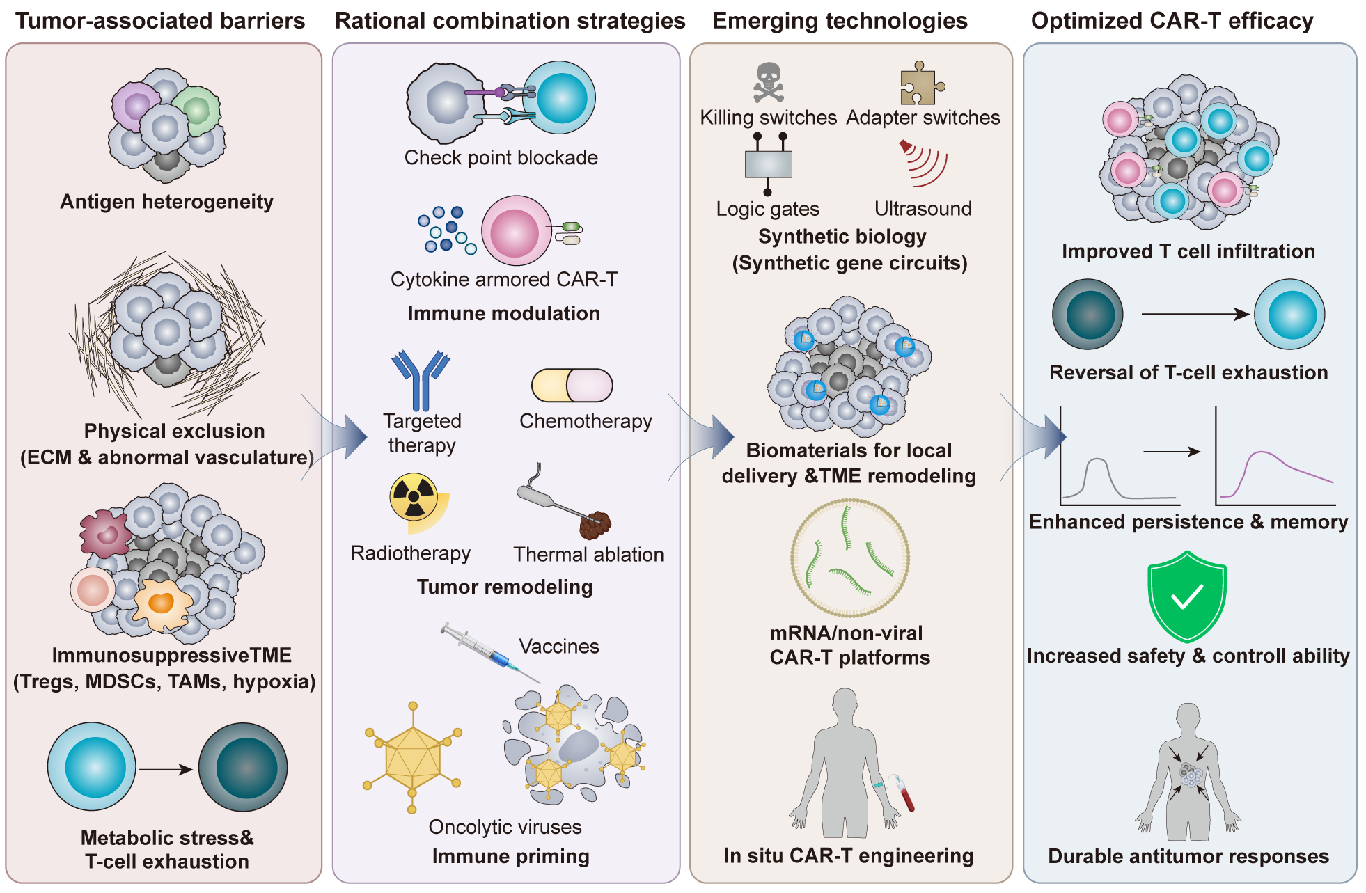
This review focuses on combination strategies to overcome the major barriers limiting CAR-T cell efficacy in solid tumors and discusses how emerging technologies may support the rational development of next-generation therapeutic platforms (Figure 1). Here, we distinguish this review from previous CAR-T solid tumor reviews by adopting a barrier-oriented framework for combination therapy. Rather than simply cataloging individual combinatorial modalities, we organize recent advances around the key biological obstacles that limit CAR-T efficacy in solid tumors, including antigen heterogeneity, impaired trafficking and infiltration, immunosuppressive TME, metabolic dysfunction, T-cell exhaustion, and limited persistence (5, 6).
This framework emphasizes that effective combination strategies should not be viewed as isolated adjuncts but as coordinated interventions designed to restore CAR-T cell function at multiple stages of the antitumor response. Importantly, the barriers encountered by CAR-T cells in solid tumors are highly interconnected rather than independent. Antigen heterogeneity promotes immune escape, inadequate infiltration limits target engagement, and persistent immunosuppressive and metabolic pressures accelerate T-cell dysfunction. Consequently, durable clinical responses will likely require multi-layered therapeutic interventions that simultaneously address several biological barriers rather than single-mechanism solutions. In doing so, this review provides a mechanistic and translational perspective for the rational development of next-generation CAR-T combination therapies for solid tumors.
2. Barriers to CAR-T efficacy in solid tumors
Tumor antigen heterogeneity and on-target/off-tumor toxicity
The immune system can identify and eliminate abnormal cells, maintain cellular homeostasis, and protect the organism from malignant transformation. However, cancer cells employ sophisticated mechanisms to evade immune surveillance (10), allowing uncontrolled proliferation and ultimately leading to life-threatening tumors. In tumor immune evasion, both intra-tumor heterogeneity (ITH) and spatial and temporal heterogeneity significantly limit CAR-T cell recognition and eradication of solid tumor cells (11). ITH refers to the genetic, phenotypic, and functional diversity within an individual tumor (12). For instance, immune cells may effectively target and eliminate subpopulations that express specific neoantigens, while failing to recognize other subclones that lack these antigens. Under selective pressure, immune-resistant clones expand, contributing to tumor progression and therapeutic resistance (13). In addition, spatial and temporal heterogeneity play critical roles in shaping tumor immune evasion, enabling tumor cells to adapt to heterogeneous microenvironmental and systemic conditions (11, 14).
Table 1. Clinical trials of CAR-T cell therapy (ClinicalTrials.gov)
| Target Antigen | National Clinical Trials Identifier | Cancer Type | Trial Phase |
|---|---|---|---|
| HER2 | NCT00924287 | Metastatic cancers | I/II |
| CEACAM5 | NCT01212887 | CEACAM5-positive malignancy | I |
| CAIX | NCT04969354 | Renal cancer | I |
| CLDN18.2 | NCT03874897 | Gastrointestinal cancers | I |
| ROR1 | NCT05694364 | CLL, MCL, ALL, DLBCL, TNBC | I |
| MSLN | NCT06885697 | Mesothelioma | I |
| GD2 | NCT02107963 | GD2-positive solid tumors | I |
| B7-H3, (CD276) | NCT07231081 | B7-H3/CD276-positive advanced solid tumors | I |
| CD70 | NCT05947487 | CD70-positive advanced/metastatic solid tumors | I/II |
| GPC3 | NCT05003895 | Hepatocellular carcinoma or other solid tumor malignancy | I |
Abbreviations: HER2, human epidermal growth factor receptor 2; CEACAM5, carcinoembryonic antigen-related cell adhesion molecule 5; CAIX, carbonic anhydrase IX; CLDN18.2, claudin 18.2; ROR1, receptor tyrosine kinase-like orphan receptor 1; CLL, chronic lymphocytic leukemia; MCL, mantle cell lymphoma; ALL, acute lymphoblastic leukemia; DLBCL, diffuse large B-cell lymphoma; TNBC, triple-negative breast cancer; MSLN, mesothelin; GD2, disialoganglioside 2; B7-H3, B7 homolog 3 (CD276); CD70, cluster of differentiation 70; GPC3, glypican-3.
This can lead to severe adverse effects known as on-target/off-tumor toxicity, as reported in several CAR-T clinical trials against solid tumors (15-17). For example, targeting HER2 has been associated with substantial toxicity, and a clinical trial involving HER2-specific CAR-T cells for colorectal cancer was terminated due to fatal pulmonary and multi-organ failure (NCT00924287) (18). Therefore, these factors collectively make CAR-T therapy more challenging to apply to solid tumors.
Poor trafficking and tumor infiltration
Effective CAR-T therapy requires efficient migration and infiltration into solid tumors. However, solid tumors impede CAR-T cell trafficking through abnormal tumor vasculature, a dense extracellular matrix (ECM), and mismatches between CAR-T cell chemokine receptors and tumor-secreted chemokines (6).
During migration, CAR-T cells initially rely on selectin-ligand interactions to mediate margination and adhesion to the vessel wall. Subsequently, chemokines activate their receptors on CAR-T cells, inducing integrin expression that enables firm adhesion to endothelial cells. Finally, intercellular adhesion molecules (ICAMs), vascular cell adhesion molecules (VCAMs), and chemokines coordinate to facilitate trans-endothelial migration (19). However, tumor vasculature poses major obstacles. Compared with normal vasculature, tumor vessels exhibit structural abnormalities and altered expression of adhesion molecules, including VCAM1 and ICAM1 (20, 21). These abnormalities cause vascular distortion and disorganized blood flow, thereby impairing CAR-T cell adhesion and extravasation (22, 23).
Additionally, the ECM further restricts infiltration. Cancer-associated fibroblasts (CAFs), abundant within the ECM, secrete large amounts of matrix components, including collagen, fibronectin, hyaluronan, glycoproteins, proteoglycans, and tenascin C (24). These components accumulate and become extensively cross-linked by lysyl oxidases (LOXs) and LOX-like proteins (LOXLs), forming a dense fibrotic barrier (25). These physical and biochemical barriers significantly hinder CAR-T cell trafficking and infiltration.
Another key limitation is chemokine-receptor mismatch. Chemokines are essential mediators of T-cell trafficking, with receptors such as CXCR3, CCR5, CCR2, and CCR6 playing critical roles (26, 27). However, solid tumors often secrete insufficient levels of T-cell-recruiting chemokines such as CCL5 (28, 29). Moreover, CAR-T cells derived from peripheral blood frequently lack expression of the corresponding receptors (30), including CCR2 and CXCR4. This mismatch between tumor-derived chemokines and CAR-T cell receptor profiles represents a major barrier to effective tumor homing (31). Furthermore, variability in chemokine expression across tumor types limits the universal adaptability of CAR-T cells.
Immunosuppressive tumor microenvironment
Beyond physical barriers, the TME exerts strong immunosuppressive effects. It contains multiple immunosuppressive immune cell populations and a range of cytokines and chemokines that drive immune cells toward an immunosuppressive phenotype. Regulatory T cells (Tregs), tumor-associated macrophages (TAMs), and myeloid-derived suppressor cells (MDSCs) are the principal immunosuppressive components within the TME (22).
Previous studies have shown that FOXP3-expressing Tregs constitute a major immunosuppressive population in tumors (32, 33). Compared with Tregs in normal tissues, tumor-associated Tregs display enhanced proliferation and activation, expressing high levels of CTLA4, TIGIT, CD25, CCR4, and CCR8 (34, 35). Highly suppressive Treg subsets are further characterized by transcription factors such as BATF and IRF4 (36, 37), as well as co-stimulatory receptors including OX40 and GITR (38). Tregs suppress CAR-T cell function through multiple mechanisms. First, they secrete inhibitory cytokines such as IL-10, TGF-β, and IL-35, which suppress effector T-cell activity (39-41). They also express inhibitory receptors, including TIGIT, LAG-3, and PD-1, thereby reinforcing immunosuppression (42-45). Second, Tregs engage in metabolic competition by consuming essential nutrients. For example, CD39/CD73 expression contributes to an immunosuppressive metabolic environment (46), while high CD25 expression reduces IL-2 levels, a key cytokine for T-cell activation and proliferation (47, 48). Additionally, Tregs promote tryptophan metabolism via IDO, producing immunosuppressive kynurenine (49). They can also suppress effector T cells through CTLA-4-mediated ligand competition (50) and direct cytotoxicity via perforin and granzyme B (51).
TAMs and MDSCs further reinforce immunosuppression. They promote Treg recruitment via chemokines such as CCL17, CCL22, and CCL8 (52) and secrete inhibitory cytokines (e.g., TGF-β, IL-10) while expressing immune checkpoint ligands such as PD-L1, B7-H4, and VISTA (53, 54). Moreover, TAMs contribute to vascular abnormalities (55), whereas MDSCs induce metabolic stress through arginine depletion and oxidative stress (54, 56). Tumor-associated hypoxia and metabolic byproducts further exacerbate immunosuppression and impair T-cell function (57).
CAR-T cell exhaustion and metabolic dysregulation
CAR-T cell exhaustion is a major factor limiting therapeutic efficacy in solid tumors. It is characterized by reduced proliferation, cytokine production, and cytotoxicity, driven by chronic antigen exposure and immunosuppressive signaling (58).
Elevated collagen in the ECM contributes to T-cell exhaustion via LAIR1 signaling, which is activated by CD18-collagen interactions and mediates inhibitory signaling via SHP-1 (59). Persistent antigen stimulation also upregulates inhibitory receptors such as PD-1, CTLA-4, TIM-3, and LAG-3. Both intrinsic and extrinsic signals contribute to this process. Extrinsic suppression arises from inhibitory cytokines produced by Tregs and MDSCs, including IL-10 and TGF-β (60). Furthermore, the metabolically hostile TME impairs T-cell bioenergetics. Abnormal tumor vasculature disrupts oxygen delivery, and rapid tumor growth leads to hypoxia (61). Stabilization of HIF-1α enhances VEGF and adenosine production, suppressing T-cell proliferation and cytotoxicity (62, 63).
Tumor cells also compete with T cells for essential nutrients, including glucose, glutamine, and arginine, while MDSCs and TAMs exacerbate nutrient depletion through arginase-1 (64). Additionally, tumor-derived lactate accumulation acidifies the TME, disrupts oxidative phosphorylation, reduces IFN-γ secretion, and promotes the recruitment of immunosuppressive cells (65). Together, these factors drive metabolic dysfunction and accelerate CAR-T cell exhaustion.
Limited persistence and memory formation
Limited CAR-T cell persistence is a major contributor to weak or transient therapeutic responses in solid tumors (66). In current applications, short-lived effector CAR-T cells predominate, whereas long-term persistence and memory formation remain insufficient.
Co-stimulatory domains shape differentiation outcomes. Second-generation CARs containing CD28 favor effector memory T cells, whereas those with 4-1BB promote central memory T cells with enhanced persistence (67-69). Mitochondrial fitness is a key determinant of T-cell persistence. Effector memory T cells rely on glycolysis, whereas central memory T cells depend on oxidative phosphorylation, which supports long-term survival and reactivation (70). Within the TME, tumor cells exhibit high rates of glycolysis, consuming large amounts of glucose and producing lactate, thereby inducing metabolic stress in CAR-T cells.
This altered metabolic environment, together with cytokine signaling, disrupts mitochondrial oxidative phosphorylation by inhibiting PDH activity and impairing CAR-T cell proliferation and survival. Signaling pathways, such as HIF-1α, further regulate the balance between mTORC1 and AMPK, thereby affecting cellular metabolism, growth, and autophagy (65, 71). Additionally, elevated cytokine levels, including IL-2, can drive CAR-T cells toward terminal effector differentiation, thereby reducing long-term persistence (72). Overall, metabolic dysregulation and cytokine signaling favor effector phenotypes over memory differentiation, limiting sustained antitumor immunity.
Collectively, these barriers form an integrated network of resistance rather than discrete obstacles. For example, poor infiltration limits tumor access, prolongs ineffective antigen exposure, and contributes to exhaustion, while immunosuppressive and metabolically hostile microenvironments further impair persistence and memory formation. This biological interdependence helps explain why single-agent CAR-T strategies have generally shown limited efficacy in solid tumors and supports combination approaches that can simultaneously target multiple resistance mechanisms.
3. Strategies of combination therapy
CAR-T plus immune checkpoint blockade
Because the major barriers limiting CAR-T activity in solid tumors stem from multiple interconnected biological processes, combinatorial approaches have emerged as a rational strategy to simultaneously enhance trafficking, overcome immunosuppression, prevent exhaustion, and improve long-term persistence (73).
Representative ongoing and completed clinical trials evaluating CAR-T combination strategies in solid tumors are summarized in Table 2.
Table 2. Clinical trials of CAR-T combination therapies in solid tumors (ClinicalTrials.gov)
| Target Antigen | National Clinical Trials Identifier | Combination Therapy | Combination Type | Cancer Type |
|---|---|---|---|---|
| Mesothelin | NCT02414269 | Pembrolizumab; cyclophosphamide | PD-1 antibody; lymphodepletion | Malignant pleural mesothelioma, lung cancer, breast cancer |
| EGFRvIII | NCT03726515 | Pembrolizumab | PD-1 antibody | EGFRvIII-positive, MGMT-unmethylated glioblastoma (GBM) |
| IL13Rα2 | NCT04003649 | Nivolumab ± ipilimumab | Immune checkpoint blockade (PD-1 and CTLA-4 antibodies) | Recurrent/refractory glioblastoma (GBM) |
| PSMA | NCT03089203 | Dominant-negative TGF-β receptor (TGFβRDN) | Armored CAR-T cells resistant to TGF-β signaling | Metastatic castration-resistant prostate cancer (mCRPC) |
| GPC3 | NCT05103631; NCT04377932 | IL-15 | IL-15-armored GPC3-targeted CAR-T cells | Hepatocellular carcinoma (HCC) and pediatric solid tumors |
| EGFR | NCT03182816 | CTLA-4 and PD-1 antibodies | Checkpoint inhibitor-secreting CAR-T cells | EGFR-positive advanced solid tumors |
| EGFRvIII | NCT05660369 | T-cell-engaging antibody molecules (TEAMs) | TEAM-secreting CAR-T cells | Glioblastoma (GBM) |
| GD2 | NCT01822652 | Pembrolizumab | PD-1 blockade in iCasp9-engineered GD2 CAR-T cells | Neuroblastoma |
| HER2 | NCT00902044 | Cyclophosphamide, fludarabine | Lymphodepletion | Advanced sarcoma |
| Mesothelin | NCT02159716 | Cyclophosphamide | Lymphodepletion | Epithelial ovarian cancer, malignant pleural mesothelioma, metastatic pancreatic ductal adenocarcinoma |
| PSCA | NCT05805371 | Radiotherapy | Radiotherapy | Prostate cancer |
| CLDN6 | NCT04503278 | CARVac | CAR-T cell-amplifying RNA vaccine | CLDN6-positive solid tumors |
| Mesothelin | NCT01897415 | In situ CAR-T | SS1 CAR-transfected autologous T cells | Metastatic pancreatic ductal adenocarcinoma (mPDAC) |
Abbreviations: EGFRvIII, epidermal growth factor receptor variant III; MGMT, O6-methylguanine-DNA methyltransferase; GBM, glioblastoma; IL13Rα2, interleukin-13 receptor alpha 2; CTLA-4, cytotoxic T-lymphocyte-associated protein 4; PD-1, programmed cell death protein 1; PSMA, prostate-specific membrane antigen; TGF-β, transforming growth factor-beta; mCRPC, metastatic castration-resistant prostate cancer; GPC3, glypican-3; IL-15, interleukin-15; HCC, hepatocellular carcinoma; TEAMs, T-cell-engaging antibody molecules; GD2, disialoganglioside 2; PSCA, prostate stem cell antigen; CARVac, CAR-T cell-amplifying RNA vaccine; CLDN6, claudin 6; mPDAC, metastatic pancreatic ductal adenocarcinoma.
As discussed above, inhibitory receptors such as PD-1, CTLA-4, TIM-3, and LAG-3 play critical roles in T-cell dysfunction (74-77). Accordingly, blockade of the PD-1/PD-L1 and CTLA-4/B7 axes has become a well-established approach and is widely used in clinical practice (78, 79). Compared with CTLA-4 blockade, inhibition of the PD-1/PD-L1 axis is generally associated with a more favorable safety profile (80).
Accumulating evidence indicates that combining ICIs with CAR-T cell therapy yields synergistic antitumor effects (81, 82). In a phase II clinical trial of 44 patients with relapsed/refractory diffuse large B-cell lymphoma (R/R DLBCL), those receiving PD-1 inhibitor maintenance therapy after CAR-T infusion had significantly improved progression-free survival (PFS) (47.26% vs. 26.67%, P = 0.0348) and overall survival (OS) (65.06% vs. 39.29%, P = 0.0227) compared with CAR-T monotherapy. Importantly, no increase in cytokine release syndrome (CRS) or immune effector cell-associated neurotoxicity syndrome (ICANS) was observed (83).
Similarly, emerging studies in solid tumors have highlighted the potential of this combination strategy. In a clinical study of malignant pleural mesothelioma, CAR-T monotherapy achieved a median survival of 17.7 months and a 1-year survival rate of 74%. Notably, patients receiving regional delivery of mesothelin-targeted CAR-T cells in combination with pembrolizumab showed improved outcomes, with a median survival of 23.9 months and a 1-year survival rate of 83% (84).
Mechanistically, ICIs enhance CAR-T cell persistence and function within the TME by alleviating inhibitory signaling, particularly via the PD-1/PD-L1 pathway, thereby reducing T-cell exhaustion and improving durability (85). Beyond restoring effector function, checkpoint blockade may also reshape CAR-T cell differentiation by preserving progenitor-like populations with greater proliferative capacity and long-term persistence. Emerging evidence suggests that maintaining these stem-like states may be particularly important for durable responses in solid tumors, where chronic antigen exposure continuously drives exhaustion programs.
However, therapeutic efficacy depends on several factors. The TME varies substantially across tumor types, and most solid tumors are considered “cold,” which may limit ICIs’ ability to promote CAR-T cell homing and accumulation at tumor sites. In addition, tumor heterogeneity may compromise the efficacy of combination therapy. For instance, PD-1 inhibitor maintenance therapy may have limited benefit when the PD-1/PD-L1 axis is not the dominant inhibitory pathway. Furthermore, a major limitation of early-generation CARs is irreversible CAR-T cell exhaustion. Because ICIs transiently block receptor-ligand interactions, they may not fully reverse an established exhausted state. Lastly, the overall impact of combining CAR-T cells with ICIs on tumor progression or regression is strongly influenced by the timing and route of administration.
CAR-T plus cytokine or costimulatory modulation
Fourth-generation CAR-T cells, also known as armored CAR-T cells, enhance antitumor activity by engineering the expression of cytokines or costimulatory molecules (86). By encoding cytokines, chemokines, or pro-inflammatory ligands (87), armored CAR-T cells can actively remodel the TME and recruit endogenous immune responses. Unlike systemic combination therapies that rely on external administration of immunomodulatory agents, armored CAR-T cells represent a cell-intrinsic combination strategy that delivers therapeutic payloads directly to tumor sites. This localized approach may improve the therapeutic index by concentrating immune activation within the tumor while minimizing systemic exposure.
Armored CAR-T cells are broadly classified into three categories: redirected universal cytokine killer (TRUCK) CAR-T cells, cytokine-regulatory CAR-T cells, and antibody-secreting CAR-T cells. TRUCK CAR-T cells secrete cytokines such as IL-12, IL-18, or IL-15 to enhance proliferation, persistence, and antitumor activity (88, 89). Cytokine-regulatory CAR-T cells are engineered to improve tumor trafficking and infiltration (90). Antibody-secreting CAR-T cells further enhance efficacy by releasing monoclonal antibodies, scFvs, or bispecific T-cell engagers (BiTEs) (91).
Several studies have demonstrated the therapeutic potential of armored CAR-T cells in solid tumors. In a phase I clinical trial (NCT03089203), PSMA-targeted, TGF-β-insensitive armored CAR-T cells showed an acceptable safety profile in patients with metastatic castration-resistant prostate cancer (mCRPC). CRS was observed, and PSA levels decreased by ≥30% in several patients (92). Similarly, IL-15-secreting CAR-T cells have demonstrated enhanced expansion and cytotoxicity. In multiple phase I trials (NCT02905188, NCT02932956, NCT05103631, NCT04377932), IL-15 co-expression significantly improved the in vivo efficacy of GPC3 CAR-T cells, achieving a disease control rate of 66% and an objective response rate of 33% (93). Furthermore, EGFRvIII-targeted CAR-T cells engineered to secrete T-cell-engaging antibody molecules (TEAMs) have shown promising early results, with sustained remissions in some patients (94).
Despite these advances, the clinical application of armored CAR-T cells is limited by increased toxicity, particularly a higher incidence of CRS. Compared with the remarkable achievements in hematologic malignancies, most studies of armored CAR-T cells for solid tumors remain in phase I/II clinical trials. A major challenge is the trade-off between antitumor potency and systemic toxicity. For example, an ongoing phase I clinical trial is evaluating the safety of different dose levels of IL-12-armored CAR-T cells in patients with recurrent MUC16-positive solid tumors. The study reported CRS at all dose levels, requiring clinical management, highlighting the potential risk of severe or even life-threatening toxicity (NCT02498912). Taken together, current strategies should focus on restricting transgene expression to tumor sites using synthetic promoters or inducible gene circuits, which will be essential for successful clinical translation.
CAR-T plus targeted therapy or chemotherapy
Combining CAR-T cell therapy with targeted agents can enhance tumor cell apoptosis through synergistic mechanisms. In addition to immune checkpoint blockade (75, 77, 84, 95, 96), kinase inhibitors (KIs) are another promising class of agents that modulate T-cell activation and reduce inhibitory signaling (97). Many KIs target aberrantly activated pathways, including the MAPK pathway (e.g., avutometinib and mirdametinib) and receptor tyrosine kinase (RTK) signaling (98). Notably, MEK1/2 inhibitors have been shown to attenuate CAR-T cell exhaustion and terminal differentiation induced by tonic signaling and antigen stimulation, thereby enhancing antitumor efficacy (99). Furthermore, specific KIs (e.g., UNC10225387B, UNC10225263A, UNC10112761A) have been reported to enrich for CD45RA⁺CCR7⁺TCF1hi T memory stem cells (TSCM)-like CAR-T cells and promote T-cell expansion (100). These findings suggest that kinase inhibition can improve CAR-T cell persistence and functionality.
Importantly, targeted therapies may serve functions beyond direct tumor cytotoxicity. Increasing evidence indicates that many targeted agents can remodel the tumor ecosystem by altering cytokine production, antigen presentation, stromal interactions, and immune cell recruitment. Consequently, their greatest value in CAR-T combinations may arise from their ability to condition tumors into more permissive immune environments rather than from direct antitumor effects alone.
Lymphodepleting chemotherapy remains a widely adopted strategy to facilitate CAR-T cell engraftment, expansion, and persistence (101). Standard regimens, typically including fludarabine and cyclophosphamide, deplete suppressive immune populations such as Tregs and MDSCs, disrupt tumor-associated barriers, and modulate chemokine signaling to enhance CAR-T cell trafficking (102). Clinical studies have demonstrated improved CAR T-cell expansion and tumor infiltration following lymphodepletion (103, 104). Beyond conventional chemotherapy, recent studies have highlighted the potential of epigenetic modulation to enhance CAR-T cell function. Wang et al. showed that CAR-T cells treated with low-dose decitabine (5-aza-2-deoxycytidine, DAC) exhibit enhanced antitumor activity, proliferation, and cytokine secretion.
Mechanistically, DAC induces DNA hypomethylation-mediated reprogramming, leading to higher expression of memory-associated genes and lower expression of exhaustion-associated genes under both in vitro and in vivo tumor stimulation conditions (105). These findings demonstrate that methylation inhibition can alleviate CAR-T cell exhaustion while promoting maintenance of the memory phenotype and effector function, thereby improving therapeutic durability.
However, chemotherapy may also have detrimental effects on CAR-T cells, including direct cytotoxicity, alterations in T-cell subsets, and increased expression of immune checkpoint molecules (106). Therefore, optimizing treatment regimens and integrating strategies such as targeted signaling modulation or epigenetic reprogramming will be critical to maximize therapeutic efficacy while minimizing adverse effects.
CAR-T plus radiotherapy or thermal ablation
Radiotherapy, a widely used cancer treatment, exerts its antitumor effects primarily by inducing DNA damage, thereby impairing tumor cell proliferation and survival (107). Beyond direct cytotoxicity, radiotherapy can enhance antitumor immunity by recruiting and activating pro-inflammatory immune cells within the irradiated tumor microenvironment, thereby potentiating the efficacy of CAR-T cell therapy (108, 109). Clinical evidence supports combining radiotherapy with CAR-T therapy. A comparative study in patients with relapsed/refractory diffuse large B-cell lymphoma found that bridging radiotherapy (BRT) significantly improved progression-free survival (PFS) and overall survival (OS) compared with CAR-T monotherapy (P = 0.001, P = 0.043), without increasing the incidence of CAR-T-related toxicities during a median follow-up of 35.27 months (110). Similarly, accumulating preclinical and clinical studies indicate that radiotherapy can improve therapeutic outcomes in solid tumors when combined with CAR-T cells.
Mechanistically, radiotherapy enhances CAR-T efficacy through multiple pathways. Radiotherapy may be particularly attractive because it addresses several resistance mechanisms simultaneously. In addition to promoting antigen release and inflammatory signaling, irradiation can transiently normalize tumor vasculature, increase chemokine production, enhance antigen presentation, and facilitate immune cell trafficking. Such pleiotropic effects position radiotherapy as a potentially powerful platform for converting immunologically cold tumors into CAR-T-permissive environments. Ultra-high-dose-rate FLASH radiotherapy has been shown to reprogram macrophage lipid metabolism, thereby reversing tumor-associated immunosuppression (111). Additionally, low-dose irradiation can sensitize antigen-negative tumor cells to TRAIL-mediated apoptosis induced by CAR-T cells, effectively overcoming antigen heterogeneity and reducing tumor relapse (112). Consistently, Quach et al. (113) demonstrated that tumor-targeted radiation administered prior to CAR-T infusion significantly enhances therapeutic efficacy in solid tumors.
These findings collectively support the potential of radiotherapy to overcome key barriers, including antigen heterogeneity and an immunosuppressive TME. Radiotherapy is often used as a bridging therapy while awaiting CAR-T cell manufacturing and infusion. However, despite these promising findings, most current evidence on the combination of CAR-T cells and radiotherapy in solid tumors is limited to case reports, and no standardized dosing protocols have been established. Furthermore, patient-specific factors, irradiation fields, and treatment timing can significantly influence therapeutic outcomes. These limitations highlight the need for further investigation into the interplay between radiotherapy dose and timing, potential toxicities, and clinical outcomes.
In parallel, minimally invasive ablative approaches, including radiofrequency (RF) or microwave ablation (MWA) and cryoablation, have emerged as effective strategies for solid tumor management. Among these, MWA has been widely used for its ability to induce rapid tumor necrosis via localized thermal effects (114, 115). Notably, emerging evidence suggests that MWA can also modulate the tumor microenvironment to enhance CAR-T cell efficacy. In our work, we demonstrated that MWA promoted the polarization of recruited macrophages toward an M1 phenotype and enhanced their phagocytic function. When combined with CV1-secreting sCAR-T cells, which block the CD47-SIRPα “don’t eat me” signal, this strategy further strengthened macrophage-mediated tumor clearance.
Together, these effects amplified antitumor immunity and induced a macrophage-dependent abscopal response against distant tumors with antigenic heterogeneity (8). In addition, combining MWA with AXL-targeted CAR-T cells in non-small cell lung cancer (NSCLC) improved CAR-T cell efficacy by remodeling the TME, alleviating hypoxia, enhancing mitochondrial oxidative metabolism, and promoting differentiation toward a central memory phenotype (9).
Overall, these combination strategies facilitate tumor antigen release, enhance local inflammation, and remodel the TME, thereby promoting antigen spreading, T-cell priming, and improved infiltration into tumor sites (116, 117). Collectively, radiotherapy and ablative therapies are promising approaches to overcome multiple barriers to CAR-T therapy in solid tumors.
CAR-T plus vaccines or oncolytic viruses
Vaccine-boosted CAR-T therapy has emerged as a promising strategy to enhance CAR-T cell function, particularly to address key limitations in solid tumors, including antigen heterogeneity, insufficient activation, and limited persistence. Tumor vaccines can be broadly categorized as whole-cell, genetically engineered, peptide/protein-based, or dendritic cell (DC) vaccines (118). Conceptually, vaccine-enhanced CAR-T therapy can be divided into two main approaches: those that leverage the host immune system to convert an immunosuppressive TME into an immunostimulatory one (119) and those that directly stimulate CAR-T cells through antigen-specific engagement (120).
Recent studies have demonstrated the potential of vaccine-mediated CAR-T activation. Ma et al. developed amphiphile CAR-T ligands (amph-ligands) that traffic to lymph nodes and decorate antigen-presenting cells, thereby priming CAR-T cells within the native lymphoid microenvironment (121). In a phase I/II clinical trial, CLDN6-targeted CAR-T cells, combined with CARVac-mediated in vivo expansion, showed a favorable safety profile and encouraging clinical activity in patients with CLDN6⁺ advanced solid tumors (122). Peptide-based vaccines offer an additional strategy to overcome tumor antigen heterogeneity by enhancing CAR-specific immune activation and sustaining CAR-T cytotoxicity. Preclinical studies have shown that peptide vaccination (e.g., CMVpp65) significantly promotes CAR-T cell proliferation and augments antitumor responses in vivo (123). Similarly, dendritic cell-based vaccines further enhance CAR-T efficacy by presenting tumor antigens and facilitating efficient T-cell priming and activation (124). Despite these encouraging findings, the clinical translation of vaccine-enhanced CAR-T therapy requires careful optimization of antigen selection, vaccine platforms, dosing regimens, and safety considerations.
Oncolytic virotherapy (OV) is another complementary strategy that exerts antitumor effects through selective tumor cell lysis and the induction of systemic antitumor immunity (125, 126). To date, several OVs have been approved for clinical use, including Rigvir (SND005), Oncorine (H101), Imlygic (Talimogene laherparepvec, T-VEC), and Delytact (teserpaturev/G47Δ) (127-130). Recent advances in OV engineering have further strengthened the synergy between OV and CAR-T therapy. For instance, Chen et al. developed a tumor-specific vaccinia virus (TTVΔTK-IL21) expressing IL-21, which demonstrated superior antitumor efficacy when combined with CAR-T therapy compared with monotherapy (131). In addition, Caroline et al. engineered a multifunctional OV (CAdTrio) that simultaneously delivers a CD44v6-targeting BiTE, IL-12, and a PD-L1-blocking antibody, thereby enhancing HER2 CAR-T cytotoxicity in vitro (132).
From a clinical maturity perspective, studies combining CAR-T cells with cancer vaccines remain largely confined in phase I/II clinical trials. To date, no CAR-T plus cancer vaccine combination regimen has been approved for commercial use, partly due to challenges posed by tumor heterogeneity. Similarly, studies combining CAR-T cells with oncolytic viruses (OVs) are mainly limited to preclinical animal models and early-phase clinical trials. Several barriers remain, including the dense extracellular matrix, which impedes intratumoral OV penetration, and the difficulty of in situ delivery to multiple metastatic lesions.
In terms of safety, CAR-T cells combined with cancer vaccines may cause local inflammation at the injection site and low-grade fever. In contrast, CAR-T cells combined with OVs may carry a higher risk of severe CRS and systemic inflammation due to their potent immune-activating mechanisms. Overall, cancer vaccines and OVs can enhance CAR-T therapy by promoting antigen presentation, amplifying immune activation, and remodeling the tumor microenvironment.
These strategies may help overcome key barriers, including antigen heterogeneity, insufficient immune priming, and immunosuppression, thereby improving CAR-T cell persistence and therapeutic efficacy. From a mechanistic perspective, vaccines and oncolytic viruses address a limitation that extends beyond CAR-T cells themselves: the absence of robust endogenous antitumor immunity. By promoting antigen spreading and broadening immune recognition beyond the CAR-targeted antigen, these approaches may reduce the risk of antigen-loss escape and generate more durable systemic immune surveillance. Nevertheless, future studies should focus on optimizing vector design, delivery strategies, and tumor-specific targeting to maximize therapeutic benefit while minimizing off-target effects.
Multi-antigen targeting strategies
Antigen heterogeneity remains a major barrier to CAR-T cell efficacy in solid tumors. Because many solid tumor antigens are heterogeneously expressed and may also be shared with normal tissues, single-antigen CAR-T strategies can promote antigen-negative immune escape or increase the risk of on-target/off-tumor toxicity (5, 6, 11, 14). Accordingly, multi-antigen targeting has emerged as a rational strategy to broaden tumor recognition while improving therapeutic specificity.
Several engineering approaches have been developed to address this challenge. Tandem CARs incorporate two antigen-binding domains within a single CAR construct, allowing a CAR-T cell to recognize either of two tumor antigens. Bicistronic CAR-T cells express two independent CAR molecules from the same vector, enabling parallel recognition of distinct tumor-associated antigens. More recently, trivalent CAR designs have extended this principle by targeting three antigens simultaneously, aiming to reduce antigen-loss relapse in highly heterogeneous tumors. These multi-targeted formats may improve tumor coverage, but their clinical translation requires careful optimization of antigen selection, receptor affinity, signaling strength, and safety.
Logic-gated CAR-T systems provide an additional layer of specificity. “AND-gate” designs require recognition of two antigens before full T-cell activation, thereby reducing the likelihood of attacking normal tissues that express only one target antigen. One representative approach integrates synNotch receptors with CAR constructs, in which engagement of a priming antigen induces CAR expression against a second antigen. This design enables spatially restricted CAR-T activation within tumors expressing the appropriate antigen combination and may reduce on-target/off-tumor toxicity (133, 134).
Conversely, “OR-gate” strategies permit activation through either of two antigens and may be particularly useful for tumors with heterogeneous or unstable antigen expression. “NOT-gate” systems, although less clinically mature, are designed to suppress CAR-T activation in the presence of antigens expressed by normal tissues, thereby improving discrimination between malignant and healthy cells.
Adapter and universal CAR platforms offer another strategy for flexible antigen recognition. Rather than permanently encoding specificity for a single tumor antigen, these systems pair a universal T-cell receptor with externally administered adaptor molecules that bridge CAR-T cells to tumor antigens. By changing the adaptor, the same CAR-T cell product can, in theory, be redirected to different targets. This modular design may enable titratable control of CAR-T activity, sequential targeting of multiple antigens, and rapid interruption of treatment in the event of toxicity. Small-molecule–regulated ON-switch systems further support this concept by enabling reversible, dose-dependent control of CAR-T activity.
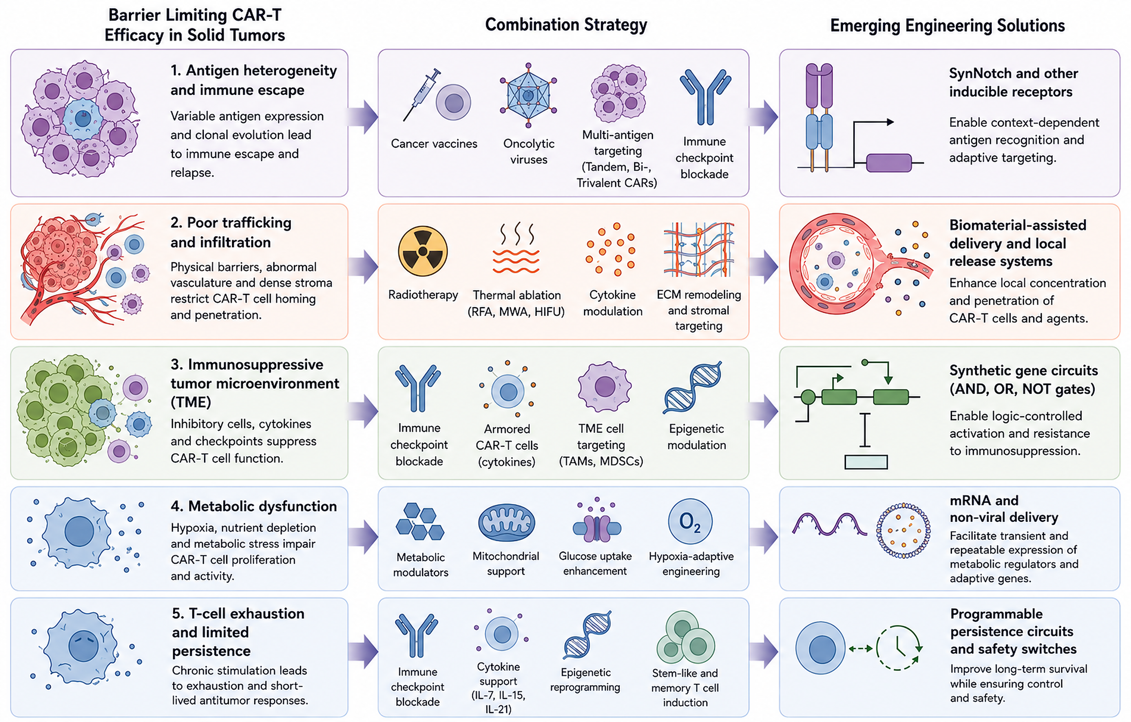
Overall, multi-antigen and adaptive recognition platforms shift CAR-T therapy from static, single-target recognition to programmable tumor sensing. These strategies directly address antigen heterogeneity, antigen-loss escape, and on-target/off-tumor toxicity, making them highly relevant to solid tumors. Future studies should determine which multi-antigen architectures provide the optimal balance among breadth of tumor recognition, functional persistence, manufacturing feasibility, and clinical safety. Collectively, these combination approaches reveal several emerging design principles for successful CAR-T therapy in solid tumors.
First, interventions that simultaneously address multiple resistance mechanisms generally outperform strategies targeting a single barrier.
Second, localized or inducible therapeutic activity appears superior to systemic immune activation because it may improve efficacy while limiting toxicity.
Third, durable responses increasingly depend on preserving stem-like and memory-associated CAR-T states rather than maximizing short-term effector activity. These principles suggest that future CAR-T platforms will likely integrate multiple complementary functions within a single programmable therapeutic system (Figure 2).
4. Emerging technologies enhancing CAR-T combination therapy
Synthetic biology and gene circuit designs
To address the complexity of the TME and reduce on-target/off-tumor toxicity, synthetic biology has emerged as a powerful approach to enhance CAR-T cell function by integrating programmable genetic circuits (133, 134). Recent advances have led to the development of engineered CAR-T systems, including hypoxia-, reactive oxygen species (ROS)-, and pH-responsive CARs, as well as cytokine-sensing designs and immune checkpoint knockout strategies (135, 136).
To improve specificity and minimize off-target effects, Kole T. Roybal et al. designed a combinatorial antigen-sensing circuit that integrates synNotch receptors with CAR constructs, creating a dual-receptor “AND-gate” system. In this design, synNotch activation by a primary antigen induces CAR expression, enabling T-cell activation only when both antigens are present on tumor cells (137).
In addition, synthetic gene circuits have been engineered to reprogram CAR-T cells in response to specific TME features, such as dense ECM, abnormal vasculature, hypoxia, oxidative stress, and elevated protease activity. For example, fusion receptors that combine the extracellular domains of TGF-β receptors with the intracellular signaling domains of IL-2/IL-7 receptors convert inhibitory signals into activating signals, thereby enhancing CAR-T cell survival and function in suppressive environments (138).
To address tumor hypoxia, hypoxia-responsive CAR-T systems have been developed to selectively express CARs under low-oxygen conditions, thereby confining activity to tumor tissues and improving safety (139). Similarly, ON-switch CAR systems enable small-molecule-dependent, titratable, and reversible control of CAR-T cell activity (140). Furthermore, thermal gene switches incorporating heat-shock elements (HSEs) enable spatial and temporal control of CAR-T activation during photothermal stimulation (141).
Collectively, these advances in synthetic biology enable precise, context-dependent control of CAR-T cell activity, improving specificity, safety, and adaptability within complex tumor environments.
CAR-T plus biomaterials
To enhance the therapeutic efficacy of CAR-T cells in solid tumors, advances in biomaterials have provided promising strategies to overcome key barriers, including physical obstruction, the immunosuppressive TME, and treatment-related toxicity (142, 143). Increasing efforts have focused on developing drug delivery systems that enable precise, controlled co-delivery of CAR-T cells and immunostimulatory molecules, thereby improving therapeutic outcomes (144-146). To date, a variety of biomaterial-based platforms have been explored to support CAR-T therapy in solid tumors (147).
To prevent systemic CAR-T cell diffusion and excessive cytokine release, Wang et al. generated GD2-specific CAR-T cells (GD2.CAR-Ts) and delivered them locally to a retinoblastoma (RB) model via an injectable hydrogel system. The study showed that combining GD2.CAR-T cells with sustained interleukin-15 release effectively eliminated RB tumor cells without impairing visual function (148). To overcome the immunosuppressive TME, hydrogel systems integrated with nanoparticles can protect CAR-T cells from immunosuppressive factors while enabling sustained release of cytokines (e.g., IL-2, IL-15) and CAR-T cells, thereby prolonging local activity and enhancing persistence. Similarly, implantable biomaterials facilitate localized administration and create a transient inflammatory niche that supports CAR-T cell expansion and function in vivo. Abigail K. Grosskopf et al. developed a self-assembled injectable biomaterial platform based on polymer-nanoparticle (PNP) hydrogels for CAR-T cell delivery (149). This system functions as a selective barrier that restricts cytokine diffusion while permitting T-cell migration, thereby establishing an optimized microenvironment that supports prolonged CAR-T cell retention and antitumor activity. In addition, abundant fibrotic components, including collagen and hyaluronic acid, contribute to the formation of a dense extracellular matrix (ECM) in solid tumors, which restricts CAR-T cell infiltration into the tumor core. To overcome this physical barrier, biomaterial-based strategies have been developed to deliver or induce the local production of ECM-degrading enzymes. For instance, hyaluronic acid (HA)-based hydrogels have been shown to remodel the ECM, thereby enhancing CAR-T cell infiltration into tumor tissues (150). In addition, biomaterial-based strategies have been explored to simplify CAR-T cell therapy and reduce manufacturing complexity by enabling in vivo CAR-T cell generation. An implantable Multifunctional Alginate Scaffold for T Cell Engineering and Release (MASTER) has been developed, enabling in situ generation and release of functional CAR-T cells in vivo (151).
Moreover, biomaterial platforms have been extended to three-dimensional (3D) in vitro systems, which allow investigation of how the mechanical properties of the TME influence CAR-T cell behavior and serve as preconditioning platforms prior to clinical administration (152, 153).
Despite these advances, most biomaterial-based CAR-T strategies remain in preclinical development. Significant challenges remain, including large-scale manufacturing, batch-to-batch consistency, and long-term safety evaluation. Therefore, further optimization and translational studies are needed to fully realize the clinical potential of biomaterial-assisted CAR-T therapy.
Nonviral or mRNA-based CAR-T delivery
Conventional viral vectors are associated with risks such as CRS, ICANS, insertional mutagenesis, and secondary malignancies (154-156). In contrast, non-viral and mRNA-based CAR-T delivery strategies have emerged as promising alternatives, offering improved safety, flexibility, and cost-effectiveness (157, 158). These strategies make a significant contribution to both ex vivo engineering and in vivo programming.
In ex vivo engineering, T cells are purified from patients, activated, and then transduced with a viral vector. After expansion, cryopreservation, and extensive quality testing (159), the CAR-T cells are infused back into the original patient. However, retroviral and lentiviral vectors are difficult to control and may inadvertently disrupt critical genes or transcriptional regulatory elements. Therefore, non-viral delivery, including transposon systems, mRNA delivery, and genome-editing technologies, plays a pivotal role in ex vivo engineering of CAR-T. Platforms such as PiggyBac and Sleeping Beauty enable stable genomic integration of CAR constructs (160, 161). Early clinical evidence demonstrates detectable CAR-T persistence and clinical responses in NSCLC patients treated with piggyBac-engineered EGFR-CAR T cells (162). Moreover, mRNA-based delivery offers a transient, controllable alternative that avoids genomic integration (163). CAR-encoding mRNA can be introduced into isolated T cells via electroporation and then infused back into the original patient (164).
In the realm of in vivo programming, mRNA is being delivered using nanocarriers, including polymer-based nanoparticles and lipid nanoparticles (LNPs) (165, 166). Notably, targeted LNP technology is the core tool for efficient in vivo CAR-T reprogramming. Building on these advances, in situ CAR-T reprogramming has emerged as a next-generation strategy that enables direct genetic engineering of immune cells in vivo, thereby bypassing ex vivo manufacturing (159, 167, 168). For instance, targeted LNPs enable the in vivo generation of anti-FAP CAR-T cells, thereby reducing cardiac fibrosis and demonstrating potential to remodel the desmoplastic stroma in solid tumors (169). Recent developments in targeted delivery systems, such as CD8-specific LNPs (e.g., L829-tLNP) and myeloid-targeted LNP-based CAR therapies (e.g., MT-302/MT-303), further enable in vivo CAR expression and effective tumor control (170, 171), highlighting their potential to expand CAR-T accessibility and scalability.
Proof-of-concept studies have demonstrated the feasibility of generating functional CAR-T cells in vivo with CD8-targeted vectors (172), and multiple platforms are advancing toward clinical translation (173). Furthermore, mRNA-based approaches have shown favorable safety profiles in early clinical studies, including mesothelin-targeted CAR-T therapy for patients with pancreatic cancer (174).
However, mRNA-based CAR-T strategies remain limited by transient expression and stability, largely determined by structural elements such as the 5′ cap, poly(A) tail, coding sequence, and untranslated regions (UTRs) (175). Consequently, repeated administration may be required, raising concerns about cumulative toxicity. To address these limitations, optimized mRNA engineering strategies, including modified cap structures (e.g., CleanCap®), extended poly(A) tails, and UTR optimization, have been developed (176). Additionally, RNA circularization technologies such as high-yield scarless PIE (Hi-Scarless-PIE) have demonstrated improved CAR expression and antitumor efficacy compared with conventional linear mRNA constructs (177).
Overall, non-viral or mRNA-based CAR-T delivery represents a complementary and evolving strategy in both ex vivo engineering and in vivo programming. These strategies enable the production of safer, more controllable, and scalable CAR-T cells, offering substantial potential for improving therapeutic outcomes in solid tumors.
5. Discussion
Biological Challenges
Despite substantial advances in CAR-T engineering and combination therapy, several biological barriers continue to constrain therapeutic efficacy in solid tumors.
When combined with ICIs, CAR-T cells may enhance CAR-T cell activity but also increase the incidence and severity of immune-related adverse events (irAEs). Importantly, different ICIs have distinct toxicity profiles, further complicating clinical management (178,179). For instance, a phase I trial (NCT03726515) evaluating EGFRvIII-targeted CAR-T cells in combination with pembrolizumab found no significant therapeutic benefit in glioblastoma (180). These findings underscore the need for more precise combinatorial strategies and alternative approaches, such as CRISPR/Cas9-mediated disruption of inhibitory receptors (e.g., PD-1), to enhance efficacy while minimizing toxicity (181).
Similarly, combination strategies that use exogenous cytokines, costimulatory modulation, or kinase inhibitors may exacerbate systemic toxicity because of their narrow therapeutic windows (154, 182). To address this limitation, approaches that enable spatiotemporally controlled activation, such as biomaterial-based delivery systems (183) and in situ expression strategies (e.g., armored CAR-T cells) (184), are actively being explored. These strategies aim to localize therapeutic activity within the TME, thereby reducing off-target effects.
In addition, combining CAR-T therapy with chemotherapy or radiotherapy introduces further complexity. These treatments can trigger innate immune responses by releasing damage-associated molecular patterns (DAMPs) from dying tumor cells (185). This may synergize with CAR-T activity but also increase the risk of systemic inflammation and toxicity. Moreover, higher radiation doses and larger treatment volumes are associated with greater damage to normal tissues (186). To mitigate these risks, engineered CAR-T cells with context-dependent activation, such as those incorporating synthetic gene circuits, have been proposed (187).
Importantly, these challenges are highly interconnected rather than independent. Antigen heterogeneity, inadequate trafficking, immunosuppressive signaling, metabolic dysfunction, and progressive exhaustion collectively drive treatment resistance in solid tumors. Consequently, future therapeutic success will likely depend on combination strategies that simultaneously overcome multiple resistance mechanisms, rather than on addressing individual barriers in isolation.
Translational and Manufacturing Challenges
Beyond biological barriers, the increasing complexity of next-generation CAR-T platforms introduces important translational, manufacturing, and regulatory challenges.
Despite these advances, several challenges remain for emerging platforms. For biomaterial-assisted CAR-T therapy, long-term safety concerns, including immunogenicity and toxicity from degradation byproducts, remain incompletely characterized (188). In parallel, most synthetic gene circuit-based approaches remain in preclinical development and lack standardized protocols for large-scale manufacturing and clinical translation.
Furthermore, critical translational challenges persist across CAR-T combination strategies, including optimizing dosing schedules, identifying predictive biomarkers, and stratifying patients. In situ CAR-T reprogramming also faces unique limitations, as the complexity of the in vivo TME cannot be fully recapitulated in current preclinical models. In addition, delivery-related challenges, including transient mRNA expression that requires repeated dosing, integration risks associated with transposon systems, and suboptimal genome-editing efficiency that may lead to genomic instability, remain to be addressed. The lack of long-term safety data and standardized manufacturing and quality-control frameworks further complicates clinical implementation.
Moreover, as CAR-T products become increasingly sophisticated through the incorporation of multi-antigen targeting, synthetic gene circuits, biomaterials, and programmable control systems, balancing therapeutic potency with manufacturing feasibility and economic sustainability will become a major consideration for widespread clinical adoption.
Toward Precision Combination CAR-T Therapy
An important emerging concept is that solid tumors differ substantially in the dominant mechanisms limiting CAR-T efficacy. While some tumors are primarily characterized by antigen heterogeneity, others are dominated by stromal exclusion, immune suppression, metabolic dysfunction, hypoxia, or inadequate persistence. Consequently, future CAR-T combination strategies will likely require biomarker-guided approaches that match specific therapeutic interventions to the underlying resistance mechanisms in individual tumors. Importantly, successful implementation of precision CAR-T therapy will require biomarkers that are not only prognostic but also actionable, enabling selection of the most appropriate combination strategy for each resistance landscape.
Advances in single-cell sequencing, spatial transcriptomics, multiplex imaging, and circulating biomarker analyses are providing unprecedented insights into tumor architecture and immune landscapes. These technologies may help identify dominant barriers within individual tumors and support the rational selection of combination therapies. In this context, precision-based frameworks that integrate molecular profiling with advanced CAR engineering may improve therapeutic efficacy while minimizing unnecessary toxicity.
Rather than applying uniform treatment strategies across all patients, future CAR-T therapy may increasingly rely on individualized combinations tailored to the specific biological characteristics of each tumor ecosystem.
Future Outlook
Overall, although CAR-T combination therapies have shown significant promise in overcoming key barriers in solid tumors, substantial challenges remain. Continued efforts to integrate advances in cellular engineering, immunology, biomaterials, and clinical oncology will be essential to developing safer, more effective, and more controllable next-generation CAR-T therapies for solid tumors.
Collectively, the field is transitioning from conventional CAR-T cell therapy toward increasingly programmable and adaptive cellular therapeutics that can sense, respond to, and remodel the tumor microenvironment. Combination strategies that integrate immune checkpoint blockade, cytokine modulation, targeted and epigenetic therapies, radiotherapy, thermal ablation, vaccines, oncolytic viruses, biomaterials, and emerging engineering technologies are beginning to address the multifaceted barriers that have historically limited success in solid tumors.
Importantly, advances in synthetic biology, multi-antigen targeting platforms, non-viral engineering technologies, and in situ CAR-T generation are expanding the therapeutic landscape, enabling more precise, controllable, and scalable cellular immunotherapies. The ultimate goal is to develop CAR-T platforms that simultaneously overcome antigen escape, improve tumor trafficking, resist immunosuppression, maintain metabolic fitness, and provide programmable safety mechanisms. Achieving this objective will require continued interdisciplinary collaboration across immunology, synthetic biology, bioengineering, biomaterials science, and clinical oncology. These efforts are expected to accelerate the translation of next-generation CAR-T therapies into durable, broadly applicable treatment options for patients with solid tumors.
References
1. Tang L, Huang Z, Mei H, Hu Y. Immunotherapy in hematologic malignancies: achievements, challenges and future prospects. Signal Transduct Target Ther. 2023;8(1):306. https://doi.org/10.1038/s41392-023-01521-5
2. Mulvey A, Trueb L, Coukos G, Arber C. Novel strategies to manage CAR-T cell toxicity. Nat Rev Drug Discov. 2025;24(5):379–397. https://doi.org/10.1038/s41573-024-01100-5
3. Bock TJ, Colonne CK, Fiorenza S, Turtle CJ. Outcome correlates of approved CD19-targeted CAR T cells for large B cell lymphoma. Nat Rev Clin Oncol. 2025;22(4):241–261. https://doi.org/10.1038/s41571-025-00992-5
4. Sterner RC, Sterner RM. CAR-T cell therapy: current limitations and potential strategies. Blood Cancer J. 2021;11(4):69. https://doi.org/10.1038/s41408-021-00459-7
5. Hou AJ, Chen LC, Chen YY. Navigating CAR-T cells through the solid-tumour microenvironment. Nat Rev Drug Discov. 2021;20(7):531–550. https://doi.org/10.1038/s41573-021-00189-2
6. Uslu U, June CH. Beyond the blood: expanding CAR T cell therapy to solid tumors. Nat Biotechnol. 2025;43(4):506–515. https://doi.org/10.1038/s41587-024-02446-2
7. Albarrán-Fernández V, Angelats L, Delgado J, Gros A, Urbano-Ispizua Á, Guedan S, et al. Unlocking the potential of engineered immune cell therapy for solid tumors. Nat Commun. 2025;16(1):1144. https://doi.org/10.1038/s41467-025-56527-0
8. Cao B, Liu M, Xiao Z, Leng D, Zhou Y, Zhang Z, et al. CV1-secreting sCAR-T cells potentiate the abscopal effect of microwave ablation in heterogeneous tumors. Cell Rep Med. 2025;6(2):101965. https://doi.org/10.1016/j.xcrm.2025.101965
9. Cao B, Liu M, Wang L, Zhu K, Cai M, Chen X, et al. Remodelling of tumour microenvironment by microwave ablation potentiates immunotherapy of AXL-specific CAR T cells against non-small cell lung cancer. Nat Commun. 2022;13(1):6203. https://doi.org/10.1038/s41467-022-33968-5
10. Galassi C, Chan TA, Vitale I, Galluzzi L. The hallmarks of cancer immune evasion. Cancer Cell. 2024;42(11):1825–1863. https://doi.org/10.1016/j.ccell.2024.09.010
11. Roerden M, Spranger S. Cancer immune evasion, immunoediting and intratumour heterogeneity. Nat Rev Immunol. 2025;25(5):353–369. https://doi.org/10.1038/s41577-024-01111-8
12. Vendramin R, Litchfield K, Swanton C. Cancer evolution: Darwin and beyond. EMBO J. 2021;40(18):e108389. https://doi.org/10.15252/embj.2021108389
13. Milo I, Bedora-Faure M, Garcia Z, Thibaut R, Périé L, Shakhar G, et al. The immune system profoundly restricts intratumor genetic heterogeneity. Sci Immunol. 2018;3(29):eaat1435. https://doi.org/10.1126/sciimmunol.aat1435
14. Tufail M, Jiang CH, Li N. Immune evasion in cancer: mechanisms and cutting-edge therapeutic approaches. Signal Transduct Target Ther. 2025;10(1):227. https://doi.org/10.1038/s41392-025-02280-1
15. Morgan RA, Yang JC, Kitano M, Dudley ME, Laurencot CM, Rosenberg SA. Case report of a serious adverse event following the administration of T cells transduced with a chimeric antigen receptor recognizing ERBB2. Mol Ther. 2010;18(4):843–851. https://doi.org/10.1038/mt.2010.24
16. Lamers CH, Sleijfer S, van Steenbergen S, van Elzakker P, van Krimpen B, Groot C, et al. Treatment of metastatic renal cell carcinoma with CAIX CAR-engineered T cells: clinical evaluation and management of on-target toxicity. Mol Ther. 2013;21(4):904–912. https://doi.org/10.1038/mt.2013.17
17. Thistlethwaite FC, Gilham DE, Guest RD, Rothwell DG, Pillai M, Burt DJ, et al. The clinical efficacy of first-generation carcinoembryonic antigen (CEACAM5)-specific CAR T cells is limited by poor persistence and transient pre-conditioning-dependent respiratory toxicity. Cancer Immunol Immunother. 2017;66(11):1425–1436. https://doi.org/10.1007/s00262-017-2034-7
18. Ahmed N, Brawley VS, Hegde M, Robertson C, Ghazi A, Gerken C, et al. Human epidermal growth factor receptor 2 (HER2)-specific chimeric antigen receptor-modified T cells for the immunotherapy of HER2-positive sarcoma. J Clin Oncol. 2015;33(15):1688–1696. https://doi.org/10.1200/JCO.2014.58.0225
19. Ley K, Laudanna C, Cybulsky MI, Nourshargh S. Getting to the site of inflammation: the leukocyte adhesion cascade updated. Nat Rev Immunol. 2007;7(9):678–689. https://doi.org/10.1038/nri2156
20. Bergers G, Benjamin LE. Tumorigenesis and the angiogenic switch. Nat Rev Cancer. 2003;3(6):401–410. https://doi.org/10.1038/nrc1093
21. Ma W, Wang Y, Zhang R, Yang F, Zhang D, Huang M, et al. Targeting PAK4 to reprogram the vascular microenvironment and improve CAR-T immunotherapy for glioblastoma. Nat Cancer. 2021;2(1):83–97. https://doi.org/10.1038/s43018-020-00147-8
22. Lamplugh ZL, Wellhausen N, June CH, Fan Y. Microenvironmental regulation of solid tumour resistance to CAR T cell therapy. Nat Rev Immunol. 2026;26(3):230–248. Epub 2025 Oct 14. https://doi.org/10.1038/s41577-025-01229-3
23. Huinen ZR, Huijbers EJM, van Beijnum JR, Nowak-Sliwinska P, Griffioen AW. Anti-angiogenic agents—overcoming tumour endothelial cell anergy and improving immunotherapy outcomes. Nat Rev Clin Oncol. 2021;18(8):527–540. https://doi.org/10.1038/s41571-021-00496-y
24. Cho Y, Doh J. The extracellular matrix in solid tumor immunotherapy. Trends Immunol. 2024;45(9):705–714. https://doi.org/10.1016/j.it.2024.07.009
25. Smith-Mungo LI, Kagan HM. Lysyl oxidase: properties, regulation and multiple functions in biology. Matrix Biol. 1998;16(7):387–398. https://doi.org/10.1016/S0945-053X(98)90012-9
26. Vilgelm AE, Richmond A. Chemokines modulate immune surveillance in tumorigenesis, metastasis, and response to immunotherapy. Front Immunol. 2019;10:333. https://doi.org/10.3389/fimmu.2019.00333
27. Harlin H, Meng Y, Peterson AC, Zha Y, Tretiakova M, Slingluff C, et al. Chemokine expression in melanoma metastases associated with CD8+ T-cell recruitment. Cancer Res. 2009;69(7):3077–3085. https://doi.org/10.1158/0008-5472.CAN-08-2281
28. Tian Y, Zhang L, Ping Y, Zhang Z, Yao C, Shen C, et al. CCR5 and IL-12 co-expression in CAR T cells improves antitumor efficacy by reprogramming tumor microenvironment in solid tumors. Cancer Immunol Immunother. 2025;74(2):55. https://doi.org/10.1007/s00262-024-03909-w
29. Mowat C, Mosley SR, Namdar A, Schiller D, Baker K. Anti-tumor immunity in mismatch repair-deficient colorectal cancers requires type I IFN-driven CCL5 and CXCL10. J Exp Med. 2021;218(9):e20210108. https://doi.org/10.1084/jem.20210108
30. Liu G, Rui W, Zhao X, Lin X. Enhancing CAR-T cell efficacy in solid tumors by targeting the tumor microenvironment. Cell Mol Immunol. 2021;18(5):1085–1095. https://doi.org/10.1038/s41423-021-00655-2
31. Foeng J, Comerford I, McColl SR. Harnessing the chemokine system to home CAR-T cells into solid tumors. Cell Rep Med. 2022;3(3):100543. https://doi.org/10.1016/j.xcrm.2022.100543
32. Hori S. FOXP3 as a master regulator of Treg cells. Nat Rev Immunol. 2021;21(10):618–619. https://doi.org/10.1038/s41577-021-00598-9
33. Tanaka A, Sakaguchi S. Regulatory T cells in cancer immunotherapy. Cell Res. 2017;27(1):109–118. https://doi.org/10.1038/cr.2016.151
34. Imianowski CJ, Chen Q, Workman CJ, Vignali DAA. Regulatory T cells in the tumour microenvironment. Nat Rev Cancer. 2025;25(9):703–722. https://doi.org/10.1038/s41568-025-00832-9
35. Sun L, Su Y, Jiao A, Wang X, Zhang B. T cells in health and disease. Signal Transduct Target Ther. 2023;8(1):235. https://doi.org/10.1038/s41392-023-01471-y
36. Itahashi K, Irie T, Yuda J, Kumagai S, Tanegashima T, Lin YT, et al. BATF epigenetically and transcriptionally controls the activation program of regulatory T cells in human tumors. Sci Immunol. 2022;7(76):eabk0957. https://doi.org/10.1126/sciimmunol.abk0957
37. Alvisi G, Brummelman J, Puccio S, Mazza EM, Tomada EP, Losurdo A, et al. IRF4 instructs effector Treg differentiation and immune suppression in human cancer. J Clin Invest. 2020;130(6):3137–3150. https://doi.org/10.1172/JCI130426
38. Crawford A, Angelosanto JM, Kao C, Doering TA, Odorizzi PM, Barnett BE, et al. Molecular and transcriptional basis of CD4+ T cell dysfunction during chronic infection. Immunity. 2014;40(2):289–302. https://doi.org/10.1016/j.immuni.2014.01.005
39. Gunderson AJ, Yamazaki T, McCarty K, Fox N, Phillips M, Alice A, et al. TGFβ suppresses CD8+ T cell expression of CXCR3 and tumor trafficking. Nat Commun. 2020;11(1):1749. https://doi.org/10.1038/s41467-020-15404-8
40. Sawant DV, Yano H, Chikina M, Zhang Q, Liao M, Liu C, et al. Adaptive plasticity of IL-10+ and IL-35+ Treg cells cooperatively promotes tumor T cell exhaustion. Nat Immunol. 2019;20(6):724–735. https://doi.org/10.1038/s41590-019-0346-9
41. Wei X, Zhang J, Gu Q, Huang M, Zhang W, Guo J, et al. Reciprocal expression of IL-35 and IL-10 defines two distinct effector Treg subsets that are required for maintenance of immune tolerance. Cell Rep. 2017;21(7):1853–1869. https://doi.org/10.1016/j.celrep.2017.10.090
42. Cillo AR, Cardello C, Shan F, Karapetyan L, Kunning S, Sander C, et al. Blockade of LAG-3 and PD-1 leads to co-expression of cytotoxic and exhaustion gene modules in CD8+ T cells to promote antitumor immunity. Cell. 2024;187(16):4373–4388.e15. https://doi.org/10.1016/j.cell.2024.06.036
43. Anderson AC, Joller N, Kuchroo VK. Lag-3, Tim-3, and TIGIT: Co-inhibitory receptors with specialized functions in immune regulation. Immunity. 2016;44(5):989–1004. https://doi.org/10.1016/j.immuni.2016.05.001
44. Chauvin JM, Zarour HM. TIGIT in cancer immunotherapy. J Immunother Cancer. 2020;8(2):e000957. https://doi.org/10.1136/jitc-2020-000957
45. Huang CT, Workman CJ, Flies D, Pan X, Marson AL, Zhou G, et al. Role of LAG-3 in regulatory T cells. Immunity. 2004;21(4):503–513. https://doi.org/10.1016/j.immuni.2004.08.010
46. Deaglio S, Dwyer KM, Gao W, Friedman D, Usheva A, Erat A, et al. Adenosine generation catalyzed by CD39 and CD73 expressed on regulatory T cells mediates immune suppression. J Exp Med. 2007;204(6):1257–1265. https://doi.org/10.1084/jem.20062512
47. Busse D, de la Rosa M, Hobiger K, Thurley K, Flossdorf M, Scheffold A, et al. Competing feedback loops shape IL-2 signaling between helper and regulatory T lymphocytes in cellular microenvironments. Proc Natl Acad Sci U S A. 2010;107(7):3058–3063. https://doi.org/10.1073/pnas.0812851107
48. Barthlott T, Moncrieffe H, Veldhoen M, Atkins CJ, Christensen J, O'Garra A, et al. CD25+ CD4+ T cells compete with naive CD4+ T cells for IL-2 and exploit it for the induction of IL-10 production. Int Immunol. 2005;17(3):279–288. https://doi.org/10.1093/intimm/dxh207
49. Seo SK, Kwon B. Immune regulation through tryptophan metabolism. Exp Mol Med. 2023;55(7):1371–1379. https://doi.org/10.1038/s12276-023-01028-7
50. Kennedy PT, Saulters EL, Duckworth AD, Lim YJ, Woolley JF, Slupsky JR, et al. Soluble CTLA-4 attenuates T cell activation and modulates anti-tumor immunity. Mol Ther. 2024;32(2):457–468. https://doi.org/10.1016/j.ymthe.2023.11.028
51. Volpe E, Sambucci M, Battistini L, Borsellino G. Fas-Fas ligand: checkpoint of T cell functions in multiple sclerosis. Front Immunol. 2016;7:382. https://doi.org/10.3389/fimmu.2016.00382
52. Cinier J, Hubert M, Besson L, Di Roio A, Rodriguez C, Lombardi V, et al. Recruitment and expansion of Treg cells in the tumor environment—how to target them? Cancers (Basel). 2021;13(8):1850. https://doi.org/10.3390/cancers13081850
53. Noy R, Pollard JW. Tumor-associated macrophages: from mechanisms to therapy. Immunity. 2014;41(1):49–61. https://doi.org/10.1016/j.immuni.2014.06.010
54. Veglia F, Perego M, Gabrilovich D. Myeloid-derived suppressor cells coming of age. Nat Immunol. 2018;19(2):108–119. https://doi.org/10.1038/s41590-017-0022-x
55. Pathria P, Louis TL, Varner JA. Targeting tumor-associated macrophages in cancer. Trends Immunol. 2019;40(4):310–327. https://doi.org/10.1016/j.it.2019.02.003
56. Veglia F, Sanseviero E, Gabrilovich DI. Myeloid-derived suppressor cells in the era of increasing myeloid cell diversity. Nat Rev Immunol. 2021;21(8):485–498. https://doi.org/10.1038/s41577-020-00490-y
57. Labanieh L, Mackall CL. CAR immune cells: design principles, resistance and the next generation. Nature. 2023;614:635–648. https://doi.org/10.1038/s41586-023-05707-3
58. Kouro T, Himuro H, Sasada T. Exhaustion of CAR T cells: potential causes and solutions. J Transl Med. 2022;20(1):239. https://doi.org/10.1186/s12967-022-03442-3
59. Peng DH, Rodriguez BL, Diao L, Chen L, Wang J, Byers LA, et al. Collagen promotes anti-PD-1/PD-L1 resistance in cancer through LAIR1-dependent CD8+ T cell exhaustion. Nat Commun. 2020;11(1):4520. https://doi.org/10.1038/s41467-020-18298-8
60. Jiang Y, Li Y, Zhu B. T-cell exhaustion in the tumor microenvironment. Cell Death Dis. 2015;6(6):e1792. https://doi.org/10.1038/cddis.2015.162
61. Barsoum IB, Koti M, Siemens DR, Graham CH. Mechanisms of hypoxia-mediated immune escape in cancer. Cancer Res. 2014;74(24):7185–7190. https://doi.org/10.1158/0008-5472.CAN-14-2598
62. Shi LZ, Wang R, Huang G, Vogel P, Neale G, Green DR, et al. HIF1alpha-dependent glycolytic pathway orchestrates a metabolic checkpoint for the differentiation of TH17 and Treg cells. J Exp Med. 2011;208(7):1367–1376. https://doi.org/10.1084/jem.20110278
63. Wu Q, You L, Nepovimova E, Heger Z, Wu W, Kuca K, et al. Hypoxia-inducible factors: master regulators of hypoxic tumor immune escape. J Hematol Oncol. 2022;15(1):77. https://doi.org/10.1186/s13045-022-01292-6
64. Cheng K, Cai N, Zhu J, Yang X, Liang H, Zhang W. Tumor-associated macrophages in liver cancer: from mechanisms to therapy. Cancer Commun (Lond). 2022;42(11):1112–1140. https://doi.org/10.1002/cac2.12345
65. Beckermann KE, Dudzinski SO, Rathmell JC. Dysfunctional T cell metabolism in the tumor microenvironment. Cytokine Growth Factor Rev. 2017;35:7–14. https://doi.org/10.1016/j.cytogfr.2017.04.003
66. Ai K, Liu B, Chen X, Huang C, Yang L, Zhang W, et al. Optimizing CAR-T cell therapy for solid tumors: current challenges and potential strategies. J Hematol Oncol. 2024;17(1):105. https://doi.org/10.1186/s13045-024-01625-7
67. Chen L, Flies DB. Molecular mechanisms of T cell co-stimulation and co-inhibition. Nat Rev Immunol. 2013;13(4):227–242. https://doi.org/10.1038/nri3405
68. Kawalekar OU, O'Connor RS, Fraietta JA, Guo L, McGettigan SE, Posey AD Jr, et al. Distinct signaling of coreceptors regulates specific metabolism pathways and impacts memory development in CAR T cells. Immunity. 2016;44(2):380–390. https://doi.org/10.1016/j.immuni.2016.01.021
69. Long AH, Haso WM, Shern JF, Wanhainen KM, Murgai M, Ingaramo M, et al. 4-1BB costimulation ameliorates T cell exhaustion induced by tonic signaling of chimeric antigen receptors. Nat Med. 2015;21(6):581–590. https://doi.org/10.1038/nm.3838
70. Xu Y, Chaudhury A, Zhang M, Savoldo B, Metelitsa LS, Rodgers J, et al. Glycolysis determines dichotomous regulation of T cell subsets in hypoxia. J Clin Invest. 2016;126(7):2678–2688. https://doi.org/10.1172/JCI85834
71. Sauer S, Bruno L, Hertweck A, Finlay D, Leleu M, Spivakov M, et al. T cell receptor signaling controls Foxp3 expression via PI3K, Akt, and mTOR. Proc Natl Acad Sci U S A. 2008;105(22):7797–7802. https://doi.org/10.1073/pnas.0800928105
72. Ross SH, Cantrell DA. Signaling and function of interleukin-2 in T lymphocytes. Annu Rev Immunol. 2018;36:411–433. https://doi.org/10.1146/annurev-immunol-042617-053352
73. Uslu U, Castelli S, June CH. CAR T cell combination therapies to treat cancer. Cancer Cell. 2024;42(8):1319–1325. https://doi.org/10.1016/j.ccell.2024.07.002
74. Day CL, Kaufmann DE, Kiepiela P, Brown JA, Moodley ES, Reddy S, et al. PD-1 expression on HIV-specific T cells is associated with T-cell exhaustion and disease progression. Nature. 2006;443(7109):350–354. https://doi.org/10.1038/nature05115
75. Agarwal S, Aznar MA, Rech AJ, Good CR, Kuramitsu S, Da T, et al. Deletion of the inhibitory co-receptor CTLA-4 enhances and invigorates chimeric antigen receptor T cells. Immunity. 2023;56(10):2388–2407.e9. https://doi.org/10.1016/j.immuni.2023.09.001
76. Das M, Zhu C, Kuchroo VK. Tim-3 and its role in regulating anti-tumor immunity. Immunol Rev. 2017;276(1):97–111. https://doi.org/10.1111/imr.12520
77. Aggarwal V, Workman CJ, Vignali DAA. LAG-3 as the third checkpoint inhibitor. Nat Immunol. 2023;24(9):1415–1422. https://doi.org/10.1038/s41590-023-01569-z
78. Sun C, Mezzadra R, Schumacher TN. Regulation and function of the PD-L1 checkpoint. Immunity. 2018;48(3):434–452. https://doi.org/10.1016/j.immuni.2018.03.014
79. Hodi FS, O'Day SJ, McDermott DF, Weber RW, Sosman JA, Haanen JB, et al. Improved survival with ipilimumab in patients with metastatic melanoma. N Engl J Med. 2010;363(8):711–723. https://doi.org/10.1056/NEJMoa1003466
80. Khoja L, Day D, Wei-Wu Chen T, Siu LL, Hansen AR. Tumour- and class-specific patterns of immune-related adverse events of immune checkpoint inhibitors: a systematic review. Ann Oncol. 2017;28(10):2377–2385. https://doi.org/10.1093/annonc/mdx286
81. Cherkassky L, Morello A, Villena-Vargas J, Feng Y, Dimitrov DS, Jones DR, et al. Human CAR T cells with cell-intrinsic PD-1 checkpoint blockade resist tumor-mediated inhibition. J Clin Invest. 2016;126(8):3130–3144. https://doi.org/10.1172/JCI83092
82. Gray KD, McCloskey JE, Vedvyas Y, Kalloo OR, Eshaky SE, Yang Y, et al. PD1 blockade enhances ICAM1-directed CAR T therapeutic efficacy in advanced thyroid cancer. Clin Cancer Res. 2020;26(22):6003–6016. https://doi.org/10.1158/1078-0432.CCR-20-1523
83. Mu J, Deng H, Lyu C, Yuan J, Li Q, Wang J, et al. Efficacy of programmed cell death 1 inhibitor maintenance therapy after combined treatment with programmed cell death 1 inhibitors and anti-CD19-chimeric antigen receptor T cells in patients with relapsed/refractory diffuse large B-cell lymphoma and high tumor burden. Hematol Oncol. 2023;41(2):275–284. https://doi.org/10.1002/hon.2981
84. Adusumilli PS, Zauderer MG, Rivière I, Solomon SB, Rusch VW, O'Cearbhaill RE, et al. A phase I trial of regional mesothelin-targeted CAR T-cell therapy in patients with malignant pleural disease, in combination with the anti-PD-1 agent pembrolizumab. Cancer Discov. 2021;11(11):2748–2763. https://doi.org/10.1158/2159-8290.CD-21-0407
85. Ni JJ, Zhang ZZ, Ge MJ, Chen JY, Zhuo W. Immune-based combination therapy to convert immunologically cold tumors into hot tumors: an update and new insights. Acta Pharmacol Sin. 2023;44(2):288–307. https://doi.org/10.1038/s41401-022-00953-z
86. Tang L, Pan S, Wei X, Xu X, Wei Q. Arming CAR-T cells with cytokines and more: innovations in the fourth-generation CAR-T development. Mol Ther. 2023;31(11):3146–3162. https://doi.org/10.1016/j.ymthe.2023.09.021
87. Prasad K, Cross RS, Jenkins MR. Progress in the development of cytokine armoured CAR T cells. Nat Rev Immunol. 2026. https://doi.org/10.1038/s41577-026-01280-8
88. Liu Y, Di S, Shi B, Zhang H, Wang Y, Wu X, et al. Armored inducible expression of IL-12 enhances antitumor activity of glypican-3-targeted chimeric antigen receptor-engineered T cells in hepatocellular carcinoma. J Immunol. 2019;203(1):198–207. https://doi.org/10.4049/jimmunol.1800033
89. Ng BD, Rajagopalan A, Kousa AI, Fischman JS, Chen S, Massa A, et al. IL-18-secreting multiantigen targeting CAR T cells eliminate antigen-low myeloma in an immunocompetent mouse model. Blood. 2024;144(2):171–186. https://doi.org/10.1182/blood.2023022293
90. Bell M, Gottschalk S. Engineered cytokine signaling to improve CAR T cell effector function. Front Immunol. 2021;12:684642. https://doi.org/10.3389/fimmu.2021.684642
91. Rafiq S, Yeku OO, Jackson HJ, Purdon TJ, van Leeuwen DG, Drakes DJ, et al. Targeted delivery of a PD-1-blocking scFv by CAR-T cells enhances anti-tumor efficacy in vivo. Nat Biotechnol. 2018;36(9):847–856. https://doi.org/10.1038/nbt.4195
92. Narayan V, Barber-Rotenberg JS, Jung IY, Lacey SF, Rech AJ, Davis MM, et al. PSMA-targeting TGFβ-insensitive armored CAR T cells in metastatic castration-resistant prostate cancer: a phase 1 trial. Nat Med. 2022;28(4):724–734. https://doi.org/10.1038/s41591-022-01726-1
93. Steffin D, Ghatwai N, Montalbano A, Rathi P, Courtney AN, Arnett AB, et al. Interleukin-15-armoured GPC3 CAR T cells for patients with solid cancers. Nature. 2025;637(8047):940–946. https://doi.org/10.1038/s41586-024-08261-8
94. Choi BD, Gerstner ER, Frigault MJ, Leick MB, Mount CW, Balaj L, et al. Intraventricular CARv3-TEAM-E T cells in recurrent glioblastoma. N Engl J Med. 2024;390(14):1290–1298. https://doi.org/10.1056/NEJMoa2314390
95. Wang DR, Wu XL, Sun YL. Therapeutic targets and biomarkers of tumor immunotherapy: response versus non-response. Signal Transduct Target Ther. 2022;7(1):331. https://doi.org/10.1038/s41392-022-01136-2
96. Heczey A, Louis CU, Savoldo B, Dakhova O, Durett A, Grilley B, et al. CAR T cells administered in combination with lymphodepletion and PD-1 inhibition to patients with neuroblastoma. Mol Ther. 2017;25(9):2214–2224. https://doi.org/10.1016/j.ymthe.2017.05.012
97. Zheng W, Jones LL, Geiger TL. Modulation of PI3K signaling to improve CAR T cell function. Oncotarget. 2018;9(88):35807–35808. https://doi.org/10.18632/oncotarget.26334
98. Roskoski R Jr. Properties of FDA-approved small molecule protein kinase inhibitors: a 2026 update. Pharmacol Res. 2026;224:108107. https://doi.org/10.1016/j.phrs.2026.108107
99. Wang X, Tao X, Chen P, Jiang P, Li W, Chang H, et al. MEK inhibition prevents CAR-T cell exhaustion and differentiation via downregulation of c-Fos and JunB. Signal Transduct Target Ther. 2024;9(1):293. https://doi.org/10.1038/s41392-024-01986-y
100. Song F, Tsahouridis O, Stucchi S, Walhart T, Mendell S, Hardy PB, et al. A multi-kinase inhibitor screen identifies inhibitors preserving stem-cell-like chimeric antigen receptor T cells. Nat Immunol. 2025;26(2):279–293. https://doi.org/10.1038/s41590-024-02042-1
101. Forsberg M, Konopleva M. AML treatment: conventional chemotherapy and emerging novel agents. Trends Pharmacol Sci. 2024;45(5):430–448. https://doi.org/10.1016/j.tips.2024.03.005
102. Hirayama AV, Gauthier J, Hay KA, Voutsinas JM, Wu Q, Gooley T, et al. The response to lymphodepletion impacts PFS in patients with aggressive non-Hodgkin lymphoma treated with CD19 CAR T cells. Blood. 2019;133(17):1876–1887. https://doi.org/10.1182/blood-2018-11-887067
103. Hegde M, Navai S, DeRenzo C, Joseph SK, Sanber K, Wu M, et al. Autologous HER2-specific CAR T cells after lymphodepletion for advanced sarcoma: a phase 1 trial. Nat Cancer. 2024;5(6):880–894. https://doi.org/10.1038/s43018-024-00749-6
104. Barber-Rotenberg JS, Haas AR, Aggarwal C, O’Hara M, Hexner E, Pequignot E, et al. Phase 1 study of autologous T cells bearing fully human chimeric antigen receptors targeting mesothelin in mesothelin-expressing cancers. Mol Ther. 2026;34(5):2653-2665. https://doi.org/10.1016/j.ymthe.2026.01.021
105. Wang Y, Tong C, Dai H, Wu Z, Han X, Guo Y, et al. Low-dose decitabine priming endows CAR T cells with enhanced and persistent antitumour potential via epigenetic reprogramming. Nat Commun. 2021;12(1):409. https://doi.org/10.1038/s41467-020-20696-x
106. Amini L, Silbert SK, Maude SL, Nastoupil LJ, Ramos CA, Brentjens RJ, et al. Preparing for CAR T cell therapy: patient selection, bridging therapies and lymphodepletion. Nat Rev Clin Oncol. 2022;19(5):342–355. https://doi.org/10.1038/s41571-022-00607-3
107. Lynch C, Pitroda SP, Weichselbaum RR. Radiotherapy, immunity, and immune checkpoint inhibitors. Lancet Oncol. 2024;25(8):e352–e362. https://doi.org/10.1016/S1470-2045(24)00075-5
108. Szlasa W, Sztuder A, Kaczmar-Dybko A, Maciejczyk A, Dybko J. Efficient combination of radiotherapy and CAR-T - a systematic review. Biomed Pharmacother. 2024;174:116532. https://doi.org/10.1016/j.biopha.2024.116532
109. Amit U, Uslu U, Verginadis II, Kim MM, Motlagh SAO, Diffenderfer ES, et al. Proton radiation boosts the efficacy of mesothelin-targeting chimeric antigen receptor T cell therapy in pancreatic cancer. Proc Natl Acad Sci U S A. 2024;121(31):e2403002121. https://doi.org/10.1073/pnas.2403002121
110. Yang Y, Xu B, Zhu H, Sun W, Liang A, Luo J. Combining CAR-T therapy with radiotherapy or not in refractory/relapsed diffuse large B-cell lymphoma: a comparative study. Clin Transl Radiat Oncol. 2025;55:101041. https://doi.org/10.1016/j.ctro.2025.101041
111. Ni H, Reitman ZJ, Zou W, Akhtar MN, Paul R, Huang M, et al. FLASH radiation reprograms lipid metabolism and macrophage immunity and sensitizes medulloblastoma to CAR-T cell therapy. Nat Cancer. 2025;6(3):460–473. https://doi.org/10.1038/s43018-025-00905-6
112. DeSelm C, Palomba ML, Yahalom J, Hamieh M, Eyquem J, Rajasekhar VK, et al. Low-dose radiation conditioning enables CAR T cells to mitigate antigen escape. Mol Ther. 2018;26(11):2542–2552. https://doi.org/10.1016/j.ymthe.2018.09.008
113. Quach HT, Skovgard MS, Villena-Vargas J, Bellis RY, Chintala NK, Amador-Molina A, et al. Tumor-targeted nonablative radiation promotes solid tumor CAR T-cell therapy efficacy. Cancer Immunol Res. 2023;11(10):1314–1331. https://doi.org/10.1158/2326-6066.CIR-22-0840
114. Ahmed M, Brace CL, Lee FT Jr, Goldberg SN. Principles of and advances in percutaneous ablation. Radiology. 2011;258(2):351–369. https://doi.org/10.1148/radiol.10081634
115. Chu KF, Dupuy DE. Thermal ablation of tumours: biological mechanisms and advances in therapy. Nat Rev Cancer. 2014;14(3):199–208. https://doi.org/10.1038/nrc3672
116. Albadawi H, Zhang Z, Altun I, Hu J, Jamal L, Ibsen KN, et al. Percutaneous liquid ablation agent for tumor treatment and drug delivery. Sci Transl Med. 2021;13(580):eabe3889. https://doi.org/10.1126/scitranslmed.abe3889
117. Huang RS, Chow R, Benour A, Chen D, Boldt G, Wallis CJD, et al. Comparative efficacy and safety of ablative therapies in the management of primary localised renal cell carcinoma: a systematic review and meta-analysis. Lancet Oncol. 2025;26(3):387–398. https://doi.org/10.1016/S1470-2045(24)00731-9
118. Grimmett E, Al-Share B, Alkassab MB, Zhou RW, Desai A, Rahim MMA, et al. Cancer vaccines: past, present and future; a review article. Discov Oncol. 2022;13(1):31. https://doi.org/10.1007/s12672-022-00491-4
119. Ma L, Hostetler A, Morgan DM, Maiorino L, Sulkaj I, Whittaker CA, et al. Vaccine-boosted CAR T crosstalk with host immunity to reject tumors with antigen heterogeneity. Cell. 2023;186(15):3148–3165.e20. https://doi.org/10.1016/j.cell.2023.06.002
120. Park AK, Fong Y, Kim SI, Yang J, Murad JP, Lu J, et al. Effective combination immunotherapy using oncolytic viruses to deliver CAR targets to solid tumors. Sci Transl Med. 2020;12(559):eaaz1863. https://doi.org/10.1126/scitranslmed.aaz1863
121. Ma L, Dichwalkar T, Chang JYH, Cossette B, Garafola D, Zhang AQ, et al. Enhanced CAR-T cell activity against solid tumors by vaccine boosting through the chimeric receptor. Science. 2019;365(6449):162–168. https://doi.org/10.1126/science.aav8692
122. Mackensen A, Haanen JBAG, Koenecke C, Alsdorf W, Wagner-Drouet E, Borchmann P, et al. CLDN6-specific CAR-T cells plus amplifying RNA vaccine in relapsed or refractory solid tumors: the phase 1 BNT211-01 trial. Nat Med. 2023;29(11):2844–2853. https://doi.org/10.1038/s41591-023-02612-0
123. Lin D, Shen Y, Liang T. Oncolytic virotherapy: basic principles, recent advances and future directions. Signal Transduct Target Ther. 2023;8(1):156. https://doi.org/10.1038/s41392-023-01407-6
124. Li YR, Lyu Z, Shen X, Fang Y, Yang L. Boosting CAR-T cell therapy through vaccine synergy. Trends Pharmacol Sci. 2025;46(2):180–199. https://doi.org/10.1016/j.tips.2024.12.004
125. Kaufman HL, Kohlhapp FJ, Zloza A. Oncolytic viruses: a new class of immunotherapy drugs. Nat Rev Drug Discov. 2015;14(9):642–662. https://doi.org/10.1038/nrd4663
126. Bommareddy PK, Shettigar M, Kaufman HL. Integrating oncolytic viruses in combination cancer immunotherapy. Nat Rev Immunol. 2018;18(8):498–513. https://doi.org/10.1038/s41577-018-0014-6
127. Alberts P, Tilgase A, Rasa A, Bandere K, Venskus D. The advent of oncolytic virotherapy in oncology: the Rigvir® story. Eur J Pharmacol. 2018;837:117–126. https://doi.org/10.1016/j.ejphar.2018.08.042
128. Liang M. Oncorine, the world first oncolytic virus medicine and its update in China. Curr Cancer Drug Targets. 2018;18(2):171–176. https://doi.org/10.2174/1568009618666171129221503
129. Andtbacka RH, Kaufman HL, Collichio F, Amatruda T, Senzer N, Chesney J, et al. Talimogene laherparepvec improves durable response rate in patients with advanced melanoma. J Clin Oncol. 2015;33(25):2780–2788. https://doi.org/10.1200/JCO.2014.58.3377
130. Todo T, Ito H, Ino Y, Ohtsu H, Ota Y, Shibahara J, et al. Intratumoral oncolytic herpes virus G47Δ for residual or recurrent glioblastoma: a phase 2 trial. Nat Med. 2022;28(8):1630–1639. https://doi.org/10.1038/s41591-022-01897-x
131. Chen T, Ding X, Liao Q, Gao N, Chen Y, Zhao C, et al. IL-21 arming potentiates the anti-tumor activity of an oncolytic vaccinia virus in monotherapy and combination therapy. J Immunother Cancer. 2021;9(1):e001647. https://doi.org/10.1136/jitc-2020-001647
132. Porter CE, Rosewell Shaw A, Jung Y, Yip T, Castro PD, Sandulache VC, et al. Oncolytic adenovirus armed with BiTE, cytokine, and checkpoint inhibitor enables CAR T cells to control the growth of heterogeneous tumors. Mol Ther. 2020;28(5):1251–1262. https://doi.org/10.1016/j.ymthe.2020.02.016
133. Marsico G, Gc S, Siciliano V. Synthetic biology and biomaterials strategies to deceive the tumor microenvironment in CAR-T immunotherapy. Small. 2025;21(39):e06429. https://doi.org/10.1002/smll.202506429
134. Cameron DE, Bashor CJ, Collins JJ. A brief history of synthetic biology. Nat Rev Microbiol. 2014;12(5):381–390. https://doi.org/10.1038/nrmicro3239
135. Vogt KC, Silberman PC, Lin Q, Han JE, Laflin A, Gellineau HA, et al. Microenvironment actuated CAR T cells improve solid tumor efficacy without toxicity. Sci Adv. 2025;11(4):eads3403. https://doi.org/10.1126/sciadv.ads3403
136. Jo Y, Shim JA, Jeong JW, Kim H, Lee SM, Jeong J, et al. Targeting ROS-sensing Nrf2 potentiates anti-tumor immunity of intratumoral CD8+ T and CAR-T cells. Mol Ther. 2024;32(11):3879–3894. https://doi.org/10.1016/j.ymthe.2024.08.019
137. Roybal KT, Rupp LJ, Morsut L, Walker WJ, McNally KA, Park JS, et al. Precision tumor recognition by T cells with combinatorial antigen-sensing circuits. Cell. 2016;164(4):770–779. https://doi.org/10.1016/j.cell.2016.01.011
138. Mikucki ME, Fisher DT, Matsuzaki J, Skitzki JJ, Gaulin NB, Muhitch JB, et al. Non-redundant requirement for CXCR3 signalling during tumoricidal T-cell trafficking across tumour vascular checkpoints. Nat Commun. 2015;6:7458. https://doi.org/10.1038/ncomms8458
139. Kosti P, Opzoomer JW, Larios-Martinez KI, Henley-Smith R, Scudamore CL, Okesola M, et al. Hypoxia-sensing CAR T cells provide safety and efficacy in treating solid tumors. Cell Rep Med. 2021;2(4):100227. https://doi.org/10.1016/j.xcrm.2021.100227
140. Wu CY, Roybal KT, Puchner EM, Onuffer J, Lim WA. Remote control of therapeutic T cells through a small molecule-gated chimeric receptor. Science. 2015;350(6258):aab4077. https://doi.org/10.1126/science.aab4077
141. Miller IC, Zamat A, Sun LK, Phuengkham H, Harris AM, Gamboa L, et al. Enhanced intratumoural activity of CAR T cells engineered to produce immunomodulators under photothermal control. Nat Biomed Eng. 2021;5(11):1348–1359. https://doi.org/10.1038/s41551-021-00781-2
142. Stephan SB, Taber AM, Jileaeva I, Pegues EP, Sentman CL, Stephan MT. Biopolymer implants enhance the efficacy of adoptive T-cell therapy. Nat Biotechnol. 2015;33(1):97–101. https://doi.org/10.1038/nbt.3104
143. Feng Y, Tang Q, Wang B, Yang Q, Zhang Y, Lei L, et al. Targeting the tumor microenvironment with biomaterials for enhanced immunotherapeutic efficacy. J Nanobiotechnology. 2024;22(1):737. https://doi.org/10.1186/s12951-024-03005-2
144. Xie YJ, Dougan M, Jailkhani N, Ingram J, Fang T, Kummer L, et al. Nanobody-based CAR T cells that target the tumor microenvironment inhibit the growth of solid tumors in immunocompetent mice. Proc Natl Acad Sci U S A. 2019;116(16):7624–7631. https://doi.org/10.1073/pnas.1817147116
145. Gamboa L, Zamat AH, Thiveaud CA, Lee HJ, Kulaksizoglu E, Zha Z, et al. Sensitizing solid tumors to CAR-mediated cytotoxicity by lipid nanoparticle delivery of synthetic antigens. Nat Cancer. 2025;6(6):1073-1087. https://doi.org/10.1038/s43018-025-00968-5
146. Ma W, Zhu D, Li J, Chen X, Xie W, Jiang X, et al. Coating biomimetic nanoparticles with chimeric antigen receptor T cell-membrane provides high specificity for hepatocellular carcinoma photothermal therapy treatment. Theranostics. 2020;10(3):1281–1295. https://doi.org/10.7150/thno.40291
147. Qin YT, Li YP, He XW, Wang X, Li WY, Zhang YK. Biomaterials promote in vivo generation and immunotherapy of CAR-T cells. Front Immunol. 2023;14:1165576. https://doi.org/10.3389/fimmu.2023.1165576
148. Wang K, Chen Y, Ahn S, Zheng M, Landoni E, Dotti G, et al. GD2-specific CAR T cells encapsulated in an injectable hydrogel control retinoblastoma and preserve vision. Nat Cancer. 2020;1(10):990–997. https://doi.org/10.1038/s43018-020-00119-y
149. Grosskopf AK, Labanieh L, Klysz DD, Roth GA, Xu P, Adebowale O, Gale EC, et al. Delivery of CAR-T cells in a transient injectable stimulatory hydrogel niche improves treatment of solid tumors. Sci Adv. 2022;8(14):eabn8264. https://doi.org/10.1126/sciadv.abn8264
150. Hu Q, Li H, Archibong E, Chen Q, Ruan H, Ahn S, et al. Inhibition of post-surgery tumour recurrence via a hydrogel releasing CAR-T cells and anti-PDL1-conjugated platelets. Nat Biomed Eng. 2021;5(9):1038–1047. https://doi.org/10.1038/s41551-021-00712-1
151. Agarwalla P, Ogunnaike EA, Ahn S, Froehlich KA, Jansson A, Ligler FS, et al. Bioinstructive implantable scaffolds for rapid in vivo manufacture and release of CAR-T cells. Nat Biotechnol. 2022;40(8):1250-1258. https://doi.org/10.1038/s41587-022-01245-x
152. Majedi FS, Hasani-Sadrabadi MM, Thauland TJ, Li S, Bouchard LS, Butte MJ. T-cell activation is modulated by the 3D mechanical microenvironment. Biomaterials. 2020;252:120058. https://doi.org/10.1016/j.biomaterials.2020.120058
153. Hyun J, Kim SJ, Cho SD, Kim HW. Mechano-modulation of T cells for cancer immunotherapy. Biomaterials. 2023;297:122101. https://doi.org/10.1016/j.biomaterials.2023.122101
154. Morris EC, Neelapu SS, Giavridis T, Sadelain M. Cytokine release syndrome and associated neurotoxicity in cancer immunotherapy. Nat Rev Immunol. 2022;22(2):85-96. https://doi.org/10.1038/s41577-021-00547-6
155. Brudno JN, Kochenderfer JN. Current understanding and management of CAR T cell-associated toxicities. Nat Rev Clin Oncol. 2024;21(7):501-521. https://doi.org/10.1038/s41571-024-00903-0
156. Verdun N, Marks P. Secondary cancers after chimeric antigen receptor T-cell therapy. N Engl J Med. 2024;390(7):584-586. https://doi.org/10.1056/NEJMp2400209
157. Sheridan C. Why gene therapies must go virus-free. Nat Biotechnol. 2023;41(6):737-739. https://doi.org/10.1038/s41587-023-01824-6
158. Wang C, Pan C, Yong H, Wang F, Bo T, Zhao Y, et al. Emerging non-viral vectors for gene delivery. J Nanobiotechnology. 2023;21(1):272. https://doi.org/10.1186/s12951-023-02044-5
159. Gehrke L, Gonçalves VDR, Andrae D, Rasko T, Ho P, Einsele H, et al. Current non-viral-based strategies to manufacture CAR-T cells. Int J Mol Sci. 2024;25(24):13685. https://doi.org/10.3390/ijms252413685
160. Kebriaei P, Singh H, Huls MH, Figliola MJ, Bassett R, Olivares S, et al. Phase I trials using Sleeping Beauty to generate CD19-specific CAR T cells. J Clin Invest. 2016;126(9):3363–3376. https://doi.org/10.1172/JCI86721
161. Zhang Y, Zhang Z, Ding Y, Fang Y, Wang P, Chu W, et al. Phase I clinical trial of EGFR-specific CAR-T cells generated by the piggyBac transposon system in advanced relapsed/refractory non-small cell lung cancer patients. J Cancer Res Clin Oncol. 2021;147(12):3725-3734. https://doi.org/10.1007/s00432-021-03613-7
162. Campelo SN, Huang PH, Buie CR, Davalos RV. Recent advancements in electroporation technologies: from bench to clinic. Annu Rev Biomed Eng. 2023;25:77-100. https://doi.org/10.1146/annurev-bioeng-110220-023800
163. Muralidharan A, Boukany PE. Electrotransfer for nucleic acid and protein delivery. Trends Biotechnol. 2024;42(6):780-798. https://doi.org/10.1016/j.tibtech.2023.11.009
164. Samal SK, Dash M, Van Vlierberghe S, Kaplan DL, Chiellini E, van Blitterswijk C, et al. Cationic polymers and their therapeutic potential. Chem Soc Rev. 2012;41(21):7147–7194. https://doi.org/10.1039/c2cs35094g
165. Sun B, Wu W, Narasipura EA, Ma Y, Yu C, Fenton OS, et al. Engineering nanoparticle toolkits for mRNA delivery. Adv Drug Deliv Rev. 2023;200:115042. https://doi.org/10.1016/j.addr.2023.115042
166. Rurik JG, Tombácz I, Yadegari A, Méndez Fernández PO, Shewale SV, Li L, et al. CAR T cells produced in vivo to treat cardiac injury. Science. 2022;375(6576):91–96. https://doi.org/10.1126/science.abm0594
167. Geng G, Xu Y, Hu Z, Wang H, Chen X, Yuan W, et al. Viral and non-viral vectors in gene therapy: current state and clinical perspectives. EBioMedicine. 2025;118:105834. https://doi.org/10.1016/j.ebiom.2025.105834
168. Bot A, Scharenberg A, Friedman K, Guey L, Hofmeister R, Andorko JI, et al. In vivo chimeric antigen receptor (CAR)-T cell therapy. Nat Rev Drug Discov. 2026;25(2):116–137. https://doi.org/10.1038/s41573-025-01291-5
169. Wang R, Yu J, Caligiuri MA, Ma S. Optimizing in vivo CAR-T cell engineering for cancer immunotherapy. Cancer Res. 2026. https://doi.org/10.1158/0008-5472.CAN-25-3748
170. Pfeiffer A, Thalheimer FB, Hartmann S, Frank AM, Bender RR, Danisch S, et al. In vivo generation of human CD19-CAR T cells results in B-cell depletion and signs of cytokine release syndrome. EMBO Mol Med. 2018;10(11):e9158. https://doi.org/10.15252/emmm.201809158
171. Yang P, Ma Z, Liu G, Shen Y, Bai Z, Zheng S, et al. Engineering viral vectors for in vivo CAR-T generation: advances, challenges, and opportunities. Crit Rev Oncol Hematol. 2026;221:105209. https://doi.org/10.1016/j.critrevonc.2026.105209
172. Beatty GL, O'Hara MH, Lacey SF, Torigian DA, Nazimuddin F, Chen F, et al. Activity of mesothelin-specific chimeric antigen receptor T cells against pancreatic carcinoma metastases in a phase 1 trial. Gastroenterology. 2018;155(1):29–32. https://doi.org/10.1053/j.gastro.2018.03.029
173. Hunter TL, Bao Y, Zhang Y, Matsuda D, Riener R, Wang A, et al. In vivo CAR T cell generation to treat cancer and autoimmune disease. Science. 2025;388(6753):1311–1317. https://doi.org/10.1126/science.ads8473
174. Argueta S, Wang Y, Zhao H, Diwanji N, Gorgievski M, Cochran E, et al. In vivo programmed myeloid cells expressing novel chimeric antigen receptors show potent anti-tumor activity in preclinical solid tumor models. Front Immunol. 2024;15:1501365. https://doi.org/10.3389/fimmu.2024.1501365
175. Metkar M, Pepin CS, Moore MJ. Tailor made: the art of therapeutic mRNA design. Nat Rev Drug Discov. 2024;23(1):67–83. https://doi.org/10.1038/s41573-023-00827-x
176. Orlandini von Niessen AG, Poleganov MA, Rechner C, Plaschke A, Kranz LM, Fesser S, et al. Improving mRNA-based therapeutic gene delivery by expression-augmenting 3′ UTRs identified by cellular library screening. Mol Ther. 2019;27(4):824–836. https://doi.org/10.1016/j.ymthe.2018.12.011
177. Hu Q, Zhao H, Zhou K, Tian X, Wang Q, Hua X, et al. Scarless circular mRNA-based CAR-T cell therapy elicits superior antitumor efficacy. Signal Transduct Target Ther. 2025;10(1):411. https://doi.org/10.1038/s41392-025-02512-4
178. Bagchi S, Yuan R, Engleman EG. Immune checkpoint inhibitors for the treatment of cancer: clinical impact and mechanisms of response and resistance. Annu Rev Pathol. 2021;16:223–249. https://doi.org/10.1146/annurev-pathol-042020-042741
179. Postow MA, Sidlow R, Hellmann MD. Immune-related adverse events associated with immune checkpoint blockade. N Engl J Med. 2018;378(2):158–168. https://doi.org/10.1056/NEJMra1703481
180. Bagley SJ, Binder ZA, Lamrani L, Marinari E, Desai AS, Nasrallah MP, et al. Repeated peripheral infusions of anti-EGFRvIII CAR T cells in combination with pembrolizumab show no efficacy in glioblastoma: a phase 1 trial. Nat Cancer. 2024;5(3):517–531. https://doi.org/10.1038/s43018-023-00709-6
181. Andreu-Saumell I, Rodriguez-Garcia A, Mühlgrabner V, Gimenez-Alejandre M, Marzal B, Castellsagué J, et al. CAR affinity modulates the sensitivity of CAR-T cells to PD-1/PD-L1-mediated inhibition. Nat Commun. 2024;15(1):3552. https://doi.org/10.1038/s41467-024-47799-z
182. Leonard JP, Sherman ML, Fisher GL, Buchanan LJ, Larsen G, Atkins MB, et al. Effects of single-dose interleukin-12 exposure on interleukin-12-associated toxicity and interferon-gamma production. Blood. 1997;90(7):2541–2548. PMID: 9326219
183. Mitchell MJ, Billingsley MM, Haley RM, Wechsler ME, Peppas NA, Langer R. Engineering precision nanoparticles for drug delivery. Nat Rev Drug Discov. 2021;20(2):101–124. https://doi.org/10.1038/s41573-020-0090-8
184. Kwon N, Chen YY. Overcoming solid-tumor barriers: armored CAR-T cell therapy. Trends Cancer. 2025;11(10):1019–1029. https://doi.org/10.1016/j.trecan.2025.08.009
185. Rock KL, Hearn A, Chen CJ, Shi Y. Natural endogenous adjuvants. Springer Semin Immunopathol. 2005;26(3):231–246. https://doi.org/10.1007/s00281-004-0173-3
186. Citrin DE, Timmerman RD. Effects of radiotherapy in normal tissue. N Engl J Med. 2026;394(10):996–1009. https://doi.org/10.1056/NEJMra2506017
187. Tian Y, Li Y, Shao Y, Zhang Y. Gene modification strategies for next-generation CAR T cells against solid cancers. J Hematol Oncol. 2020;13(1):54. https://doi.org/10.1186/s13045-020-00890-6
188. Salthouse D, Novakovic K, Hilkens CMU, Ferreira AM. Interplay between biomaterials and the immune system: challenges and opportunities in regenerative medicine. Acta Biomater. 2023;155:1–18. https://doi.org/10.1016/j.actbio.2022.11.003
Declarations
Funding Statement
This research was supported by the National Natural Science Foundation of China (No. 82203380 to WC, No. 82402409 to BHC), Guangdong Basic and Applied Basic Research Foundation (No. 2023A1515011416 to WC), and Guangzhou Basic and Applied Basic Research (No. 2024A04J4419 to WC).
Competing Interest
The authors declare no financial or personal relationships with other individuals or organizations that could inappropriately influence or bias the content of this work. All authors have read the final version of the manuscript and confirm that there are no competing interests.
Consent for Publication
Consent for publication: All authors have approved the final version of the manuscript.
Use of Artificial Intelligence Disclosure
This article was written by human contributors. Artificial intelligence-based tools were used to improve grammar, language, and readability without affecting the article's scientific content, data interpretation, or conclusions. The authors reviewed and verified all content to ensure its accuracy and integrity.
Data Availability Statement
“No datasets were generated or analyzed in the current study.”
Ethics approval and consent to participate
Not applicable, as this study did not involve the conduct of research.
Authors’ affiliations
1. Guangzhou Municipal and Guangdong Provincial Key Laboratory of Protein Modification and Degradation, School of Basic Medical Science, Guangzhou Medical University, Guangzhou 511436, China.
2. The Third Clinical School of Guangzhou Medical University, Guangzhou Medical University, Guangzhou 511436, China.
3. Department of Dermatology, Burns and Plastic Surgery, Xijing 986 Hospital Department, Fourth Military Medical University, Xi'an 710054, China.
4. The Third Clinical School of Guangzhou Medical University, Guangzhou Medical University, Guangzhou 511436, China.
5. KingMed School of Laboratory Medicine, Guangzhou Medical University, Guangzhou 511436, China.
6. Department of Radiology, Central Laboratory, the Second Affiliated Hospital, Guangzhou Medical University, Guangzhou, Guangdong, 510260, China.
7. Department of Traditional Chinese Medicine, the Second Affiliated Hospital of Guangzhou Medical University, Guangzhou, Guangdong, 510260, China.
8. China Scientific Research Center, Guangzhou Medical University, Guangzhou 511436, China.
9. Guangdong Cardiovascular Institute, Guangdong Provincial People’s Hospital, Guangdong Academy of Medical Sciences, Southern Medical University, Guangzhou 510080, China.
CRediT authorship contribution statement
WC, MLL, BKC, YPW, CZ, and MTL: Writing - review & editing. JYC: Conceptualization, Funding acquisition, Writing – review & editing. BHC: Conceptualization, Funding acquisition, Writing – review & editing. All authors contributed to the work and approved the final version of the manuscript.
ORCID ID
Jian-Yu Cai: https://orcid.org/0009-0005-3785-5605
Bihui Cao: https://orcid.org/0000-0003-1161-7463