Patient-derived organoids in pancreatic cancer: Advances and applications in precision oncology
1 Division of Gastroenterology, Department of Internal Medicine, Yonsei University College of Medicine, Seoul, Republic of Korea
2 Institute of Gastroenterology, Yonsei University College of Medicine, Seoul, Republic of Korea
3 Center for Genome Engineering, Institute for Basic Sciences, Daejeon, Republic of Korea
4 Department of Life Sciences, Pohang University of Science and Technology (POSTECH), Pohang, Republic of Korea
5 Graduate School of Stem Cell and Regenerative Biology, KAIST, Daejeon 34141, Republic of Korea
Correspondence: Hee Seung Lee (lhs6865@gmail.com)
Received: March 31, 2026
Accepted: June 2, 2026
Published: August 12, 2026
© 2026 The Author(s). Published by GCINC Press, Spokane, Washington, United States. Open Access licensed under a Creative Commons Attribution 4.0 International License (CC BY 4.0), which permits unrestricted use, distribution, and reproduction in any medium, provided the original author(s) and source are credited. To view a copy of this license, visit: Creative Commons Attribution 4.0 International License (CC BY 4.0)
Abstract
Pancreatic ductal adenocarcinoma (PDAC) remains one of the deadliest malignancies owing to late diagnosis, extensive intratumoral heterogeneity, a dense desmoplastic tumor microenvironment, and limited therapeutic responsiveness. Although current treatment regimens, including FOLFIRINOX and gemcitabine plus nab-paclitaxel, have modestly improved survival, chemoresistance and the lack of reliable predictive biomarkers continue to hinder precision oncology. Conventional two-dimensional cell lines and many in vivo models do not fully recapitulate the complex architecture, cellular interactions, and biological heterogeneity of human PDAC. Patient-derived organoids (PDOs) have emerged as physiologically relevant three-dimensional models that preserve the genetic, molecular, and phenotypic characteristics of the original tumor while enabling rapid ex vivo pharmacotyping and functional therapeutic evaluation. This review summarizes recent advances in PDAC organoid technology, including tissue acquisition, organoid establishment, molecular characterization, biobanking, and integration with cancer-associated fibroblasts, immune cells, endothelial cells, CRISPR/Cas9 genome editing, assembloid systems, and microfluidic organ-on-chip platforms. We further discuss the applications of PDOs in drug screening, molecular subtype characterization, modeling therapeutic resistance, and supporting precision treatment strategies, together with emerging evidence from prospective clinical studies. Finally, we critically examine the remaining translational challenges, including culture-induced phenotypic drift, limited microenvironmental complexity, interlaboratory variability, assay standardization, and regulatory implementation. Although PDOs represent promising platforms for functional precision oncology, widespread clinical adoption will require standardized methodologies, prospective multicenter validation, and regulatory qualification before routine integration into clinical decision-making.
Keywords
Pancreatic ductal adenocarcinoma (PDAC); patient-derived organoids (PDO); precision oncology; tumor microenvironment; drug resistance; personalized therapy; pharmacotyping
1. Introduction
Pancreatic ductal adenocarcinoma (PDAC) is a major medical problem worldwide, with an increasing incidence and persistently poor outcomes. PDAC is currently the third leading cause of cancer-related death in the United States and is projected to become the second cause by 2030 (1, 2). PDAC lethality is caused by late-stage diagnosis, with 80–85% of patients presenting with locally advanced or metastatic disease ineligible for curative resection (3, 4). Current standards of care, such as the leucovorin, 5-fluorouracil, irinotecan, and oxaliplatin (FOLFIRINOX) regimen and gemcitabine plus nab-paclitaxel, have resulted in modest improvements in survival but are frequently limited by toxicity and chemoresistance (5, 6). The clinical application of precision medicine in PDAC has been challenged by significant intratumoral heterogeneity, dense desmoplastic stroma, and early chemoresistance.
Two-dimensional (2D) cell lines are the mainstay of cancer research (7). However, they undergo genetic drift over long-term culture and fail to recapitulate the three-dimensional (3D) architecture, cell polarity, and cell-to-cell interactions of the parental tumors (8). Critically, 2D monolayers lack a complex tumor microenvironment (TME), specifically dense desmoplastic stroma and immune cell infiltrates, which act as biophysical barriers to drug delivery in PDAC (9).
To address these limitations, in vivo models such as cell-line-derived xenografts and genetically engineered mouse models (GEMMs) have been developed (10). Although GEMMs such as the KPC model (KrasLSL-G12D/+; Trp53LSL-R172H/+; Pdx1-Cre) faithfully mimic the histopathology and stepwise progression of human PDAC, they are expensive, time-consuming, and limited by species-specific differences in immune biology and drug metabolism (11). Although patient-derived xenografts (PDX) preserve human tumor genomic fidelity, they are limited by low engraftment rates and long establishment times (3–5 months), which are incompatible with clinical decision-making (12).
The development of organoid technology, initially pioneered by the Clevers laboratory, has significantly advanced oncology research (13). This method leverages the self-organizing capacity of Lgr5+ adult stem cells (ASCs) within a 3D extracellular matrix (ECM) to generate "mini organs" that retain the histological and functional characteristics of their tissue of origin (14). In the context of gastrointestinal cancers, patient-derived organoids (PDOs) have emerged as promising ex vivo models. Unlike induced pluripotent stem cells (iPSCs), ASC-derived PDOs can be rapidly established directly from resected tumors or fine-needle biopsies with high efficiency (>70–90%), thus preserving the genetic heterogeneity and mutational landscape of the donor tumor (15). Landmark studies have demonstrated that PDAC organoids maintain key driver mutations (e.g., KRAS, TP53, SMAD4, and CDKN2A) over long-term culture, providing a renewable resource for biobanking and high-throughput screening (16).
Organoids have increasingly been explored as translational platforms, enabling the rapid generation of PDOs for ex vivo pharmacotyping that extends beyond static genomic profiling to capture functional drug responses and TME influences. Emerging technologies, including organ-on-a-chip systems, CRISPR–Cas9 editing, and AI-based analytics, have further strengthened their clinical relevance (17–20). Therefore, this review examines recent advances in PDAC organoid research, evaluates its predictive validity and translational limitations, and discusses future strategies for clinical implementation.
2. Patient-derived organoids
Historical development of pancreatic cancer organoids
The development of 3D organoid technology has marked a significant advance in biomedical research, transitioning cell cultures from static monolayers to complex, self-organizing tissue avatars (21). The foundational work was laid by the Clevers laboratory in 2009, with the identification of Lgr5+ stem cells in the murine intestine and subsequent establishment of long-term intestinal organoid cultures driven by Wnt signaling (13). This principle was successfully adapted to the pancreas by Huch et al., who demonstrated that murine pancreatic ductal cells could be expanded indefinitely in vitro as cyst-like structures capable of differentiating into ductal and endocrine lineages (22).
However, a pivotal translation to human pancreatic oncology occurred in 2015, with a landmark study by Boj et al. published in Cell (15). This study established the first robust protocol for deriving organoids from human PDAC resected specimens. By optimizing a chemically defined medium containing Wnt agonists (Wnt3a and R-spondin 1), bone morphogenetic protein (BMP) inhibitors (noggin), and transforming growth factor-β (TGF-β) inhibitors (A83-01), they successfully propagated both normal and neoplastic pancreatic tissues. These PDOs retained the cytoarchitecture, differentiation status, and genetic landscape of primary tumors, effectively overcoming the senescence limits of primary cell cultures.
Tissue sources
Surgical specimens
Historically, surgical resection has been the primary source of organoids, providing abundant viable tissue with preserved histological architecture. Early cohorts reported establishment rates exceeding 60–80% from resected tissues (15, 23, 24). Although these models have provided a comprehensive genomic archive of operable diseases, they have also introduced a significant selection bias, as only 15–20% of patients with PDAC present with resectable tumors at diagnosis. Consequently, resection-derived libraries fail to capture the biology of locally advanced and metastatic disease, which constitutes the vast majority of the clinical burden (3, 4).
However, the reported success of these models is highly dependent on the standardized definition of establishment and specific methods of tissue procurement (25, 26). In one study, a 75% success rate was reported for PDAC PDO establishment (Table 1). Table 1. Studies reporting success rates for establishing PDAC organoid cultures by specimen source (15, 16, 23, 24, 27–30).
Table 1. Establishment success rates, methodologies, and clinical applications of pancreatic ductal adenocarcinoma patient-derived organoids according to specimen source
| Source | Success rate | Country | Key methodology | Major findings | Ref. |
|---|---|---|---|---|---|
| Surgical resection | 75–85% | USA | Mouse/human PDO culture; NGS | Established PDOs recapitulating PDAC progression and identified therapeutic targets | (15) |
| ~70% | Netherlands | High-throughput drug screening from PDO lines | Histologic and genomic fidelity; personalized drug screening | (23) | |
| 68% | Germany | CFTR molecular subtyping | Defined molecular subtypes based on CFTR expression | (28) | |
| 50% | Germany | PDO establishment; multidrug pharmacotyping | Stricter validation reduced establishment rate; multidrug testing improved response prediction | (24) | |
| EUS-FNA/FNB | 66–72% | USA/Canada | PDO library; drug screening | Drug responses correlated with clinical outcomes | (16) |
| ~30% | Netherlands | High-throughput drug screening | Successful PDO generation from biopsy samples | (23) | |
| 53–56% | USA | Neoadjuvant response prediction | PDOs predicted response to neoadjuvant chemotherapy | (27) | |
| 74% | Japan | Molecular subtype analysis | Reflected intrinsic PDAC molecular subtypes | (29) | |
| Metastasis | 89% | Germany | Multidrug pharmacotyping | Highest establishment rate from metastatic tissue | (24) |
| Ascites/Pleural fluid | 41–78% | Germany | Precision medicine review | Demonstrated feasibility of fluid-derived PDOs and preservation of tumor heterogeneity | (30) |
Abbreviations: CFTR, cystic fibrosis transmembrane conductance regulator; EUS-FNA/FNB, endoscopic ultrasound-guided fine-needle aspiration/fine-needle biopsy; NGS, next-generation sequencing; PDAC, pancreatic ductal adenocarcinoma; PDO, patient-derived organoid.
However, this result was derived using less stringent criteria that primarily emphasized initial organoid growth in early passages, with limited validation to confirm the presence of a high proportion of malignant cells (27). In contrast, the same researchers demonstrated a 50% success rate under stricter establishment requirements, including rigorous histological and molecular validation. Furthermore, tissue acquisition methods significantly influence outcomes (25). Although conventional tissue slices yield variable results, tumor scraping techniques have shown higher success rates, offering a more consistent approach for harvesting aggressive neoplastic cells from the invasive front.
]Direct comparisons among published establishment rates should be interpreted with caution because definitions of successful organoid generation, quality-control criteria, and culture protocols remain heterogeneous across studies. In addition to the collection technique, specimen viability is often compromised by transport time from the surgical suite to the laboratory, during which prolonged cold ischemia can cause significant tissue necrosis and reduced establishment rates.
EUS-FNA/FNB biopsies
To address the limitations of surgical cohorts, protocols have been optimized to derive PDOs from endoscopic ultrasound-guided fine-needle aspiration (EUS-FNA) and fine-needle biopsy (FNB). Tiriac et al. and subsequent studies demonstrated that PDOs can be successfully established using EUS-guided sampling, with success rates ranging from 60–80% (31). Importantly, FNB-derived organoids enable longitudinal sampling at clinically defined time points, including serial endoscopic biopsy specimens and matched surgical resection samples, thereby facilitating investigation of tumor evolution and emerging acquired resistance in patients undergoing neoadjuvant therapy.
Metastatic lesions
Given that metastasis is the primary cause of PDAC-related mortality, establishing models based on metastatic sites is critical. PDOs were successfully generated from percutaneous core-needle biopsies of liver metastases (16, 24). These models are instrumental for elucidating the mechanisms of metastatic colonization and organotropism (32). Comparative genomic analyses between primary and metastatic organoids from the same patient have revealed concordant driver mutations, but divergent transcriptional programs driven by epigenetic plasticity (33).
Ascites and pleural effusion-derived cultures
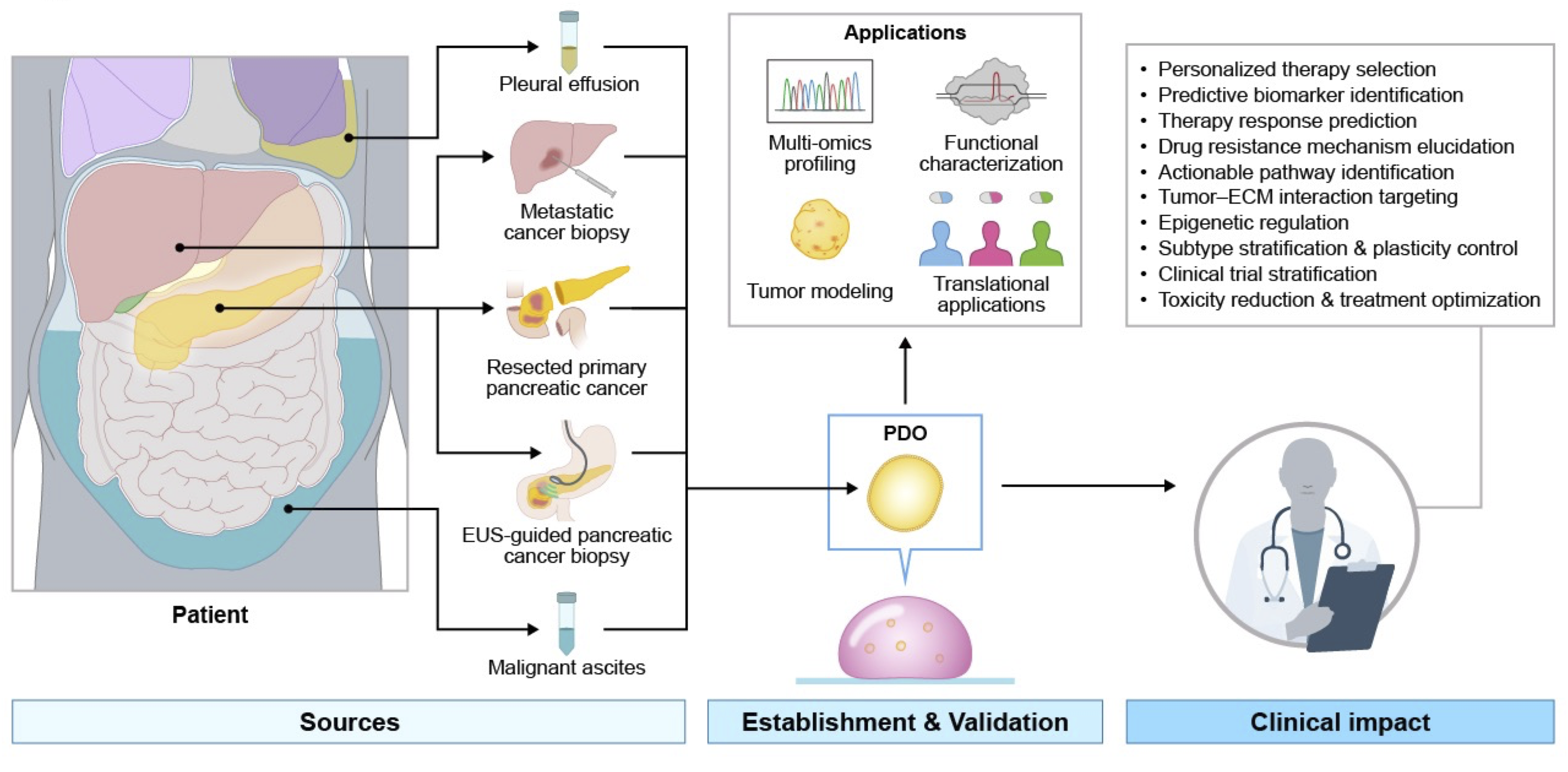
Liquid biopsy is a minimally invasive alternative for patients with advanced disease. Choi et al. reported the generation of PDOs from malignant ascites and pleural effusion (34). These fluid-derived organoids capture exfoliated tumor cells and circulating tumor clusters, often representing a highly aggressive anoikis-resistant subpopulation. These models provide a unique insight into the biology of carcinomatosis and late-stage disease progression (Figure 1) (35).
PSC-derived and tissue-resident-derived organoids
PDOs can be categorized by cellular origin into those derived from adult tissue-resident stem cells (ASCs) and those generated from pluripotent stem cells (PSCs), including embryonic stem cells (ESCs) and induced pluripotent stem cells (iPSCs).
PSC-derived organoids
These models rely on the stepwise differentiation of PSCs through the definitive endoderm and pancreatic progenitor stages using developmental morphogens (e.g., activin A, FGF10, and retinoic acid) (36, 37). Their primary advantages lie in the modeling of developmental biology and early carcinogenesis. Huang et al. used iPSCs to model KRAS- and TP53-driven tumorigenesis in a genetically defined background (38). However, PSC-derived organoids often exhibit an immature, fetal-like phenotype and require prolonged culture periods (months) to reach maturity, limiting their utility in rapid clinical decision-making (17, 39).
2.3.2. Tissue-resident ASC organoids
In contrast to PSC-derived models, ASC-derived PDOs are generated directly from neoplastic tissue or from the patient's normal epithelial samples and harbor transformed ASC-like cancer stem cells or progenitor cells (15). By bypassing the need for exogenous reprogramming factors, ASC-derived organoids avoid introducing potential epigenetic artifacts and fetal-like immature characteristics frequently observed in iPSC-derived systems (40). Consequently, they retain mature epigenetic memory, intrinsic mutational burden, and intricate clonal architecture specific to an individual patient's tumor, without the confounding effects of reprogramming (16, 41).
Furthermore, ASC organoids can be established and expanded within a clinically relevant timeframe of 2–6 weeks, whereas PSC differentiation protocols typically require several months (27). This rapid turnaround time, combined with their ability to preserve patient-specific physiological characteristics, establishes them as promising platforms for real-time personalized medicine applications, such as high-throughput pharmacotyping (24, 42). Although ASC organoids are primarily epithelial in nature and inherently lack mesenchymal or stromal cellular compartments that can sometimes co-differentiate in PSC systems, their high phenotypic fidelity and genetic stability make them a widely used platform for ex vivo translational oncology (43).
Therefore, given their strong ability to recapitulate patient-specific disease biology within a clinically relevant time window, this review primarily focuses on tissue-resident ASC-derived organoids and their potential applications in functional precision cancer therapy (44, 45).
Genetic fidelity and clonal architecture preservation
A fundamental prerequisite for the clinical utility of PDOs is their ability to maintain the parental tumor's genomic landscape (46). Comprehensive multi-omics analyses have confirmed that PDAC organoids retain more than 90% of the somatic mutations, copy-number variations (CNVs), and structural variants present in the original tissue (16,30). Key driver mutations in KRAS, TP53, SMAD4, and CDKN2A are preserved with high fidelity even after extended passaging. In fact, eliminating contaminating stromal DNA in PDO cultures significantly increases overall tumor purity compared with highly desmoplastic primary tumors, which paradoxically enhances the detection sensitivity of these critical driver alterations by enriching for their variant allele frequencies (42, 47).
Furthermore, PDOs effectively recapitulated the pronounced intratumoral heterogeneity characteristic of PDAC. Landmark single-cell RNA sequencing studies, such as those by Krieger et al., have demonstrated that individual PDOs maintain distinct subclonal populations and a conserved developmental hierarchy, allowing for the coexistence of both "classical" and "basal-like" transcriptional phenotypes within a single culture (41). Unlike traditional 2D monolayer cell lines that are prone to rapid genetic drift, loss of wild-type TP53 function, and clonal selection of highly proliferative subpopulations (8), PDOs largely preserve the intricate clonal architecture and phenotypic plasticity of primary tumors. The utility of patient-derived pancreatic organoids has also been extended beyond PDAC, with recent studies demonstrating their application in modeling chronic pancreatitis (48).
However, maintaining this dynamic heterogeneity requires the meticulous optimization of culture conditions, as exogenous niche factors can inadvertently exert selective evolutionary pressure on tumor cells. For instance, it has been widely documented that standard organoid media rich in Wnt and R-spondin can selectively drive the culture toward a Wnt-dependent, classical-like transcriptomic state over time, potentially masking the emergence of more aggressive basal-like subclones and altering intrinsic drug sensitivities (19, 49). Therefore, careful control of these microenvironmental cues and validation using early-passage PDOs are important for minimizing phenotypic drift and maintaining the biological characteristics of the original patient tumor. Whether culture-induced transcriptomic shifts materially influence clinical drug prediction remains incompletely resolved and warrants systematic investigation.
Biobanking and scalability
The intrinsic self-renewing capacity and genomic stability of PDOs have catalyzed a paradigm shift from static tissue repositories to dynamic living biobanks (50). Global collaborative initiatives, most notably the Human Cancer Model Initiative (HCMI) and the Hubrecht Organoid Technology (HUB) Foundation, have successfully aggregated hundreds of well-characterized PDAC cell lines (51). These large-scale repositories are comprehensively annotated with matched clinical outcomes, multi-omics profiles, and functional drug sensitivity data, offering an unprecedented renewable resource for high-throughput screening, off-label drug repurposing, and the discovery of novel genotype-phenotype associations (23). Importantly, optimized cryopreservation protocols utilizing specialized freezing media and controlled-rate freezing techniques ensure high post-thaw viability (>80%) while preserving the epigenetic memory, mutational landscape, and intricate clonal architecture of the primary tumor (15).
Despite the substantial translational potential of PDOs, their widespread clinical implementation will depend on overcoming key engineering challenges related to scalability, cost, and assay standardization (30). Traditional Matrigel-based dome cultures are highly labor-intensive and prone to interoperator variability, rendering them suboptimal for rapid clinical decision-making. Recent bioengineering advancements have aggressively addressed these limitations by integrating automated liquid-handling robotics and miniaturized assay formats, which exponentially increase throughput while minimizing the required tumor biomass and the need for expensive reagents (52). For instance, automated picking and seeding platforms, such as the Yamaha Cell Handler, enable precise dispensing of a controlled number of organoids per well, thereby significantly standardizing HTS pipelines and reducing the amount of starting material required (53).
Furthermore, the field is rapidly advancing toward next-generation scalable platforms, including 3D bioprinting and microfluidic organoid-on-a-chip systems. Automated droplet-based microfluidics and magnetic 3D bioprinting enable the rapid, high-throughput generation of uniform organoids, facilitating the screening of thousands of therapeutic compounds within an actionable clinical window of less than two weeks (54). Concurrently, microphysiological systems provide dynamic fluid flow, enabling the delivery of nutrients and therapeutics that closely mimic in vivo pharmacokinetics, interstitial shear stress, and oxygen gradients (55). These scalable, bioengineered platforms not only enhance the reproducibility and physiological relevance of pharmacotyping assays but also facilitate the integration of PDO-guided functional precision medicine into routine clinical oncology practice.
3. Establishment and characterization of pancreatic cancer organoids
Isolation protocols and enzymatic dissociation
The successful derivation of PDOs from PDAC is contingent on the efficient dissociation of the dense, desmoplastic stroma characteristic of these malignancies (15). Although early protocols have relied on surgical resection specimens, in clinical practice, more than 80% of patients present with unresectable disease at diagnosis, necessitating protocols optimized for low-input samples, such as EUS-FNB (31, 56). The current standard operating procedures involve a meticulous two-step digestion process. Initially, mechanical mincing is followed by enzymatic digestion using a highly optimized cocktail of collagenases (types II, IV, or XI) and dispases to efficiently degrade the collagen-rich ECM. This is often supplemented with DNase I to prevent cell clumping caused by the release of free DNA after tissue disruption, thereby enhancing single-cell or small-cluster yields and suspension homogeneity (15, 16, 24, 27).
Recent protocol optimizations have emphasized that digestion times must be strictly monitored (typically 15-60 min, depending on tissue cellularity and biopsy type) to maximize cellular yield while preserving cell viability. Prolonged enzymatic exposure can inadvertently induce anoikis, compromise stem cell marker expression, and exert a selective pressure that favors resilient but potentially nonrepresentative clonal subpopulations, thereby altering the phenotypic fidelity of the resulting organoid cultures (56, 57).
Culture conditions
The robust maintenance and expansion of PDAC PDOs rely heavily on a chemically defined medium that mimics the in vivo stem cell niche, primarily by modulating the Wnt, Notch, BMP, and TGF-β signaling pathways (58). The core culture medium typically utilizes a basal formulation of Advanced DMEM/F12 supplemented with essential mitogens, such as epidermal growth factor and fibroblast growth factor 10, to sustain continuous epithelial proliferation (15).
Dependence on Wnt signaling represents a major axis of functional heterogeneity in PDAC. Although normal pancreatic ductal organoids require exogenous Wnt3a and R-spondin 1 (an LGR5 ligand that dramatically potentiates canonical Wnt signaling) for survival, transformed PDAC organoids exhibit varying degrees of Wnt autonomy (19, 57, 59). Landmark studies have revealed that tumor organoids with GATA6 amplification or high expression (classical subtype) often remain intrinsically Wnt-dependent (19). Conversely, cells harboring GATA6 loss, which correlates with a highly aggressive basal-like or squamous phenotype, frequently acquire Wnt-independence through epigenetic adaptation and endogenous Wnt ligand secretion. Consequently, to capture the full spectrum of inter- and intra-tumoral heterogeneity without inadvertently selecting specific subclonal populations, it is important to culture and test PDOs in both Wnt-enriched and Wnt-deprived media conditions (19, 59).
Furthermore, the TGF-β signaling pathway exerts a dual, context-dependent role in pancreatic carcinogenesis, acting as a potent tumor suppressor in the normal ductal epithelium but paradoxically promoting epithelial-to-mesenchymal transition and metastatic dissemination in late-stage cancer (60). In standard organoid protocols, the ALK4/5/7 (TGF-β type I receptor) inhibitor A83-01 is routinely added to cultures to suppress this pathway, thereby maintaining epithelial stemness and preventing premature differentiation or apoptosis. However, to establish tumor-specific cultures, strategically withdrawing inhibitors such as A83-01 or adding exogenous TGF-β can selectively eliminate non-neoplastic ductal contaminants, as only PDAC clones harboring mutations in the TGF-β signaling axis (SMAD4 inactivation) will evade growth arrest and survive this negative selection pressure (18).
The addition of the Rho-associated protein kinase (ROCK) inhibitor Y-27632 prevented anoikis (apoptosis induced by the loss of cell–matrix attachment) during initial plating and subsequent passaging, thereby significantly enhancing establishment efficiency. However, prolonged exposure to Y-27632 may alter cellular phenotypes, potentially promoting a shift from a mesenchymal-like state toward a more epithelial-like phenotype by modulating cytoskeletal tension and Rho/ROCK signaling dynamics (61, 62). Accordingly, culture conditions should be regarded as experimental variables rather than as neutral components of organoid maintenance, as they may influence both tumor phenotype and pharmacologic response.
Molecular characterization
Comprehensive multi-omics profiling is an essential prerequisite for validating that PDOs faithfully retain the intricate molecular landscape of the parental tumor (46, 63). Extensive genomic characterizations using whole-exome and whole-genome sequencing have consistently demonstrated that PDAC organoids preserve the vast majority (>80%) of somatic driver mutations, such as KRAS, TP53, SMAD4, and CDKN2A, as well as the copy-number variations and structural variants inherent to the primary tumor (16, 47). Unlike conventional 2D cell lines, which are notoriously susceptible to rapid genetic drift and clonal selection under culture stress, PDOs maintain profound genomic stability during long-term expansion, ensuring their reliability as preclinical avatars (44).
Beyond the static genome, transcriptomic profiling (RNA-seq) is critical for delineating molecular subtypes (classical vs. basal-like) that dictate clinical behavior and therapeutic vulnerability (41). However, as elucidated by Raghavan et al. and others, the phenotypic state of PDAC cells is highly plastic, and exogenous components of the culture medium can inadvertently drive transcriptomic drift. For instance, the routine inclusion of specific niche factors or inhibitors, such as TGF-β pathway antagonists (e.g., A83-01), may artificially enforce a Wnt-dependent classical epithelial phenotype, thereby suppressing the emergence of aggressive, basal-like states that rely on TGF-β signaling for epithelial-to-mesenchymal transition (49, 64). Single-cell RNA sequencing has become an indispensable technology for overcoming the limitations of bulk sequencing and for capturing phenotypic fluidity. It provides the necessary resolution to dissect profound intratumoral heterogeneity, identify rare drug-tolerant subclones, and map the dynamic trajectories of epithelial plasticity that, in bulk transcriptomics approaches, may otherwise be obscured (65).
Furthermore, the integration of epigenomic profiling, such as Assays for Transposase-Accessible Chromatin using sequencing (ATAC-seq), has increasingly been used to assess chromatin accessibility and functional genomic states associated with drug sensitivity (66). Advanced epigenetic analyses have revealed that PDAC organoids robustly preserve the epigenetic memory of the primary tumor, including critical transcription factor-driven enhancer landscapes (FOXA1 and EN1), which actively orchestrate metastatic evolution and adaptive chemoresistance (32, 67).
4. Tumor microenvironment integration in organoid platforms
Limitations of epithelial-only models
Conventional epithelial PDOs incompletely model the tumor microenvironment, thereby driving the development of next-generation organoid platforms that integrate stromal, immune, and vascular components.
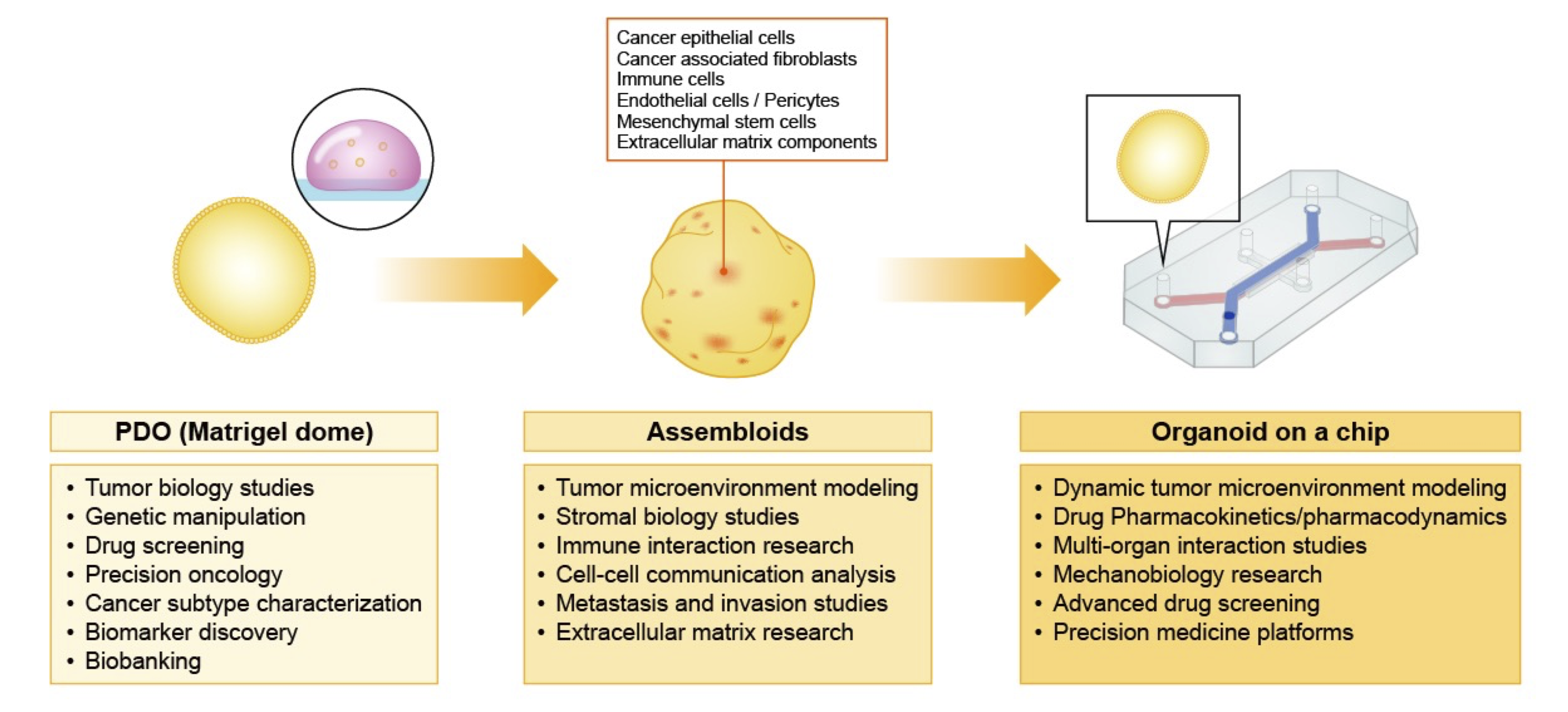
The 2026 update of the 'Hallmarks of Cancer' by Douglas Hanahan advances a four-dimensional framework in which malignant progression arises not only from cell-intrinsic alterations but also from dynamic interactions within the TME and broader systemic contexts (68, 69). In this multidimensional model, the TME, the third dimension of cancer, actively orchestrates hallmark-enabling processes, such as immune evasion, phenotypic plasticity, and therapeutic resistance. This paradigm highlights a major limitation of conventional epithelial-only PDO systems, which fail to capture essential heterotypic interactions between CAFs and immune cells (44). To bridge this translational gap, next-generation organoid platforms must integrate defined stromal and immune components to faithfully recapitulate the spatial and functional complexity of human diseases (70) (Figure 2).
PDAC is histologically characterized by a profound desmoplastic reaction in which the dense ECM and stromal cells constitute up to 90% of the total tumor volume (9, 30). Traditional Matrigel-embedded PDOs predominantly propagate in ductal epithelial cells, inadvertently selecting stromal and immune components that critically drive tumor progression, metastasis, and therapeutic resistance. Consequently, these epithelial-only models cannot recapitulate the physical barriers to drug delivery imposed by interstitial hypertension and also cannot properly model paracrine signaling networks that confer extrinsic chemoresistance (9, 63). Furthermore, the lack of immune effector cell components in typical PDOs prevents meaningful assessment of immunotherapies, which are characterized by a profoundly immunologically cold and immune-excluded TME (70, 71).
Cancer-associated fibroblasts
To address this translational gap, next-generation organoid platforms are increasingly incorporating patient-matched cancer-associated fibroblasts (CAFs) to more faithfully model the desmoplastic stroma. Foundational co-culture studies have demonstrated that CAFs are not a uniform population but rather comprise distinct, functionally plastic subtypes shaped by tumor-derived signaling cues (44, 72). Using organoid and CAF co-culture systems, Öhlund et al. characterized two principal CAF subsets: myofibroblastic CAFs (myCAFs), driven by TGF-β signaling and localized near neoplastic epithelial cells, and inflammatory CAFs (iCAFs), driven by IL-1/JAK/STAT pathway signaling and typically situated more distally within the tumor stroma (72).
More recently, innovative platforms such as the Fused Pancreatic Cancer Organoid (FPCO) system, described by Takeuchi et al., have advanced this paradigm by generating composite organoids through fusion of epithelial spheroids with iPSC-derived mesenchymal cells (37, 73). These FPCOs spontaneously recapitulate the spatial organization of myCAFs and iCAFs and include antigen-presenting CAFs (apCAFs), thereby reproducing key aspects of in vivo stromal heterogeneity without the need for exogenous scaffolding matrices.
Functionally, CAF integration within organoid systems enhances stemness-associated programs, promotes epithelial-mesenchymal transition, and confers substantial resistance to cytotoxic regimens, including gemcitabine and FOLFIRINOX, compared with epithelial monocultures (43).
Immune cells
Reconstituting the immune microenvironment is essential for identifying strategies to overcome the immune evasion in PDAC. Two primary methodologies have been developed: the air-liquid interface (ALI) method and reconstituted co-cultures (70). The ALI approach, in which mechanically minced tumor fragments are cultured on permeable inserts, preserves the native architecture, including endogenous tumor-infiltrating lymphocytes (TILs), macrophages, and stromal matrix, for several weeks. This method successfully retains T-cell receptor heterogeneity and demonstrates PD-1/PD-L1 dependent immune checkpoint responses ex vivo (74).
Conversely, reconstituted models involve the addition of autologous peripheral blood mononuclear cells or expanded T cells to the established PDOs. Tsai et al. and Schuth et al. developed patient-matched triple co-cultures (organoids, CAFs, and T cells) that demonstrated tumor-dependent lymphocyte infiltration and activation (75). More recently, the InterOMaX (i.e., Interaction with Organoid in Matrix) platform described by Lahusen et al. enables 3D visualization of T-cell-mediated cytotoxicity, revealing that pancreatic stellate cells actively exclude cytotoxic T cells from the tumor core, thereby mechanically protecting the organoids from immune attack. These platforms are now being used to screen for bispecific antibodies and epigenetic modulators that can enhance antigen presentation and T-cell infiltration (76).
These advanced immune-augmented platforms are now being used to actively screen novel immunotherapeutics at high throughput. For instance, complex co-culture systems are instrumental in evaluating T-cell bispecific antibodies and identifying epigenetic modulators, such as BET and HDAC inhibitors, which can epigenetically reprogram tumor cells to enhance antigen presentation and significantly potentiate T-cell infiltration and cytotoxicity (77).
Endothelial cells
The hypovascular and hypoxic nature of PDAC is a key determinant of the failure of drug delivery. To model this, organoid-on-a-chip platforms have been engineered to incorporate perfusable endothelial channels (63). The InVADE (i.e., Integrated Vasculature for Assessing Dynamic Events) platform, developed by Lai et al., co-cultures patient-derived PDAC organoids with fibroblasts and endothelial cells within a microfluidic device (55). This system demonstrated that the presence of fibroblasts increased matrix stiffness and collagen deposition, which in turn impeded the diffusion of gemcitabine perfused through the vascular channel, thereby effectively replicating the clinically observed drug-delivery barrier. Similarly, Haque et al. developed a PDAC-on-a-chip model separated by a porous membrane, allowing the study of endothelial-tumor crosstalk (78). These vascularized models have shown that endothelial cells are not merely passive conduits but actively maintain the cancer stem cell pool via angiocrine signaling, specifically via the Notch and Wnt pathways (79). These microphysiological systems enable precise control of fluid dynamics and shear stress, providing a more accurate prediction of therapeutic efficacy than static 3D cultures.
Mechanical and metabolic niche modeling
The physical properties of the TME, particularly the matrix stiffness, profoundly influence tumor biology. LeSavage et al. recently used engineered hydrogels with tunable stiffness to demonstrate that increased matrix rigidity is sufficient to induce chemoresistance in PDAC organoids independent of chemical signaling (80). This mechanotransduction-driven resistance is primarily mediated by CD44 receptor interactions with hyaluronan within the stiffened matrix, thereby upregulating drug efflux transporters and activating the YAP/TAZ pathway.
Metabolic gradients, such as oxygen, glucose, and pH, are critical regulators of phenotypic plasticity. Randriamanantsoa et al. and Papargyriou et al. showed that culturing organoids in collagen gels rather than in soft Matrigel promotes branching morphogenesis and a star-like mesenchymal phenotype associated with hypoxia and aggressive behavior (65, 81). These mesenchymal-like organoids exhibit distinct metabolic dependencies, relying more on glycolysis than on oxidative phosphorylation, and show differential sensitivity to metabolic inhibitors compared to their cystic and epithelial counterparts. Thus, advanced organoid platforms must integrate both biomechanical ECM cues and metabolic gradients to accurately model basal-like and classical transcriptional subtypes of PDAC.
5. PDO Applications in personalized treatment
Drug sensitivity testing
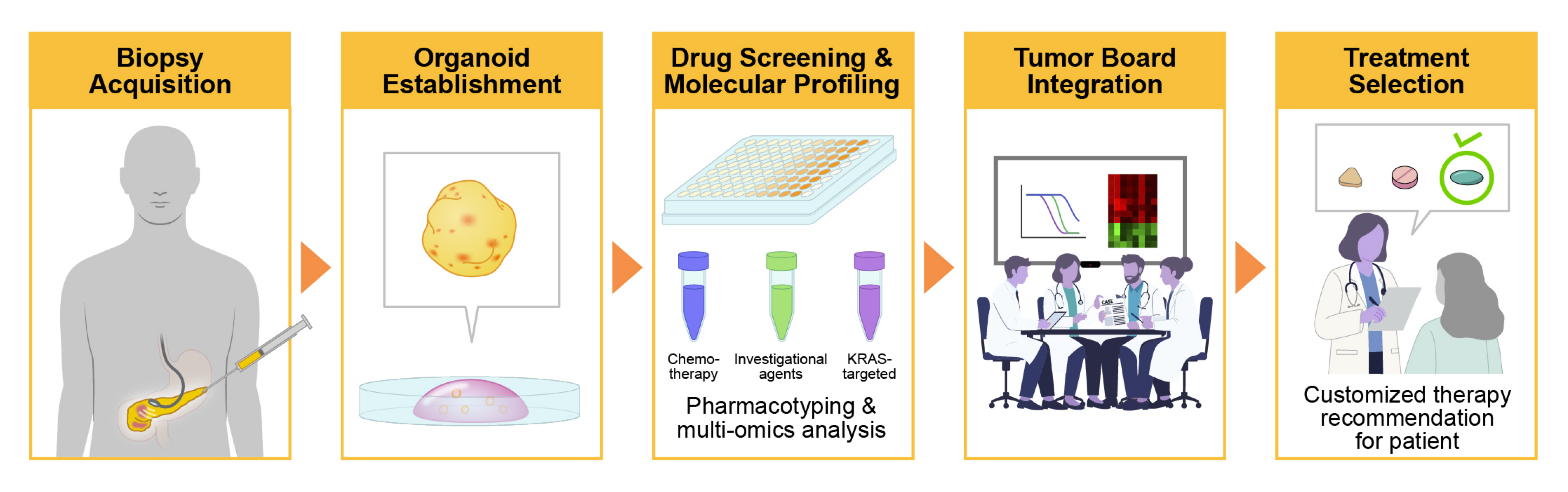
The therapeutic paradigm of PDAC is transitioning from empirical cytotoxic regimens to functional precision oncology strategies. Although conventional genomic profiling has historically identified actionable alterations in a limited subset of patients, such as BRCA1/2 mutations, the recent development of allele-specific and pan-KRAS inhibitors has substantially expanded the population potentially eligible for targeted therapy, given that oncogenic KRAS mutations occur in over 90% of PDAC cases. However, genomic alterations alone cannot reliably predict therapeutic responsiveness or mechanisms of resistance. In this context, PDOs provide a functional platform for ex vivo pharmacotyping, enabling empirical assessment of tumor sensitivity to standard-of-care chemotherapy, KRAS-targeted agents, and investigational compounds (Figure 3) (44).
To align with clinical timelines, PDO platforms have evolved from low-throughput manual assays to automated high-throughput screening pipelines. Early studies by Hou et al. and Driehuis et al. established the feasibility of screening libraries for more than 70 compounds in established organoid lines using cell viability endpoints (23, 52). Recent advancements have utilized acoustic liquid handling and miniaturized 384- or 1536-well formats to screen FDA-approved drug libraries for PDOs within 2–4 weeks of biopsy. Importantly, Wansch et al. demonstrated that assay optimization shifts the focus from the half-maximal inhibitory concentration (IC50) to area-under-the-curve metrics. This significantly enhances the robustness of these pipelines, allowing for the classification of PDOs into sensitive, intermediate, and resistant pharmacotypes with greater physiological relevance (82).
Target discovery and molecular subtype characterization
PDO biobanks serve as renewable resources for identifying novel vulnerabilities in specific molecular subtypes (30). Driehuis et al. performed extensive drug screening and identified that MTAP-deficient PDAC organoids were uniquely sensitive to the PRMT5 inhibitor EZP01556, exploiting a synthetic lethal interaction derived from the co-deletion of CDKN2A and MTAP (23). Within the same screening platform, the integration of CRISPR-Cas9 genome editing has uncovered a novel synthetic lethal interaction in which missense mutations in the SWI/SNF chromatin remodeling complex subunit ARID1A confer increased susceptibility to the kinase inhibitors dasatinib and VE-821 (83).
In addition to identifying targeted vulnerabilities, large-scale screening of >1,000 FDA-approved non-oncology drugs on PDAC PDOs revealed that the antimicrobial agent emetine and the cardiac glycoside ouabain exhibited potent antitumor activity by targeting hypoxia-inducible factors (83). Organoid platforms have been instrumental in identifying vulnerabilities to histone modifiers in the epigenetic landscape of PDAC. For instance, PDO screening revealed that tumors exhibiting high levels of H3K27me3 repression are uniquely sensitive to UNC1999, a specific EZH2 inhibitor that demonstrates potent antitumor effects, especially when combined with standard gemcitabine therapy (67).
Furthermore, PDOs have accelerated the development of direct KRAS-targeting therapies. Duan et al. used a sophisticated library of isogenic organoids to screen thousands of compounds and identified perhexiline maleate as a specific inhibitor of KRAS G12D-driven tumors, targeting the metabolic reprogramming of cholesterol biosynthesis associated with the mutation (84). As highly selective non-covalent KRAS G12D inhibitors have entered clinical evaluation, early resistance mechanisms of PDOs have been critically elucidated. Gulay et al. used PDOs to demonstrate that prolonged exposure to MRTX1133 induces compensatory upregulation of EGFR and HER2 (ERBB pathways), identifying a potent synthetic lethal interaction when combining KRAS G12D with pan-ERBB inhibitors to circumvent acquired resistance (85). Organoid-based multi-omics integration continues to yield innovative strategies to overcome intrinsic chemoresistance. Screening of FDA-approved compound libraries has recently identified the angiotensin receptor antagonist irbesartan as a novel agent capable of reversing gemcitabine resistance by suppressing the Hippo/YAP1 signaling axis and maintaining stemness (86).
Mechanisms of drug resistance
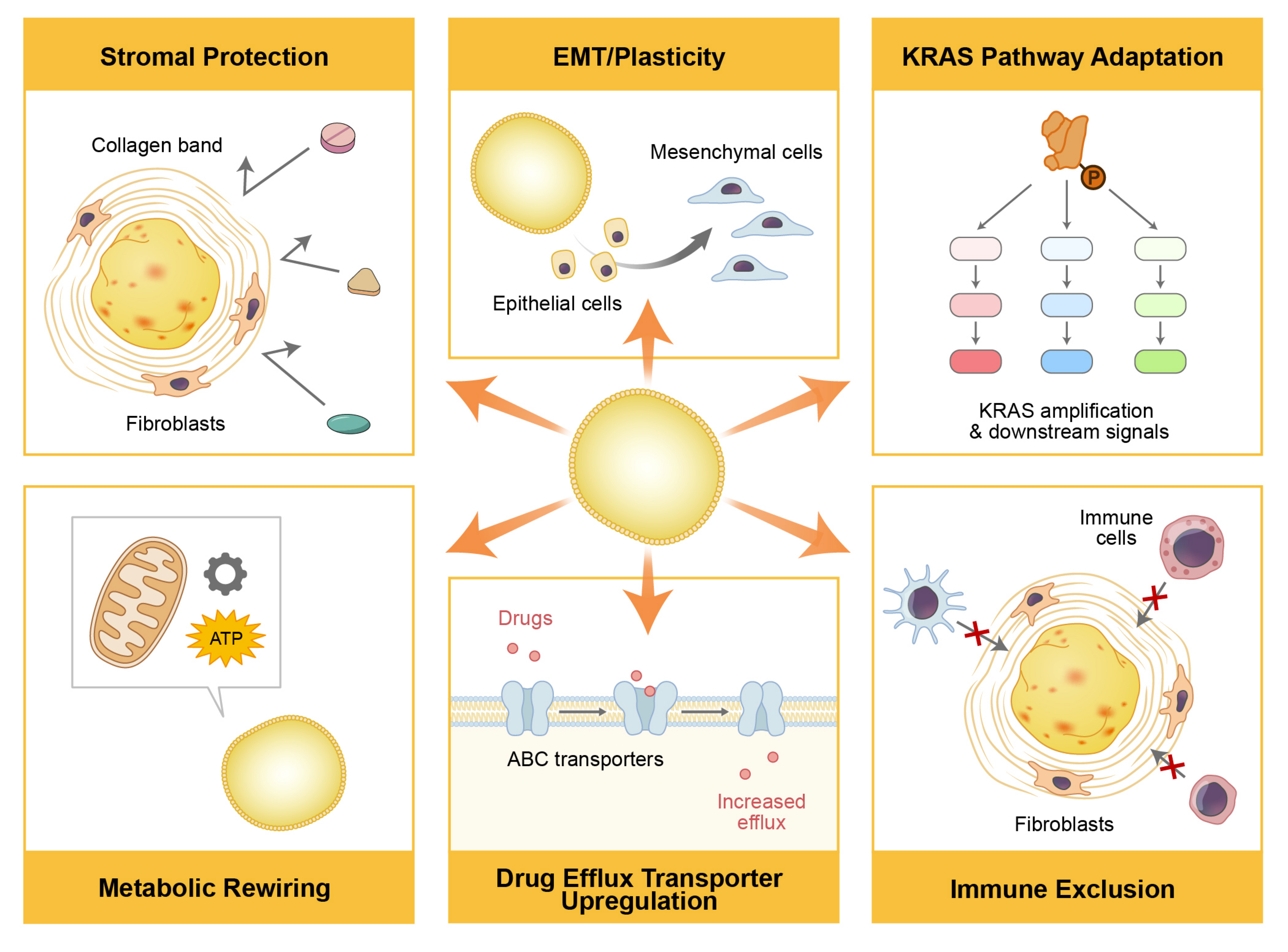
Acquired resistance, driven by tumor evolution under therapeutic pressure, remains a major challenge in the longitudinal management of PDAC (Figure 4). PDOs provide a practical ex vivo platform for modeling these adaptive dynamics, enabling investigation of clonal selection and emergent drug-resistance states. Resistance-matched organoid pairs can be generated by exposing PDOs to increasing concentrations of chemotherapeutic agents over time. Bachir et al. successfully established FOLFIRINOX-resistant PDO models that recapitulated the molecular signatures observed in patients who progressed to therapy (87). These models revealed that resistance is often driven not by a single mutation but by transcriptional reprogramming, including upregulation of drug efflux pumps, such as ABCC1/ABCC2, and metabolic shifts toward oxidative phosphorylation (65, 80).
This concept was further validated by the longitudinal collection of PDOs from the same patient across different clinical time points (at initial diagnosis, following post-neoadjuvant therapy, and at metastatic progression), which uniquely enabled high-resolution tracking of clonal evolution. A landmark longitudinal study by Tiriac et al. illustrated this phenomenon, wherein PDOs derived from a patient’s initial biopsy were highly sensitive to chemotherapy, whereas subsequent autopsy PDOs following treatment failure exhibited profound resistance, accompanied by KRAS amplification and a transcriptomic class switch from a classical to a highly aggressive basal-like phenotype (16).
Conventional bulk sequencing often obscures the subclonal dynamics underlying clinical relapse. To address this limitation, Le Compte et al. employed a high-content live-imaging platform with a single-organoid resolution to track treatment responses longitudinally. They demonstrated that rare drug-tolerant subclones and highly invasive phenotypes existed as minor populations at the time of diagnosis. Under chemotherapeutic pressure, such as paclitaxel exposure, these subclones can be preferentially selected, driving tumor recurrence, highlighting the intratumoral heterogeneity masked by bulk analyses and the necessity of single-organoid resolution to better understand therapeutic resistance (88).
Clinical trial applications and translational studies
The goal of PDO pharmacotyping is its clinical implementation as a functional precision-medicine platform that guides individualized treatment decisions. Clinical studies ranging from retrospective validation to prospective trials have increasingly demonstrated its feasibility and predictive value in patients with PDAC. A retrospective analysis by Tiriac et al. has demonstrated that PDO sensitivity parallels patient responses to gemcitabine/nab-paclitaxel and FOLFIRINOX (16). This finding has been substantiated by recent prospective studies. This landmark study provided one of the earliest demonstrations that ex vivo PDO pharmacotyping could recapitulate patient-specific responses to standard chemotherapy, establishing the foundation for subsequent clinical validation studies.
Building upon these retrospective observations, the PASS-01 randomized phase II trial incorporated patient-derived organoid pharmacotyping into a prospective clinical trial evaluating first-line modified FOLFIRINOX versus gemcitabine/nab-paclitaxel for metastatic PDAC. Pretreatment biopsies underwent comprehensive molecular profiling, PDO establishment, and drug sensitivity testing, and the resulting data were reviewed by a multidisciplinary molecular tumor board to support biomarker-driven treatment decisions (89).
The ORGANOPREDICT trial reported 91.2% concordance between PDO pharmacotyping and clinical outcomes, supporting the predictive potential of PDO pharmacotyping for patients with advanced PDAC (96). These findings demonstrated that PDO-based pharmacotyping can be incorporated into prospective clinical workflows with a high degree of concordance between ex vivo drug responses and clinical outcomes. Similarly, Seppälä et al. demonstrated that PDO sensitivity profiles generated from pre-treatment biopsies were significant predictors of objective response and progression-free survival in patients undergoing neoadjuvant chemotherapy.
Importantly, these results suggested that PDO pharmacotyping performed before treatment initiation may facilitate patient stratification for neoadjuvant therapy (42). A recent randomized study suggested that integrating genomic and organoid-based stratification significantly improved median overall survival (19.3 months vs. 8.6 months) compared with standard physician choice, representing an important step toward clinical translation of organoid-guided therapy (97).
6. Different organoid platforms
Patient-derived organoid xenograft (PDOX) models
Transplanting PDOs into immunodeficient mice (patient-derived organoid xenografts [PDOX]) creates robust in vivo models that retain the histology and metastatic patterns of the donor tumor (90). Unlike traditional PDX established from bulk tissue fragments, PDOX models can be generated from limited biopsy materials following in vitro expansion (15). Boj et al. and Romero-Calvo et al. demonstrated that PDOX models recapitulate key features of PDAC, including the characteristic desmoplastic stroma, following orthotopic implantation. These models therefore provide a valuable in vivo platform for validating candidate therapeutics identified through in vitro drug screening (15, 91).
Tumor microenvironment-integrated assembloids
Assembloids are spatially organized models formed by the directed fusion of epithelial organoids with distinct stromal, immune, or neural components to model intertissue interactions (Figure 4) (73).
Takeuchi et al. described a protocol for generating fused pancreatic cancer organoids (FPCOs) that spontaneously organize into tumor and stromal compartments, mimicking the desmoplastic reaction (37, 73). Additionally, Zhou et al. developed a T cell-engaging platform by co-culturing PDOs with autologous lymphocytes to screen bispecific antibodies, demonstrating the potential of assembloids in immuno-oncology (77).
In pancreatic cancer, this involves the spatial integration of tumor organoids with islet organoids, neural spheroids, and vascular networks to create a mini-pancreas or tumor-in-a-dish. Future assembloid systems aim to incorporate innervation, since perineural invasion is a hallmark of PDAC, and to link pancreatic and liver organoids in multi-organ-on-a-chip devices to study metastatic colonization and systemic drug metabolism.
These models provide an effective means for bridging the gap between the systemic complexity of the human body and conventional in vitro experiments.
CRISPR/Cas9 functional genomics
PDOs are highly amenable to precise genetic engineering and provide a robust platform for functional genomics and target validation. Utilizing CRISPR/Cas9, researchers have modeled the stepwise progression of PDAC. For instance, Seino et al. demonstrated that the sequential introduction of driver mutations (KRAS, TP53, SMAD4, and CDKN2A) into normal organoids confers independence from essential stem cell niche factors, such as Wnt and R-spondin, through non-genetic epigenetic adaptation during tumorigenesis (19, 59). More recently, Hirt et al. employed CRISPR screens in PDOs to validate drug-gene interactions, identifying synthetic lethality between ARID1A loss and bromodomain inhibitors (83). Furthermore, the generation of isogenic organoids via CRISPR-Cas9 enables the high-throughput screening of mutation-specific vulnerabilities. Duan et al. used CRISPR-engineered isogenic organoids harboring specific combinations of KRAS, TP53, and SMAD4 alterations to identify perhexiline maleate as a potent inhibitor specific to KRAS G12D-driven tumors that targets cholesterol metabolism (84).
Organoid-on-chip and microfluidic systems
Microfluidic organ-on-a-chip devices address the lack of vascular perfusion and dynamic fluid flow inherent in static 3D dome cultures (92). Vascularized PDAC-on-a-chip models incorporating endothelial channels allow the study of drug delivery across vascular barriers and the influence of interstitial fluid shear stress on tumor biology (55). These microphysiological systems demonstrate that the dense stroma and resulting high interstitial pressure in PDAC significantly impede the penetration of chemotherapeutics, such as intravascularly perfused gemcitabine, compared with standard static cultures.
Furthermore, these microfluidic platforms enable ex vivo evaluation of stroma-targeting strategies and tumor-immune interactions. For instance, Haque et al. utilized a dual-channel microfluidic system to co-culture PDAC organoids with pancreatic stellate cells (PSCs) and macrophages and demonstrated that combining gemcitabine with stroma-targeting agents significantly increased cancer cell apoptosis by overcoming stromal barriers (78). Similarly, Geyer et al. developed a tri-culture model comprising PDAC organoids, PSCs, and endothelial cells to study immune cell trafficking (92). Their study revealed that PSCs establish physical and biochemical barriers that actively sequester peripheral blood mononuclear cells, thereby restricting their direct interactions with tumor cells (93).
Automated microfluidic systems have been increasingly employed to precisely regulate dynamic drug delivery to align with rapid clinical decision-making timelines. These advanced platforms support high-throughput combinatorial drug screening and the real-time monitoring of organoid viability, thereby significantly bridging the translational gap in functional precision oncology (94).
7. Discussion
Current limitations and future challenges
Despite their potential, several limitations remain for the clinical implementation of PDO-based precision medicine. Organoid establishment rates vary with tumor characteristics and specimen quality, and contamination with normal ductal epithelium may affect experimental results. In addition, standardized culture protocols and reproducible pharmacotyping assays across laboratories have not yet been established. The cost and infrastructure required for individualized drug testing, together with the need for clinically relevant turnaround times in patients with PDAC, may also limit routine clinical use. Furthermore, regulatory requirements, reimbursement policies, and implementation considerations in CLIA/CAP-certified clinical laboratories should be taken into account when applying PDO-guided therapeutic decision-making in clinical practice. A recent systematic review that applied a translatability scoring framework to 95 PDAC organoid studies revealed significant differences between biological fidelity and clinical utility. Notably, only 4.2% of the evaluated studies met the criteria for good translational potential, largely due to the lack of multi-omic integration, pharmacogenomic modeling, and formal embedding in clinical trials (45).
Developmental immaturity and phenotypic drift
Although PDOs preserve genomic characteristics more faithfully than conventional 2D cell lines, they remain inherently susceptible to culture-induced selective pressures over time. Longitudinal passaging often leads to clonal drift, in which faster-proliferating subclones dominate the culture. This dynamic can inadvertently mask rare quiescent subpopulations, such as drug-tolerant persister cells, which are critical drivers of therapeutic resistance and clinical recurrence (88). Furthermore, the absence of complex stromal and immune signaling in standard Matrigel-based monocultures can induce profound transcriptomic drift. Exogenous culture media can determine the phenotypic state of PDAC cells. For instance, standard media rich in Wnt and R-spondin tend to artificially induce a well-differentiated classical epithelial phenotype (49, 64). This media-driven selection actively suppresses the emergence or maintenance of more aggressive basal-like or quasi-mesenchymal transcriptional subtypes frequently observed in vivo (65). Consequently, this phenotypic skewing can yield discordant drug sensitivity profiles, particularly when evaluating targeted agents that exploit vulnerabilities in epithelial-to-mesenchymal transition or metabolic dependencies specific to the basal-like state.
Lack of inter-laboratory standardization
A major barrier to the regulatory approval and widespread clinical integration of PDO-based assays is the lack of interlaboratory standardization (45). Heterogeneity exists at every step of the pipeline, from the tissue digestion enzymes used to the definition of drug responses (30, 57). Major discordance arises in the readout metrics. Although some laboratories use ATP-based metabolic assays such as CellTiter-Glo, others employ live-cell imaging or morphological sizing to evaluate cytotoxicity (30). Beutel et al. emphasized that differences in defining drug sensitivity thresholds, specifically the use of IC50 versus the area under the curve, prevent the meta-analysis of data across institutions. Finally, rigorous quality control is essential to ensure that PDOs are reliable and authentic clinical avatars. This includes verification of identity via short tandem repeat profiling or single nucleotide polymorphism fingerprinting to exclude cross-contamination, and regular screening for Mycoplasma (57).
Undefined extracellular matrix components
Current PDO studies rely on basement membrane extracts derived from Engelbreth-Holm-Swarm mouse sarcomas (Matrigel) as the scaffolding matrix (30). These matrices exhibit significant batch-to-batch variability in growth factor content and stiffness, introducing experimental noise. Importantly, the murine origin of these matrices poses a risk of xenogeneic pathogen transfer, making them incompatible with Good Manufacturing Practice standards required for clinical applications such as regenerative medicine or adoptive immunotherapy co-cultures (95). Furthermore, standard basement membrane extracts are mechanically soft (~400 Pa) and fail to recapitulate the profound stiffness of PDAC stroma (2-5 kPa), a known driver of chemoresistance via mechanotransduction pathways (80). A recent study by LeSavage et al., which utilized engineered polyethylene glycol-based hydrogels, demonstrated that matrix stiffness directly influenced the therapeutic response. PDAC organoids cultured in stiff biomimetic matrices exhibit increased resistance to gemcitabine and paclitaxel compared with those cultured in soft matrices, which are driven by mechanotransduction pathways that upregulate drug efflux transporters (80).
Insufficient prospective clinical validation
Although retrospective concordance between PDO drug sensitivity and patient clinical outcomes is well documented, prospective validation remains scarce (30). Only a few recent selective trials, such as the observational ORGANOPREDICT study and the interventional phase III AVATAR trial, have demonstrated clinical benefits (96, 97). Furthermore, the turnaround time required for organoid establishment and subsequent pharmacotyping remains a logistical Achilles' heel in PDO screening for PDAC. The 2–4 weeks required to establish and screen organoids often exceed the clinical window for decision-making in patients with aggressive metastatic PDAC (3, 4). Consequently, to align with the practical realities of oncology, PDO-based pharmacotyping is currently more feasible and is frequently used to select adjuvant regimens during the postoperative recovery window or to guide second-line therapies following initial empiric treatment failure, rather than to direct upfront therapy in treatment-naïve patients with advanced PDAC. Although accumulating evidence supports their utility for disease modeling and functional drug testing, additional prospective, multicenter clinical studies are required before PDO-guided treatment strategies can be routinely incorporated into clinical practice.
Conclusions
PDOs have successfully transitioned from exploratory in vitro models to promising translational platforms for determining functional precision oncology in PDAC. Prospective trials, notably the ORGANOPREDICT and the Phase III AVATAR studies, have demonstrated that PDO-guided therapy significantly improves survival outcomes compared with standard care. Furthermore, the methodological shift toward multidrug pharmacotyping using area-under-the-curve metrics has improved the prediction of synergistic effects in combinatorial regimens such as FOLFIRINOX. Despite this progress, its widespread clinical adoption has been hindered by translational gaps. Critical barriers include the lack of standardization in culture protocols and readout metrics, which limits inter-institutional reproducibility. Additionally, the turnaround time remains a logistical challenge in treating metastatic patients, and standard epithelial-only models fail to capture the stroma-mediated drug-resistance mechanisms driven by the dense pancreatic TME. To overcome these limitations, future studies should focus on comprehensive ecosystems that integrate TME components via assembloids and microfluidic organ-on-a-chip platforms. Ultimately, the systematic integration of advanced PDO platforms into clinical workflows may facilitate the implementation of functional precision oncology and improve therapeutic outcomes in PDAC. Despite these advances, widespread clinical implementation will require standardized organoid culture and pharmacotyping protocols, prospective multicenter validation, and regulatory qualification to establish the clinical utility of PDO-guided therapeutic decision-making.
References
1. Siegel RL, Giaquinto AN, Jemal A. Cancer statistics, 2024. CA Cancer J Clin. 2024;74(1):12–49. https://doi.org/10.3322/caac.21820
2. Rahib L, Smith BD, Aizenberg R, Rosenzweig AB, Fleshman JM, Matrisian LM. Projecting cancer incidence and deaths to 2030: the unexpected burden of thyroid, liver, and pancreas cancers in the United States. Cancer Res. 2014;74(11):2913–2921. https://doi.org/10.1158/0008-5472.CAN-14-0155
3. Dreyer SB, Beer P, Hingorani SR, Biankin AV. Improving outcomes of patients with pancreatic cancer. Nat Rev Clin Oncol. 2025;22(6):439–456. https://doi.org/10.1038/s41571-025-01019-9
4. Mizrahi JD, Surana R, Valle JW, Shroff RT. Pancreatic cancer. Lancet. 2020;395(10242):2008–2020. https://doi.org/10.1016/S0140-6736(20)30974-0
5. Conroy T, Desseigne F, Ychou M, Bouché O, Guimbaud R, Bécouarn Y, et al. FOLFIRINOX versus gemcitabine for metastatic pancreatic cancer. N Engl J Med. 2011;364(19):1817–1825. https://doi.org/10.1056/NEJMoa1011923
6. Von Hoff DD, Ervin T, Arena FP, Chiorean EG, Infante J, Moore M, et al. Increased survival in pancreatic cancer with nab-paclitaxel plus gemcitabine. N Engl J Med. 2013;369(18):1691–1703. https://doi.org/10.1056/NEJMoa1304369
7. Masters JR. Human cancer cell lines: fact and fantasy. Nat Rev Mol Cell Biol. 2000;1(3):233–236. https://doi.org/10.1038/35043102
8. Ben-David U, Siranosian B, Ha G, Tang H, Oren Y, Hinohara K, et al. Genetic and transcriptional evolution alters cancer cell line drug response. Nature. 2018;560(7718):325–330. https://doi.org/10.1038/s41586-018-0409-3
9. Feig C, Gopinathan A, Neesse A, Chan DS, Cook N, Tuveson DA. The pancreas cancer microenvironment. Clin Cancer Res. 2012;18(16):4266–4276. https://doi.org/10.1158/1078-0432.CCR-11-3114
10. Ho WJ, Jaffee EM, Zheng L. The tumour microenvironment in pancreatic cancer—clinical challenges and opportunities. Nat Rev Clin Oncol. 2020;17(9):527-540. https://doi.org/10.1038/s41571-020-0363-5
11. Hingorani SR, Wang L, Multani AS, Combs C, Deramaudt TB, Hruban RH, et al. Trp53R172H and KrasG12D cooperate to promote chromosomal instability and widely metastatic pancreatic ductal adenocarcinoma in mice. Cancer Cell. 2005;7(5):469–483. https://doi.org/10.1016/j.ccr.2005.04.023
12. Tentler JJ, Tan AC, Weekes CD, Jimeno A, Leong S, Pitts TM, et al. Patient-derived tumour xenografts as models for oncology drug development. Nat Rev Clin Oncol. 2012;9(6):338–350. https://doi.org/10.1038/nrclinonc.2012.61
13. Sato T, Vries RG, Snippert HJ, van de Wetering M, Barker N, Stange DE, et al. Single Lgr5 stem cells build crypt-villus structures in vitro without a mesenchymal niche. Nature. 2009;459(7244):262–265. https://doi.org/10.1038/nature07935
14. Lancaster MA, Knoblich JA. Organogenesis in a dish: modeling development and disease using organoid technologies. Science. 2014;345(6194):1247125. https://doi.org/10.1126/science.1247125
15. Boj SF, Hwang C-I, Baker LA, Chio IIC, Engle DD, Corbo V, et al. Organoid models of human and mouse ductal pancreatic cancer. Cell. 2015;160(1–2):324–338. https://doi.org/10.1016/j.cell.2014.12.021
16. Tiriac H, Belleau P, Engle DD, Plenker D, Deschênes A, Somerville TDD, et al. Organoid profiling identifies common responders to chemotherapy in pancreatic cancer. Cancer Discov. 2018;8(9):1112–1129. https://doi.org/10.1158/2159-8290.CD-18-0349
17. Hofer M, Lutolf MP. Engineering organoids. Nat Rev Mater. 2021;6(5):402–420. https://doi.org/10.1038/s41578-021-00279-y
18. Matano M, Date S, Shimokawa M, Takano A, Fujii M, Ohta Y, et al. Modeling colorectal cancer using CRISPR-Cas9-mediated engineering of human intestinal organoids. Nat Med. 2015;21(3):256–262. https://doi.org/10.1038/nm.3802
19. Seino T, Kawasaki S, Shimokawa M, Tamagawa H, Toshimitsu K, Fujii M, et al. Human pancreatic tumor organoids reveal loss of stem cell niche factor dependence during disease progression. Cell Stem Cell. 2018;22(3):454-467.e6. https://doi.org/10.1016/j.stem.2017.12.009
20. Lukonin I, Serra D, Challet Meylan L, Volkmann K, Baaten J, Zhao R, et al. Phenotypic landscape of intestinal organoid regeneration. Nature. 2020;586(7828):275–280. https://doi.org/10.1038/s41586-020-2776-9
21. Kim J, Koo B-K, Knoblich JA. Human organoids: model systems for human biology and medicine. Nat Rev Mol Cell Biol. 2020;21(10):571–584. https://doi.org/10.1038/s41580-020-0259-3
22. Huch M, Bonfanti P, Boj SF, Sato T, Loomans CJM, van de Wetering M, et al. Unlimited in vitro expansion of adult bi-potent pancreas progenitors through the Lgr5/R-spondin axis. EMBO J. 2013;32(20):2708–2721. https://doi.org/10.1038/emboj.2013.204
23. Driehuis E, van Hoeck A, Moore K, Kolders S, Francies HE, Gulersonmez MC, et al. Pancreatic cancer organoids recapitulate disease and allow personalized drug screening. Proc Natl Acad Sci U S A. 2019;116(52):26580–26590. https://doi.org/10.1073/pnas.1911273116
24. Wansch K, Schneider F, Dölvers F, Kühn A, Dragomir MP, Joosten M, et al. Identifying factors of organoid establishment in pancreatic cancer: A prospective observational study. Cancer Med. 2026;15(1):e71490. https://doi.org/10.1002/cam4.71490
25. Tsang CF, Patel H, Kouassi FM, Khandakar B, St Surin LG, Rouse JA, et al. Utility of pancreatic tumor scrapings for organoid development and precision medicine strategies. J Pathol. 2026;268(4):428-444. https://doi.org/10.1002/path.70025
26. Sljukic A, Green Jenkinson J, Niksic A, Prior N, Huch M. Advances in liver and pancreas organoids: how far we have come and where we go next. Nat Rev Gastroenterol Hepatol. 2026;23(1):44-64. https://doi.org/10.1038/s41575-025-01116-1
27. Demyan L, Habowski AN, Plenker D, King DA, Standring OJ, Tsang C, et al. Pancreatic cancer patient-derived organoids can predict response to neoadjuvant chemotherapy. Ann Surg. 2022;276(3):450–462. https://doi.org/10.1097/SLA.0000000000005558
28. Hennig A, Wolf L, Jahnke B, Polster H, Seidlitz T, Werner K, et al. CFTR expression analysis for subtyping of human pancreatic cancer organoids. Stem Cells Int. 2019;2019:1024614. https://doi.org/10.1155/2019/1024614
29. Matsumoto K, Fujimori N, Ichihara K, Takeno A, Murakami M, Ohno A, et al. Patient-derived organoids of pancreatic ductal adenocarcinoma for subtype determination and clinical outcome prediction. J Gastroenterol. 2024;59(7):629–640. https://doi.org/10.1007/s00535-024-02103-0
30. Beutel AK, Ekizce M, Ettrich TJ, Seufferlein T, Lindenmayer J, Gout J, et al. Organoid-based precision medicine in pancreatic cancer. United European Gastroenterol J. 2025;13(1):21–33. https://doi.org/10.1002/ueg2.12701
31. Tiriac H, Bucobo JC, Tzimas D, Grewel S, Lacomb JF, Rowehl LM, et al. Successful creation of pancreatic cancer organoids by means of EUS-guided fine-needle biopsy sampling for personalized cancer treatment. Gastrointest Endosc. 2018;87(6):1474–1480. https://doi.org/10.1016/j.gie.2017.12.032
32. Roe J-S, Hwang C-I, Somerville TDD, Milazzo JP, Lee EJ, Da Silva B, et al. Enhancer Reprogramming Promotes Pancreatic Cancer Metastasis. Cell. 2017;170(5):875-888.e20. https://doi.org/10.1016/j.cell.2017.07.007
33. Makohon-Moore AP, Zhang M, Reiter JG, Bozic I, Allen B, Kundu D, et al. Limited heterogeneity of known driver gene mutations among the metastases of individual patients with pancreatic cancer. Nat Genet. 2017;49(3):358–366. https://doi.org/10.1038/ng.3764
34. Choi W, Kim Y-H, Woo SM, Yu Y, Lee MR, Lee WJ, et al. Establishment of patient-derived organoids using ascitic or pleural fluid from cancer patients. Cancer Res Treat. 2023;55(4):1077-1086. https://doi.org/10.4143/crt.2022.1630
35. Seghers S, Le Compte M, Hendriks JMH, Van Schil P, Janssens A, Wener R, et al. A systematic review of patient-derived tumor organoids generation from malignant effusions. Crit Rev Oncol Hematol. 2024;195(104285):104285. https://doi.org/10.1016/j.critrevonc.2024.104285
36. D’Amour KA, Bang AG, Eliazer S, Kelly OG, Agulnick AD, Smart NG, et al. Production of pancreatic hormone-expressing endocrine cells from human embryonic stem cells. Nat Biotechnol. 2006;24(11):1392–1401. https://doi.org/10.1038/nbt1259
37. Takeuchi K, Tabe S, Takahashi K, Aoshima K, Matsuo M, Ueno Y, et al. Incorporation of human iPSC-derived stromal cells creates a pancreatic cancer organoid with heterogeneous cancer-associated fibroblasts. Cell Rep. 2023;42(11):113420. https://doi.org/10.1016/j.celrep.2023.113420
38. Huang L, Desai R, Conrad DN, Leite NC, Akshinthala D, Lim CM, et al. Commitment and oncogene-induced plasticity of human stem cell-derived pancreatic acinar and ductal organoids. Cell Stem Cell. 2021;28(6):1090-1104.e6. https://doi.org/10.1016/j.stem.2021.03.022
39. Takebe T, Wells JM. Organoids by design. Science. 2019;364(6444):956–959. https://doi.org/10.1126/science.aaw7567
40. Casamitjana J, Espinet E, Rovira M. Pancreatic organoids for regenerative medicine and cancer research. Front Cell Dev Biol. 2022;10:886153. https://doi.org/10.3389/fcell.2022.886153
41. Krieger TG, Le Blanc S, Jabs J, Ten FW, Ishaque N, Jechow K, et al. Single-cell analysis of patient-derived PDAC organoids reveals cell state heterogeneity and a conserved developmental hierarchy. Nat Commun. 2021;12(1):5826. https://doi.org/10.1038/s41467-021-26059-4
42. Seppälä TT, Zimmerman JW, Suri R, Zlomke H, Ivey GD, Szabolcs A, et al. Precision medicine in pancreatic cancer: patient-derived organoid pharmacotyping is a predictive biomarker of clinical treatment response. Clin Cancer Res. 2022;28(15):3296–3307. https://doi.org/10.1158/1078-0432.CCR-21-4165
43. Schuth S, Le Blanc S, Krieger TG, Jabs J, Schenk M, Giese NA, et al. Patient-specific modeling of stroma-mediated chemoresistance of pancreatic cancer using a three-dimensional organoid-fibroblast co-culture system. J Exp Clin Cancer Res. 2022;41(1):312. https://doi.org/10.1186/s13046-022-02519-7
44. Chew CA, Wun CM, Lee YF, Chee CE, Ho KY, Bonney GK. Functional precision in pancreatic cancer: redefining biomarkers with patient-derived organoids. Int J Mol Sci. 2025;26(18):9083. https://doi.org/10.3390/ijms26189083
45. Malik DA, Schmieder EAS, Genova G, Gaskins J, Martin RCG II. Organoids in translation: a bench-to-bedside framework for pancreatic cancer precision medicine. J Transl Med. 2026;24(1):136. https://doi.org/10.1186/s12967-025-07596-8
46. Ludwig TE, Andrews PW, Barbaric I, Benvenisty N, Bhattacharyya A, Crook JM, et al. ISSCR standards for the use of human stem cells in basic research. Stem Cell Reports. 2023;18(9):1744–1752. https://doi.org/10.1016/j.stemcr.2023.08.003
47. Farshadi EA, Wang W, Mohammad F, van der Oost E, Doukas M, van Eijck CHJ, et al. Tumor organoids improve mutation detection of pancreatic ductal adenocarcinoma. Sci Rep. 2024;14(1):25468. https://doi.org/10.1038/s41598-024-75888-y
48. Osorio-Vasquez V, Zhu J, Lumibao JC, Lande K, Peck KL, Stamp MK, et al. Patient-derived organoids reveal ductal dysfunction and CFTR-modulator responses in chronic pancreatitis. Cell Stem Cell. 2026 Jun 30. https://doi.org/10.1016/j.stem.2026.06.002
49. Raghavan S, Winter PS, Navia AW, Williams HL, DenAdel A, Lowder KE, et al. Microenvironment drives cell state, plasticity, and drug response in pancreatic cancer. Cell. 2021;184(25):6119-6137.e26. https://doi.org/10.1016/j.cell.2021.11.017
50. Huang W, Xu Z, Li S, Zhou J, Zhao B. Living biobanks of organoids: Valuable resource for translational research. Biopreserv Biobank. 2024;22(6):543–549. https://doi.org/10.1089/bio.2023.0142
51. Yao J, Yang M, Atteh L, Liu P, Mao Y, Meng W, et al. A pancreas tumor derived organoid study: from drug screen to precision medicine. Cancer Cell Int. 2021;21(1):398. https://doi.org/10.1186/s12935-021-02044-1
52. Hou S, Tiriac H, Sridharan BP, Scampavia L, Madoux F, Seldin J, et al. Advanced development of primary pancreatic organoid tumor models for high-throughput phenotypic drug screening. SLAS Discov. 2018;23(6):574–584. https://doi.org/10.1177/2472555218766842
53. Abouleila Y, Smabers LP, Voskuilen T, Doorn M, Verkerk R, Harada G, et al. Accelerating personalized medicine: miniaturized patient-derived organoid drug screening for predicting cancer treatment responses and beyond. NPJ Biomed Innov. 2026;3(1):16. https://doi.org/10.1038/s44385-026-00067-9
54. Song T, Zhang H, Luo Z, Shang L, Zhao Y. Primary human pancreatic cancer cells cultivation in microfluidic hydrogel microcapsules for drug evaluation. Adv Sci (Weinh). 2023;10(12):e2206004. https://doi.org/10.1002/advs.202206004
55. Lai Benjamin FL, Lu Rick X, Hu Y, Davenport HL, Dou W, Wang EY, et al. Recapitulating pancreatic tumor microenvironment through synergistic use of patient organoids and organ-on-a-chip vasculature. Adv Funct Mater. 2020;30(48):2000545. https://doi.org/10.1002/adfm.202000545
56. Wang M-J, Gao C, Huang X, Wang M, Zhang S, Gao X-P, et al. Establishing pancreatic cancer organoids from EUS-guided fine-needle biopsy specimens. Cancers (Basel). 2025;17(4):692. https://doi.org/10.3390/cancers17040692
57. Driehuis E, Gracanin A, Vries RGJ, Clevers H, Boj SF. Establishment of pancreatic organoids from normal tissue and tumors. STAR Protoc. 2020;1(3):100192. https://doi.org/10.1016/j.xpro.2020.100192
58. Broutier L, Andersson-Rolf A, Hindley CJ, Boj SF, Clevers H, Koo B-K, et al. Culture and establishment of self-renewing human and mouse adult liver and pancreas 3D organoids and their genetic manipulation. Nat Protoc. 2016;11(9):1724–1743. https://doi.org/10.1038/nprot.2016.097
59. Tamagawa H, Fujii M, Togasaki K, Seino T, Kawasaki S, Takano A, et al. Wnt-deficient and hypoxic environment orchestrates squamous reprogramming of human pancreatic ductal adenocarcinoma. Nat Cell Biol. 2024;26(10):1759-1772. https://doi.org/10.1038/s41556-024-01498-5
60. Seoane J, Gomis RR. TGF-β family signaling in tumor suppression and cancer progression. Cold Spring Harb Perspect Biol. 2017;9(12):a022277. https://doi.org/10.1101/cshperspect.a022277
61. Loza AJ, Koride S, Schimizzi GV, Li B, Sun SX, Longmore GD. Cell density and actomyosin contractility control the organization of migrating collectives within an epithelium. Mol Biol Cell. 2016;27(22):3459–3470. https://doi.org/10.1091/mbc.E16-05-0329
62. Watanabe K, Ueno M, Kamiya D, Nishiyama A, Matsumura M, Wataya T, et al. A ROCK inhibitor permits survival of dissociated human embryonic stem cells. Nat Biotechnol. 2007;25(6):681–686. https://doi.org/10.1038/nbt1310
63. Morceau F, El-Khoury V, Lee K, Berna MJ, Kwon Y-J. Pancreatic cancer organoids: modeling disease and guiding therapy. Cancers (Basel). 2025;17(23):3850. https://doi.org/10.3390/cancers17233850
64. Hogenson TL, Xie H, Phillips WJ, Toruner MD, Li JJ, Horn IP, et al. Culture media composition influences patient-derived organoid ability to predict therapeutic responses in gastrointestinal cancers. JCI Insight. 2022;7(22):e158060. https://doi.org/10.1172/jci.insight.158060
65. Papargyriou A, Najajreh M, Cook DP, Maurer CH, Bärthel S, Messal HA, et al. Heterogeneity-driven phenotypic plasticity and treatment response in branched-organoid models of pancreatic ductal adenocarcinoma. Nat Biomed Eng. 2025;9(6):836–864. https://doi.org/10.1038/s41551-024-01273-9
66. Shi X, Li Y, Yuan Q, Tang S, Guo S, Zhang Y, et al. Integrated profiling of human pancreatic cancer organoids reveals chromatin accessibility features associated with drug sensitivity. Nat Commun. 2022;13(1):2169. https://doi.org/10.1038/s41467-022-29857-6
67. Xu J, Pham MD, Corbo V, Ponz-Sarvise M, Oni T, Öhlund D, et al. Advancing pancreatic cancer research and therapeutics: the transformative role of organoid technology. Exp Mol Med. 2025;57(1):50–58. https://doi.org/10.1038/s12276-024-01378-w
68. Hanahan D. Hallmarks of cancer-Then and now, and beyond. Cell. 2026;189(8):2254-2277. https://doi.org/10.1016/j.cell.2025.12.049
69. Hanahan D, Weinberg RA. Hallmarks of cancer: the next generation. Cell. 2011;144(5):646–674. https://doi.org/10.1016/j.cell.2011.02.013
70. Polak R, Zhang ET, Kuo CJ. Cancer organoids 2.0: modelling the complexity of the tumour immune microenvironment. Nat Rev Cancer. 2024;24(8):523–539. https://doi.org/10.1038/s41568-024-00706-6
71. Bear AS, Vonderheide RH, O'Hara MH. Challenges and opportunities for pancreatic cancer immunotherapy. Cancer Cell. 2020;38(6):788-802. https://doi.org/10.1016/j.ccell.2020.08.004
72. Öhlund D, Handly-Santana A, Biffi G, Elyada E, Almeida AS, Ponz-Sarvise M, et al. Distinct populations of inflammatory fibroblasts and myofibroblasts in pancreatic cancer. J Exp Med. 2017;214(3):579–596. https://doi.org/10.1084/jem.20162024
73. Takeuchi K, Tabe S, Yamamoto Y, Takahashi K, Matsuo M, Ueno Y, et al. Protocol for generating a pancreatic cancer organoid associated with heterogeneous tumor microenvironment. STAR Protoc. 2025;6(1):103539. https://doi.org/10.1016/j.xpro.2024.103539
74. Neal JT, Li X, Zhu J, Giangarra V, Grzeskowiak CL, Ju J, et al. Organoid modeling of the tumor immune microenvironment. Cell. 2018;175(7):1972-1988.e16. https://doi.org/10.1016/j.cell.2018.11.021
75. Tsai S, McOlash L, Palen K, Johnson B, Duris C, Yang Q, et al. Development of primary human pancreatic cancer organoids, matched stromal and immune cells and 3D tumor microenvironment models. BMC Cancer. 2018;18(1):335. https://doi.org/10.1186/s12885-018-4238-4
76. Lahusen A, Cai J, Schirmbeck R, Wellstein A, Kleger A, Seufferlein T, et al. A pancreatic cancer organoid-in-matrix platform shows distinct sensitivities to T cell killing. Sci Rep. 2024;14(1):9377. https://doi.org/10.1038/s41598-024-60107-5
77. Zhou Z, Van der Jeught K, Li Y, Sharma S, Yu T, Moulana I, et al. A T cell-engaging tumor organoid platform for pancreatic cancer immunotherapy. Adv Sci (Weinh). 2023;10(23):e2300548. https://doi.org/10.1002/advs.202300548
78. Haque MR, Wessel CR, Leary DD, Wang C, Bhushan A, Bishehsari F. Patient-derived pancreatic cancer-on-a-chip recapitulates the tumor microenvironment. Microsyst Nanoeng. 2022;8(1):36. https://doi.org/10.1038/s41378-022-00370-6
79. Choi J-I, Jang SI, Hong J, Kim CH, Kwon SS, Park JS, et al. Cancer-initiating cells in human pancreatic cancer organoids are maintained by interactions with endothelial cells. Cancer Lett. 2021;498:42–53. https://doi.org/10.1016/j.canlet.2020.10.012
80. LeSavage BL, Zhang D, Huerta-López C, Gilchrist AE, Krajina BA, Karlsson K, et al. Engineered matrices reveal stiffness-mediated chemoresistance in patient-derived pancreatic cancer organoids. Nat Mater. 2024;23(8):1138-1149. https://doi.org/10.1038/s41563-024-01908-x
81. Randriamanantsoa S, Papargyriou A, Maurer HC, Peschke K, Schuster M, Zecchin G, et al. Spatiotemporal dynamics of self-organized branching in pancreas-derived organoids. Nat Commun. 2022;13(1):5219. https://doi.org/10.1038/s41467-022-32806-y
82. Wansch K, Pelzer U, Schneider F, Dölvers F, Kühn A, Dragomir MP, et al. Multi-drug pharmacotyping improves therapy prediction in pancreatic cancer organoids. Cancer Cell Int. 2025;25(1):321. https://doi.org/10.1186/s12935-025-03969-7
83. Hirt CK, Booij TH, Grob L, Simmler P, Toussaint NC, Keller D, et al. Drug screening and genome editing in human pancreatic cancer organoids identifies drug-gene interactions and candidates for off-label treatment. Cell Genom. 2022;2(2):100095. https://doi.org/10.1016/j.xgen.2022.100095
84. Duan X, Zhang T, Feng L, de Silva N, Greenspun B, Wang X, et al. A pancreatic cancer organoid platform identifies an inhibitor specific to mutant KRAS. Cell Stem Cell. 2024;31(1):71-88.e8. https://doi.org/10.1016/j.stem.2023.11.011
85. Gulay KCM, Zhang X, Pantazopoulou V, Patel J, Esparza E, Pran Babu DS, et al. Dual inhibition of KRASG12D and pan-ERBB is synergistic in pancreatic ductal adenocarcinoma. Cancer Res. 2023;83(18):3001–3012. https://doi.org/10.1158/0008-5472.CAN-23-1313
86. Zhou T, Xie Y, Hou X, Bai W, Li X, Liu Z, et al. Irbesartan overcomes gemcitabine resistance in pancreatic cancer by suppressing stemness and iron metabolism via inhibition of the Hippo/YAP1/c-Jun axis. J Exp Clin Cancer Res. 2023;42(1):111. https://doi.org/10.1186/s13046-023-02671-8
87. Hadj Bachir E, Poiraud C, Paget S, Stoup N, El Moghrabi S, Duchêne B, et al. A new pancreatic adenocarcinoma-derived organoid model of acquired chemoresistance to FOLFIRINOX: First insight of the underlying mechanisms. Biol Cell. 2022;114(1):32–55. https://doi.org/10.1111/boc.202100003
88. Le Compte M, De La Hoz EC, Peeters S, Fortes FR, Hermans C, Domen A, et al. Single-organoid analysis reveals clinically relevant treatment-resistant and invasive subclones in pancreatic cancer. NPJ Precis Oncol. 2023;7(1):128. https://doi.org/10.1038/s41698-023-00480-y
89. Knox JJ, O’Kane G, King D, Laheru D, Habowski AN, Yu K, et al. PASS-01: randomized phase II trial of modified FOLFIRINOX versus gemcitabine/nab-paclitaxel and molecular correlatives for previously untreated metastatic pancreatic cancer. J Clin Oncol. 2025;43(31):3355–3368. https://doi.org/10.1200/JCO-25-00436
90. Wang E, Xiang K, Zhang Y, Wang X-F. Patient-derived organoids (PDOs) and PDO-derived xenografts (PDOXs): new opportunities in establishing faithful pre-clinical cancer models. J Natl Cancer Cent. 2022;2(4):263–276. https://doi.org/10.1016/j.jncc.2022.10.001
91. Romero-Calvo I, Weber CR, Ray M, Brown M, Kirby K, Nandi RK, et al. Human organoids share structural and genetic features with primary pancreatic adenocarcinoma tumors. Mol Cancer Res. 2019;17(1):70–83. https://doi.org/10.1158/1541-7786.MCR-18-0531
92. Geyer M, Queiroz K. Microfluidic platforms for high-throughput pancreatic ductal adenocarcinoma organoid culture and drug screening. Front Cell Dev Biol. 2021;9:761807. https://doi.org/10.3389/fcell.2021.761807
93. Geyer M, Gaul L-M, D Agosto SL, Corbo V, Queiroz K. The tumor stroma influences immune cell distribution and recruitment in a PDAC-on-a-chip model. Front Immunol. 2023;14:1155085. https://doi.org/10.3389/fimmu.2023.1155085
94. Schuster B, Junkin M, Kashaf SS, Romero-Calvo I, Kirby K, Matthews J, et al. Automated microfluidic platform for dynamic and combinatorial drug screening of tumor organoids. Nat Commun. 2020;11(1):5271. https://doi.org/10.1038/s41467-020-19058-4
95. Jiao C, Karakaya OF, Dadgar N, Wehrle CJ, Massoud Z, Hong H, et al. Mastering organoid growth: a complete guide to overcoming methodological challenges. MedComm. 2026;7(1):e70571. https://doi.org/10.1002/mco2.70571
96. Boilève A, Cartry J, Goudarzi N, Bedja S, Mathieu JRR, Bani M-A, et al. Organoids for functional precision medicine in advanced pancreatic cancer. Gastroenterology. 2024;167(5):961-976.e13. https://doi.org/10.1053/j.gastro.2024.05.032
97. Sarno F, Tenorio J, Perea S, Medina L, Pazo-Cid R, Juez I, et al. A phase III randomized trial of integrated genomics and avatar models for personalized treatment of pancreatic cancer: the AVATAR trial. Clin Cancer Res. 2025;31(2):278–287. https://doi.org/10.1158/1078-0432.CCR-23-4026
Declarations
Funding Statement
This work was supported by the National Research Foundation of Korea (NRF) grant funded by the Korea government (MSIT) (RS-2024-00342475), the Korea Health Technology R&D Project through the Korea Health Industry Development Institute (KHIDI), funded by the Ministry of Health & Welfare, Republic of Korea (RS-2025-25458830)
Competing Interest
The authors declare no financial or personal relationships with other individuals or organizations that could inappropriately influence or bias the content of this work. All authors have read the final version of the manuscript and confirm that there are no competing interests.
Consent for Publication
Consent for publication: All authors have approved the final version of the manuscript.
Use of Artificial Intelligence Disclosure
This article was written by human contributors. Artificial intelligence-based tools were used to improve grammar, language, and readability without affecting the article's scientific content, data interpretation, or conclusions. The authors reviewed and verified all content to ensure its accuracy and integrity.
Data Availability Statement
“No datasets were generated or analyzed in the current study.”
Ethics approval and consent to participate
Not applicable, as this study did not involve the conduct of research.
Authors’ affiliations
1. Division of Gastroenterology, Department of Internal Medicine, Yonsei University College of Medicine, Seoul, Republic of Korea
2. Institute of Gastroenterology, Yonsei University College of Medicine, Seoul, Republic of Korea
3. Center for Genome Engineering, Institute for Basic Sciences, Daejeon, Republic of Korea
4. Department of Life Sciences, Pohang University of Science and Technology (POSTECH), Pohang, Republic of Korea
5. Graduate School of Stem Cell and Regenerative Biology, KAIST, Daejeon 34141, Republic of Korea
CRediT authorship contribution statement
Bon-Kyoung Koo: Writing – review & editing. Hee Seung Lee: Conceptualization, Writing – review & editing. All authors contributed to the work and approved the final version of the manuscript.
ORCID ID
Hee Seung Lee: https://orcid.org/0000-0002-2825-3160