Targeting Metabolic Heterogeneity in the Tumor Microenvironment: From Metabolic Plasticity to Therapeutic Vulnerability
1 Université de Lille, Inserm, CHU Lille, Institut Pasteur de Lille, U1011-EGID, F-59000 Lille, France.
2 Université de Lille, CNRS, Inserm, CHU Lille, UMR9020-U1277-CANTHER-Cancer Heterogeneity Plasticity and Resistance to Therapies, F-59000 Lille, France.
Correspondence: Anna Rita Cantelmo (anna-rita.cantelmo@univ-lille.fr)
Received: April 2, 2026
Accepted: August 26, 2026
Published: September 15, 2026
© 2026 The Author(s). Published by GCINC Press, Spokane, Washington, United States. Open Access licensed under a Creative Commons Attribution 4.0 International License (CC BY 4.0), which permits unrestricted use, distribution, and reproduction in any medium, provided the original author(s) and source are credited. To view a copy of this license, visit: Creative Commons Attribution 4.0 International License (CC BY 4.0)
Abstract
Metabolic heterogeneity has emerged as a defining feature of cancer, yet its origins and therapeutic implications are often considered in a cancer cell-centric manner. Here, we summarize evidence that metabolic diversity is instead dynamically co-regulated by cancer cells, stromal and immune compartments, and by the abnormal vasculature within the tumor microenvironment (TME). We discuss how spatial and temporal fluctuations in oxygen and nutrient availability generate distinct metabolic niches that select for phenotypic plasticity and adaptive cell states. Across these niches, cancer cells differentially engage glycolysis, oxidative phosphorylation (OXPHOS), glutamine and lipid metabolism, and stress-response pathways, generating metabolic states associated with proliferation, survival, metastatic capacity, and therapy resistance. Particular attention is given to tumor endothelial cells (TECs), whose metabolic states influence vascular function and nutrient distribution and may thereby contribute to intratumoral metabolic heterogeneity. In parallel, oncogenic mutations, tissue context, and treatment-induced stress remodel pathway usage and foster the emergence of metabolically adaptable, stem-like, and drug-tolerant populations. Thus, cancer metabolic heterogeneity is shaped by cellular and environmental crosstalk rather than as a static consequence of genetic variation alone. This framework has important translational implications: effective metabolic therapies will require context-aware strategies that target specific niche liabilities while accounting for vascular state, immune function, and adaptive rewiring. Defining when and where these metabolic states create selective dependencies will be essential for translating metabolic heterogeneity into therapeutically actionable vulnerabilities.
Keywords
Tumor microenvironment; metabolic plasticity; cancer metabolism; therapy resistance; metabolic crosstalk.
1. Introduction
Cancer cell metabolism has been recognized as a central driver of tumor development since the original description of the Warburg effect (1) and is now understood as a key determinant of tumor complexity across molecular, cellular, and spatial scales (2). While early-stage tumors may arise from relatively homogeneous genetic lesions, oncogenic evolution rapidly generates phenotypically diverse cancer cell populations. Importantly, this diversity cannot be fully explained by genetic variation alone. Even among genetically similar cells, metabolic programs can diverge substantially, leading to heterogeneous proteomic, epigenomic, and metabolomic states that are not directly predictable from genomic alterations (3). This functional metabolic heterogeneity can support phenotypic plasticity and thereby contribute to therapeutic resistance and treatment failure (4).
Within this complexity, a broad distinction can be made between intertumoral heterogeneity, reflecting differences between tumor types or individual patients, and intratumoral heterogeneity, defined by distinct, spatially organized cellular compartments within a single tumor. Advances in multi-omics technologies, including transcriptomics and metabolomics, now permit metabolic heterogeneity to be examined at increasingly high cellular and spatial resolution (5). However, these advances have not yet translated into a fully integrated understanding of the multicellular interactions that shape the TME.
In tumors, cancer cells are embedded in a complex TME composed of the extracellular matrix (ECM), stromal cells, immune cells, and vasculature. Besides providing structural support and migration routes, the ECM also actively regulates signaling pathways, including integrin-mediated adhesion signaling, growth factor receptor signaling, and hypoxia-responsive pathways. Through these mechanisms, the ECM influences the phenotype and metabolic behavior of surrounding cells (6, 7). In parallel, neighboring stromal and tissue-resident immune cells influence tumor onset and development through paracrine signaling, metabolic competition and cooperation, direct cell-cell interactions and vesicle-mediated communication, and the establishment of hypoxic and inflammatory niches that collectively activate pro-tumorigenic signaling (8–10). Consequently, the TME is organized into spatially distinct niches characterized by differences in nutrient availability, oxygenation, ECM composition, and inflammatory cues. These microenvironmental gradients contribute to phenotypic plasticity and generate diverse functional and metabolic states across the tumor. Some of these states may create therapeutic dependencies, although distinguishing a targetable dependency from a reversible adaptive state remains an important experimental challenge.
Although cancer metabolism research has increasingly shifted from cancer cell-intrinsic metabolic pathways towards the multicellular metabolic interactions within the TME, the contribution of tumor endothelial cell (TEC) metabolism to spatial metabolic heterogeneity remains less well defined than cancer cell-intrinsic metabolic adaptation. As the cellular interface between circulating blood and tumor tissue, ECs are uniquely positioned to integrate mechanical, metabolic, and inflammatory cues while regulating vascular perfusion, barrier function, and immune cell trafficking. In tumors, ECs acquire profound metabolic and transcriptional adaptations that drive the formation of a structurally and functionally abnormal vasculature (11–13). The resulting defects in perfusion generate spatial gradients of nutrients, thereby establishing distinct metabolic niches throughout the tumor (11). TECs therefore participate in the spatial and metabolic organization of the TME rather than responding passively to tumor-derived cues. Defining how EC metabolism governs these processes may therefore uncover new therapeutic vulnerabilities with broad relevance across cancer types.
This review considers metabolic heterogeneity as an emergent property of interactions among malignant, stromal, immune, and vascular compartments rather than as a cancer cell-intrinsic phenotype alone. This review places particular emphasis on TEC metabolism and how vascular state intersects with nutrient availability, metabolic plasticity, and therapeutic response.
2. Metabolic heterogeneity in the TME
Cancer metabolism is not solely an intrinsic property of malignant cells but results from a complex, spatially organized ecosystem. Early observations of altered nutrient and gas fluxes in tumors led to the description of the Warburg effect, describing the high rates of glucose uptake and lactate production observed in many cancers despite oxygen availability (1). This metabolic phenotype, characterized by elevated glucose uptake and increased lactate production, is now recognized as a hallmark of cancer (14). Rather than reflecting complete suppression of mitochondrial function, the shift toward glycolysis in cancer cells is associated with a rewiring of central carbon metabolism, in which tricarboxylic acid (TCA) cycle activity and oxidative phosphorylation (OXPHOS) are partially redirected away from ATP production. This metabolic configuration supports diverting glycolytic and mitochondrial intermediates into anabolic pathways, sustaining the biosynthetic demands of rapidly proliferating cancer cells while profoundly affecting redox balance, biomass production, and cellular signaling (15).
Yet the Warburg effect, as originally conceived, captures only a fraction of the metabolic complexity now recognized within tumors. Cancer cells can shift metabolic pathway usage in response to oxygen availability, nutrient constraints, and the physical properties of the TME. As an example, lung cancer cells can exploit mannose when glucose availability or glycolysis is restricted, redirecting it toward glycolysis and the synthesis of glycosylation precursors, thereby partially sustaining proliferation under metabolic stress (16). Metabolic adaptation can also involve intracellular nutrient recycling: under metabolic stress, neuroendocrine prostate cancer relies on lysosomal programs to maintain lipid availability, while its inhibition creates a vulnerability to fatty acid synthase (FASN) blockade (17). At the interface with the TME, hyperglycemia and tissue mechanics engage heat shock factor 1 (HSF1)-dependent metabolic programs that remodel the cancer cell glycocalyx and alter immune cell surveillance (18).
Critically, this metabolic flexibility is not uniform across cells within the same tumor. Single-cell and spatial profiling approaches indicate substantial cell-to-cell and regional variation in metabolic pathway activity, energy state, and redox programs, generating intratumoral metabolic heterogeneity in addition to the metabolic variation observed between tumors (19–20). Using clustering approaches, Iqbal and colleagues identified three metabolic subtypes of breast cancer with progressively increasing degrees of metabolic deregulation, each characterized by distinct pathway dependencies and closely associated with tumor aggressiveness and patient outcome (21). In gastric cancer, integrated lipidomics and spatial transcriptomics provided a comprehensive spatial map of intratumoral metabolic heterogeneity and cell-cell metabolic interactions within the same tissue sample. By combining spatial information on metabolites and genes, this multi-omics framework revealed how metabolic reprogramming is coupled to transcriptional states, cellular architecture, and immunometabolic remodeling at the tumor-normal interface (22). More broadly, spatial metabolomics studies have shown that nutrient availability, perfusion differences, and local cell composition can generate metabolically distinct niches within tumors (23–25). For instance, in several solid tumors, cells located in well-perfused peripheral regions preferentially rely on oxidative metabolism and display enhanced biosynthetic activity, whereas cells in hypoxic cores undergo hypoxia-dependent metabolic reprogramming and increased glycolytic activity (26). Spatial transcriptomic analyses of glioblastoma have further revealed regional differences in the expression of hypoxia-responsive and metabolic genes according to their proximity to necrotic areas (27). Similarly, osteosarcomas exhibit compartment-specific metabolic adaptations, where hypoxic regions activate alternative pathways, including proline metabolism, to maintain redox homeostasis under nutrient stress (28). These spatial studies support a model in which local oxygenation, nutrient supply, and cellular composition contribute to distinct metabolic states within the same tumor (27).
Cancer stem cells (CSCs) represent a metabolically adaptable cancer cell subpopulation whose contribution to metabolic heterogeneity stems from their capacity to switch between glycolytic and oxidative states in response to microenvironmental cues to sustain self-renewal, survival, and tumor-regenerating capacity (29). This flexibility is evident across malignancies, including leukemia, breast cancer, pancreatic cancer, and glioblastoma (30–33). Importantly, CSC metabolic plasticity also limits therapeutic efficacy: OXPHOS inhibition can impair CSC survival but may favor the emergence of compensatory glycolytic states, indicating that inhibiting one metabolic state can select for alternative pathway use. Whether combined or sequential targeting can suppress this adaptation without unacceptable toxicity remains context-dependent and requires further evaluation (34, 35). Beyond bioenergetics, CSC metabolism is linked to epigenetic maintenance, antioxidant defense, ferroptosis resistance, and immune evasion (36–39).
3. Lactate-driven TME remodeling
Lactate as a metabolic substrate
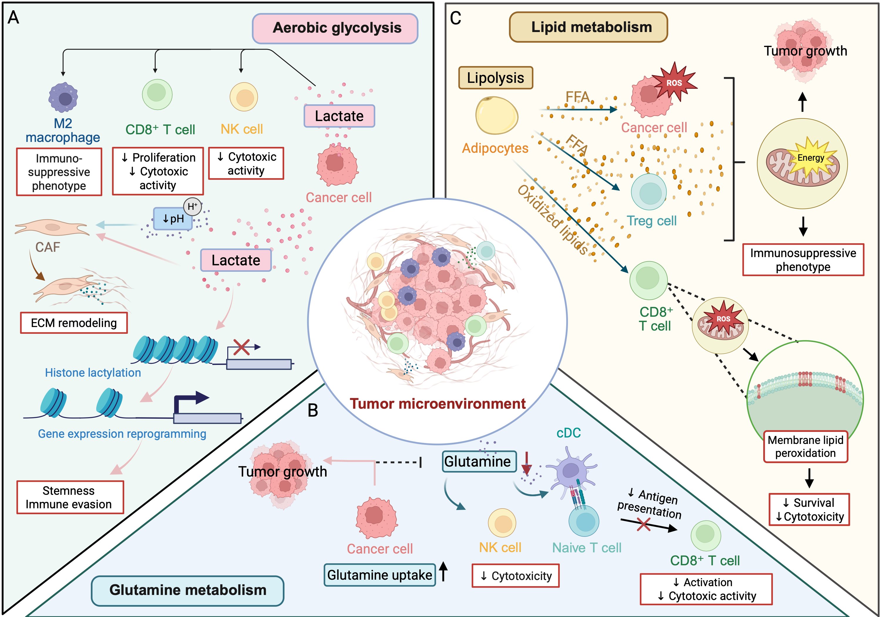
Among the metabolic intermediates that actively structure TME heterogeneity, lactate has emerged as a key molecule (Figure 1A). Lactate production is largely driven by lactate dehydrogenase A (LDHA), which catalyzes the conversion of pyruvate to lactate. Clinical studies have associated elevated LDHA expression with poorer survival in several malignancies, including lung adenocarcinoma, thyroid cancer, hematological cancers, and clear cell renal cell carcinoma (40–43). In contrast, genetic ablation of LDHA isoforms under hypoxic conditions markedly impairs tumor growth, underscoring the functional importance of fermentative glycolysis in cancer progression (44). Associations between lactate-related metabolic features and clinical outcome, together with functional studies of LDHA, support a role for lactate metabolism in tumor progression. However, these observations do not establish lactate as a broadly validated prognostic biomarker across cancer types, nor do they distinguish its effects from those of the accompanying acidic and glycolytic state.
Lactate is imported via monocarboxylate transporter 1 (MCT1), converted back to pyruvate by LDH, and oxidized in the TCA cycle to fuel mitochondrial ATP production and biosynthetic processes (45). Through this metabolic coupling, lactate shuttling between glycolytic and oxidative cancer cell populations, notably between cancer cells and cancer-associated fibroblasts (CAFs), promotes ECM remodeling, invasion, and angiogenesis by sustaining oxidative metabolism in recipient cells (46, 47). Recent evidence from non-tumor experimental systems further refines this paradigm by showing that mitochondria can generate lactate within the matrix (48). This mitochondrial lactate is exported to the cytosol, where it modulates reactive oxygen species (ROS) production, thereby revealing a previously unrecognized role for mitochondrial lactate metabolism in redox homeostasis.
Lactate as a signaling molecule
Beyond its role as a carbon source, lactate can influence the tumor niche through receptor-mediated signaling and through changes associated with extracellular acidification. Lactate-driven acidification of the TME facilitates invasion and metastasis through the activation of matrix-degrading proteases, weakening of cell-matrix adhesion, induction of epithelial-to-mesenchymal transition (EMT), and the selection of aggressive, acid-tolerant cancer cell clones (49).
Lactate also acts as a potent immunomodulatory signal shaping immune cell polarization and function. Tumor-derived lactate promotes macrophage polarization toward non-inflammatory, tumor-supportive phenotypes and suppresses the proliferation and cytotoxic activity of T cells and natural killer (NK) cells, thereby conferring a competitive advantage to immunosuppressive phenotypes (50–52). These immunosuppressive effects are partly mediated by lactate-sensing receptors such as GPR81/HCAR1, expressed on immune and stromal cells, which transduce extracellular lactate signals into intracellular programs. Through this receptor-mediated mechanism, lactate functions not merely as a metabolic competitor but as an active ligand that reprograms immune cell behavior independently of pH changes (53, 54). Intratumoral dendritic cells (DCs) require MCT4-mediated lactate efflux to maintain intracellular pH and cGAS-STING signaling, thereby preserving their anti-tumor activity (55). In NSCLC cells, lactate has also been reported to increase CD38 expression through OXPHOS–Hippo-TAZ signaling, promoting adenosine receptor signaling, Snail induction, and EMT (56).
Lactate as an epigenetic regulator
Lactate also influences TME heterogeneity at the epigenetic level. Lactate-driven protein and histone lactylation, the covalent modification of lysine residues by lactyl groups, represents a post-translational modification that links metabolic state to transcriptional reprogramming (57). Histone lactylation marks, including H3K18la, accumulate under conditions of high glycolytic flux and have been shown to activate gene expression programs associated with tumor progression, stemness, and immune evasion. In bladder cancer-associated macrophages, lactate-induced histone lactylation promotes the expression of homeostatic and anti-inflammatory genes, contributing to the establishment of immunosuppressive tumor-associated macrophage (TAM) phenotypes in a manner distinct from classical cytokine-driven polarization (58). Lactate-associated lactylation therefore provides one mechanism by which metabolic state can influence transcriptional programs in cancer and immune cells. However, the persistence, reversibility, and relative contribution of these modifications to intratumoral heterogeneity remain incompletely defined.
4. Glutamine metabolism and immunometabolic competition
Glutamine addiction and metabolic competition
Glutamine is one of the most abundant and versatile amino acids in the human body and plays a central role in maintaining cellular and systemic metabolic homeostasis (Figure 1B). In tumors, glutamine serves as a critical carbon and nitrogen source, supporting biosynthetic processes, redox balance, and energy production through its contribution to the TCA cycle via conversion to α-ketoglutarate (αKG). This reliance on glutamine-derived carbon has led to the concept of “glutamine addiction”, whereby cancer cells become highly dependent on glutamine metabolism to sustain proliferation, survival, and metastatic spread (59–61).
Importantly, glutamine addiction is not merely a consequence of nutrient availability but reflects a profound metabolic rewiring. When extracellular glutamine becomes limiting, cancer cells can exploit alternative metabolic pathways to preserve glutamate and αKG pools. For example, N-acetyl-aspartyl-glutamate can be converted into glutamate through enzymatic pathways involving glutamate carboxylase and glutamine transaminase K (GTK) (62). Consistent with the functional importance of this pathway, genetic ablation of GTK significantly impairs tumor growth in pancreatic cancer models (63), highlighting glutamine-dependent anaplerosis as a critical metabolic vulnerability.
At the tissue level, the high glutamine demand of cancer cells profoundly reshapes nutrient availability. Using in vivo metabolic tracing, Reinfeld and colleagues demonstrated that cancer cells preferentially import glutamine, whereas immune cells preferentially import glucose, a nutrient-partitioning program driven by intrinsic mTORC1-dependent metabolic circuits rather than nutrient scarcity itself (64). Nevertheless, immune cells remain highly dependent on glutamine to sustain activation, proliferation, and effector differentiation (65). Competition for glutamine can therefore influence immune-cell function in nutrient-limited tumors, although the direction and magnitude of this competition depend on cell type, tumor context, and the availability of alternative substrates.
Glutamine metabolism in anti-tumor immunity: a double-edged sword
As mentioned, the metabolic competition for glutamine within the TME has important consequences for anti-tumor immunity. A compelling example is provided by type-1 conventional DCs (cDC1s), which are essential for priming cytotoxic CD8⁺ T cell responses. Guo and colleagues showed that glutamine acts as an intercellular metabolic checkpoint regulating cancer cell-cDC1 crosstalk. Competition for glutamine uptake through SLC38A2 limits cDC1 activation and CD8⁺ T cell priming, whereas restoring intratumoral glutamine availability enhances anti-tumor immunity and sensitizes tumors to immune checkpoint blockade and T cell-based immunotherapies (66). Additional evidence indicates that glutamine deprivation disrupts immunosuppressive TAM polarization (67).
Beyond nutrient availability, glutamine-derived glutamate can also regulate immune responses through glutamatergic signaling. In particular, loss of mGluR4 activity in DCs enhances their migration, maturation, and antigen-presenting capacity, thereby strengthening CD8⁺ T cell and NK cell responses and suppressing tumor growth and metastasis by limiting adenyl cyclase/PKA signaling (68).
Effector T cells, NK cells and DCs rely on glutamine not only as a bioenergetic substrate but also for nucleotide synthesis, protein production and maintenance of mTORC1-dependent activation programs. Consequently, indiscriminate glutamine depletion can impair lymphocyte proliferation and cytokine production (69). In vitro, restriction of glutamine-dependent nucleotide synthesis during human T cell activation enriches FOXP3-high CD4+ populations with regulatory properties (70), raising the possibility that indiscriminate glutamine deprivation could have context-dependent immunosuppressive consequences.
Adding further complexity, glutamine metabolism can intrinsically threaten T cell survival. Zhang and colleagues recently showed that glutaminolysis in activated CD8⁺ T cells generates ammonia that accumulates within lysosomes. Excessive ammonia accumulation disrupts lysosomal homeostasis, promotes mitochondrial dysfunction, and impairs autophagic flux, ultimately triggering a distinct form of T cell death characterized by lysosomal alkalinization and mitochondrial swelling (71). Thus, glutamine catabolism represents not only a metabolic resource for activated T cells but also a potential source of endogenous metabolic toxicity.
5. Lipid metabolic adaptation, ferroptosis and immune dysfunction
Lipid metabolism plays a central role in cellular homeostasis by providing structural components for membranes, signaling molecules, and bioenergetic substrates, and is strongly shaped by the spatial heterogeneity of the TME. Hypoxic niches promote lipid uptake and neutral lipid storage in lipid droplets, buffering lipotoxic stress while preserving substrates for membrane remodeling and oxidative metabolism upon reoxygenation (72). In this way, metabolic adaptation fosters compartmentalization of lipid resources, thereby shaping lipid availability and exchange between cancer cells, the circulation, and surrounding adipose tissue (73).
Beyond passive storage, cancer cells undergo profound metabolic adaptations that allow them to secure lipids through de novo synthesis, extracellular uptake, and lipid exchange with neighboring stromal and immune cells. These processes support cancer cell fitness and help maintain a metabolically resilient, tumor-supportive microenvironment (74). Among the lipid exchanges, one of the best-characterized lipid symbioses occurs between cancer cells and adipocytes. Tumor-derived signals induce adipocyte lipolysis and exosome secretion, resulting in the transfer of free fatty acids (FFA), cholesterol, and lipid-associated proteins to cancer cells (Figure 1C). These lipids are imported predominantly through CD36- and FABP-dependent pathways, fueling fatty acid oxidation (FAO), sustaining mitochondrial metabolism, and promoting invasive and metastatic phenotypes (75).
Paradoxically, the dependence on lipid remodeling can either protect against or create vulnerability to ferroptosis, a form of regulated cell death driven by iron-dependent peroxidation of polyunsaturated fatty acid (PUFA)-containing phospholipids (76, 77). De novo lipogenesis promotes membrane saturation and protects cancer cells from oxidative stress, whereas increased incorporation of PUFAs into membrane phospholipids enhances susceptibility to lipid peroxidation and ferroptosis. Thus, the effect of lipid remodeling depends on whether it favors oxidation-resistant saturated and monounsaturated lipids or oxidation-prone PUFA-containing phospholipids. Accordingly, increased lipid availability cannot be interpreted uniformly as pro- or anti-ferroptotic; its effect depends on lipid species, membrane incorporation, antioxidant capacity, and the metabolic state of the recipient cell.
This balance may vary across metabolically distinct TME niches, generating spatial heterogeneity in ferroptosis sensitivity (78). Ferroptosis is controlled by the balance between pro-peroxidative lipid remodeling and antioxidant defense systems. On the pro-ferroptotic side, Acyl-CoA synthetase long-chain family member 4 (ACSL4) promotes the activation of PUFAs, including arachidonic acid and adrenic acid, into acyl-CoA derivatives, whereas lysophosphatidylcholine acyltransferase 3 (LPCAT3) incorporates these PUFA-containing species into membrane phospholipids (79, 80). Together, ACSL4 and LPCAT3 increase the abundance of oxidation-prone PUFA-phospholipids, thereby sensitizing cancer cells to ferroptosis. Conversely, reduced ACSL4 or LPCAT3 activity limits the availability of peroxidizable membrane substrates and can confer resistance to ferroptotic death (81).
Cancer cells counteract this vulnerability through several anti-ferroptotic defense mechanisms. The solute carrier family 7 member 11 (SLC7A11)-glutathione- glutathione peroxidase 4 (GPX4) axis represents a central protective pathway: SLC7A11 imports cystine to sustain glutathione synthesis, and GPX4 uses glutathione to detoxify phospholipid hydroperoxides before they propagate lipid peroxidation cascades (82). In parallel, GPX4-independent systems provide additional protection. Ferroptosis suppressor protein 1 (FSP1) suppresses ferroptosis by reducing coenzyme Q10 (CoQ10) to ubiquinol at the plasma membrane, generating a lipophilic antioxidant shield that neutralizes lipid peroxyl radicals independently of glutathione (81, 83). This protective pathway is also regulated by the metabolic TME. Microenvironmental prostaglandin F2α (PGF2α) directly binds FSP1 and inhibits its enzymatic activity, promoting lipid peroxidation and sensitizing cancer cells to ferroptosis. In preclinical models, PGF2α supplementation also enhances CD8⁺ T cell-mediated immunity and responses to immunotherapy (84). Ferroptosis sensitivity therefore reflects the interaction between membrane lipid composition, cell-intrinsic antioxidant defenses, and metabolic inputs from the surrounding microenvironment.
The lipid-centered symbiosis also profoundly reshapes anti-tumor immunity. In tumor-associated macrophages, lipid-droplet-dependent long-chain fatty acid metabolism supports an immunosuppressive phenotype (85). In Tregs, tumor-driven lipid metabolic adaptation, including fatty acid synthesis and FAO, supports expansion, survival, and suppressive function under nutrient-limited conditions (86, 87). Lipid availability can also directly shape neutrophil function in a tissue-specific manner. In liver metastases, breast and colorectal cancer (CRC) cells exploit the local abundance of palmitate to promote palmitoyltransferase 17-dependent stabilization and secretion of laminin-511, which suppresses neutrophil cancer cell-killing capacity and thereby facilitates metastatic growth (88). In cytotoxic T cells, excessive lipid uptake can become detrimental. CD8+ T cells take up oxidized phospholipids and oxidized low-density lipoproteins (LDL) through CD36-dependent mechanisms (89, 90). Accumulation of these oxidized lipids promotes membrane lipid peroxidation, impairs CD8⁺ T cell metabolic fitness, and can ultimately drive ferroptotic cell death (90, 91). When lipid peroxide detoxification is insufficient, CD8⁺ T cells become particularly vulnerable to ferroptotic damage; accordingly, preservation of GPX4-dependent lipid peroxide detoxification is critical for their survival, expansion, and persistence (92, 93). Thus, lipid metabolic remodeling represents a central determinant of both ferroptotic control and immune cell escape in the TME.
6. Energy charge and redox regulation as integrators of metabolic heterogeneity
Beyond nutrient availability and metabolic pathway engagement, cellular energy charge and redox balance represent fundamental layers of metabolic heterogeneity within the TME. Energy supply within cells is primarily mediated by adenine and guanine nucleotides, whose relative abundance reflects metabolic state and dictates cellular behavior (94). In rapidly growing tumors, spatial differences in nutrient and oxygen availability generate regional disparities in ATP levels, resulting in heterogeneous energetic states across tumor regions (95). Hypoxic conditions further exacerbate this heterogeneity by promoting a shift toward glycolytic ATP production, which yields less ATP per glucose molecule than OXPHOS and alters the ATP/ADP balance. This changes cellular energy kinetics and contributes to metabolic stress (96).
Alternative nuclear and cytosolic pathways, including poly(ADP-ribose) (PAR) metabolism, have been proposed to contribute to local ATP pools under specific conditions, further diversifying energetic states within tumors. In the nucleus, activation of PARP1 and PARP2 during DNA damage responses consumes large amounts of NAD⁺ to synthesize PAR, rapidly depleting nuclear and cellular NAD⁺ and indirectly suppressing ATP production by limiting NAD⁺-dependent glycolysis and TCA cycle flux (97, 98). Excessive PAR accumulation, such as that triggered by calcium-induced mitochondrial ROS, can lead to severe energy depletion and PARP1-dependent cell death in pancreatic cancer cells, illustrating how dysregulated PAR turnover can collapse ATP levels in stressed tumor regions (99). Conversely, coordinated activity of PAR glycohydrolase (PARG) and ADP-ribose-processing enzymes has been proposed to contribute to local ATP generation in the nucleus, potentially supporting DNA repair and replication independently of bulk cytosolic ATP availability (100, 101). The energetic consequences of PAR metabolism are further modulated by genetic context: in isocitrate dehydrogenase (IDH)-mutant tumors, inhibition of PARG during alkylating chemotherapy sequesters NAD⁺ in PAR polymers, leading to global NAD⁺ depletion, impaired ATP production, and metabolic collapse, revealing a genotype-specific vulnerability rooted in nucleotide and redox imbalance (102).
Beyond the nucleus, cytosolic and extracellular purine-handling pathways also shape local energy landscapes within the TME. Cancer and stromal cells express ecto-adenylate kinase and nucleoside diphosphate kinases capable of regenerating ATP from AMP or ADP in the pericellular space (103). Together, these nuclear, cytosolic, and extracellular nucleotide cycles add a fine-grained layer of energetic heterogeneity on top of classical glycolysis vs. OXPHOS differences, linking DNA damage responses, redox balance, and microenvironmental signaling to spatial control of ATP availability (104). Guanine nucleotides contribute to metabolic heterogeneity by linking energy status to transcriptional and translational control. GTP availability regulates protein synthesis and growth signaling, and perturbations in GTP biosynthesis have been shown to impair RNA polymerase I activity (94). In MYC-driven small cell lung cancer (SCLC), disruption of GTP synthesis alters transcriptional output and contributes to chemoresistance (105).
In parallel, redox cofactors such as NADH, NADPH, and flavin adenine dinucleotide (FAD/FADH₂) play central roles in determining cellular fate. These cofactors integrate metabolic inputs from glycolysis, glutaminolysis, FAO, and mitochondrial respiration, enabling cancer cells to balance ATP production with biosynthesis and oxidative stress management (106, 107). NADPH is particularly critical for anabolic reactions and antioxidant defense, as it fuels glutathione and thioredoxin recycling. Under nutrient- or glucose-limited conditions, cancer cells exploit alternative substrates, including lactate and glutamine, to sustain NADPH production via IDH1- and malic enzyme 1 (ME1)-dependent pathways, thereby preserving redox homeostasis despite metabolic stress (108). Consistently, glucose-6-phosphate dehydrogenase (G6PD)-dependent NADPH generation and NADP⁺ pool expansion support metastatic potential and therapy resistance (109). ROS buffering through the glutathione system further reinforce this heterogeneity (110). Maintenance of reduced glutathione (GSH) pools through NADPH-dependent recycling is essential for buffering oxidative stress, regulating redox-sensitive signaling, and preventing ROS-induced cell death, thereby contributing to divergent metabolic and survival phenotypes within the same tumor (111).
7. Systemic, genetic, and tissue-context determinants of metabolic heterogeneity
Systemic metabolic inputs
Beyond cell-intrinsic metabolic programs and local nutrient competition within the TME, systemic nutrient availability adds another, often underappreciated, layer of metabolic heterogeneity by shaping spatially heterogeneous metabolite availability within the tumor interstitium. Systemic nutrient availability, shaped in part by diet and host metabolic state, can influence substrate availability and metabolic pathway usage within tumors in a context-dependent manner. Circulating nutrients such as glucose, fatty acids, and amino acids are heterogeneously distributed across tumors and dynamically sensed by cancer cells, leading to spatially distinct metabolic programs (112). Metabolic reprogramming in cancer also involves activation of the mTORC1-SREBP axis, which promotes de novo lipid synthesis and supports cancer cell growth and proliferation (113). As a result, metabolic heterogeneity is shaped not only by cancer progression stage and microenvironmental constraints, but also by inter-individual differences in systemic dietary inputs and host metabolic status (114, 115). Epidemiological studies associate dietary patterns, obesity, and metabolic disorders with the incidence and, in some settings, progression of particular cancers (116), although such associations do not establish a direct tumor-metabolic mechanism. For example, obesity has been associated with an increased incidence of several malignancies, although its impact on overall survival appears to depend on complex interactions between diet composition, nutrient excess, and the metabolic properties of the TME rather than adiposity alone (117).
Experimental studies using dietary interventions in cancer models have provided mechanistic insight into how nutrient availability modulates cancer metabolism. High-fat, Western, ketogenic, or nutrient-restricted diets have been shown to differentially reprogram metabolic pathways, either promoting or restraining tumor growth depending on tumor type and genetic context (118). For instance, high-fat diets exacerbate hyperglycemia and hyperinsulinemia in CRC models, whereas ketogenic diets can create therapeutic vulnerabilities when combined with targeted inhibitors, such as PI3K pathway blockade (119). Importantly, the metabolic impact of specific nutrients also depends on tumor stage and genotype. Replacing exogenous glucose with fructose can limit tumor initiation, while paradoxically accelerating cancer progression once the tumors are established (114).
Oncogenic and tumor suppressor-driven metabolic reprogramming
MYC activation, a potent driver of carcinogenesis across multiple cancer types, promotes the transcription of glycolytic and glutaminolytic enzymes, sustaining high proliferative capacity through enhanced glucose and glutamine catabolism (120).
KRAS mutations drive metabolic reprogramming toward increased utilization of glucose- and glutamine-derived carbon to support TCA cycle activity and biosynthesis (121). Mutant KRAS promotes hypoxia-inducible factor (HIF)-1α-dependent metabolic rewiring, increasing glucose uptake through GLUT1 and lactate production (122, 123). KRAS simultaneously reprograms glutamine metabolism by favoring non-canonical transamination pathways that replenish TCA cycle intermediates and support anabolic growth (124), while also stimulating auxiliary nutrient-consuming pathways including the pentose phosphate pathway (PPP), hexosamine biosynthesis, and folate cycling (123).
PIK3CA mutations confer differential sensitivity to glutamine availability. Compared with wild-type (WT) cells, PIK3CA-mutant CRC cells convert more glutamine to αKG to replenish the TCA cycle and generate ATP, resulting in subpopulations with distinct survival thresholds under nutrient-limited conditions (125).
Loss or mutation of TP53 enforces wide-ranging metabolic dependencies. p53 deficiency enhances glucose uptake and glycolytic flux (126). Loss of p53-mediated repression can increase PHGDH expression and serine biosynthetic capacity (127). Pan-cancer analyses have associated TP53 mutational status with hypoxia-related metabolic programs and differences in immune-cell composition (128). In breast cancer brain metastasis (BCBM), p53 loss functionally increases metastatic formation and growth in part by upregulating SCD1 and de novo FAS, with astrocytes further amplifying the lipid-anabolic state via secreted metabolites. Notably, p53-deficient tumors display vulnerability to FASN inhibition ex vivo and in vivo, highlighting FAS and SCD1 as therapeutically targetable metabolic liabilities in BCBM (129).
APC loss, an early and frequent event in CRC, has been linked to a Warburg-like shift, with increased expression of glycolytic genes (e.g., PKM2 and LDHA), elevated glucose consumption, and lactate secretion (130). APC mutations can also engage nutrient-sensing circuitry such as mTORC1, coupling oncogenic signaling to metabolite-availability cues (131).
Tissue-of-origin effects on mutation-driven metabolic phenotypes
A fundamental principle emerging from comparative metabolic analyses is that oncogenic mutations do not operate in a metabolic vacuum; instead, they redirect and amplify pre-existing tissue-specific metabolic programs, critically shaping mutation-driven metabolic phenotypes.
This principle is exemplified by MYC-driven liver tumors, in which MYC imposes a selective metabolic dependence on alanine catabolism. Montoya et al. used in vivo isotope tracing to show that MYC-driven liver cancers catabolize alanine in a GPT2-dependent manner, with alanine-derived carbon contributing to the TCA cycle, gluconeogenesis, and biosynthesis of non-essential amino acids and nucleotides (132). This dependency was functionally relevant: genetic ablation of Gpt2 limited MYC-driven liver tumorigenesis, whereas pharmacological inhibition with L-cycloserine reduced tumor formation and slowed the growth of established liver tumors. A complementary comparison by Yuneva et al. further illustrates that metabolic phenotypes cannot be inferred from oncogene or enzyme expression alone (133). By following labeled glucose fate in MYC- and MET-driven tumors, they found increased glucose catabolism across the tumors examined, but increased lactate production only in MYC-induced liver and lung tumors, not in MET-induced liver tumors, despite elevated LDHA expression. These comparisons demonstrate that the metabolic consequences of an oncogenic driver are not fixed but depend on the signaling and tissue context in which the alteration occurs.
The influence of tissue context on metabolic heterogeneity has been further demonstrated by quantitative metabolomic analyses. In a seminal study, Sullivan and colleagues introduced identical genetic alterations, KRAS activation combined with loss of p53, into lung and pancreatic ductal adenocarcinoma cells, and implanted them subcutaneously into the same host environment. Despite identical mutations and implantation sites, tumors displayed distinct branched-chain amino acid metabolic profiles, highlighting the critical contribution of the tissue of origin to metabolic heterogeneity within the TME. Thus, genotype is insufficient to predict metabolic behavior, as tissue of origin and microenvironmental factors strongly influence metabolic wiring (112).
Metabolic enzyme mutations and oncometabolites
Beyond oncogenes and tumor suppressors, mutations in core metabolic enzymes profoundly reshape cancer metabolism. Loss-of-function mutations in fumarate hydratase (FH) or succinate dehydrogenase (SDH) lead to accumulation of fumarate or succinate, respectively, which act as oncometabolites that disrupt αKG-dependent dioxygenase activity, alter chromatin and epigenetic regulation, and impair DNA repair pathways. Mechanistically, fumarate and succinate can inhibit αKG-dependent dioxygenases (including prolyl hydroxylases), stabilize HIF, and promote a pseudohypoxic transcriptional program (134, 135). Broader analyses of IDH-, FH-, and SDH-mutant tumors highlighted how oncometabolite accumulation reshapes chromatin states and signaling networks, with emerging evidence that these metabolites also modulate immune cell behavior (136). In IDH1/2-mutant tumors, the neomorphic production of (R)-2-hydroxyglutarate (2-HG) provides a clear example of an oncometabolite that competitively inhibits αKG-dependent dioxygenases, impairing histone demethylation and TET-mediated DNA demethylation (137). With this mechanism, IDH-mutant acute myeloid leukemia cells display a characteristic DNA hypermethylation phenotype linked to disrupted TET function (138). Together, these observations provide direct evidence that oncometabolites can enforce durable epigenetic states and stress-response programs that may shape cancer cell phenotypes within the tumor niche.
8. Stromal metabolic crosstalk in the TME
CAF-cancer cell metabolic interactions
CAFs are among the most abundant and functionally diverse stromal populations in solid tumors and undergo profound metabolic reprogramming in response to tumor-derived signals. Single-cell and spatial transcriptomic analyses have revealed metabolically distinct CAF subpopulations, including myofibroblastic CAFs (myCAFs) with high contractile and glycolytic activity and inflammatory CAFs (iCAFs) with distinct cytokine secretion and amino acid metabolic profiles (139). This metabolic heterogeneity contributes to spatial compartmentalization within the TME, with CAFs acting as metabolic intermediaries between cancer cell populations exposed to different levels of oxygen and nutrient availability. These categories should not be viewed as fixed metabolic lineages: CAF states can overlap, vary among tumor types, and change with spatial location and treatment exposure.
A well-characterized feature of CAF metabolism is the induction of a Warburg-like glycolytic phenotype, driven in part by cancer cell-secreted factors including TGF-β, exosomal miRNAs, and ROS (140–142). This cancer cell-imposed metabolic reprogramming suppresses mitochondrial oxidative metabolism in CAFs while upregulating glycolytic flux and lactate secretion, effectively converting CAFs into metabolic supporters that fuel oxidative cancer cells through metabolic symbiosis (143, 144).
Beyond lactate, CAFs serve as major sources of glutamine within the TME. Under nutrient-limited conditions, CAFs upregulate glutamine synthetase (GS) activity, enabling de novo glutamine synthesis from glutamate and ammonia, and export glutamine to neighboring cancer cells. This CAF-to-cancer glutamine relay sustains cancer cell anaplerosis, supports TCA cycle activity, and buffers cancer cells against the metabolic consequences of glutamine depletion (145, 146). CAFs also play a central role in lipid metabolic crosstalk, releasing lipid mediators including lysophosphatidylcholine, prostaglandins, and FFA that are taken up by cancer cells. In pancreatic and ovarian cancers, CAF-derived lipids promote cancer cell invasiveness and chemoresistance by sustaining mitochondrial metabolism and suppressing apoptotic signaling (147). Complementing these findings, metabolomics and fatty acid tracing experiments in CRC demonstrated that cancer cells can take up CAF-secreted lipids and lipid-like metabolites to compensate for low endogenous stearoyl-CoA desaturase activity, regulating membrane fluidity and invasive potential (148).
Adipocyte-cancer cell metabolic interactions
Adipocytes represent an important stromal population in cancers arising in or metastasizing to adipose-rich environments, including CRC, breast and ovarian cancers. Tumor-derived signals (TNF-α, IL-6, and Wnt ligands) stimulate adipocyte triglyceride (TG) hydrolysis. Among these lipolytic cues, TNF-α potently stimulates adipocyte lipolysis through an adipose triglyceride lipase (ATGL)-dominant mechanism, with hormone-sensitive lipase (HSL) also contributing to TG hydrolysis. In 3T3-L1 adipocytes, ATGL silencing almost completely abolished basal and TNF-α-induced glycerol release, whereas HSL knockdown only partially reduced lipolysis, supporting a major role for ATGL in TNF-α-driven release of FFA and glycerol (149).
In ovarian cancer peritoneal metastasis, adipocyte-derived FFA accelerate metastatic outgrowth by fueling FAO-dependent energy production, with CD36 blockade significantly impairing metastatic colonization (150). In breast cancer, peritumoral adipocytes undergo delipidation and reprogram toward a cancer-associated adipocyte (CAA) phenotype characterized by reduced lipid storage, increased lipolysis, and secretion of pro-tumorigenic adipokines, including leptin and IL-6, that further stimulate cancer cell proliferation and survival signaling (151). Adipocyte-derived acetate can additionally be incorporated into acetyl-CoA pools in cancer cells, influencing histone acetylation and transcriptional states that reinforce proliferative and stem-like phenotypes (38).
9. TECs as metabolic gatekeepers of TME heterogeneity
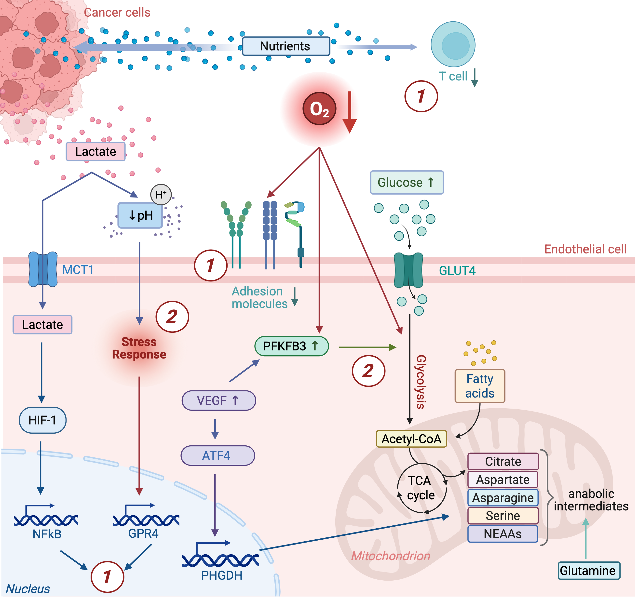
Because TECs lie at the interface between the circulation and tumor tissue, their metabolic state can influence vascular function, nutrient delivery, and exchange between systemic and local compartments. This metabolic plasticity enables TECs to coordinate vascular function while regulating the delivery and exchange of nutrients, oxygen, growth factors, and waste products (152–154). Through heterogeneous expression of nutrient transporters and their effects on vascular perfusion and exchange, TECs can contribute to spatial metabolic gradients within tumors (12, 13). Conversely, TEC metabolism and function are themselves shaped by metabolic and environmental cues within the TME (Figure 2). TECs therefore occupy a strategically important position in tumor metabolic organization and may provide context-specific therapeutic targets where pathological endothelial states create dependencies that can be separated from normal vascular metabolism.
State-dependent metabolic specialization in endothelial subpopulations
ECs comprise a spectrum of phenotypically distinct subpopulations. Across independent single-cell atlases, a conserved set of states has emerged, including highly migratory tip cells at the leading edge of sprouting, stalk cells that elongate nascent vessels, and quiescent ECs that maintain mature vessels (12, 155, 156). Single-cell studies associate these endothelial states with distinct metabolic gene-expression programs, although transcriptional signatures do not necessarily establish metabolic flux or dependency. Glycolytic bias, for example, appears to be an intrinsic feature of the migratory tip phenotype, providing rapid ATP generation independent of oxygen availability to meet the high local energy demands of cell migration (13). This metabolic program fuels the dynamic remodeling of the actin cytoskeleton required to generate the filopodial protrusions that guide angiogenic sprouting (154, 157). Consistent with this concept, TECs highly express actin-binding genes, including the cytoskeletal scaffold protein PDLIM5, which promotes filopodia formation and tumor angiogenesis by regulating ACTN1/ACTN4-dependent actin bundling. Accordingly, endothelial-specific deletion of Pdlim5 disrupts filopodia formation and suppresses sprouting angiogenesis (158).
The glycolytic phenotype of TECs is functionally relevant, although its therapeutic consequences depend strongly on the degree of pathway inhibition. Partial pharmacological inhibition of the glycolytic activator PFKFB3 by 3PO attenuates the hyperglycolytic phenotype of TECs, normalizes the disorganized tumor vasculature, reinforces endothelial barrier integrity, and reduces both intratumoral hypoxia and metastatic dissemination (13). By contrast, complete PFKFB3 inhibition at higher doses of 3PO destabilizes rather than normalizes tumor vessels, indicating that the vascular response depends on the extent of glycolytic inhibition and highlighting a defined therapeutic window (159). This dependence on the degree of glycolytic inhibition has subsequently been exploited in combination therapies. In glioblastoma, dual PFKFB3/VEGF blockade prolongs the vascular normalization window achieved only transiently with anti-VEGF therapy, thereby improving chemotherapy delivery (160), while in CRC, PFKFB3 inhibition alleviates tumor hypoxia and enhances radiotherapy response through vascular normalization (161). These studies identify endothelial glycolysis as a potentially targetable stromal dependency, but they also show that outcome depends on the degree of pathway inhibition: partial suppression can promote vascular normalization, whereas stronger inhibition may destabilize the vasculature.
In contrast to the highly glycolytic tip cell phenotype, stalk cells that trail the leading edge operate under a distinct metabolic program tailored to biomass production rather than migration (162). DLL4-Notch lateral inhibition holds these cells in a proliferative fate (163, 164), and single-cell profiling of tumor endothelium consistently shows that they downregulate the glycolytic program in favor of OXPHOS, TCA cycle activity, and one-carbon metabolism (12, 155). Consistent with this metabolic state, glioblastoma-derived TECs selectively upregulate the serine synthesis pathway. Tumor-derived signals induce ATF4-dependent expression of PHGDH, the rate-limiting enzyme of this pathway, thereby sustaining nucleotide biosynthesis and EC proliferation. Endothelial-specific deletion of PHGDH reduces pathological sprouting (165), normalizes the tumor vasculature, alleviates intratumoral hypoxia, and enhances T cell infiltration and responsiveness to CAR-T therapy (166).
Proliferating TECs also adapt their metabolism to nutrient availability within the TME. In IDH1/2-mutant gliomas, cancer cells release the oncometabolite 2-hydroxyglutarate (2-HG), which ECs preferentially import via SLC1A1 and transport to mitochondria, where it remodels mitochondrial respiration to sustain angiogenesis, revealing a vascular vulnerability not targeted by IDH1 inhibitors alone (167). Interestingly, other studies have reported distinct endothelial responses to 2-HG, including reduced endothelial proliferation, migration, and tube formation through suppression of mTOR/STAT3 signaling (168). These apparently opposing effects caution against assigning a uniform endothelial phenotype to 2-HG exposure. Differences in tumor context, 2-HG concentration, endothelial state, exposure duration, or experimental system may determine whether the net response is pro- or anti-angiogenic.
TEC metabolic adaptation to locally available nutrients extends beyond tumor-derived oncometabolites. In hepatocellular carcinoma, proliferating TECs upregulate the fructose transporter SLC2A5 and ketohexokinase, enabling fructose to fuel mitochondrial respiration through AMPK activation and thereby promote angiogenesis and metastasis (169).
Lactate as a central metabolic signaling nexus in the tumor endothelium
Lactate, produced by glycolytic TECs and Warburg-active cancer cells, can be taken up by the tumor endothelium, where it functions as both a metabolic substrate and a signaling molecule. Mechanistically, TECs import lactate through MCT1, triggering the NF-κB/IL-8 signaling axis that drives autocrine angiogenic gene expression and endothelial migration (170). Lactate uptake through MCT1 also activates HIF-1 under normoxic conditions, while pharmacological MCT1 inhibition suppresses lactate-induced HIF-1 activation and tumor angiogenesis in vivo (46).
TECs are also adapted to survive in the lactic-acidic TME. Unlike normal ECs, TECs maintain proliferation at interstitial lactate concentrations that impair normal endothelial growth. This tolerance is mediated by carbonic anhydrase II (CAII), which is upregulated in TECs downstream of VEGFA/VEGFR2 signaling and maintains intracellular pH homeostasis under acidic conditions. Accordingly, CAII knockdown selectively reduces TEC survival under lactic acidosis, whereas pharmacological CA inhibition in vivo promotes vessel maturation and increases pericyte coverage, recapitulating features observed upon bevacizumab treatment (171).
Lactate can additionally regulate TEC function through protein lactylation. In melanoma, tumor-derived lactate increases histone lactylation at the ST2 promoter in TECs, enhancing IL-33 responsiveness and promoting angiogenesis; combined LDHA and ST2 inhibition suppresses tumor growth in vivo (172). Beyond TECs, lactylation-dependent mechanisms in other cellular compartments may further influence the tumor vasculature. In ovarian cancer, AARS1-driven ENO1-K71 lactylation enhances lactate production, which increases H3K9la and H3K14la lactylation at the ESM1 locus and promotes ESM1 expression in TECs. Secreted ESM1 subsequently acts on TECs to promote angiogenesis (173).
These studies support several mechanisms through which lactate can influence tumor endothelial behavior, including MCT1-dependent signaling, adaptation to acidosis, and lactylation-associated transcriptional regulation. Their relative contribution likely varies with tumor type, vascular state, and local lactate exposure.
Amino acid metabolism in TECs
Glutamine is the most abundant circulating amino acid and a major anaplerotic substrate for proliferating endothelium. ECs rely on glutamine metabolism predominantly for proliferation, consistent with the distinct metabolic requirements of stalk and tip cells during angiogenic sprouting (174, 175). Genetic or pharmacological inhibition of glutaminase 1 (GLS1), which converts glutamine to glutamate, impairs EC proliferation and angiogenic function across experimental models (175–177). This dependency extends to the tumor vasculature, where endothelial-specific GLS deletion reduces tumor growth and metastasis while increasing pericyte coverage and perfusion, alleviating hypoxia, and improving chemotherapy delivery (178).
Glutamine metabolism also supports endothelial growth through aspartate production. Endothelial aspartate activates mTORC1, sustaining translation of the pro-angiogenic receptors VEGFR2 and FGFR1 while supplying pyrimidines required for nucleotide synthesis and proliferation (179). Glutamine catabolism therefore couples anaplerotic and biosynthetic demands to the translational control of angiogenic signaling.
Importantly, however, these pathways can have different consequences in endothelial versus malignant compartments. Beyond glutamine, amino acid metabolism intersects with other metabolic pathways that shape TEC function, as illustrated by the PHGDH-dependent serine synthesis program discussed above. These pathways also contribute to metabolic adaptation in malignant cells. Increased PHGDH activity in TECs supports pathological angiogenesis (166), whereas PHGDH-dependent serine synthesis in breast cancer cells can promote tumor growth and metastatic progression, highlighting the importance of this metabolic pathway across distinct tumor compartments (180).
Lipid metabolism and endothelial homeostasis
Lipid metabolism contributes to endothelial homeostasis not only by supplying carbon for TCA cycle replenishment, aspartate production, and nucleotide synthesis (162), but also by maintaining redox homeostasis (181) and endothelial identity (182), and by generating metabolites that act as pro-angiogenic signals (183).
Single-cell profiling of breast tumor endothelium identified a lipid-processing endothelial subpopulation (LIPEC), enriched in the peritumoral vasculature and characterized by expression of PPARγ-regulated lipid-handling genes (184). A retrospective analysis of more than 4,000 patients with breast cancer associated the LIPEC signature with long-term outcome among patients receiving metformin (184). This association is hypothesis-generating; retrospective data cannot establish that the endothelial state predicts metformin benefit or that PPARγ-dependent endothelial metabolism mediates the clinical association.
This metabolic feature becomes particularly relevant in lipid-rich metastatic environments. In CRC liver metastases, TECs acquire a proliferative, FAO-active phenotype characterized by increased lipid utilization. The TEC-enriched factor F3 inhibits the autophagy-lysosome pathway through JNK-ERK-p53 signaling, preventing CPT1A degradation and enhancing lipid uptake and lipophagy. This FAO-dependent state confers resistance to anti-VEGFA therapy, whereas CPT1A inhibition restores therapeutic sensitivity in preclinical models (185). Consistent with a role for endothelial FAO in therapeutic resistance, CPT1A inhibition has been shown to restore sensitivity to anti-VEGF therapy (186), supporting endothelial FAO as a potential target for overcoming resistance to angiogenesis blockade.
Lipid-dependent resistance can also arise through metabolic crosstalk between TECs and cancer cells. In hepatocellular carcinoma, apolipoprotein A2 (APOA2) promotes endothelial-to-mesenchymal transition (EndMT) through TGF-β signaling, reducing VEGFR2 expression in TECs while simultaneously inducing FAO reprogramming in adjacent cancer cells. APOA2-enriched tumors are refractory to anti-VEGFA therapy, whereas TGF-β inhibition suppresses both EndMT and cancer cell lipid reprogramming (187). These findings support a lipid-associated intercellular circuit linking endothelial plasticity and cancer cell metabolism to resistance to anti-angiogenic therapy (AAT) in the reported hepatocellular carcinoma models.
Additional regulators of endothelial lipid handling directly influence tumor vascular function. Endothelial ANGPTL4 restrains lipoprotein lipase-dependent fatty acid uptake, and its deletion increases endothelial fatty acid uptake while reducing pathological neovascularization and vascular permeability and increasing pericyte coverage (188). FABP4, an intracellular lipid chaperone enriched in tumor endothelium, similarly supports tumor angiogenesis, whereas endothelial FABP4 depletion increases oxidative stress and suppresses tumor vascularization in vivo (189).
10. Metabolic plasticity as a driver of therapeutic resistance
Therapeutic pressure can actively reshape metabolic interactions within the TME, selecting adaptive states that enable cancer cells and stromal populations to survive treatment and sustain disease progression. This is particularly evident after AAT, which restricts vascular supply while simultaneously imposing hypoxia and nutrient limitation. Rather than simply starving the tumor, these conditions promote metabolic adaptation across multiple cellular compartments and create alternative routes of therapeutic escape. Importantly, metabolic changes detected after therapy may represent either causal resistance mechanisms or secondary adaptations in surviving cells; functional perturbation is required to distinguish the two.
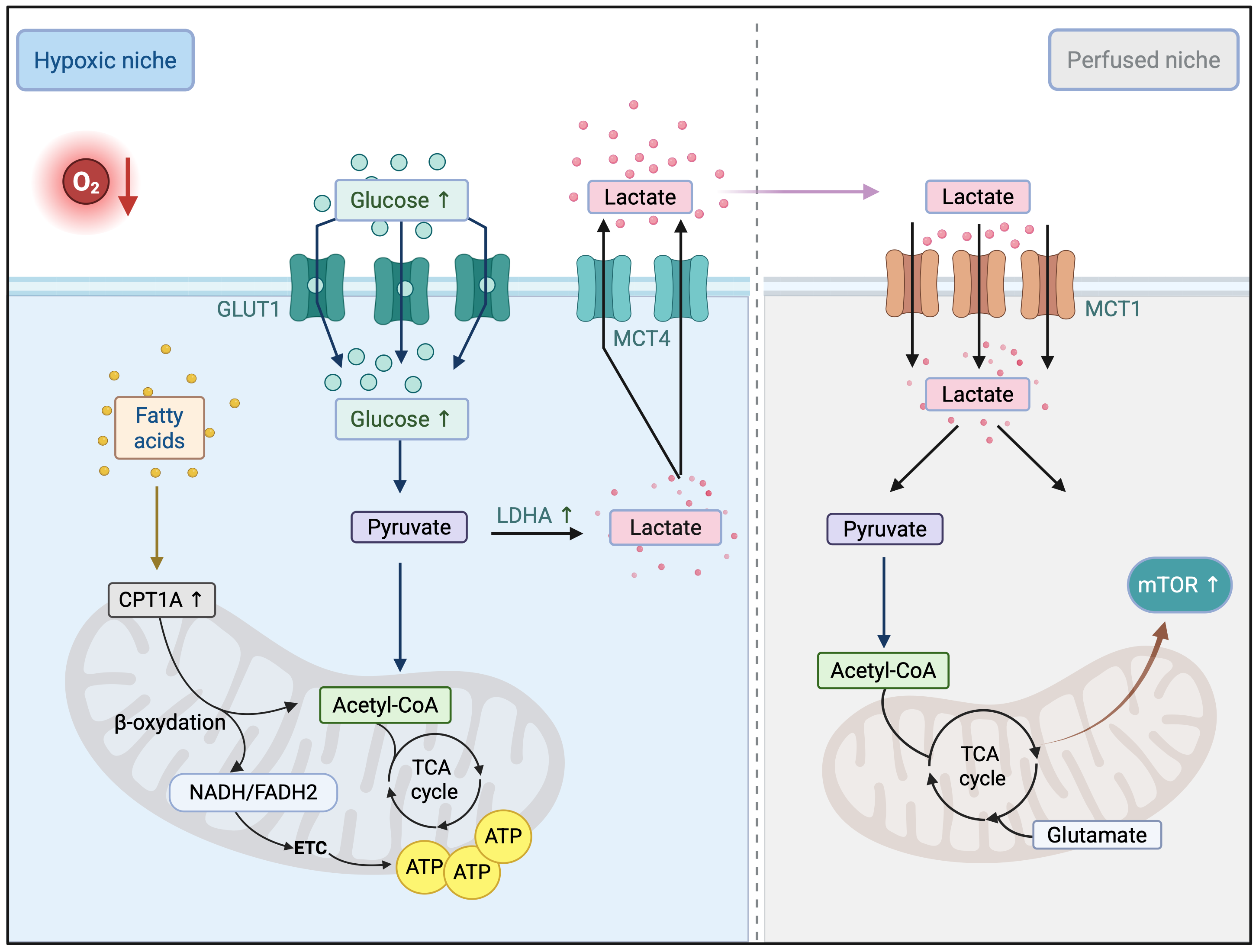
One prominent adaptation to AAT is the redistribution of cancer cell metabolism according to oxygen and nutrient availability. In bevacizumab-treated glioblastoma, increased hypoxia drives a shift toward anaerobic glycolysis and lactate accumulation (190). Under these conditions, metabolic symbiosis can emerge between hypoxic MCT4⁺ glycolytic cancer cells and MCT1⁺ oxidative cells located closer to residual vessels, allowing spatially distinct cancer cell populations to exchange metabolites and sustain growth despite restricted vascular supply through the upregulation of mTOR signaling (191–193) (Figure 3). This adaptive response extends beyond glucose metabolism. In sorafenib-resistant hepatocellular carcinoma, PHGDH upregulation increases serine biosynthesis and glutathione production (194). Similarly, AAT withdrawal triggers a lipogenic rebound that promotes metastatic growth and can be suppressed by FASN or acetyl-CoA carboxylase (ACC) inhibition, revealing an acquired metabolic vulnerability after therapy (195).
Metabolic barriers to anti-tumor immunity
Metabolic reprogramming within the TME can also limit the efficacy of anti-tumor immunity. In poorly perfused and highly glycolytic tumor regions, nutrient depletion and lactate accumulation impair effector T cell metabolism and function (196). By competing for glucose, cancer cells reduce T cell glycolytic capacity, mTOR activity, and IFN-γ production, thereby promoting T cell dysfunction and tumor progression (197). Elevated lactate levels further suppress cytotoxic T cell activity and reduce responsiveness to PD-1/PD-L1 blockade (196). Thus, enhanced cancer cell glycolysis can sustain malignant-cell metabolism while creating a metabolically restrictive environment for effector lymphocytes, contributing to resistance to immune checkpoint blockade.
Additional metabolic pathways further reinforce immune suppression. Tumor-driven tryptophan catabolism through IDO1 depletes local tryptophan and generates immunosuppressive kynurenine metabolites that promote T cell dysfunction and immune tolerance (198). Similarly, extracellular adenosine produced through CD39/CD73 activity suppresses T cell responses via A2A receptor signaling and impairs NK cell function and DC activation, thereby establishing a profoundly immunosuppressive microenvironment (199). These metabolic mechanisms therefore contribute not only to cancer cell survival but also to immune escape, limiting the efficacy of anti-tumor immune responses.
Redox and biosynthetic adaptation to cytotoxic stress
Chemotherapy and radiotherapy impose substantial oxidative stress on cancer cells. In response, resistant tumors frequently reprogram redox and biosynthetic pathways to enhance antioxidant capacity and maintain survival (200). In non-small cell lung cancer (NSCLC), cisplatin exposure induces ROS-dependent upregulation of PGC-1α, promoting mitochondrial biogenesis and OXPHOS, which supports survival and limits apoptosis (201). Chemotherapy-induced mitochondrial adaptation can also involve calcium homeostasis: in triple-negative breast cancer, the transient receptor potential melastatin 2 (TRPM2) ion channel limits mitochondrial calcium overload after chemotherapy, promoting cancer cell persistence, whereas its inhibition increases mitochondrial calcium accumulation and sensitizes cells to apoptosis (202).
Cisplatin resistance is also associated with the activation of the oxidative PPP, increasing NADPH production and glutathione-dependent ROS detoxification through enzymes such as G6PD (203, 204). Inhibition of these pathways can restore chemosensitivity, highlighting their role in acquired resistance. Similar adaptations contribute to radioresistance. By increasing antioxidant capacity through NADPH and GSH production, irradiated tumors enhance ROS detoxification and limit radiation-induced damage, with PPP activation representing a major source of antioxidant support (205, 206).
Metabolic adaptation and therapeutic resistance in cancer stem cells (CSCs)
CSCs provide a particularly clear example of metabolic plasticity under therapeutic selection. Rather than exhibiting a universal metabolic phenotype, CSCs can adopt glycolytic, oxidative, and lipid-dependent states according to tumor type, niche, and environmental pressure (207). In pancreatic ductal adenocarcinoma, the MYC/PGC-1α balance regulates switching between glycolytic and OXPHOS states, enabling CSCs to adapt their metabolic program to changing conditions (208). In AML, ROS-low leukemic stem cells depend on BCL-2-supported OXPHOS and are initially sensitive to venetoclax-based therapy (31, 209), whereas resistant populations can subsequently exploit nicotinamide and fatty acid metabolism to restore mitochondrial function (34, 35).
Tumor type and niche further shape these adaptive states. In breast cancer and glioma, CSCs maintain low intracellular ROS through elevated antioxidant capacity and NADPH production, conferring radioresistance (210). In hepatocellular and CRC, CSC states have been associated with increased lipid utilization and CD36-dependent fatty acid uptake (211), whereas in melanoma, slow-cycling CSC populations shift toward OXPHOS under targeted-therapy pressure (212).
Hypoxia adds another layer of CSC metabolic regulation, with HIF-1α-dependent programs coordinating stemness, energy metabolism, and redox homeostasis (213, 214). Spatially distinct niches, including the perivascular niche, also shape CSC metabolic plasticity by providing metabolic substrates that support persistence and therapy tolerance. Recent single-cell and organoid studies reinforce this dynamic view, indicating that therapy-resistant stem-like populations transition between metabolic states rather than relying on a single conserved metabolic program (215, 216). This plasticity may create transient windows of metabolic vulnerability, supporting sequential rather than simultaneous targeting as resistant states emerge during therapy.
Therapeutic strategies targeting TME metabolic heterogeneity: clinical translation and challenges
The metabolic heterogeneity and plasticity described above create both opportunities and major challenges for therapeutic intervention. Unlike oncogenic signaling pathways, metabolic programs are extensively shared between malignant and normal cells, vary across cellular compartments and spatial niches, and can be rapidly reconfigured under therapeutic pressure. Effective metabolic targeting therefore requires moving beyond pathway inhibition alone toward strategies that define the relevant metabolic dependency, the cellular compartment in which it operates, and the context in which it becomes therapeutically limiting. Table 1 summarizes selected metabolism-targeting agents that have reached clinical evaluation, with representative examples discussed below.
Genetically-defined and pathway-level metabolic vulnerabilities
Clinical translation has been most successful when a defined genetic alteration creates a measurable metabolic dependency and a corresponding biomarker-selected patient population. IDH1/2 mutations generate the oncometabolite 2-HG and create a discrete metabolic dependency (217). Targeting mutant IDH1 with ivosidenib produces durable responses in IDH1-mutant AML (218), while the brain-penetrant IDH1/2 inhibitor vorasidenib prolonged progression-free survival in patients with grade 2 IDH-mutant glioma (219). Notably, beyond its direct therapeutic activity, recent preclinical evidence indicates that vorasidenib pretreatment can enhance response to subsequent chemoradiation in IDH-mutant glioma (220).
Other genetic alterations similarly generate targetable metabolic dependencies. Von Hippel-Lindau (VHL) loss drives constitutive HIF-2α activity in renal cell carcinoma (RCC), enabling therapeutic targeting with the HIF-2α antagonist belzutifan (221, 222). Likewise, loss of methylthioadenosine phosphorylase (MTAP) creates a metabolic dependency on methionine adenosyltransferase 2A (MAT2A), supporting selective MAT2A inhibition in MTAP-deleted tumors. The MAT2A inhibitor AG-270/S095033 has entered early clinical evaluation in MTAP-deleted tumors, with first-in-human studies reporting pharmacodynamic target engagement and initial evidence of antitumor activity (223).
A similar genotype-associated vulnerability has recently been identified in KRAS-driven AML, where mitochondrial NAD⁺ homeostasis represents a selective metabolic dependency. Combined targeting of the mitochondrial NAD⁺ transporter SLC25A51 and SDH selectively depletes mitochondrial NAD⁺, inducing energetic collapse and suppressing leukemia progression (224). This vulnerability remains preclinical, however, and its therapeutic window will depend on whether mitochondrial NAD⁺ depletion can be achieved selectively in malignant cells.
By contrast, pathway-level metabolic inhibitors have produced more variable clinical outcomes. The GLS inhibitor telaglenastat failed to improve progression-free survival when combined with cabozantinib in advanced RCC despite a strong preclinical rationale (225), while AZD3965 confirmed pharmacological inhibition of MCT1 but encountered dose-limiting toxicity during early clinical development (226). Similarly, the complex I inhibitor IACS-010759 showed potent preclinical activity in OXPHOS-dependent tumors (227), but phase I studies in advanced solid tumors and AML revealed lactic acidosis and peripheral neuropathy at exposures required for sustained target inhibition (228).
Therapeutic pressure can also generate new metabolic dependencies. In chemoresistant NSCLC, C/EBPβ-dependent metabolic reprogramming promotes reliance on FAO, increasing mitochondrial ATP production that sustains ATP-dependent multidrug efflux. Genetic or pharmacological inhibition of FAO restores chemosensitivity and additionally reprograms NK cell metabolism to enhance anti-tumor activity, identifying FAO as an actionable vulnerability of the chemoresistant state (229). Together, these experiences suggest that clinical tractability depends less on whether a metabolic pathway is altered in cancer than on whether inhibition exposes a sufficiently selective dependency that alternative pathways or normal tissues cannot rapidly compensate for.
Broader approaches are also being explored, including the glutamine antagonist sirpiglenastat (DRP-104), which targets multiple glutamine-dependent pathways and has shown both cancer-intrinsic and immunomodulatory activity in preclinical models (230, 231). The contrast between strong preclinical activity and variable clinical performance indicates that pathway dependence alone is insufficient for successful metabolic therapy; target engagement, normal-tissue dependence, compensatory pathway usage, and patient selection also determine the therapeutic window.
Extending metabolic targeting across the TME
The effects of metabolically targeted therapies are not necessarily confined to cancer cells. As illustrated by DRP-104, metabolic interventions can simultaneously affect malignant and non-malignant cell populations, potentially reshaping metabolic interactions across the TME. Whether such multicompartment activity improves efficacy or instead increases toxicity will depend on the selectivity of the targeted dependency. TECs provide a particularly relevant example, as targeting endothelial glycolysis, amino acid metabolism, or lipid utilization can alter vascular function and influence therapeutic response, as discussed above. Importantly, these approaches may exploit metabolic dependencies associated with pathological endothelial states without requiring complete suppression of pathways essential for normal vascular function.
Other stromal and immune populations provide complementary metabolic targets. TAMs undergo metabolic adaptation to hypoxia and tumor-derived metabolites such as lactate (50, 232), while CAFs can sustain cancer cell metabolism through nutrient exchange, including glutamine production under nutrient-limited conditions (233). Targeting these stromal metabolic programs may therefore disrupt both their intrinsic functions and the metabolic support they provide to neighboring cancer cells.
Immune-metabolic pathways have progressed further toward clinical translation. IDO1 inhibition with epacadostat failed to improve the efficacy of pembrolizumab in metastatic melanoma (234), highlighting the difficulty of translating inhibition of a single metabolic checkpoint into effective restoration of anti-tumor immunity. By contrast, CD73 blockade with oleclumab, alone or with durvalumab, has demonstrated tolerability and biomarker activity (235), and adenosine-axis combinations, including with radiotherapy and immunotherapy, remain among the most actively developed metabolic strategies (236). The NeoCOAST trial further evaluated durvalumab-based neoadjuvant combinations, including oleclumab, in resectable NSCLC (237, 238). Biomarker modulation and tolerability should not, however, be equated with clinical efficacy; whether adenosine-axis inhibition provides durable clinical benefit is likely to depend on tumor context, combination partner, and patient selection.
Adoptive cell therapies provide another setting in which TME metabolism may become therapeutically relevant. In solid tumors, CAR-T cell persistence and function are constrained by hypoxia, nutrient competition, and the accumulation of immunosuppressive metabolites. Strategies to improve the metabolic fitness of transferred T cells, including enhanced mitochondrial function and adaptation to harsh environments, may therefore complement interventions that remodel the metabolic composition of the TME (239).
The timing and sequence of interventions targeting different TME compartments may also influence therapeutic efficacy. In preclinical NSCLC, sequential anti-PD-L1 followed by anti-VEGFR2 treatment was more effective than concurrent administration (240), indicating that treatment sequence can influence efficacy in at least some preclinical settings and warrants prospective evaluation rather than assuming that concurrent administration is optimal.
Toxicity and patient selection
Toxicity remains a major constraint on the clinical development of metabolism-directed therapies because many metabolic targets are also required for normal tissue homeostasis. Biguanides carry a risk of lactic acidosis and gastrointestinal toxicity, HIF-2α antagonists can cause anemia and hypoxia-related adverse events, and inhibition of glutamine metabolism may affect highly proliferative normal tissues (228). Thus, toxicity is not simply a consequence of metabolic inhibition, but a key determinant of which metabolic vulnerabilities can be therapeutically exploited and at what level of pathway inhibition.
Patient selection is therefore critical for maximizing the therapeutic window. Beyond established genomic biomarkers, functional metabolic markers may be particularly important for TME-directed strategies, where therapeutic vulnerabilities are often defined by cellular and metabolic states rather than genetic alterations. Metabolic imaging, circulating metabolites, and single-cell profiling could complement these approaches by identifying these states and monitoring their evolution during treatment. Endothelial metabolic signatures associated with therapeutic response (241) and GLUT1 expression associated with response to neoadjuvant chemoradiotherapy in CRC (242) illustrate the potential of metabolic phenotypes for patient stratification, although prospective validation remains required. A key unresolved question is whether such functional metabolic biomarkers provide predictive information beyond tumor genotype, histology, and established clinical biomarkers which will need to be addressed in biomarker-stratified prospective studies.
| Metabolic axis | Drug | Molecular target | Clinical setting / trial phase | Treatment regimen | ClinicalTrials.gov ID |
|---|---|---|---|---|---|
| Immuno-metabolism | Oleclumab (MEDI9447) |
Adenosine axis/CD73 | Phase II (resectable stage III NSCLC) |
Durvalumab (anti-PD-L1) |
NCT03794544 |
| Phase III (unresectable stage III NSCLC) | Durvalumab (anti-PD-L1) | NCT05221840 | |||
| Immuno-metabolism | Etrumadenant (AB928) |
Adenosine-A2A/A2B | Phase Ib/II (mCRC) | Zimberelimab (anti-PD-1) + mFOLFOX6 + bevacizumab (anti-VEGF) | NCT04660812 |
| Phase II (advanced NSCLC) | Zimberelimab + domvanalimab | NCT04791839 | |||
| Phase II (mCRPC) | SRF617 + Zimberelimab | NCT05177770 | |||
| Immuno-metabolism | Epacadostat (INCB024360) |
IDO1 / Tryptophan-kynurenine axis |
Phase I/II (advanced solid tumors) | Pembrolizumab (anti-PD-1) | NCT02178722 |
| Phase I/II (advanced solid tumors, B cell NHL, including DLBCL, and HL) | Nivolumab (anti-PD-1) | NCT02327078 | |||
| Phase I/II (advanced solid tumors) | Ipilimumab (anti-CTLA-4) | NCT01604889 | |||
| Phase I/II (advanced solid tumors) | Durvalumab (anti-PD-L1) | NCT02318277 | |||
| Phase III (unresectable or metastatic melanoma) | Pembrolizumab (anti-PD-1) | NCT02752074 | |||
| Glutamine metabolism |
Telaglenastat (CB-839) |
GLS1 | Phase II (advanced/metastatic ccRCC) | Everolimus | NCT03163667 |
| Phase II (advanced/metastatic ccRCC) | Cabozantinib | NCT03428217 | |||
| Phase II (ccRCC and NSCLC) | Cabozantinib, everolimus | NCT02771626 | |||
| Phase I/II (EGFR-mutated stage IV NSCLC) | Nivolumab (anti-PD-1) | NCT03831932 | |||
| Arginine metabolism | Pegargiminase (ADI-PEG20) |
Arginine metabolism | Phase II/III (nonepithelioid pleural mesothelioma) | Pemetrexed + cisplatin/carboplatin | NCT02709512 |
| Phase II (ASS1-deficient malignant pleural mesothelioma) | Monotherapy | NCT01279967 | |||
| Phase II (ASS1-deficient mesothelioma and non-squamous NSCLC) | Pemetrexed + cisplatin/carboplatin | NCT02029690 | |||
| Lactate metabolism | AZD3965 | MCT1 | Phase I (advanced solid tumors/lymphoma, DLBCL, BL) | Monotherapy | NCT01791595 |
| Hypoxia | Belzutifan (MK-6482) |
HIF-2α | Phase II (VHL-associated ccRCC) | Monotherapy | NCT03401788 |
| Phase III (advanced ccRCC after PD-1/PD-L1 and VEGF-TKI) | Monotherapy | NCT04195750 | |||
| Phase II (PPGL, pNET, VHL-associated tumors, wt GIST, HIF-2α-altered solid tumors) | Monotherapy | NCT04924075 | |||
| OXPHOS | Metformin hydrochloride |
ETC complex I / AMPK-mTOR | Phase III (early-stage breast cancer) | Standard chemotherapy | NCT01101438 |
| Phase II (stage IIIa/IIIb locally advanced NSCLC) | Chemoradiotherapy | NCT02115464 | |||
| Phase II (HNSCC) | Surgery ± cisplatin/radiotherapy | NCT03048500 | |||
| OXPHOS | Lixumistat (IM156) |
ETC complex I | Phase I (advanced solid tumor and lymphoma) | Monotherapy | NCT03272256 |
| Phase Ib (advanced pancreatic cancer) | Gemcitabine + Nab-paclitaxel | NCT05497778 | |||
| OXPHOS | IACS-010759 | ETC complex I | Phase I (relapsed/refractory AML) | Monotherapy | NCT02882321 |
| Phase I (advanced solid tumors / lymphoma) | Monotherapy | NCT03291938 | |||
| TCA cycle metabolism |
Ivosidenib (AG-120) |
Mutant IDH1 / 2-HG production |
Phase I (R/R AML, untreated AML, MDS, MDS/MPN) | Monotherapy | NCT02074839 |
| Phase III (IDH1-mutated newly diagnosed AML) | Azacitidine | NCT03173248 | |||
| Phase III (advanced cholangiocarcinoma) | Monotherapy | NCT02989857 | |||
| TCA cycle metabolism |
Vorasidenib (AG-881) |
Mutant IDH1/2 / 2-HG production |
Phase III (grade 2 IDH-mutant glioma) | Monotherapy | NCT04164901 |
| Phase I (low-grade glioma) | Monotherapy | NCT03343197 | |||
| Phase I (advanced solid tumors, including glioma) | Monotherapy | NCT02481154 | |||
| Lipid metabolism | Denifanstat (TVB-2640) |
FASN | Phase II (HER2+ metastatic breast cancer) | Trastuzumab (anti-HER2) + paclitaxel (or endocrine therapy) | NCT03179904 |
| Phase II (advanced/metastatic KRAS-mutant NSCLC) | Monotherapy | NCT03808558 | |||
| Phase II (first relapse of high-grade astrocytoma) | Bevacizumab (anti-VEGF) | NCT03032484 | |||
| Glycolysis | PFK-158 (ACT-PFK-158) |
PFKFB3 | Phase I (advanced solid malignancies) | Monotherapy | NCT02044861 |
| Glycolysis | 2-Deoxy-D-glucose (2-DG) |
HK | Phase I (lung, breast, pancreatic, HNC, and gastric cancers) | Monotherapy or docetaxel | NCT00096707 |
Table 1. Selected metabolism-targeting agents evaluated in clinical trials. Representative clinical trials of anticancer strategies targeting metabolic pathways, showing molecular targets, clinical settings and trial phases, treatment regimens, and ClinicalTrials.gov identifiers. Cancer types and clinical terms: AML, acute myeloid leukemia; BL, Burkitt lymphoma; B cell NHL, B cell non-Hodgkin lymphoma; ccRCC, clear cell renal cell carcinoma; CNS, central nervous system; DLBCL, diffuse large B-cell lymphoma; HL, Hodgkin lymphoma; HNC, head and neck cancer; HNSCC, head and neck squamous cell carcinoma; MDS, myelodysplastic syndrome; MPN, myeloproliferative neoplasm; mCRPC, metastatic castration-resistant prostate cancer; mCRC, metastatic colorectal cancer; NSCLC, non-small cell lung cancer; pNET, pancreatic neuroendocrine tumor; PPGL, pheochromocytoma and paraganglioma; RCC, renal cell carcinoma; R/R, relapsed/refractory; wt GIST, wild-type gastrointestinal stromal tumor. Molecular targets, pathways and biological terms: 2-HG, 2-hydroxyglutarate; A2A/A2B, adenosine A2A/A2B receptors; AMPK, AMP-activated protein kinase; ASS1, argininosuccinate synthetase 1; CD73, cluster of differentiation 73; CTLA-4, cytotoxic T-lymphocyte-associated protein 4; EGFR, epidermal growth factor receptor; ETC, electron transport chain; FASN, fatty acid synthase; GLS1, glutaminase 1; HER2, human epidermal growth factor receptor 2; HIF-2α, hypoxia-inducible factor 2 alpha; HK, hexokinase; IDH, isocitrate dehydrogenase; IDO1, indoleamine 2,3-dioxygenase 1; KRAS, Kirsten rat sarcoma viral oncogene homolog; MCT1, monocarboxylate transporter 1; mTOR, mechanistic target of rapamycin; OXPHOS, oxidative phosphorylation; PD-1, programmed cell death protein 1; PD-L1, programmed death-ligand 1; PFKFB3, 6-phosphofructo-2-kinase/fructose-2,6-bisphosphatase 3; TCA, tricarboxylic acid cycle; VEGF, vascular endothelial growth factor; VHL, von Hippel–Lindau. Drugs, treatment regimens and therapeutic terms: 2-DG, 2-deoxy-D-glucose; mFOLFOX6, modified folinic acid, fluorouracil, and oxaliplatin; TKI, tyrosine kinase inhibitor.
Discussion
Evidence across experimental systems supports a view of metabolic heterogeneity as a multicellular property of the TME, arising from interactions among malignant, stromal, immune, and vascular compartments rather than from cancer cells alone. Several priorities now emerge for translating TME metabolic heterogeneity into experimentally and therapeutically actionable strategies.
First, tumor metabolism needs to be resolved at spatial and cellular resolution. Spatial metabolomics can map metabolite distributions across perivascular niches, hypoxic regions, and immune-excluded areas, whereas single-cell transcriptomics can identify the cellular populations associated with these metabolic states (241). Neither approach alone resolves metabolic flux: transcript abundance does not necessarily predict pathway activity, while spatial metabolite measurements may not identify the cells responsible for metabolite production or consumption.
Genetically encoded metabolite biosensors can complement these approaches by capturing metabolic dynamics in real time. For example, the glutamine biosensor iGlo enables monitoring of glutamine uptake, production, and consumption at cellular and subcellular resolution and can be multiplexed with lactate sensors to resolve temporal crosstalk between glucose and glutamine metabolism in individual cells (243).
A major unresolved question is whether the metabolite exchange inferred from co-localization, transcriptional programs, and ex vivo experiments occurs at quantitatively meaningful rates in vivo. Stable-isotope infusion has demonstrated the feasibility of tracing nutrient utilization directly in human tumors (244). Compartment-resolved in vivo tracing should now be used to determine the direction and magnitude of substrate exchange between cancer cells, TECs, immune cells, and fibroblasts, and how these fluxes change during treatment. Such measurements could distinguish genuine intercellular metabolite exchange from parallel adaptation to the same microenvironmental constraint and determine whether disrupting such metabolic interactions produces a selective therapeutic effect.
TECs represent a potentially important, but incompletely validated, compartment for metabolism-directed therapeutic development. Future studies should further establish which endothelial metabolic dependencies are conserved across tumor types and which are restricted to specific vascular states, tumor regions, or metastatic niches. TEC-selective delivery strategies and dose optimization will be important to determine whether these dependencies can be targeted while preserving normal vascular function. Direct comparison with ECs from matched normal tissues will be particularly important because many of the pathways discussed here, including glycolysis, glutaminolysis, serine synthesis, and FAO, also support physiological endothelial function. Future studies should also define which metabolic interventions enhance responses to AAT, chemotherapy, radiotherapy, or immunotherapy (245, 246).
Clinical translation will further require biomarkers that identify the relevant metabolic dependency in the appropriate cellular compartment. Spatial metabolic signatures, metabolic imaging, circulating metabolites, and cell type-specific molecular profiles should therefore be prospectively evaluated for patient stratification and pharmacodynamic monitoring. Integrating these measurements may be particularly important for targeted therapies, for which tumor genotype may be insufficient to predict response when the relevant metabolic dependency is strongly influenced by cellular state or microenvironmental context.
Treatment scheduling also warrants systematic investigation during metabolic therapy development. Metabolic dependencies can change during treatment, creating vulnerabilities that are absent at baseline. Where treatment induces a reproducible metabolic dependency, sequential or staggered therapy could be compared directly with concurrent treatment. Longitudinal metabolic profiling would be required to define when the dependency emerges, whether it persists, and whether its therapeutic window is sufficiently stable for clinical exploitation.
The therapeutic significance of metabolic heterogeneity therefore lies not in the existence of metabolic differences per se, but in identifying states that create selective, measurable, and sufficiently stable dependencies. Many of the mechanisms discussed in this review derive from specific tumor types, experimental models, or treatment contexts and should not be assumed to generalize across cancers. Translation will require functional validation of metabolic dependencies in the relevant cellular compartment, demonstration of a therapeutic window relative to normal tissues, and biomarkers that can identify when those dependencies are present. Framed in this way, metabolic plasticity represents both a major obstacle to pathway-directed therapy and a potential source of context-dependent vulnerabilities that may be exploited when their spatial and temporal boundaries are defined.
References
1. Warburg O. On the origin of cancer cells. Science. 1956;123(3191):309–314. https://doi.org/10.1126/science.123.3191.309
2. Hanahan D. Hallmarks of cancer-then and now, and beyond. Cell. 2026;189(8):2254–2277. https://doi.org/10.1016/j.cell.2025.12.049
3. Marusyk A, Almendro V, Polyak K. Intra-tumour heterogeneity: a looking glass for cancer? Nat Rev Cancer. 2012;12(5):323–334. https://doi.org/10.1038/nrc3261
4. Jamal-Hanjani M, Quezada SA, Larkin J, Swanton C. Translational implications of tumor heterogeneity. Clin Cancer Res. 2015;21(6):1258–1266. https://doi.org/10.1158/1078-0432.CCR-14-1429
5. Chen S, Jiang W, Du Y, Yang M, Pan Y, Li H, et al. Single-cell analysis technologies for cancer research: from tumor-specific single cell discovery to cancer therapy. Front Genet. 2023;14:1276959. https://doi.org/10.3389/fgene.2023.1276959
6. Lu P, Weaver VM, Werb Z. The extracellular matrix: a dynamic niche in cancer progression. J Cell Biol. 2012;196(4):395–406. https://doi.org/10.1083/jcb.201102147
7. Desgrosellier JS, Cheresh DA. Integrins in cancer: biological implications and therapeutic opportunities. Nat Rev Cancer. 2010;10(1):9–22. https://doi.org/10.1038/nrc2748
8. Lucchetti D, Ricciardi Tenore C, Colella F, Sgambato A. Extracellular vesicles and cancer: a focus on metabolism, cytokines, and immunity. Cancers (Basel). 2020;12(1):171. https://doi.org/10.3390/cancers12010171
9. Mun JY, Leem SH, Lee JH, Kim HS. Dual relationship between stromal cells and immune cells in the tumor microenvironment. Front Immunol. 2022;13:864739. https://doi.org/10.3389/fimmu.2022.864739
10. Zhao Y, Shen M, Wu L, Yang H, Yao Y, Yang Q, et al. Stromal cells in the tumor microenvironment: accomplices of tumor progression? Cell Death Dis. 2023;14(9):587. https://doi.org/10.1038/s41419-023-06110-6
11. Jain RK. Antiangiogenesis strategies revisited: from starving tumors to alleviating hypoxia. Cancer Cell. 2014;26(5):605–622. https://doi.org/10.1016/j.ccell.2014.10.006
12. Rohlenova K, Goveia J, García-Caballero M, Subramanian A, Kalucka J, Treps L, et al. Single-cell RNA sequencing maps endothelial metabolic plasticity in pathological angiogenesis. Cell Metab. 2020;31(4):862–877.e14. https://doi.org/10.1016/j.cmet.2020.03.009
13. Cantelmo AR, Conradi LC, Brajic A, Goveia J, Kalucka J, Pircher A, et al. Inhibition of the glycolytic activator PFKFB3 in endothelium induces tumor vessel normalization, impairs metastasis, and improves chemotherapy. Cancer Cell. 2016;30(6):968–985. https://doi.org/10.1016/j.ccell.2016.10.006
14. Hanahan D, Weinberg RA. Hallmarks of cancer: the next generation. Cell. 2011;144(5):646–674. https://doi.org/10.1016/j.cell.2011.02.013
15. Vander Heiden MG, DeBerardinis RJ. Understanding the intersections between metabolism and cancer biology. Cell. 2017;168(4):657–669. https://doi.org/10.1016/j.cell.2016.12.039
16. Haitzmann T, Thompson NB, Shah H, Schindlmaier K, Jelwan J, Eichmann TO, et al. Mannose supports lung cancer metabolism during glycolytic limitation. EMBO Rep. 2026;27(18):5365–5381. https://doi.org/10.1038/s44319-026-00874-6
17. Zheng Y, Cheng C, Cao Y, Cruz G, Zhang Y, Paturu R, et al. A stress-adaptive lipid kinase axis defines metabolic vulnerabilities in neuroendocrine prostate cancer. Cancer Cell. 2026;44(9):1761–1778.e12. https://doi.org/10.1016/j.ccell.2026.07.003
18. Tharp KM, Park S, Timblin GA, Richards AL, Berg JA, Twells NM, et al. The microenvironment dictates glyco-immune surveillance via HSF1-mediated metabolism. Sci Adv. 2026;12(32):eaeb1136. https://doi.org/10.1126/sciadv.aeb1136
19. Flint LE, Hamm G, Ready JD, Ling S, Duckett CJ, Cross NA, et al. Characterization of an aggregated three-dimensional cell culture model by multimodal mass spectrometry imaging. Anal Chem. 2020;92(18):12538–12547. https://doi.org/10.1021/acs.analchem.0c02389
20. Demicco M, Liu XZ, Leithner K, Fendt SM. Metabolic heterogeneity in cancer. Nat Metab. 2024;6(1):18–38. https://doi.org/10.1038/s42255-023-00963-z
21. Iqbal MA, Siddiqui S, Smith K, Singh P, Kumar B, Chouaib S, et al. Metabolic stratification of human breast tumors reveal subtypes of clinical and therapeutic relevance. iScience. 2023;26(10):108059. https://doi.org/10.1016/j.isci.2023.108059
22. Sun C, Wang A, Zhou Y, Chen P, Wang X, Huang J, et al. Spatially resolved multi-omics highlights cell-specific metabolic remodeling and interactions in gastric cancer. Nat Commun. 2023;14(1):2692. https://doi.org/10.1038/s41467-023-38360-5
23. Hsieh WC, Budiarto BR, Wang YF, Lin CY, Gwo MC, So DK, et al. Spatial multi-omics analyses of the tumor immune microenvironment. J Biomed Sci. 2022;29(1):96. https://doi.org/10.1186/s12929-022-00879-y
24. Andersson A, Larsson L, Stenbeck L, Salmén F, Ehinger A, Wu SZ, et al. Spatial deconvolution of HER2-positive breast cancer delineates tumor-associated cell type interactions. Nat Commun. 2021;12(1):6012. https://doi.org/10.1038/s41467-021-26271-2
25. Schmitt AM, Gätje FB, Tran MYR, Midelashvili T, Xu X, Fleischer JR, et al. Spatially-resolved subtype progression reveals metabolic vulnerabilities in pancreatic ductal adenocarcinoma. Mol Cancer. 2026;25(1):112. https://doi.org/10.1186/s12943-026-02628-3
26. Lee P, Chandel NS, Simon MC. Cellular adaptation to hypoxia through hypoxia inducible factors and beyond. Nat Rev Mol Cell Biol. 2020;21(5):268–283. https://doi.org/10.1038/s41580-020-0227-y
27. Kueckelhaus J, Frerich S, Kada-Benotmane J, Koupourtidou C, Ninkovic J, Dichgans M, et al. Inferring histology-associated gene expression gradients in spatial transcriptomic studies. Nat Commun. 2024;15(1):7280. https://doi.org/10.1038/s41467-024-50904-x
28. Miallot R, Galland F, Millet V, Blay JY, Naquet P. Metabolic landscapes in sarcomas. J Hematol Oncol. 2021;14(1):114. https://doi.org/10.1186/s13045-021-01125-y
29. Shen YA, Wang CY, Hsieh YT, Chen YJ, Wei YH. Metabolic reprogramming orchestrates cancer stem cell properties in nasopharyngeal carcinoma. Cell Cycle. 2015;14(1):86–98. https://doi.org/10.4161/15384101.2014.974419
30. Sancho P, Burgos-Ramos E, Tavera A, Bou Kheir T, Jagust P, Schoenhals M, et al. MYC/PGC-1α balance determines the metabolic phenotype and plasticity of pancreatic cancer stem cells. Cell Metab. 2015;22(4):590–605. https://doi.org/10.1016/j.cmet.2015.08.015
31. Lagadinou ED, Sach A, Callahan K, Rossi RM, Neering SJ, Minhajuddin M, et al. BCL-2 inhibition targets oxidative phosphorylation and selectively eradicates quiescent human leukemia stem cells. Cell Stem Cell. 2013;12(3):329–341. https://doi.org/10.1016/j.stem.2012.12.013
32. Vlashi E, Lagadec C, Vergnes L, Matsutani T, Masui K, Poulou M, et al. Metabolic state of glioma stem cells and nontumorigenic cells. Proc Natl Acad Sci U S A. 2011;108(38):16062–16067. https://doi.org/10.1073/pnas.1106704108
33. Vlashi E, Lagadec C, Vergnes L, Reue K, Frohnen P, Chan M, et al. Metabolic differences in breast cancer stem cells and differentiated progeny. Breast Cancer Res Treat. 2014;146(3):525–534. https://doi.org/10.1007/s10549-014-3051-2
34. Stevens BM, Jones CL, Pollyea DA, Culp-Hill R, D'Alessandro A, Winters A, et al. Fatty acid metabolism underlies venetoclax resistance in acute myeloid leukemia stem cells. Nat Cancer. 2020;1(12):1176–1187. https://doi.org/10.1038/s43018-020-00126-z
35. Jones CL, Stevens BM, D'Alessandro A, Reisz JA, Culp-Hill R, Nemkov T, et al. Inhibition of amino acid metabolism selectively targets human leukemia stem cells. Cancer Cell. 2018;34(5):724–740.e4. https://doi.org/10.1016/j.ccell.2018.10.005
36. Musella M, Guarracino A, Manduca N, Galassi C, Ruggiero E, Potenza A, et al. Type I IFNs promote cancer cell stemness by triggering the epigenetic regulator KDM1B. Nat Immunol. 2022;23(9):1379–1392. https://doi.org/10.1038/s41590-022-01290-3
37. Deng J, Li Y, Yin L, Liu S, Li Y, Liao W, et al. Histone lactylation enhances GCLC expression and thus promotes chemoresistance of colorectal cancer stem cells through inhibiting ferroptosis. Cell Death Dis. 2025;16(1):193. https://doi.org/10.1038/s41419-025-07498-z
38. Huang CK, Chang PH, Kuo WH, Chen CL, Jeng YM, Chang KJ, et al. Adipocytes promote malignant growth of breast tumours with monocarboxylate transporter 2 expression via β-hydroxybutyrate. Nat Commun. 2017;8:14706. https://doi.org/10.1038/ncomms14706
39. Ma L, Chen C, Zhao C, Li T, Ma L, Jiang J, et al. Targeting carnitine palmitoyl transferase 1A (CPT1A) induces ferroptosis and synergizes with immunotherapy in lung cancer. Signal Transduct Target Ther. 2024;9(1):64. https://doi.org/10.1038/s41392-024-01772-w
40. Bozgul SMK, Kurtulmus IA, Gunes A, Koymen G, Bozkurt D, Karaman ZT, et al. Lactate level is an independent predictor of mortality in patients with hematologic malignancy receiving urgent chemotherapy in intensive care unit. Curr Res Transl Med. 2024;72(4):103451. https://doi.org/10.1016/j.retram.2024.103451
41. Pan D, Zou B, Wang Y, Yu L. LDHA associated with telomerase-related genes promotes tumorigenesis and progression of lung adenocarcinoma. Sci Rep. 2025;15(1):41677. https://doi.org/10.1038/s41598-025-25626-9
42. Ban EJ, Kim D, Kim JK, Kang SW, Lee J, Jeong JJ, et al. Lactate dehydrogenase A as a potential new biomarker for thyroid cancer. Endocrinol Metab (Seoul). 2021;36(1):96–105. https://doi.org/10.3803/EnM.2020.819
43. Girgis H, Masui O, White NM, Scorilas A, Rotondo F, Seivwright A, et al. Lactate dehydrogenase A is a potential prognostic marker in clear cell renal cell carcinoma. Mol Cancer. 2014;13:101. https://doi.org/10.1186/1476-4598-13-101
44. Le A, Cooper CR, Gouw AM, Dinavahi R, Maitra A, Deck LM, et al. Inhibition of lactate dehydrogenase A induces oxidative stress and inhibits tumor progression. Proc Natl Acad Sci U S A. 2010;107(5):2037–2042. https://doi.org/10.1073/pnas.0914433107
45. Hui S, Ghergurovich JM, Morscher RJ, Jang C, Teng X, Lu W, et al. Glucose feeds the TCA cycle via circulating lactate. Nature. 2017;551(7678):115–118. https://doi.org/10.1038/nature24057
46. Sonveaux P, Copetti T, De Saedeleer CJ, Végran F, Verrax J, Kennedy KM, et al. Targeting the lactate transporter MCT1 in endothelial cells inhibits lactate-induced HIF-1 activation and tumor angiogenesis. PLoS One. 2012;7(3):e33418. https://doi.org/10.1371/journal.pone.0033418
47. Fiaschi T, Marini A, Giannoni E, Taddei ML, Gandellini P, De Donatis A, et al. Reciprocal metabolic reprogramming through lactate shuttle coordinately influences tumor-stroma interplay. Cancer Res. 2012;72(19):5130–5140. https://doi.org/10.1158/0008-5472.CAN-12-1949
48. Rauseo D, Contreras-Baeza Y, Salazar M, Galarza AJ, Holtheuer-Gallardo S, Faurand H, et al. Mitochondrial lactate venting limits oxidative stress. Cell Metab. 2026;38(6):1130–1140.e6. https://doi.org/10.1016/j.cmet.2026.02.020
49. Estrella V, Chen T, Lloyd M, Wojtkowiak J, Cornnell HH, Ibrahim-Hashim A, et al. Acidity generated by the tumor microenvironment drives local invasion. Cancer Res. 2013;73(5):1524–1535. https://doi.org/10.1158/0008-5472.CAN-12-2796
50. Colegio OR, Chu NQ, Szabo AL, Chu T, Rhebergen AM, Jairam V, et al. Functional polarization of tumour-associated macrophages by tumour-derived lactic acid. Nature. 2014;513(7519):559–563. https://doi.org/10.1038/nature13490
51. Brand A, Singer K, Koehl GE, Kolitzus M, Schoenhammer G, Thiel A, et al. LDHA-associated lactic acid production blunts tumor immunosurveillance by T and NK cells. Cell Metab. 2016;24(5):657–671. https://doi.org/10.1016/j.cmet.2016.08.011
52. Husain Z, Huang Y, Seth P, Sukhatme VP. Tumor-derived lactate modifies antitumor immune response: effect on myeloid-derived suppressor cells and NK cells. J Immunol. 2013;191(3):1486–1495. https://doi.org/10.4049/jimmunol.1202702
53. Lundø K, Dmytriyeva O, Spøhr L, Goncalves-Alves E, Yao J, Blasco LP, et al. Lactate receptor GPR81 drives breast cancer growth and invasiveness through regulation of ECM properties and Notch ligand DLL4. BMC Cancer. 2023;23(1):1136. https://doi.org/10.1186/s12885-023-11631-6
54. Liu C, Wu J, Zhu J, Kuei C, Yu J, Shelton J, et al. Lactate inhibits lipolysis in fat cells through activation of an orphan G-protein-coupled receptor, GPR81. J Biol Chem. 2009;284(5):2811–2822. https://doi.org/10.1074/jbc.M806409200
55. Ding R, Yuan Y, Xu T, Zhang J, Zhu Y, Yao B, et al. MCT4-mediated lactate efflux promotes STING-dependent dendritic cell antitumor immunity. Cell Rep. 2026;45(8):117778. https://doi.org/10.1016/j.celrep.2026.117778
56. Lu Y, Yang Y, Chang T, Jiang Q, Yang C, Fu C, et al. Lactate drives CD38 signaling to promote epithelial-mesenchymal transition through Snail induction in non-small cell lung cancer cells. J Cell Commun Signal. 2024;18(1):e12018. https://doi.org/10.1002/ccs3.12018
57. Zhang D, Tang Z, Huang H, Zhou G, Cui C, Weng Y, et al. Metabolic regulation of gene expression by histone lactylation. Nature. 2019;574(7779):575–580. https://doi.org/10.1038/s41586-019-1678-1
58. Deng X, Huang Y, Zhang J, Chen Y, Jiang F, Zhang Z, et al. Histone lactylation regulates PRKN-mediated mitophagy to promote M2 macrophage polarization in bladder cancer. Int Immunopharmacol. 2025;148:114119. https://doi.org/10.1016/j.intimp.2025.114119
59. Bhutia YD, Babu E, Ramachandran S, Ganapathy V. Amino acid transporters in cancer and their relevance to "glutamine addiction": novel targets for the design of a new class of anticancer drugs. Cancer Res. 2015;75(9):1782–1788. https://doi.org/10.1158/0008-5472.CAN-14-3745
60. Cluntun AA, Lukey MJ, Cerione RA, Locasale JW. Glutamine metabolism in cancer: understanding the heterogeneity. Trends Cancer. 2017;3(3):169–180. https://doi.org/10.1016/j.trecan.2017.01.005
61. Leone RD, Zhao L, Englert JM, Sun IM, Oh MH, Sun IH, et al. Glutamine blockade induces divergent metabolic programs to overcome tumor immune evasion. Science. 2019;366(6468):1013–1021. https://doi.org/10.1126/science.aav2588
62. Nguyen T, Kirsch BJ, Asaka R, Nabi K, Quinones A, Tan J, et al. Uncovering the role of N-acetyl-aspartyl-glutamate as a glutamate reservoir in cancer. Cell Rep. 2019;27(2):491–501.e6. https://doi.org/10.1016/j.celrep.2019.03.036
63. Udupa S, Nguyen S, Hoang G, Nguyen T, Quinones A, Pham K, et al. Upregulation of the glutaminase II pathway contributes to glutamate production upon glutaminase 1 inhibition in pancreatic cancer. Proteomics. 2019;19(21–22):e1800451. https://doi.org/10.1002/pmic.201800451
64. Reinfeld BI, Madden MZ, Wolf MM, Chytil A, Bader JE, Patterson AR, et al. Cell-programmed nutrient partitioning in the tumour microenvironment. Nature. 2021;593(7858):282–288. https://doi.org/10.1038/s41586-021-03442-1
65. Cruzat V, Rogero MM, Keane KN, Curi R, Newsholme P. Glutamine: metabolism and immune function, supplementation and clinical translation. Nutrients. 2018;10(11):1564. https://doi.org/10.3390/nu10111564
66. Guo C, You Z, Shi H, Sun Y, Du X, Palacios G, et al. SLC38A2 and glutamine signalling in cDC1s dictate anti-tumour immunity. Nature. 2023;620(7972):200–208. https://doi.org/10.1038/s41586-023-06299-8
67. Liu PS, Wang H, Li X, Chao T, Teav T, Christen S, et al. α-ketoglutarate orchestrates macrophage activation through metabolic and epigenetic reprogramming. Nat Immunol. 2017;18(9):985–994. https://doi.org/10.1038/ni.3796
68. Ju X, Putz EM, Liu Y, Yuan D, Sun G, Koda S, et al. Metabotropic glutamate receptor 4-mediated glutamatergic signaling reshapes the tumor microenvironment by regulating dendritic cell maturation. Nat Commun. 2025;16(1):5874. https://doi.org/10.1038/s41467-025-60922-y
69. Carr EL, Kelman A, Wu GS, Gopaul R, Senkevitch E, Aghvanyan A, et al. Glutamine uptake and metabolism are coordinately regulated by ERK/MAPK during T lymphocyte activation. J Immunol. 2010;185(2):1037–1044. https://doi.org/10.4049/jimmunol.0903586
70. Metzler B, Gfeller P, Guinet E. Restricting glutamine or glutamine-dependent purine and pyrimidine syntheses promotes human T cells with high FOXP3 expression and regulatory properties. J Immunol. 2016;196(9):3618–3630. https://doi.org/10.4049/jimmunol.1501756
71. Zhang H, Liu J, Yuan W, Zhang Q, Luo X, Li Y, et al. Ammonia-induced lysosomal and mitochondrial damage causes cell death of effector CD8+ T cells. Nat Cell Biol. 2024;26(11):1892–1902. https://doi.org/10.1038/s41556-024-01503-x
72. Munir R, Lisec J, Swinnen JV, Zaidi N. Lipid metabolism in cancer cells under metabolic stress. Br J Cancer. 2019;120(12):1090–1098. https://doi.org/10.1038/s41416-019-0451-4
73. Koizume S, Miyagi Y. Lipid droplets: a key cellular organelle associated with cancer cell survival under normoxia and hypoxia. Int J Mol Sci. 2016;17(9):1430. https://doi.org/10.3390/ijms17091430
74. Corn KC, Windham MA, Rafat M. Lipids in the tumor microenvironment: from cancer progression to treatment. Prog Lipid Res. 2020;80:101055. https://doi.org/10.1016/j.plipres.2020.101055
75. Gyamfi J, Yeo JH, Kwon D, Min BS, Cha YJ, Koo JS, et al. Interaction between CD36 and FABP4 modulates adipocyte-induced fatty acid import and metabolism in breast cancer. NPJ Breast Cancer. 2021;7(1):129. https://doi.org/10.1038/s41523-021-00324-7
76. Rysman E, Brusselmans K, Scheys K, Timmermans L, Derua R, Munck S, et al. De novo lipogenesis protects cancer cells from free radicals and chemotherapeutics by promoting membrane lipid saturation. Cancer Res. 2010;70(20):8117–8126. https://doi.org/10.1158/0008-5472.CAN-09-3871
77. Zhou Q, Meng Y, Li D, Yao L, Le J, Liu Y, et al. Ferroptosis in cancer: from molecular mechanisms to therapeutic strategies. Signal Transduct Target Ther. 2024;9(1):55. https://doi.org/10.1038/s41392-024-01769-5
78. Ubellacker JM, Tasdogan A, Ramesh V, Shen B, Mitchell EC, Martin-Sandoval M, et al. Lymph protects metastasizing melanoma cells from ferroptosis. Nature. 2020;585(7823):113–118. https://doi.org/10.1038/s41586-020-2623-z
79. Qiu B, Zandkarimi F, Bezjian CT, Reznik E, Soni RK, Gu W, et al. Phospholipids with two polyunsaturated fatty acyl tails promote ferroptosis. Cell. 2024;187(5):1177–1190.e18. https://doi.org/10.1016/j.cell.2024.01.030
80. Kagan VE, Mao G, Qu F, Angeli JP, Doll S, Croix CS, et al. Oxidized arachidonic and adrenic PEs navigate cells to ferroptosis. Nat Chem Biol. 2017;13(1):81–90. https://doi.org/10.1038/nchembio.2238
81. Doll S, Porto Freitas F, Shah R, Aldrovandi M, Costa da Silva M, Ingold I, et al. FSP1 is a glutathione-independent ferroptosis suppressor. Nature. 2019;575(7784):693–698. https://doi.org/10.1038/s41586-019-1707-0
82. Yang WS, SriRamaratnam R, Welsch ME, Shimada K, Skouta R, Viswanathan VS, et al. Regulation of ferroptotic cancer cell death by GPX4. Cell. 2014;156(1–2):317–331. https://doi.org/10.1016/j.cell.2013.12.010
83. Bersuker K, Hendricks JM, Li Z, Magtanong L, Ford B, Tang PH, et al. The CoQ oxidoreductase FSP1 acts parallel to GPX4 to inhibit ferroptosis. Nature. 2019;575(7784):688–692. https://doi.org/10.1038/s41586-019-1705-2
84. Meng Y, Zhou Q, Zhao D, Su H, Le J, Li D, et al. Microenvironmental prostaglandin F2α antagonizes FSP1 in cancer cells to sensitize tumors to ferroptosis and immunotherapy. Cancer Res. 2026. https://doi.org/10.1158/0008-5472.CAN-25-5697
85. Wu H, Han Y, Rodriguez Sillke Y, Deng H, Siddiqui S, Treese C, et al. Lipid droplet-dependent fatty acid metabolism controls the immune suppressive phenotype of tumor-associated macrophages. EMBO Mol Med. 2019;11(11):e10698. https://doi.org/10.15252/emmm.201910698
86. Pacella I, Procaccini C, Focaccetti C, Miacci S, Timperi E, Faicchia D, et al. Fatty acid metabolism complements glycolysis in the selective regulatory T cell expansion during tumor growth. Proc Natl Acad Sci U S A. 2018;115(28):E6546–E6555. https://doi.org/10.1073/pnas.1720113115
87. Wang H, Franco F, Tsui YC, Xie X, Trefny MP, Zappasodi R, et al. CD36-mediated metabolic adaptation supports regulatory T cell survival and function in tumors. Nat Immunol. 2020;21(3):298–308. https://doi.org/10.1038/s41590-019-0589-5
88. Vandekeere A, Fernández-García J, Peng-Winkler Y, Martín Pérez M, Liu M, Fernandes SI, et al. Palmitate promotes liver metastases by decreasing neutrophil antitumour behaviour. Nat Metab. 2026;8(8):1730–1750. https://doi.org/10.1038/s42255-026-01582-0
89. Xu S, Chaudhary O, Rodríguez-Morales P, Sun X, Chen D, Zappasodi R, et al. Uptake of oxidized lipids by the scavenger receptor CD36 promotes lipid peroxidation and dysfunction in CD8+ T cells in tumors. Immunity. 2021;54(7):1561–1577.e7. https://doi.org/10.1016/j.immuni.2021.05.003
90. Ma X, Xiao L, Liu L, Ye L, Su P, Bi E, et al. CD36-mediated ferroptosis dampens intratumoral CD8+ T cell effector function and impairs their antitumor ability. Cell Metab. 2021;33(5):1001–1012.e5. https://doi.org/10.1016/j.cmet.2021.02.015
91. Drijvers JM, Gillis JE, Muijlwijk T, Nguyen TH, Gaudiano EF, Harris IS, et al. Pharmacologic screening identifies metabolic vulnerabilities of CD8+ T cells. Cancer Immunol Res. 2021;9(2):184–199. https://doi.org/10.1158/2326-6066.CIR-20-0384
92. Chen S, Fan J, Xie P, Ahn J, Fernandez M, Billingham LK, et al. CD8+ T cells sustain antitumor response by mediating crosstalk between adenosine A2A receptor and glutathione/GPX4. J Clin Invest. 2024;134(8):e170071. https://doi.org/10.1172/JCI170071
93. Matsushita M, Freigang S, Schneider C, Conrad M, Bornkamm GW, Kopf M. T cell lipid peroxidation induces ferroptosis and prevents immunity to infection. J Exp Med. 2015;212(4):555–568. https://doi.org/10.1084/jem.20140857
94. Mullen NJ, Singh PK. Nucleotide metabolism: a pan-cancer metabolic dependency. Nat Rev Cancer. 2023;23(5):275–294. https://doi.org/10.1038/s41568-023-00557-7
95. Carmona-Fontaine C, Deforet M, Akkari L, Thompson CB, Joyce JA, Xavier JB. Metabolic origins of spatial organization in the tumor microenvironment. Proc Natl Acad Sci U S A. 2017;114(11):2934–2939. https://doi.org/10.1073/pnas.1700600114
96. Nisar H, Sanchidrián González PM, Brauny M, Labonté FM, Schmitz C, Roggan MD, et al. Hypoxia changes energy metabolism and growth rate in non-small cell lung cancer cells. Cancers (Basel). 2023;15(9):2472. https://doi.org/10.3390/cancers15092472
97. Martí JM, Fernández-Cortés M, Serrano-Sáenz S, Zamudio-Martinez E, Delgado-Bellido D, Garcia-Diaz A, et al. The Multifactorial Role of PARP-1 in Tumor Microenvironment. Cancers (Basel). 2020;12(3):739. https://doi.org/10.3390/cancers12030739
98. Vida A, Márton J, Mikó E, Bai P. Metabolic roles of poly(ADP-ribose) polymerases. Semin Cell Dev Biol. 2017;63:135–143. https://doi.org/10.1016/j.semcdb.2016.12.009
99. Jeong KY, Sim JJ, Park M, Kim HM. Accumulation of poly (adenosine diphosphate-ribose) by sustained supply of calcium inducing mitochondrial stress in pancreatic cancer cells. World J Gastroenterol. 2022;28(27):3422–3434. https://doi.org/10.3748/wjg.v28.i27.3422
100. Tanuma SI, Shibui Y, Oyama T, Uchiumi F, Abe H. Targeting poly(ADP-ribose) glycohydrolase to draw apoptosis codes in cancer. Biochem Pharmacol. 2019;167:163–172. https://doi.org/10.1016/j.bcp.2019.06.004
101. Demény MA, Virág L. The PARP enzyme family and the hallmarks of cancer part 1. cell intrinsic hallmarks. Cancers (Basel). 2021;13(9):2042. https://doi.org/10.3390/cancers13092042
102. Nagashima H, Lee CK, Tateishi K, Higuchi F, Subramanian M, Rafferty S, et al. Poly(ADP-ribose) glycohydrolase inhibition sequesters NAD+ to potentiate the metabolic lethality of alkylating chemotherapy in IDH-mutant tumor cells. Cancer Discov. 2020;10(11):1672–1689. https://doi.org/10.1158/2159-8290.CD-20-0226
103. Vultaggio-Poma V, Sarti AC, Di Virgilio F. Extracellular ATP: a feasible target for cancer therapy. Cells. 2020;9(11):2496. https://doi.org/10.3390/cells9112496
104. Yegutkin GG, Boison D. ATP and adenosine metabolism in cancer: exploitation for therapeutic gain. Pharmacol Rev. 2022;74(3):797–822. https://doi.org/10.1124/pharmrev.121.000528
105. Huang F, Huffman KE, Wang Z, Wang X, Li K, Cai F, et al. Guanosine triphosphate links MYC-dependent metabolic and ribosome programs in small-cell lung cancer. J Clin Invest. 2021;131(1):e139929. https://doi.org/10.1172/JCI139929
106. Pramono AA, Rather GM, Herman H, Lestari K, Bertino JR. NAD- and NADPH-contributing enzymes as therapeutic targets in cancer: an overview. Biomolecules. 2020;10(3):358. https://doi.org/10.3390/biom10030358
107. Ju HQ, Lin JF, Tian T, Xie D, Xu RH. NADPH homeostasis in cancer: functions, mechanisms and therapeutic implications. Signal Transduct Target Ther. 2020;5(1):231. https://doi.org/10.1038/s41392-020-00326-0
108. Ying M, You D, Zhu X, Cai L, Zeng S, Hu X. Lactate and glutamine support NADPH generation in cancer cells under glucose deprived conditions. Redox Biol. 2021;46:102065. https://doi.org/10.1016/j.redox.2021.102065
109. Zhang Y, Xu Y, Lu W, Li J, Yu S, Brown EJ, et al. G6PD-mediated increase in de novo NADP+ biosynthesis promotes antioxidant defense and tumor metastasis. Sci Adv. 2022;8(29):eabo0404. https://doi.org/10.1126/sciadv.abo0404
110. Martínez-Reyes I, Chandel NS. Mitochondrial TCA cycle metabolites control physiology and disease. Nat Commun. 2020;11(1):102. https://doi.org/10.1038/s41467-019-13668-3
111. Bhowmick R, Sarkar RR. Differential suitability of reactive oxygen species and the role of glutathione in regulating paradoxical behavior in gliomas: a mathematical perspective. PLoS One. 2020;15(6):e0235204. https://doi.org/10.1371/journal.pone.0235204
112. Sullivan MR, Danai LV, Lewis CA, Chan SH, Gui DY, Kunchok T, et al. Quantification of microenvironmental metabolites in murine cancers reveals determinants of tumor nutrient availability. Elife. 2019;8:e44235. https://doi.org/10.7554/eLife.44235
113. Ricoult SJH, Yecies JL, Ben-Sahra I, Manning BD. Oncogenic PI3K and K-Ras stimulate de novo lipid synthesis through mTORC1 and SREBP. Oncogene. 2016;35(10):1250–1260. https://doi.org/10.1038/onc.2015.179
114. Goncalves MD, Lu C, Tutnauer J, Hartman TE, Hwang SK, Murphy CJ, et al. High-fructose corn syrup enhances intestinal tumor growth in mice. Science. 2019;363(6433):1345–1349. https://doi.org/10.1126/science.aat8515
115. McIntyre CL, Temesgen A, Lynch L. Diet, nutrient supply, and tumor immune responses. Trends Cancer. 2023;9(9):752–763. https://doi.org/10.1016/j.trecan.2023.06.003
116. Albini A, Cantelmo AR, Mortara L, Noonan DM, Corso G. Cancer prevention and interception with antidiabetic and anti-obesity drugs: current and future perspectives. Semin Cancer Biol. 2025;115:40–52. https://doi.org/10.1016/j.semcancer.2025.07.008
117. Mei L, He L, Song Y, Lv Y, Zhang L, Hao F, et al. Association between obesity with disease-free survival and overall survival in triple-negative breast cancer: a meta-analysis. Medicine (Baltimore). 2018;97(19):e0719. https://doi.org/10.1097/MD.0000000000010719
118. Menyhárt O, Győrffy B. Dietary approaches for exploiting metabolic vulnerabilities in cancer. Biochim Biophys Acta Rev Cancer. 2024;1879(2):189062. https://doi.org/10.1016/j.bbcan.2023.189062
119. Hopkins BD, Pauli C, Du X, Wang DG, Li X, Wu D, et al. Suppression of insulin feedback enhances the efficacy of PI3K inhibitors. Nature. 2018;560(7719):499–503. https://doi.org/10.1038/s41586-018-0343-4
120. Dang CV. MYC on the path to cancer. Cell. 2012;149(1):22–35. https://doi.org/10.1016/j.cell.2012.03.003
121. Wu R, Zhu H, He Q, Yuan T, Yang B. Metabolic reprogramming in KRAS-mutant cancers: proven targetable vulnerabilities and potential therapeutic strategies. Drug Discov Today. 2024;29(12):104220. https://doi.org/10.1016/j.drudis.2024.104220
122. Liu H, Liang Z, Zhou C, Zeng Z, Wang F, Hu T, et al. Mutant KRAS triggers functional reprogramming of tumor-associated macrophages in colorectal cancer. Signal Transduct Target Ther. 2021;6(1):144. https://doi.org/10.1038/s41392-021-00534-2
123. Ying H, Kimmelman AC, Lyssiotis CA, Hua S, Chu GC, Fletcher-Sananikone E, et al. Oncogenic Kras maintains pancreatic tumors through regulation of anabolic glucose metabolism. Cell. 2012;149(3):656–670. https://doi.org/10.1016/j.cell.2012.01.058
124. Son J, Lyssiotis CA, Ying H, Wang X, Hua S, Ligorio M, et al. Glutamine supports pancreatic cancer growth through a KRAS-regulated metabolic pathway. Nature. 2013;496(7443):101–105. https://doi.org/10.1038/nature12040
125. Hao Y, Samuels Y, Li Q, Krokowski D, Guan BJ, Wang C, et al. Oncogenic PIK3CA mutations reprogram glutamine metabolism in colorectal cancer. Nat Commun. 2016;7:11971. https://doi.org/10.1038/ncomms11971
126. Kawauchi K, Araki K, Tobiume K, Tanaka N. p53 regulates glucose metabolism through an IKK-NF-kappaB pathway and inhibits cell transformation. Nat Cell Biol. 2008;10(5):611–618. https://doi.org/10.1038/ncb1724
127. Ou Y, Wang SJ, Jiang L, Zheng B, Gu W. p53 protein-mediated regulation of phosphoglycerate dehydrogenase (PHGDH) is crucial for the apoptotic response upon serine starvation. J Biol Chem. 2015;290(1):457–446. https://doi.org/10.1074/jbc.M114.616359
128. Liu T, Du J, Cheng X, Wei J. Integrative analysis of the role of TP53 in human pan-cancer. Curr Issues Mol Biol. 2023;45(12):9606–9633. https://doi.org/10.3390/cimb45120601
129. Laue K, Pozzi S, Zerbib J, Bertolio R, Eliezer Y, Cohen-Sharir Y, et al. p53 inactivation drives breast cancer metastasis to the brain through SCD1 upregulation and increased fatty acid metabolism. Nat Genet. 2026;58(1):116–131. https://doi.org/10.1038/s41588-025-02446-1
130. Cha PH, Hwang JH, Kwak DK, Koh E, Kim KS, Choi KY. APC loss induces Warburg effect via increased PKM2 transcription in colorectal cancer. Br J Cancer. 2021;124(3):634–644. https://doi.org/10.1038/s41416-020-01118-7
131. O'Malley C, Pidgeon GP. The mTOR pathway in obesity driven gastrointestinal cancers: potential targets and clinical trials. BBA Clin. 2015;5:29–40. https://doi.org/10.1016/j.bbacli.2015.11.003
132. Montoya T, Lee JV, Qiu L, Krall A, Matulionis N, Seo Y, et al. Alanine catabolism as a targetable vulnerability for MYC-driven liver cancer. Cell Rep. 2026;45(4):117107. https://doi.org/10.1016/j.celrep.2026.117107
133. Yuneva MO, Fan TWM, Allen TD, Higashi RM, Ferraris DV, Tsukamoto T, et al. The metabolic profile of tumors depends on both the responsible genetic lesion and tissue type. Cell Metab. 2012;15(2):157–170. https://doi.org/10.1016/j.cmet.2011.12.015
134. Xiao M, Yang H, Xu W, Ma S, Lin H, Zhu H, et al. Inhibition of α-KG-dependent histone and DNA demethylases by fumarate and succinate that are accumulated in mutations of FH and SDH tumor suppressors. Genes Dev. 2012;26(12):1326–1338. https://doi.org/10.1101/gad.191056.112
135. Sulkowski PL, Oeck S, Dow J, Economos NG, Mirfakhraie L, Liu Y, et al. Oncometabolites suppress DNA repair by disrupting local chromatin signalling. Nature. 2020;582(7813):586–591. https://doi.org/10.1038/s41586-020-2363-0
136. Masui K, Harachi M, Cavenee WK, Mischel PS, Shibata N. Codependency of metabolism and epigenetics drives cancer progression: a review. Acta Histochem Cytochem. 2020;53(1):1–10. https://doi.org/10.1267/ahc.20002
137. Dang L, White DW, Gross S, Bennett BD, Bittinger MA, Driggers EM, et al. Cancer-associated IDH1 mutations produce 2-hydroxyglutarate. Nature. 2009;462(7274):739–744. https://doi.org/10.1038/nature08617
138. Figueroa ME, Abdel-Wahab O, Lu C, Ward PS, Patel J, Shih A, et al. Leukemic IDH1 and IDH2 mutations result in a hypermethylation phenotype, disrupt TET2 function, and impair hematopoietic differentiation. Cancer Cell. 2010;18(6):553–567. https://doi.org/10.1016/j.ccr.2010.11.015
139. Elyada E, Bolisetty M, Laise P, Flynn WF, Courtois ET, Burkhart RA, et al. Cross-species single-cell analysis of pancreatic ductal adenocarcinoma reveals antigen-presenting cancer-associated fibroblasts. Cancer Discov. 2019;9(8):1102–1123. https://doi.org/10.1158/2159-8290.CD-19-0094
140. Guido C, Whitaker-Menezes D, Capparelli C, Balliet R, Lin Z, Pestell RG, et al. Metabolic reprogramming of cancer-associated fibroblasts by TGF-β drives tumor growth: connecting TGF-β signaling with "Warburg-like" cancer metabolism and L-lactate production. Cell Cycle. 2012;11(16):3019–3035. https://doi.org/10.4161/cc.21384
141. Yan W, Wu X, Zhou W, Fong MY, Cao M, Liu J, et al. Cancer-cell-secreted exosomal miR-105 promotes tumour growth through the MYC-dependent metabolic reprogramming of stromal cells. Nat Cell Biol. 2018;20(5):597–609. https://doi.org/10.1038/s41556-018-0083-6
142. Li X, Xu Q, Wu Y, Li J, Tang D, Han L, et al. A CCL2/ROS autoregulation loop is critical for cancer-associated fibroblasts-enhanced tumor growth of oral squamous cell carcinoma. Carcinogenesis. 2014;35(6):1362–1370. https://doi.org/10.1093/carcin/bgu046
143. Pavlides S, Whitaker-Menezes D, Castello-Cros R, Flomenberg N, Witkiewicz AK, Frank PG, et al. The reverse Warburg effect: aerobic glycolysis in cancer associated fibroblasts and the tumor stroma. Cell Cycle. 2009;8(23):3984–4001. https://doi.org/10.4161/cc.8.23.10238
144. Bertero T, Oldham WM, Grasset EM, Bourget I, Boulter E, Pisano S, et al. Tumor-stroma mechanics coordinate amino acid availability to sustain tumor growth and malignancy. Cell Metab. 2019;29(1):124–140.e10. https://doi.org/10.1016/j.cmet.2018.09.012
145. Cormerais Y, Massard PA, Vucetic M, Giuliano S, Tambutté E, Durivault J, et al. The glutamine transporter ASCT2 (SLC1A5) promotes tumor growth independently of the amino acid transporter LAT1 (SLC7A5). J Biol Chem. 2018;293(8):2877–2887. https://doi.org/10.1074/jbc.RA117.001342
146. Chen T, Xu Y, Yang F, Pan Y, Ji N, Li J, et al. Crosstalk of glutamine metabolism between cancer-associated fibroblasts and cancer cells. Cell Signal. 2025;133:111874. https://doi.org/10.1016/j.cellsig.2025.111874
147. Papavassiliou KA, Papavassiliou AG. Hungry for fat: metabolic crosstalk with lipid-rich CAFs fuels pancreatic cancer. Cell Metab. 2024;36(6):1172–1174. https://doi.org/10.1016/j.cmet.2024.05.007
148. Peng S, Li Y, Huang M, Tang G, Xie Y, Chen D, et al. Metabolomics reveals that CAF-derived lipids promote colorectal cancer peritoneal metastasis by enhancing membrane fluidity. Int J Biol Sci. 2022;18(5):1912–1932. https://doi.org/10.7150/ijbs.68484
149. Yang X, Zhang X, Heckmann BL, Lu X, Liu J. Relative contribution of adipose triglyceride lipase and hormone-sensitive lipase to tumor necrosis factor-α (TNF-α)-induced lipolysis in adipocytes. J Biol Chem. 2011;286(47):40477–40485. https://doi.org/10.1074/jbc.M111.257923
150. Cai Q, Yang J, Shen H, Xu W. Cancer-associated adipocytes in the ovarian cancer microenvironment. Am J Cancer Res. 2024;14(7):3259–3279. https://doi.org/10.62347/XZRI9189
151. Dirat B, Bochet L, Dabek M, Daviaud D, Dauvillier S, Majed B, et al. Cancer-associated adipocytes exhibit an activated phenotype and contribute to breast cancer invasion. Cancer Res. 2011;71(7):2455–2465. https://doi.org/10.1158/0008-5472.CAN-10-3323
152. Eelen G, de Zeeuw P, Treps L, Harjes U, Wong BW, Carmeliet P. Endothelial cell metabolism. Physiol Rev. 2018;98(1):3–58. https://doi.org/10.1152/physrev.00001.2017
153. Du W, Ren L, Hamblin MH, Fan Y. Endothelial cell glucose metabolism and angiogenesis. Biomedicines. 2021;9(2):147. https://doi.org/10.3390/biomedicines9020147
154. De Bock K, Georgiadou M, Schoors S, Kuchnio A, Wong BW, Cantelmo AR, et al. Role of PFKFB3-driven glycolysis in vessel sprouting. Cell. 2013;154(3):651–663. https://doi.org/10.1016/j.cell.2013.06.037
155. Goveia J, Rohlenova K, Taverna F, Treps L, Conradi LC, Pircher A, et al. An integrated gene expression landscape profiling approach to identify lung tumor endothelial cell heterogeneity and angiogenic candidates. Cancer Cell. 2020;37(1):21–36.e13. https://doi.org/10.1016/j.ccell.2019.12.001
156. Qian J, Olbrecht S, Boeckx B, Vos H, Laoui D, Etlioglu E, et al. A pan-cancer blueprint of the heterogeneous tumor microenvironment revealed by single-cell profiling. Cell Res. 2020;30(9):745–762. https://doi.org/10.1038/s41422-020-0355-0
157. Cruys B, Wong BW, Kuchnio A, Verdegem D, Cantelmo AR, Conradi LC, et al. Glycolytic regulation of cell rearrangement in angiogenesis. Nat Commun. 2016;7:12240. https://doi.org/10.1038/ncomms12240
158. Xu Z, Shi Y, Yang Y, Tang B, Zhao J, Xiao M, et al. Endothelial PDLIM5 promotes tip cell filopodia formation and tumor angiogenesis by regulating ACTN1/ACTN4-dependent actin bundling. Nat Commun. 2026;17(1):2103. https://doi.org/10.1038/s41467-026-68765-x
159. Conradi LC, Brajic A, Cantelmo AR, Bouché A, Kalucka J, Pircher A, et al. Tumor vessel disintegration by maximum tolerable PFKFB3 blockade. Angiogenesis. 2017;20(4):599–613. https://doi.org/10.1007/s10456-017-9573-6
160. Matsumoto K, Noda T, Kobayashi S, Sakano Y, Yokota Y, Iwagami Y, et al. Inhibition of glycolytic activator PFKFB3 suppresses tumor growth and induces tumor vessel normalization in hepatocellular carcinoma. Cancer Lett. 2021;500:29–40. https://doi.org/10.1016/j.canlet.2020.12.011
161. Edelmann M, Fan S, De Oliveira T, Goldhardt T, Sartorius D, Midelashvili T, et al. Tumor vessel normalization via PFKFB3 inhibition alleviates hypoxia and increases tumor necrosis in rectal cancer upon radiotherapy. Cancer Res Commun. 2024;4(8):2008–2024. https://doi.org/10.1158/2767-9764.CRC-24-0077
162. Schoors S, Bruning U, Missiaen R, Queiroz KC, Borgers G, Elia I, et al. Fatty acid carbon is essential for dNTP synthesis in endothelial cells. Nature. 2015;520(7546):192–197. https://doi.org/10.1038/nature14362
163. Hellström M, Phng LK, Hofmann JJ, Wallgard E, Coultas L, Lindblom P, et al. Dll4 signalling through Notch1 regulates formation of tip cells during angiogenesis. Nature. 2007;445(7129):776–780. https://doi.org/10.1038/nature05571
164. Suchting S, Freitas C, le Noble F, Benedito R, Bréant C, Duarte A, et al. The Notch ligand Delta-like 4 negatively regulates endothelial tip cell formation and vessel branching. Proc Natl Acad Sci U S A. 2007;104(9):3225–3230. https://doi.org/10.1073/pnas.0611177104
165. Vandekeere S, Dubois C, Kalucka J, Sullivan MR, García-Caballero M, Goveia J, et al. Serine synthesis via PHGDH is essential for heme production in endothelial cells. Cell Metab. 2018;28(4):573–587.e13. https://doi.org/10.1016/j.cmet.2018.06.009
166. Zhang D, Li AM, Hu G, Huang M, Yang F, Zhang L, et al. PHGDH-mediated endothelial metabolism drives glioblastoma resistance to chimeric antigen receptor T cell immunotherapy. Cell Metab. 2023;35(3):517–534.e8. https://doi.org/10.1016/j.cmet.2023.01.010
167. Wang X, Chen Z, Xu J, Tang S, An N, Jiang L, et al. SLC1A1-mediated cellular and mitochondrial influx of R-2-hydroxyglutarate in vascular endothelial cells promotes tumor angiogenesis in IDH1-mutant solid tumors. Cell Res. 2022;32(7):638–658. https://doi.org/10.1038/s41422-022-00650-w
168. Cao C, Zhang L, Sorensen MD, Reifenberger G, Kristensen BW, McIntyre TM, et al. D-2-hydroxyglutarate regulates human brain vascular endothelial cell proliferation and barrier function. J Neuropathol Exp Neurol. 2023;82(11):921–933. https://doi.org/10.1093/jnen/nlad072
169. Fang JH, Chen JY, Zheng JL, Zeng HX, Chen JG, Wu CH, et al. Fructose metabolism in tumor endothelial cells promotes angiogenesis by activating AMPK signaling and mitochondrial respiration. Cancer Res. 2023;83(8):1249–1263. https://doi.org/10.1158/0008-5472.CAN-22-1844
170. Végran F, Boidot R, Michiels C, Sonveaux P, Feron O. Lactate influx through the endothelial cell monocarboxylate transporter MCT1 supports an NF-κB/IL-8 pathway that drives tumor angiogenesis. Cancer Res. 2011;71(7):2550–2560. https://doi.org/10.1158/0008-5472.CAN-10-2828
171. Annan DA, Maishi N, Soga T, Dawood R, Li C, Kikuchi H, et al. Carbonic anhydrase 2 (CAII) supports tumor blood endothelial cell survival under lactic acidosis in the tumor microenvironment. Cell Commun Signal. 2019;17(1):169. https://doi.org/10.1186/s12964-019-0478-4
172. Zhao M, Qian Y, He L, Peng T, Wang H, Wang X, et al. Lactate-mediated histone lactylation promotes melanoma angiogenesis via IL-33/ST2 axis. Cell Death Dis. 2025;16(1):701. https://doi.org/10.1038/s41419-025-08023-y
173. Zeng T, Wang Q, Li L, Tan X, He J, Liu Y, et al. ENO1 lactylation drives bevacizumab resistance through metabolic reprogramming and angiogenesis in ovarian cancer. Drug Resist Updat. 2026;87:101413. https://doi.org/10.1016/j.drup.2026.101413
174. Kim B, Li J, Jang C, Arany Z. Glutamine fuels proliferation but not migration of endothelial cells. EMBO J. 2017;36(16):2321–2333. https://doi.org/10.15252/embj.201796436
175. Huang H, Vandekeere S, Kalucka J, Bierhansl L, Zecchin A, Brüning U, et al. Role of glutamine and interlinked asparagine metabolism in vessel formation. EMBO J. 2017;36(16):2334–2352. https://doi.org/10.15252/embj.201695518
176. Peyton KJ, Liu XM, Yu Y, Yates B, Behnammanesh G, Durante W. Glutaminase-1 stimulates the proliferation, migration, and survival of human endothelial cells. Biochem Pharmacol. 2018;156:204–214. https://doi.org/10.1016/j.bcp.2018.08.032
177. Schoonjans CA, Mathieu B, Joudiou N, Zampieri LX, Brusa D, Sonveaux P, et al. Targeting endothelial cell metabolism by inhibition of pyruvate dehydrogenase kinase and glutaminase-1. J Clin Med. 2020;9(10):3308. https://doi.org/10.3390/jcm9103308
178. Ngwa VM, Edwards DN, Hwang Y, Karno B, Wang X, Yan C, et al. Loss of vascular endothelial glutaminase inhibits tumor growth and metastasis, and increases sensitivity to chemotherapy. Cancer Res Commun. 2022;2(7):694–705. https://doi.org/10.1158/2767-9764.CRC-22-0048
179. Oberkersch RE, Pontarin G, Astone M, Spizzotin M, Arslanbaeva L, Tosi G, et al. Aspartate metabolism in endothelial cells activates the mTORC1 pathway to initiate translation during angiogenesis. Dev Cell. 2022;57(10):1241–1256.e8. https://doi.org/10.1016/j.devcel.2022.04.018
180. Lee G, Wong C, Cho A, West JJ, Crawford AJ, Russo GC, et al. E-cadherin induces serine synthesis to support progression and metastasis of breast cancer. Cancer Res. 2024;84(17):2820–2835. https://doi.org/10.1158/0008-5472.CAN-23-3082
181. Kalucka J, Bierhansl L, Vasconcelos Conchinha N, Missiaen R, Elia I, Brüning U, et al. Quiescent endothelial cells upregulate fatty acid β-oxidation for vasculoprotection via redox homeostasis. Cell Metab. 2018;28(6):881–894.e13. https://doi.org/10.1016/j.cmet.2018.07.016
182. Xiong J, Kawagishi H, Yan Y, Liu J, Wells QS, Edmunds LR, et al. A metabolic basis for endothelial-to-mesenchymal transition. Mol Cell. 2018;69(4):689–698.e7. https://doi.org/10.1016/j.molcel.2018.01.010
183. Brüning U, Morales-Rodriguez F, Kalucka J, Goveia J, Taverna F, Queiroz KCS, et al. Impairment of angiogenesis by fatty acid synthase inhibition involves mTOR malonylation. Cell Metab. 2018;28(6):866–880.e15. https://doi.org/10.1016/j.cmet.2018.07.019
184. Geldhof V, de Rooij LPMH, Sokol L, Amersfoort J, De Schepper M, Rohlenova K, et al. Single cell atlas identifies lipid-processing and immunomodulatory endothelial cells in healthy and malignant breast. Nat Commun. 2022;13(1):5511. https://doi.org/10.1038/s41467-022-33052-y
185. Huang N, Ren J, Deng X, Bao Q, Huang G, Zhi S, et al. Endothelial F3-mediated autolysosome and lipid metabolism promote resistance to anti-VEGFA therapy in metastatic colorectal cancer. Autophagy. 2025;21(12):3004–3023. https://doi.org/10.1080/15548627.2025.2551720
186. Iwamoto H, Abe M, Yang Y, Cui D, Seki T, Nakamura M, et al. Cancer lipid metabolism confers antiangiogenic drug resistance. Cell Metab. 2018;28(1):104–117.e5. https://doi.org/10.1016/j.cmet.2018.05.005
187. Zhang S, Fu Z, Zhu F, Gu X, Wang H, Cao M, et al. APOA2-mediated endothelial mesenchymal transition and cancer lipid metabolism reprogramming confers antiangiogenic drug resistance through TGF-β. Cell Death Discov. 2026;12(1):119. https://doi.org/10.1038/s41420-026-02984-5
188. Chaube B, Citrin KM, Sahraei M, Singh AK, Saenz de Urturi D, Ding W, et al. Suppression of angiopoietin-like 4 reprograms endothelial cell metabolism and inhibits angiogenesis. Nat Commun. 2023;14(1):8251. https://doi.org/10.1038/s41467-023-43900-0
189. Harjes U, Bridges E, Gharpure KM, Roxanis I, Sheldon H, Miranda F, et al. Antiangiogenic and tumour inhibitory effects of downregulating tumour endothelial FABP4. Oncogene. 2017;36(7):912–921. https://doi.org/10.1038/onc.2016.256
190. Fack F, Espedal H, Keunen O, Golebiewska A, Obad N, Harter PN, et al. Bevacizumab treatment induces metabolic adaptation toward anaerobic metabolism in glioblastomas. Acta Neuropathol. 2015;129(1):115–131. https://doi.org/10.1007/s00401-014-1352-5
191. Allen E, Miéville P, Warren CM, Saghafinia S, Li L, Peng MW, et al. Metabolic symbiosis enables adaptive resistance to anti-angiogenic therapy that is dependent on mTOR signaling. Cell Rep. 2016;15(6):1144–1160. https://doi.org/10.1016/j.celrep.2016.04.029
192. Pisarsky L, Bill R, Fagiani E, Dimeloe S, Goosen RW, Hagmann J, et al. Targeting metabolic symbiosis to overcome resistance to anti-angiogenic therapy. Cell Rep. 2016;15(6):1161–1174. https://doi.org/10.1016/j.celrep.2016.04.028
193. Jiménez-Valerio G, Martínez-Lozano M, Bassani N, Vidal A, Ochoa-de-Olza M, Suárez C, et al. Resistance to antiangiogenic therapies by metabolic symbiosis in renal cell carcinoma PDX models and patients. Cell Rep. 2016;15(6):1134–1143. https://doi.org/10.1016/j.celrep.2016.04.015
194. Wei L, Lee D, Law CT, Zhang MS, Shen J, Chin DWC, et al. Genome-wide CRISPR/Cas9 library screening identified PHGDH as a critical driver for sorafenib resistance in HCC. Nat Commun. 2019;10(1):4681. https://doi.org/10.1038/s41467-019-12606-7
195. Sounni NE, Cimino J, Blacher S, Primac I, Truong A, Mazzucchelli G, et al. Blocking lipid synthesis overcomes tumor regrowth and metastasis after antiangiogenic therapy withdrawal. Cell Metab. 2014;20(2):280–294. https://doi.org/10.1016/j.cmet.2014.05.022
196. Zeng Y, Huang Y, Tan Q, Peng L, Wang J, Tong F, et al. Influence of lactate in resistance to anti-PD-1/PD-L1 therapy: mechanisms and clinical applications (Review). Mol Med Rep. 2025;31(2):48. https://doi.org/10.3892/mmr.2024.13413
197. Chang CH, Qiu J, O'Sullivan D, Buck MD, Noguchi T, Curtis JD, et al. Metabolic competition in the tumor microenvironment is a driver of cancer progression. Cell. 2015;162(6):1229–1241. https://doi.org/10.1016/j.cell.2015.08.016
198. Platten M, Nollen EAA, Röhrig UF, Fallarino F, Opitz CA. Tryptophan metabolism as a common therapeutic target in cancer, neurodegeneration and beyond. Nat Rev Drug Discov. 2019;18(5):379–401. https://doi.org/10.1038/s41573-019-0016-5
199. Jin D, Fan J, Wang L, Thompson LF, Liu A, Daniel BJ, et al. CD73 on tumor cells impairs antitumor T-cell responses: a novel mechanism of tumor-induced immune suppression. Cancer Res. 2010;70(6):2245–2255. https://doi.org/10.1158/0008-5472.CAN-09-3109
200. Desbats MA, Giacomini I, Prayer-Galetti T, Montopoli M. Metabolic plasticity in chemotherapy resistance. Front Oncol. 2020;10:281. https://doi.org/10.3389/fonc.2020.00281
201. Cruz-Bermúdez A, Laza-Briviesca R, Vicente-Blanco RJ, García-Grande A, Coronado MJ, Laine-Menéndez S, et al. Cisplatin resistance involves a metabolic reprogramming through ROS and PGC-1α in NSCLC which can be overcome by OXPHOS inhibition. Free Radic Biol Med. 2019;135:167–181. https://doi.org/10.1016/j.freeradbiomed.2019.03.009
202. Soussi M, Hasselsweiller A, Gordienko D, Courmont E, Nazıroğlu A, Nazıroğlu M, et al. TRPM2 channel increases cell viability of triple negative breast cancer cells following chemotherapy through altered mitochondrial calcium homeostasis. Cancer Cell Int. 2026. https://doi.org/10.1186/s12935-026-04427-8
203. Yamawaki K, Mori Y, Sakai H, Kanda Y, Shiokawa D, Ueda H, et al. Integrative analyses of gene expression and chemosensitivity of patient-derived ovarian cancer spheroids link G6PD-driven redox metabolism to cisplatin chemoresistance. Cancer Lett. 2021;521:29–38. https://doi.org/10.1016/j.canlet.2021.08.018
204. Hong W, Cai P, Xu C, Cao D, Yu W, Zhao Z, et al. Inhibition of glucose-6-phosphate dehydrogenase reverses cisplatin resistance in lung cancer cells via the redox system. Front Pharmacol. 2018;9:43. https://doi.org/10.3389/fphar.2018.00043
205. Lewis JE, Forshaw TE, Boothman DA, Furdui CM, Kemp ML. Personalized genome-scale metabolic models identify targets of redox metabolism in radiation-resistant tumors. Cell Syst. 2021;12(1):68–81.e11. https://doi.org/10.1016/j.cels.2020.12.001
206. Cruz-Gregorio A, Martínez-Ramírez I, Pedraza-Chaverri J, Lizano M. Reprogramming of energy metabolism in response to radiotherapy in head and neck squamous cell carcinoma. Cancers (Basel). 2019;11(2):182. https://doi.org/10.3390/cancers11020182
207. Peiris-Pagès M, Martinez-Outschoorn UE, Pestell RG, Sotgia F, Lisanti MP. Cancer stem cell metabolism. Breast Cancer Res. 2016;18(1):55. https://doi.org/10.1186/s13058-016-0712-6
208. Valle S, Alcalá S, Martin-Hijano L, Cabezas-Sáinz P, Navarro D, Muñoz ER, et al. Exploiting oxidative phosphorylation to promote the stem and immunoevasive properties of pancreatic cancer stem cells. Nat Commun. 2020;11(1):5265. https://doi.org/10.1038/s41467-020-18954-z
209. Pollyea DA, Stevens BM, Jones CL, Winters A, Pei S, Minhajuddin M, et al. Venetoclax with azacitidine disrupts energy metabolism and targets leukemia stem cells in patients with acute myeloid leukemia. Nat Med. 2018;24(12):1859–1866. https://doi.org/10.1038/s41591-018-0233-1
210. Diehn M, Cho RW, Lobo NA, Kalisky T, Dorie MJ, Kulp AN, et al. Association of reactive oxygen species levels and radioresistance in cancer stem cells. Nature. 2009;458(7239):780–783. https://doi.org/10.1038/nature07733
211. Bort A, Sánchez BG, de Miguel I, Mateos-Gómez PA, Diaz-Laviada I. Dysregulated lipid metabolism in hepatocellular carcinoma cancer stem cells. Mol Biol Rep. 2020;47(4):2635–2647. https://doi.org/10.1007/s11033-020-05352-3
212. Roesch A, Vultur A, Bogeski I, Wang H, Zimmermann KM, Speicher D, et al. Overcoming intrinsic multidrug resistance in melanoma by blocking the mitochondrial respiratory chain of slow-cycling JARID1B(high) cells. Cancer Cell. 2013;23(6):811–825. https://doi.org/10.1016/j.ccr.2013.05.003
213. Mimeault M, Batra SK. Hypoxia-inducing factors as master regulators of stemness properties and altered metabolism of cancer- and metastasis-initiating cells. J Cell Mol Med. 2013;17(1):30–54. https://doi.org/10.1111/jcmm.12004
214. De Francesco EM, Sotgia F, Lisanti MP. Cancer stem cells (CSCs): metabolic strategies for their identification and eradication. Biochem J. 2018;475(9):1611–1634. https://doi.org/10.1042/BCJ20170164
215. Papargyriou A, Najajreh M, Cook DP, Maurer CH, Bärthel S, Messal HA, et al. Heterogeneity-driven phenotypic plasticity and treatment response in branched-organoid models of pancreatic ductal adenocarcinoma. Nat Biomed Eng. 2025;9(6):836–864. https://doi.org/10.1038/s41551-024-01273-9
216. Park JH, Hothi P, de Lomana ALG, Pan M, Calder R, Turkarslan S, et al. Gene regulatory network topology governs resistance and treatment escape in glioma stem-like cells. Sci Adv. 2024;10(23):eadj7706. https://doi.org/10.1126/sciadv.adj7706
217. Dang L, Jin S, Su SM. IDH mutations in glioma and acute myeloid leukemia. Trends Mol Med. 2010;16(9):387–397. https://doi.org/10.1016/j.molmed.2010.07.002
218. DiNardo CD, Stein EM, de Botton S, Roboz GJ, Altman JK, Mims AS, et al. Durable remissions with ivosidenib in IDH1-mutated relapsed or refractory AML. N Engl J Med. 2018;378(25):2386–2398. https://doi.org/10.1056/NEJMoa1716984
219. Mellinghoff IK, van den Bent MJ, Blumenthal DT, Touat M, Peters KB, Clarke J, et al. Vorasidenib in IDH1- or IDH2-mutant low-grade glioma. N Engl J Med. 2023;389(7):589–601. https://doi.org/10.1056/NEJMoa2304194
220. Shi DD, Calvo Fernández E, Puliyappadamba VT, Lanman TA, Xiao Y, Wen Z, et al. Vorasidenib improves response to subsequent chemoradiation in a genetically engineered mouse model of IDH-mutant glioma. Sci Transl Med. 2026;18(861):eaee5876. https://doi.org/10.1126/scitranslmed.aee5876
221. Chen W, Hill H, Christie A, Kim MS, Holloman E, Pavia-Jimenez A, et al. Targeting renal cell carcinoma with a HIF-2 antagonist. Nature. 2016;539(7627):112–117. https://doi.org/10.1038/nature19796
222. Jonasch E, Donskov F, Iliopoulos O, Rathmell WK, Narayan VK, Maughan BL, et al. Belzutifan for renal cell carcinoma in von Hippel-Lindau disease. N Engl J Med. 2021;385(22):2036–2046. https://doi.org/10.1056/NEJMoa2103425
223. Gounder M, Johnson M, Heist RS, Shapiro GI, Postel-Vinay S, Wilson FH, et al. MAT2A inhibitor AG-270/S095033 in patients with advanced malignancies: a phase I trial. Nat Commun. 2025;16(1):423. https://doi.org/10.1038/s41467-024-55316-5
224. Jia A, Zhang X, Zhou JH, Ma Y, Wang J, Gong Y, et al. Dual targeting of SLC25A51 and succinate dehydrogenase selectively depletes mitochondrial NAD+ to eradicate KRAS-driven AML. Cell Metab. 2026;38(6):1113–1129.e12. https://doi.org/10.1016/j.cmet.2026.01.001
225. Tannir NM, Agarwal N, Porta C, Lawrence NJ, Motzer R, McGregor B, et al. Efficacy and safety of telaglenastat plus cabozantinib vs placebo plus cabozantinib in patients with advanced renal cell carcinoma: the CANTATA randomized clinical trial. JAMA Oncol. 2022;8(10):1411–1418. https://doi.org/10.1001/jamaoncol.2022.3511
226. Halford S, Veal GJ, Wedge SR, Payne GS, Bacon CM, Sloan P, et al. A phase I dose-escalation study of AZD3965, an oral monocarboxylate transporter 1 inhibitor, in patients with advanced cancer. Clin Cancer Res. 2023;29(8):1429–1439. https://doi.org/10.1158/1078-0432.CCR-22-2263
227. Molina JR, Sun Y, Protopopova M, Gera S, Bandi M, Bristow C, et al. An inhibitor of oxidative phosphorylation exploits cancer vulnerability. Nat Med. 2018;24(7):1036–1046. https://doi.org/10.1038/s41591-018-0052-4
228. Yap TA, Daver N, Mahendra M, Zhang J, Kamiya-Matsuoka C, Meric-Bernstam F, et al. Complex I inhibitor of oxidative phosphorylation in advanced solid tumors and acute myeloid leukemia: phase I trials. Nat Med. 2023;29(1):115–126. https://doi.org/10.1038/s41591-022-02103-8
229. Fontana S, Belisario DC, Giannotta C, Alessandri L, Digiovanni S, Meoli A, et al. Targeting a novel metabolic vulnerability of non-small cell lung cancer to fatty acid oxidation surmounts chemoresistance. Drug Resist Updat. 2026;89:101462. https://doi.org/10.1016/j.drup.2026.101462
230. Yokoyama Y, Estok TM, Wild R. Sirpiglenastat (DRP-104) induces antitumor efficacy through direct, broad antagonism of glutamine metabolism and stimulation of the innate and adaptive immune systems. Mol Cancer Ther. 2022;21(10):1561–1572. https://doi.org/10.1158/1535-7163.MCT-22-0282
231. Encarnación-Rosado J, Sohn ASW, Biancur DE, Lin EY, Osorio-Vasquez V, Rodrick T, et al. Targeting pancreatic cancer metabolic dependencies through glutamine antagonism. Nat Cancer. 2024;5(1):85–99. https://doi.org/10.1038/s43018-023-00647-3
232. Fang X, Zhao P, Gao S, Liu D, Zhang S, Shan M, et al. Lactate induces tumor-associated macrophage polarization independent of mitochondrial pyruvate carrier-mediated metabolism. Int J Biol Macromol. 2023;237:123810. https://doi.org/10.1016/j.ijbiomac.2023.123810
233. Yang L, Achreja A, Yeung TL, Mangala LS, Jiang D, Han C, et al. Targeting stromal glutamine synthetase in tumors disrupts tumor microenvironment-regulated cancer cell growth. Cell Metab. 2016;24(5):685–700. https://doi.org/10.1016/j.cmet.2016.10.011
234. Long GV, Dummer R, Hamid O, Gajewski TF, Caglevic C, Dalle S, et al. Epacadostat plus pembrolizumab versus placebo plus pembrolizumab in patients with unresectable or metastatic melanoma (ECHO-301/KEYNOTE-252): a phase 3, randomised, double-blind study. Lancet Oncol. 2019;20(8):1083–1097. https://doi.org/10.1016/S1470-2045(19)30274-8
235. Bendell J, LoRusso P, Overman M, Noonan AM, Kim DW, Strickler JH, et al. First-in-human study of oleclumab, a potent, selective anti-CD73 monoclonal antibody, alone or in combination with durvalumab in patients with advanced solid tumors. Cancer Immunol Immunother. 2023;72(7):2443–2458. https://doi.org/10.1007/s00262-023-03430-6
236. Allard B, Allard D, Buisseret L, Stagg J. The adenosine pathway in immuno-oncology. Nat Rev Clin Oncol. 2020;17(10):611–629. https://doi.org/10.1038/s41571-020-0382-2
237. Cascone T, Bonanno L, Guisier F, Insa A, Liberman M, Bylicki O, et al. Perioperative durvalumab plus chemotherapy plus new agents for resectable non-small-cell lung cancer: the platform phase 2 NeoCOAST-2 trial. Nat Med. 2025;31(8):2788–2796. https://doi.org/10.1038/s41591-025-03746-z
238. Cascone T, Kar G, Spicer J, García-Campelo R, Weder W, Daniel DB, et al. Neoadjuvant durvalumab alone or combined with novel immuno-oncology agents in resectable lung cancer: the phase II NeoCOAST platform trial. Cancer Discov. 2023;13(11):2394–2411. https://doi.org/10.1158/2159-8290.CD-23-0436
239. McPhedran SJ, Carleton GA, Lum JJ. Metabolic engineering for optimized CAR-T cell therapy. Nat Metab. 2024;6(3):396–408. https://doi.org/10.1038/s42255-024-00976-2
240. Lin QX, Song W-W, Xie W-X, Deng Y-T, Gong Y-N, Liu Y-R, et al. Sequential treatment of anti-PD-L1 therapy prior to anti-VEGFR2 therapy contributes to more significant clinical benefits in non-small cell lung cancer. Neoplasia. 2025;59:101077. https://doi.org/10.1016/j.neo.2024.101077
241. Pan X, Li X, Dong L, Liu T, Zhang M, Zhang L, et al. Tumour vasculature at single-cell resolution. Nature. 2024;632(8024):429–436. https://doi.org/10.1038/s41586-024-07698-1
242. Hellkamp M, Gertken S, Buchloh J, Hellwig JL, Kowitzke LB, Duan N, et al. Targeting metabolic dependencies to reverse chemoradiotherapy resistance in colorectal cancer. J Exp Clin Cancer Res. 2026;45(1):143. https://doi.org/10.1186/s13046-026-03755-x
243. Scully JM, Sun MJ, Das S, Arias NJ, Kapelczak ED, Robinson TD, et al. Illuminating spatial dynamics of glutamine metabolism with a sensitive genetically encoded biosensor. RSC Chem Biol. 2026;7(9):1839–1855. https://doi.org/10.1039/d6cb00164e
244. Bartman CR, Faubert B, Rabinowitz JD, DeBerardinis RJ. Metabolic pathway analysis using stable isotopes in patients with cancer. Nat Rev Cancer. 2023;23(12):863–878. https://doi.org/10.1038/s41568-023-00632-z
245. De Palma M, Hanahan D. Milestones in tumor vascularization and its therapeutic targeting. Nat Cancer. 2024;5(6):827–843. https://doi.org/10.1038/s43018-024-00780-7
246. Kuo HY, Khan KA, Kerbel RS. Antiangiogenic-immune-checkpoint inhibitor combinations: lessons from phase III clinical trials. Nat Rev Clin Oncol. 2024;21(6):468–482. https://doi.org/10.1038/s41571-024-00886-y
Declarations
Funding Statement
This work is supported by ITMO Cancer of Aviesan within the framework of the 2021-2030 Cancer Control Strategy on funds administered by INSERM (Institut National de la Santé et de la Recherche Médicale), the French National Cancer Institute (PRT-K 2023-113 and INCa 20174) and by an ACIP grant (ACIP 1768-24) from Institut Pasteur Paris. LB is supported by Inserm-ICMR and CB by the Université de Lille. EC was supported by the Institut Pasteur de Lille/Région Hauts-de-France. AE was supported by the Erasmus+ Traineeship Program. ARC is supported by INSERM. We acknowledge the institutional support of the Institut Pasteur de Lille.
Competing Interest
The authors declare no financial or personal relationships with other individuals or organizations that could inappropriately influence or bias the content of this work. All authors have read the final version of the manuscript and confirm that there are no competing interests.
Consent for Publication
Consent for publication: All authors have approved the final version of the manuscript.
Use of Artificial Intelligence Disclosure
This article was written by human contributors. Artificial intelligence-based tools were used to improve grammar, language, and readability without affecting the article's scientific content, data interpretation, or conclusions. The authors reviewed and verified all content to ensure its accuracy and integrity.
Data Availability Statement
No datasets were generated or analyzed in the current study.
Ethics approval and consent to participate
Not applicable, as this study did not involve the conduct of research.
Authors’ affiliations
1. Université de Lille, Inserm, CHU Lille, Institut Pasteur de Lille, U1011-EGID, F-59000 Lille, France.
2. Université de Lille, CNRS, Inserm, CHU Lille, UMR9020-U1277-CANTHER-Cancer Heterogeneity Plasticity and Resistance to Therapies, F-59000 Lille, France.
CRediT authorship contribution statement
LB and ARC: Conceptualization, Writing - original draft, Writing - review & editing. CB: Visualization. All authors: Literature review, Writing - review & editing. All authors read and approved the final version of the manuscript. All authors contributed to the work and approved the final version of the manuscript.
ORCID ID
Louay Bettaieb: https://orcid.org/0000-0002-1680-7891
Corina Birzoi: https://orcid.org/0009-0002-8745-5974
Evan Courmont: https://orcid.org/0009-0008-5624-8933
Alina Errerd: https://orcid.org/0009-0003-8854-6189
Anna Rita Cantelmo: https://orcid.org/0000-0002-2748-264X