The Role and Mechanistic Effects of Gut Microbiota Metabolites in Immunotherapy for Hepatocellular Carcinoma
1 School of Chinese Medicine, LKS Faculty of Medicine, The University of Hong Kong, 3 Sassoon Road, Pokfulam, Hong Kong
2 Department of Chinese Medicine, the University of Hong Kong-Shenzhen Hospital (HKU-SZH), Shenzhen, China.
Correspondence: Ning Wang (ckwang@hku.hk)
Received: March 3, 2026
Accepted: June 17, 2026
Published: August 12, 2026
© 2026 The Author(s). Published by GCINC Press, Spokane, Washington, United States. Open Access licensed under a Creative Commons Attribution 4.0 International License (CC BY 4.0), which permits unrestricted use, distribution, and reproduction in any medium, provided the original author(s) and source are credited. To view a copy of this license, visit: Creative Commons Attribution 4.0 International License (CC BY 4.0)
Abstract
Immune checkpoint inhibitors (ICIs) have revolutionized the treatment of advanced hepatocellular carcinoma (HCC), but low response rates and drug resistance remain major clinical challenges. Recent data underscore the gut-liver axis as a major determinant of therapeutic outcomes, particularly amid an epidemiological transition towards chronic liver diseases. In this review, we summarize the effects of gut microbiota-derived metabolites on the hepatic tumor immune microenvironment (TIME) and the efficacy of ICIs. Beneficial metabolites such as short-chain fatty acids and primary bile acids promote antitumor immunity by enhancing the infiltration and cytotoxicity of CD8+ T cells, NK cells, and NKT cells. On the contrary, immunosuppressive metabolites such as secondary bile acids and kynurenine promote exhausted TIME, induce M2 polarization of macrophages, and immune evasion. In addition, structural differences between lipopolysaccharides and tryptophan-derived indoles directly modulate toll-like receptor activation and signaling pathways, including NF-κB, PI3K/Akt, and JAK/STAT3. Elucidation of these complex metabolic networks provides critical insights into mechanisms of ICI responsiveness. Collectively, current evidence supports gut-derived metabolites as promising candidates for predictive biomarkers and therapeutic modulation; however, prospective validation, mechanistic confirmation in humans, and standardized microbiome profiling remain necessary before routine clinical implementation.
Keywords
Hepatocellular carcinoma; gut microbiota; microbiota-derived metabolites; tumor immune microenvironment; immune checkpoint inhibitors; gut-liver axis
1. Introduction
Global Burden of Hepatocellular Carcinoma
According to the latest global cancer statistics, there were approximately 865,269 new liver cancer cases and 757,948 deaths in 2022 (1). Hepatocellular carcinoma (HCC) accounts for over 80% of primary liver cancer cases, making it the most prevalent subtype of liver malignancy (2). The global burden of HCC is substantial, with geographical variation in incidence closely tied to the distribution of risk factors. In East Asia, Southeast Asia, and sub-Saharan Africa, HCC is a major public health problem, mainly due to the high prevalence of chronic hepatitis B virus (HBV) infection (3). For instance, China has approximately 79.7 million HBV carriers, accounting for about 50% of the global total (4), and HBV-related HCC accounts for 60%-70% of all HCC cases in China (5). While viral etiologies (HBV and HCV) historically dominated the HCC landscape, there is a clear epidemiological shift toward metabolic dysfunction-associated steatotic liver disease (MASLD/NAFLD) (6). It is estimated that by 2030, NAFLD will be the leading cause of HCC in the USA (7). Emerging trends indicate a significant rise in NAFLD‑related HCC in Western regions, with an estimated 23% increase in NAFLD‑driven HCC incidence in Western Europe over the last decade (8). This shift from viral to metabolic etiologies highlights the growing importance of the gut-liver-immune axis, as metabolic dysfunction-associated steatotic liver disease is tightly linked to intestinal dysbiosis and altered metabolic profiles that may directly modulate immunotherapy responses.
The HCC 5-year survival rate has been below 20% worldwide (9), largely due to late diagnosis and limited therapeutic options for advanced HCC. At the time of diagnosis, only about 30% of HCC patients are candidates for curative treatments such as surgical resection, transplantation, or radiofrequency ablation. For advanced or metastatic HCC, systemic therapies are the primary treatment options, yet their efficacy remains far from satisfactory (10). Chronic liver diseases often lead to progressive liver fibrosis, dysregulation of the immune microenvironment, and tumor immune escape, creating a favorable microenvironment for HCC initiation and progression. For example, liver cirrhosis, regardless of etiology, increases the risk of HCC by 10-20-fold compared with the general population (10), and recent studies have shown that gut microbiota dysbiosis promotes progression from cirrhosis to HCC in high-risk populations (11).
Current Status of Immunotherapy for HCC
The management of non-resectable HCC has been fundamentally altered by the introduction of immune checkpoint inhibitors (ICIs) targeting the PD-1/PD-L1 and CTLA-4 pathways on cytotoxic T cells (12). Clinical approval for these therapies began in 2017, when the FDA authorized nivolumab as a second-line treatment for advanced cases (13), followed by approvals of agents such as pembrolizumab, durvalumab, and atezolizumab (14). A landmark shift occurred with the IMbrave150 trial, which established the combination of atezolizumab and bevacizumab as the preferred first-line intervention. This dual therapy demonstrated a median overall survival (OS) of 19.2 months and an objective response rate (ORR) of 36%, significantly surpassing the 13.4-month median OS observed with sorafenib (15). Furthermore, long-term data indicate that the three-year OS rate for patients with advanced HCC has now reached 25.3% (16).
However, durable responses are achieved only in a subset of patients, and primary or acquired resistance remains a major clinical obstacle (12). Clinical studies consistently report that approximately 60-80% of patients derive limited benefit from current ICI-based regimens, although response rates vary according to treatment combinations, disease stage, and patient selection (17). The mechanisms of ICI resistance in HCC are complex, encompassing tumor cell-intrinsic factors such as PD-L1 overexpression, loss of MHC class I expression, and aberrant activation of oncogenic signaling pathways, as well as tumor microenvironment (TME)-related factors such as immune cell exhaustion, infiltration by immunosuppressive cells, and abnormal cytokine secretion (18). The lack of reliable predictive biomarkers and the complexity of the tumor immune microenvironment (TIME) contribute to the inconsistent efficacy of ICIs, highlighting the urgent need to identify novel modulators of antitumor immunity (19).
In recent years, the gut microbiota has attracted extensive attention as a key regulator of the immune system, and its role in modulating ICI efficacy has become a research hotspot (20). A study concluded that disrupting the gut microbiome with antibiotics (HR: 1.61, 95% CI, 1.44-1.81) or proton pump inhibitors (HR: 1.28, 95% CI, 1.17-1.41) significantly shortened OS in cancer patients receiving ICI therapy, whereas probiotic supplementation was associated with improved treatment outcomes (HR: 0.67, 95% CI, 0.55-0.82) (21), underscoring their clinical significance.
Although these associations are compelling, observational studies cannot fully distinguish microbiome-mediated effects from confounding variables, including antibiotic indication, nutritional status, liver function, and concomitant medications.
The Gut-Liver Axis and HCC Pathogenesis
The intestinal microbiome is linked to hepatic physiology and immune function through the gut-liver axis, a two-way signaling system facilitated by bile acids (BAs), immune cells, and portal blood flow (22). Because the portal vein provides roughly 70% to 80% of the liver’s blood supply, the liver is the primary organ through which materials originating in the gastrointestinal tract pass (23). In a healthy state, the gut microbiota supports immune equilibrium and maintains the integrity of the intestinal lining by releasing beneficial metabolites and regulating epithelial cell activity (24). As illustrated in Figure 1, this protective boundary, comprising the mucus layer, tight junctions, and epithelial cells, is essential for preventing the systemic migration of gut-derived pathogens, antigens, and metabolic byproducts (25).
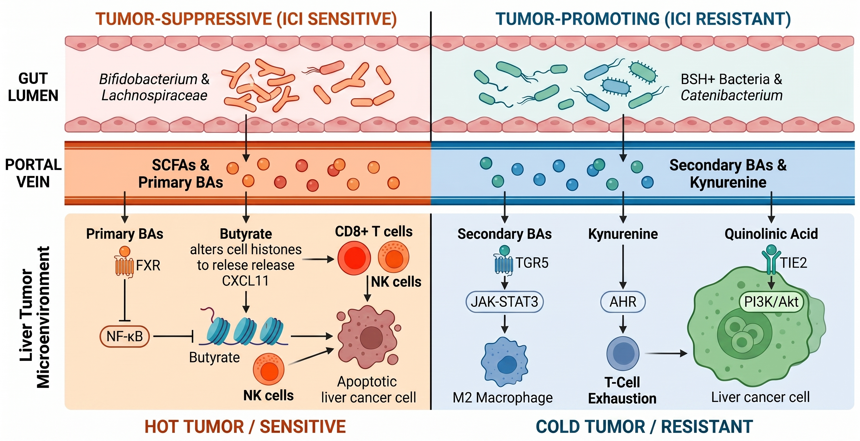
In chronic liver disease, intestinal dysbiosis leads to intestinal barrier dysfunction (26). For example, HCC patients often show a marked reduction in the abundance of short-chain fatty acid (SCFA)-producing bacteria (e.g., Lachnospiraceae, Ruminococcaceae) and an increase in pathogenic bacteria (e.g., Escherichia-Shigella, Klebsiella) (27). This dysbiosis pattern is more pronounced in ICI-resistant patients. Intestinal barrier dysfunction allows microbial metabolites, including LPS, secondary BAs, antigens, and bacteria, to translocate to the liver via the portal vein, thereby triggering hepatic inflammation, fibrosis, and immune dysregulation (24). These gut-derived factors modulate hepatic inflammation by activating pattern recognition receptors (PRRs), such as toll-like receptors (TLRs) and nucleotide-binding oligomerization domain (NOD)-like receptors (NLRs) on hepatic immune cells (28, 29). For example, LPS binds to TLR4 on Kupffer cells, activating the NF-κB signaling pathway and inducing the production of pro-inflammatory cytokines that promote liver fibrosis and HCC development (30).
Recent data further revealed that Enterococcus faecalis in liver tumors promotes HCC progression by delivering Obg GTPase via extracellular vesicles to activate the host mTOR pathway, identifying a direct cross-kingdom mechanism of bacterial-driven carcinogenesis with potential therapeutic implications for mTOR inhibitor treatment in affected patients (31).
Emerging evidence indicates that gut microbiota metabolites are key messengers in gut-liver axis crosstalk, directly shaping the hepatic TIME and ICI responsiveness (32). Ma et al. demonstrate that the gut microbiome regulates hepatic antitumor immunity by modulating the balance between primary and secondary BAs, thereby modulating the CXCL16-dependent recruitment of NKT cells to the liver microenvironment (33). These observations suggest that targeting gut microbial metabolism may represent a complementary strategy to improve immunotherapy, although prospective interventional studies remain limited (34, 35).
2. Key Gut Microbiota Metabolites Involved in HCC Immunotherapy
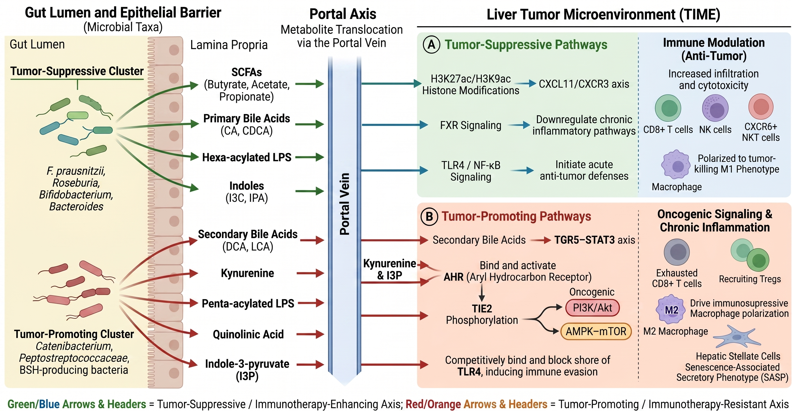
Gut microbiota metabolites, including SCFAs, BAs, lipopolysaccharides (LPS), tryptophan (Trp) metabolites, indole derivatives, and bacterial toxins, are the primary mediators of microbiota-liver-immune crosstalk. Their production is shaped by microbial composition, dietary substrates, host genetics, and environmental factors, and they exert distinct regulatory effects on antitumor immunity and ICI efficacy in HCC. Importantly, metabolite abundance is highly dynamic and may vary according to diet, liver function, medication exposure, geography, and analytical methodology, complicating direct comparisons across studies.
As illustrated in Figure 2, the biological functions and immunomodulatory effects of these key metabolites are crucial for developing microbiota-targeted strategies to enhance HCC immunotherapy. Recent multi-omics studies integrating bulk and single-cell transcriptomics have established a novel polyamine metabolism-related signature (PMRS) comprising 9 genes, which is closely associated with prognosis and immunotherapy sensitivity in HCC, thereby identifying potential biomarkers for personalized treatment strategies (36).
Short-Chain Fatty Acids
SCFAs are the end products of anaerobic fermentation of dietary fiber by gut bacteria. The three primary SCFAs are acetate, propionate, and butyrate, with acetate the most abundant in the colon, followed by propionate and butyrate in a molar ratio of approximately 60:20:20, though these ratios can vary with diet and microbiota composition (37). The main producers of SCFAs are anaerobic bacteria in the phyla Firmicutes and Bacteroidetes, including the genera Ruminococcus, Butyricicoccus, Roseburia, Faecalibacterium, and Bacteroides (38). Once produced, SCFAs are readily absorbed by the colonic epithelium (39). Their metabolic fate, however, differs significantly. Butyrate is preferentially taken up and used as the primary energy source by colonocytes (40). In contrast, acetate and propionate, which are not metabolized in the colon, largely enter the portal circulation and are transported to the liver (41), where propionate serves as a substrate for gluconeogenesis and acetate is used for the synthesis of lipids such as long-chain fatty acids and cholesterol (42, 43).
Multiple independent studies consistently report that HCC patients exhibit significant reductions in fecal and plasma SCFA levels, particularly butyrate, which correlates with poor clinical outcomes and shorter PFS (44). A cross-sectional study of the fecal microbiota in HCC patients found that the abundance of butyrate-producing bacteria, including Roseburia, Eubacterium rectale, and Faecalibacterium prausnitzii, was significantly lower than in healthy controls (45). This reduction in butyrate-producing bacteria is associated with significantly lower plasma levels of acetate, propionate, and butyrate in HCC patients. Furthermore, a recent study showed that in HCC patients receiving immunotherapy, fecal acetate levels were a significant predictor of durable response, PFS, and OS; those with higher acetate levels had significantly longer OS (median: 25.2 vs. 11.3 months; p < 0.001) and PFS (median: 15.3 vs. 4.2 months; p < 0.001) (46). These observations support the potential of SCFAs as prognostic biomarkers and therapeutic targets, although prospective validation remains limited.
Butyrate, the most extensively studied SCFA in HCC immunotherapy, has been shown to enhance antitumor immunity by promoting NK cell infiltration and function. Butyrate upregulates CXCL11 expression in HCC cells through epigenetic remodeling, increasing H3K27ac and H3K9ac at the CXCL11 enhancer and recruiting the STAT4 transcription factor, which activates the CXCL11/CXCR3 signaling axis to induce NK cell chemotaxis and cytotoxicity (44). Beyond NK cell modulation, butyrate exerts broad immunoregulatory effects through G protein-coupled receptor signaling (47) and histone deacetylase inhibition (48), influencing the differentiation and function of regulatory T cells (Tregs), effector T cells, and antigen-presenting cells (49). Experimental evidence indicates that butyrate enhances the efficacy of anticancer therapies by directly boosting cytotoxic CD8+ T cell responses through an ID2-dependent mechanism, suggesting potential synergy with immunotherapy (50). The main G protein-coupled receptors (GPCRs) activated by SCFAs include GPR41 (FFAR3), GPR43 (FFAR2), and GPR109A (51). GPR41 and GPR43 are widely expressed on immune cells and intestinal epithelial cells, whereas GPR109A is mainly expressed on dendritic cells, macrophages, and intestinal epithelial cells (52). Activation of these GPCRs by SCFAs can regulate immune cell activation, proliferation, and cytokine secretion, thereby modulating antitumor immunity.
Acetate and propionate also contribute to immune regulation in HCC. Acetate can enhance the cytotoxic activity of CD8⁺ T cells by regulating cellular metabolism (53). Specifically, acetate derived from Bacteroides thetaiotaomicron has been shown to promote M1 macrophage polarization and enhance the cytotoxic function of CD8⁺ T cells, thereby inhibiting HCC progression and recurrence (42). Propionate inhibits hepatic inflammation by activating PPARα and reducing the secretion of pro-inflammatory cytokines, such as TNF-α (54). Furthermore, propionate has been shown to suppress Ras activity and downregulate PI3K, VEGF, and inflammation-associated pathways, thereby inhibiting cancer cell proliferation and delaying the pathogenesis of HBV-associated HCC (55). Depletion of SCFA-producing bacteria in HCC patients disrupts these protective mechanisms, potentially leading to immune suppression and resistance to ICIs (56).
Whether systemic SCFA supplementation can recapitulate the localized effects observed in experimental models remains uncertain, as circulating concentrations differ substantially from those in the intestinal microenvironment.
Bile Acids
The composition of BAs, the main metabolites of hepatic cholesterol, is greatly influenced by gut microbiota. BAs undergo enterohepatic circulation, which involves secretion into the intestine and reabsorption by the liver (57). Recent clinical observations have shown a link between changes in the BA profile and HCC incidence, with various BA species exerting distinct and often conflicting effects on immunity and tumor progression (57, 58).
In HCC, primary and secondary BAs generally exhibit opposing immunological effects, although their biological activities remain highly context-dependent and influenced by receptor expression, concentration, and disease stage. The liver synthesizes conjugated primary BAs, including taurocholic acid (TCA), glycocholic acid (GCA), taurochenodeoxycholic acid (TCDCA), and glycochenodeoxycholic acid (GCDCA), by conjugating primary BAs such as cholic acid (CA) and chenodeoxycholic acid (CDCA) with taurine or glycine (57, 59). Conjugated BAs are released into the intestine, where the gut microbiota, especially bacteria that produce bile salt hydrolase (BSH), deconjugate them and convert them into secondary BAs, including deoxycholic acid (DCA), lithocholic acid (LCA), ursodeoxycholic acid (UDCA), and their conjugated derivatives such as glycodeoxycholic acid (GDCA) (57, 58). The immunological impact of BAs is determined not only by the presence of specific species but also by the homeostatic balance, specifically the ratio of primary to secondary BAs. A prospective cohort study showed that a lower secondary-to-primary BA ratio was associated with increased tumor promotion, whereas higher levels of conjugated PBAs, especially taurine-conjugated CDCA, were associated with a higher risk of HCC (60). Primary BAs such as CDCA promote antitumor environments by facilitating the recruitment of CXCR6⁺ NKT cells via CXCL16 expression in liver cancer (33). Conversely, an overabundance of secondary BAs can trigger a senescence-associated secretory phenotype (SASP) in hepatic stellate cells, leading to an immunosuppressive milieu (61). The host balances these signals through the bile acid interactome, and a 'tipping point' occurs when gut dysbiosis accelerates the conversion of primary to secondary BAs, effectively switching the hepatic microenvironment from an immune-active to an immune-exhausted state. Monitoring this ratio may ultimately prove more informative than measuring individual bile acids, although standardized clinical thresholds have yet to be established.
The antitumor immunity is regulated by the bile acid-gut microbiota-immune axis. A 2026 study by Xu et al. indicates that AF6, a polarity protein, regulates the expression of CYP7A1, the rate-limiting enzyme in bile acid synthesis, thereby serving as a key regulator of primary bile acid synthesis in hepatocytes (62). AF6 knockout increased primary BA levels, which subsequently altered gut microbiota composition and elevated butyrate production, an SCFA. Butyrate, delivered via enterohepatic circulation, upregulated CXCL14 expression and secretion in hepatocytes. This chemokine recruited activated DCs and enhanced CD8+ T-cell cytotoxicity within the TME, thereby creating a tumor-suppressive immune milieu and significantly inhibiting HCC progression. This study established a "BA-metabolism-gut microbiota-immunity" axis as a critical regulator of antitumor immune responses in HCC.
Specific BAs mechanistically modulate immune cell function in HCC through distinct mechanisms. DCA and LCA, two hydrophobic secondary BAs, are associated with the formation of an immunosuppressive tumor microenvironment. DCA suppresses CD8+ T-cell cytotoxicity, promotes M2-like tumor-associated macrophage polarization, and induces hepatic stellate cells to adopt a senescence-associated secretory phenotype, thereby accelerating HCC progression (63). LCA suppresses pro-inflammatory cytokine secretion, promotes M2 polarization via TGR5, and inhibits hepatic NKT cell accumulation by downregulating CXCL16, thereby facilitating tumor immune evasion (64). Lipoteichoic acid (LTA) and DCA can act synergistically to drive macrophage polarization through dual receptor signaling: LTA activates the TLR2-NF-κB pathway to promote M1 activation, while DCA activates the TGR5-STAT3 pathway to promote M2 polarization (65). This "double polarization" creates a unique tumor microenvironment characterized by both pro-inflammatory and immunosuppressive features, paradoxically enhancing HCC progression. The study demonstrated that combined DCA and LTA treatment increased tumor invasiveness and VEGF expression in obesity-associated HCC models. The PNMA1 gene, identified as a novel immune modulator linked to bile acid metabolism, promotes a suppressive TME in HCC upon overexpression, correlating with reduced M1 macrophage and CD8⁺ T cell infiltration (66). Experimental knockdown of PNMA1 inhibited HCC cell migration and proliferation, indicating that PNMA1 is a potential therapeutic target.
Bile acid-mediated dysbiosis of the gut microbiota further exacerbates immunosuppression. Depletion of beneficial bacteria and enrichment of pro-inflammatory taxa alter the BA pool composition, leading to accumulation of immunosuppressive secondary BAs and resistance to ICIs. Targeting the bile acid-gut microbiota-immunity axis represents an attractive therapeutic concept that now requires rigorous clinical validation.
Lipopolysaccharides
Lipopolysaccharide (LPS) is a component of the outer membrane of Gram-negative bacteria. In chronic liver disease, intestinal barrier dysfunction may enhance LPS translocation to the liver via the portal vein, thereby facilitating the pathogenesis and progression of HCC (67). Studies have shown that modulating the gut microbiota to control LPS leakage can inhibit the TLR4/NF-κB pathway and reduce inflammation in HCC. TLR4 is a key component of innate immunity and plays a significant role in the progression of liver disease and tumorigenesis (68). In HCC, TLR4 is functionally expressed and can either promote or inhibit tumor growth, depending on the surrounding cells and environment. In hepatoma cell lines, LPS-mediated TLR4 activation may indirectly facilitate the recruitment of Tregs to the tumor site and augment intrahepatic metastasis through interactions with macrophages (69). A study investigated the effects of LPS on macrophage polarization in the context of HCC (70, 71). The research indicated that LPS at concentrations exceeding 10 ng/mL markedly facilitated M1 polarization of macrophages derived from THP-1 monocytes. LPS treatment redirected the mitochondrial oxidative metabolism of M1 macrophages toward aerobic glycolysis. The study found that LPS-induced M1 macrophages inhibit HCC growth by activating T cells and inducing tumor cell death, which could aid HCC immunotherapy.
New evidence shows that the structure of gut microbiota-derived LPS affects whether immunotherapy works. A groundbreaking study in Nature Microbiology found that patients who responded to anti-PD-1 therapy had more bacterial species with genes for producing hexa-acylated LPS, whereas patients who did not respond had more bacteria producing penta-acylated LPS (72). This difference is important for how things work: hexa-acylated LPS selectively activates TLR4 and NF-κB, prompting macrophages to release cytokines and boosting antitumor immunity. By contrast, penta-acylated LPS blocks immune activation. In murine models of cancer, oral administration of purified hexa-acylated LPS improved the efficacy of anti-PD-1 therapy, leading to enhanced tumor regression and increased infiltration of tumor-specific cytotoxic CD8⁺ T cells, an effect not observed in mice treated with penta-acylated LPS. Blocking TLR4 with a small-molecule antagonist abolished the beneficial effects of hexa-acylated LPS on anti-PD-1 therapy. These findings support an important role for TLR4 signaling in microbiota-mediated immunotherapy responses, although confirmation in human HCC remains limited.
Modulating the LPS-TLR4 pathway represents a potential treatment strategy for HCC. A study showed that Echinacea purpurea polysaccharide (EPP) treatment in mice with HCC enhanced the proliferation of beneficial gut bacteria that produce butyrate, increased the expression of intestinal tight junction proteins, and repaired the intestinal barrier (67). This reduced LPS leakage, thereby suppressing the TLR4/NF-κB pathway and diminishing the expression of inflammatory mediators such as IL-6 and migratory factors like MMP-2. The research demonstrated that EPP efficiently controlled LPS leakage by altering the gut microbiota, modulating the TLR4/NF-κB pathway, and ultimately hindering HCC-induced tumor cell survival in murine models.
Tryptophan Metabolites
Tryptophan metabolism in HCC proceeds through three major pathways with distinct immunological consequences: (1) the kynurenine (Kyn) pathway, which is predominantly immunosuppressive; (2) the serotonin pathway, which supports tumor growth and metastasis; and (3) the indole pathway, which generates both pro‑tumorigenic and anti‑tumorigenic metabolites depending on the specific derivative and context (73). Understanding how these pathways interact with the gut microbiota is essential for exploiting the antitumor immunity in HCC.
The Kyn pathway promotes immunosuppression and tumor progression in HCC. Indoleamine 2,3-dioxygenase (IDO1, IDO2) and Trp 2,3-dioxygenase (TDO2) are the enzymes that initiate and regulate the Kyn pathway (74). In liver cancer, TDO2 expression correlates with tumor size, stage, differentiation, and recurrence, thereby promoting cell proliferation and tumor growth by regulating p21 and p27 (75). This primary metabolite activates the aryl hydrocarbon receptor, a transcription factor that controls the expression of pro-tumorigenic genes and suppresses antitumor immunity (76). Kyn 3-monooxygenase (KMO) expression is associated with TNM stage, tumor differentiation, and prognosis in HCC patients, and it promotes cell proliferation, migration, and invasion (77). Additionally, 3-hydroxyanthranilic acid (3-HAA), another Kyn pathway metabolite, induces apoptosis in HCC cells through YY1-dependent mechanisms (78).
Serotonin signaling influences HCC progression. It is derived from Trp via Trp hydroxylase and has been implicated in HCC pathogenesis. In HCC patients, elevated levels are associated with worse prognosis, higher Edmondson grade, microvascular invasion, tumor multiplicity, and TNM stage. Serotonin mechanistically promotes cell proliferation, epithelial-mesenchymal transition, and metastasis via the PI3K/AKT/FoxO6 signaling pathway (79).
The indole pathway produces metabolites that play dual roles in HCC. Gut microbiota metabolize dietary Trp into various indole derivatives that exert context-dependent effects on tumor immunity (73). Indole-3-carbinol (I3C), a naturally occurring compound from cruciferous vegetables, has shown substantial immunomodulatory effects in HCC. A study found that I3C prevents PD-L1 from helping HCC evade the immune system by inhibiting the ubiquitination of NF-κB p105 (80). I3C treatment reduced PD-L1 expression in HepG2 cells and increased CD8⁺ T cell infiltration into tumor tissues. The combination of I3C and anti-PD-L1 monoclonal antibodies produced stronger antitumor effects in mouse HCC models, suggesting it could be used as an immunotherapeutic adjuvant.
On the other hand, indole 3-pyruvate (I3P) acts as an oncometabolite in MYC-driven liver tumorigenesis. A pivotal study demonstrated that MYC-driven liver tumors depend on increased Trp uptake, whereas utilization of Trp to produce Kyn pathway metabolites is diminished (74). Instead, these tumors produce I3P through the activity of interleukin-4-induced 1 (IL4I1). I3P activates AHR, which promotes cell and tumor growth. A No-Trp diet that deprived MYC-driven tumors of Trp not only halted tumor growth but also restored the transcriptional profile of normal liver cells. Conversely, adding I3P to the diet helped Trp-starved cancer cells grow again. These findings identify I3P as a promising therapeutic target in MYC-driven HCC, although the relevance of this pathway across broader HCC subtypes remains to be determined.
New evidence suggests that circulating indole metabolites could help predict how well ICI will work in people with HCC. A study by Jia and colleagues (2024) shows that microbial indole metabolites, such as indole-3-propionic acid (IPA), are important for tumor growth and can serve as adjuvants in tumor immunotherapy (81). Data presented at the American Association for Cancer Research Annual Meeting support this. The data showed that 50% of unresectable HCC patients with high IPA and 10% with low IPA achieved a time-to-progression (TTP) > 8 months (82). TrpNet analysis indicates that the Peptostreptococcaceae family is most likely responsible for producing IPA. The Pasteurellaceae and Lachnospiraceae families, responsible for synthesizing the IPA precursor indole pyruvate, were significantly elevated in individuals who first responded to ICIs. These preliminary clinical observations suggest that circulating indoles may serve as predictive biomarkers, although validation in independent prospective cohorts remains necessary.
Among the enzymes that gate these pathways, interleukin‑4‑induced‑1 (IL4I1) has emerged as a critical metabolic immune checkpoint. IL4I1 catalyzes the production of indole derivatives and kynurenic acid, which potently activate AHR, thereby promoting tumor cell motility, dampening adaptive immunity, and accelerating tumor progression (83). Notably, IL4I1 expression is induced by immune checkpoint blockade and appears to compensate for IDO1 inhibition, which may explain the disappointing results of clinical trials combining PD‑1/PD‑L1 inhibitors with IDO1 inhibitors alone. This makes IL4I1 an attractive target for next‑generation combination strategies in HCC.
Other Metabolites
Bacterial toxins, polyamines, and vitamins are additional classes of metabolites derived from the gut microbiota that influence HCC progression and immunotherapy response (84, 85).
Bacterial toxins, particularly LPS from Gram-negative bacteria, contribute to HCC pathogenesis via the gut-liver axis. LPS activates TLR4 signaling, driving chronic inflammation and creating a tumor-permissive microenvironment (84, 85). Dysbiosis-induced intestinal barrier dysfunction increases the translocation of bacterial toxins via the portal vein, generating an inflammatory environment that causes liver damage and may promote hepatocarcinogenesis. Beyond LPS, other bacterial genotoxins, such as cytolethal distending toxin (CDT) produced by certain Enterobacteriaceae species, have been implicated in inducing DNA damage and genomic instability, though their specific role in HCC requires further investigation.
Polyamines, including spermidine and spermine, are produced by gut bacteria such as Bifidobacterium and Lactobacillus and have context-dependent effects on HCC (86). Polyamines can regulate cell proliferation, differentiation, and immune function through multiple mechanisms (87). Recent reviews highlight that polyamines modulate the TIME by influencing T-cell and natural killer (NK) cell function, with potential implications for immunotherapy efficacy (84). Spermidine has been shown to enhance autophagy in immune cells, which may promote antitumor immunity, though clinical data in HCC remain limited. Whether modulation of polyamine metabolism should involve supplementation or inhibition likely depends on tumor context and immune status, highlighting an important area for future investigation.
Gut microbiota-derived vitamins, including vitamins B6, B12, and K, play important roles in regulating antitumor immunity and liver health (84). Vitamin K, particularly vitamin K2, has been studied for its ability to inhibit hepatic inflammation and fibrosis, both of which are critical drivers of HCC progression (88). Protein induced by vitamin K absence or antagonist-II (PIVKA-II) is a well-established clinical biomarker for HCC, though it reflects vitamin K status in tumor tissue rather than direct immunomodulation (89). Vitamin B6 supplementation in HCC patients after resection improved antioxidant capacity primarily by reducing plasma homocysteine, rather than through a direct antioxidant effect (90).
Although these metabolites are presented individually, their biological effects are highly interconnected, and multiple metabolites frequently converge on shared immune and signaling pathways within the hepatic tumor microenvironment. Table 1 summarizes the major gut microbiota‑derived metabolites discussed in this review, their principal microbial sources, dominant immunological mechanisms in HCC, and overall impact on ICI efficacy.
Table 1. Gut Microbiota-Derived Metabolites and Their Roles in HCC Immunotherapy
| Metabolite Class | Key Metabolite | Major Producing Bacteria / Biological Source | Primary Mechanism in HCC Immunotherapy | Effect on ICI Efficacy |
|---|---|---|---|---|
| Short-Chain Fatty Acids (SCFAs) | Butyrate | Roseburia, Eubacterium rectale, Faecalibacterium prausnitzii, Butyricicoccus | Enhances NK- and CD8⁺ T-cell antitumor activity through CXCL11/CXCR3 activation, epigenetic regulation, and NF-κB suppression. | Enhance |
| Acetate | Firmicutes and Bacteroidetes (e.g., Bacteroides thetaiotaomicron) | Regulates cellular metabolism to enhance CD8⁺ T cell cytotoxicity; promotes M1 macrophage polarization to inhibit HCC progression. | Enhance | |
| Propionate | Anaerobic phyla (Firmicutes, Bacteroidetes, e.g., Ruminococcus, Bacteroides) | Activates PPARα to reduce pro-inflammatory TNF-α; suppresses Ras activity and downregulates PI3K, VEGF, and NF-κB pathways. | Enhance | |
| Isobutyrate | Bifidobacterium | Stimulates CD8⁺ T cells, reduces oncogenic JAK/STAT3 signaling pathway activity, and increases IFN-γ secretion. | Enhance | |
| Bile Acids (BAs) | Primary BAs (e.g., CDCA, CA, TCA, GCA) | Host-derived (composition modulated by the gut microbiota) | Promotes NKT-cell recruitment and suppresses NF-κB-mediated inflammation via FXR activation. | Enhance |
| Secondary BAs (e.g., DCA, LCA) | Gut microbiota (BSH-producing bacteria) | Promotes M2 macrophage polarization and suppresses CD8⁺ and NKT-cell antitumor immunity. | Inhibit | |
| Lipopolysaccharides (LPS) | Hexa-acylated LPS | Gut bacteria enriched in anti-PD-1 responders | Activates TLR4/NF-κB signaling to enhance macrophage activation and CD8⁺ T-cell infiltration. | Enhance |
| Penta-acylated LPS | Gut bacteria enriched in anti-PD-1 non-responders | Antagonizes TLR4 signaling and suppresses antitumor immunity. | Inhibit | |
| Tryptophan Metabolites | Kynurenine (Kyn Pathway) | Host and microbial tryptophan metabolism (IDO1/2, TDO2) | Activates AHR signaling to promote immune evasion and tumor progression. | Inhibit |
| Indole-3-carbinol (I3C) | Cruciferous vegetables (microbiota-derived) | Suppresses PD-L1 expression through NF-κB inhibition and promotes CD8⁺ T-cell infiltration. | Enhance | |
| Indole-3-pyruvate (I3P) | IL4I1-mediated Trp metabolism in MYC-driven tumors | Activates AHR to promote tumor cell growth and motility while actively dampening adaptive immunity. | Enhance | |
| Indole-3-propionic acid (IPA) | Peptostreptococcaceae (supported by Pasteurellaceae and Lachnospiraceae) | Limits tumor growth and serves as a circulating metabolic driver of tumor immunotherapy response. | Enhance | |
| Other Metabolites | Quinolinic acid | Catenibacterium mitsuokai | Activates TIE2-mediated PI3K-AKT signaling to promote tumor survival. | Inhibit |
| Polyamines (e.g., Spermidine, Spermine) | Gut commensals (e.g., Bifidobacterium, Lactobacillus) | Enhances T-cell and NK-cell antitumor function and immune-cell autophagy. | Enhance |
3. Mechanisms of Gut Microbiota Metabolites Modulating HCC Immunotherapy Efficacy
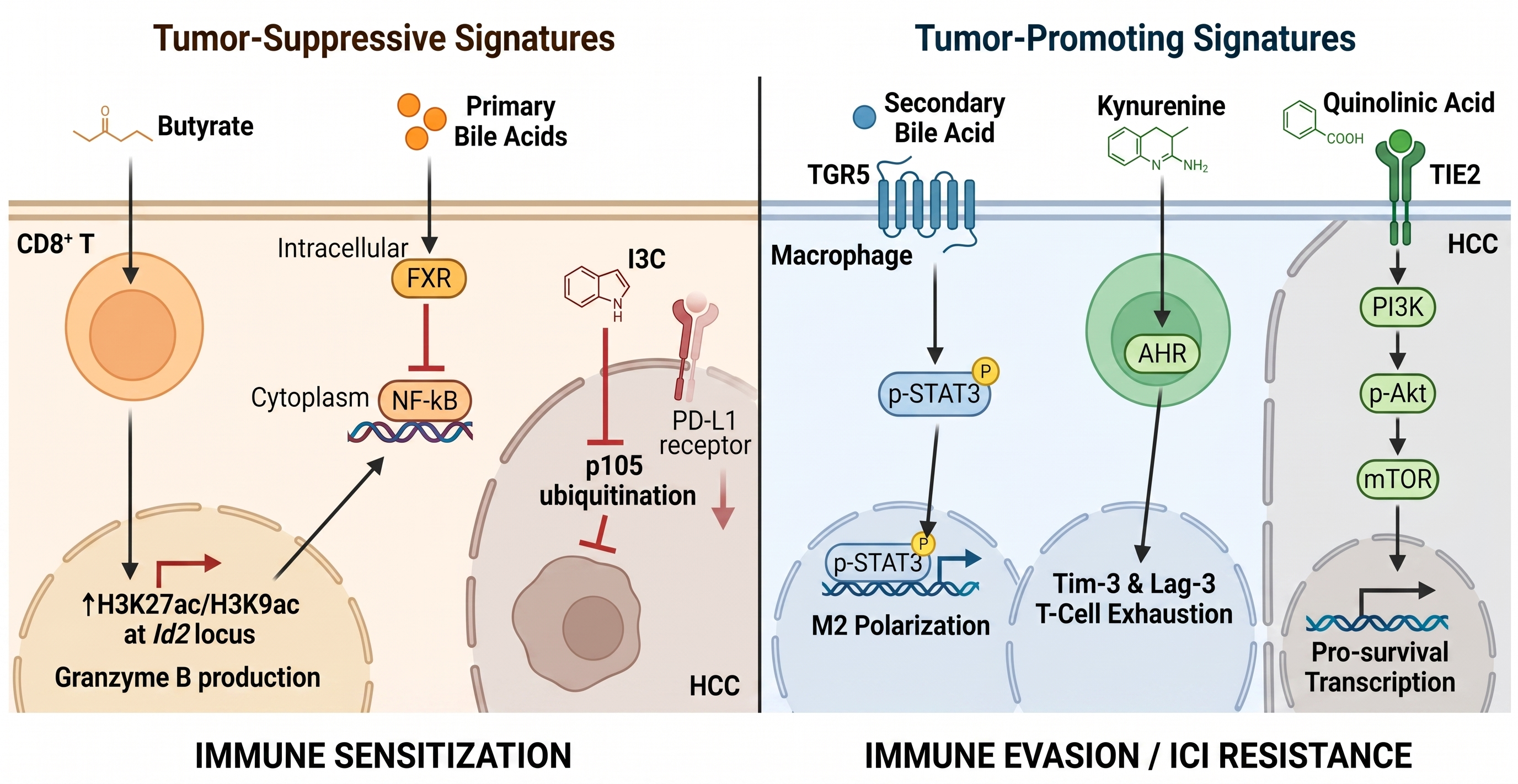
Gut microbiota metabolites regulate ICI efficacy by reshaping the TIME and targeting multiple immune cell subsets and signaling pathways (32, 50). The core mechanisms include modulating immune cell activation and function, regulating inflammatory signaling pathways, and influencing tumor cell immune escape (Figure 3). Improved understanding of these pathways may facilitate the development of microbiota-targeted therapeutic strategies (32). Recent single-cell sequencing and spatial metabolomics studies have provided new insights into the spatiotemporal dynamics of these mechanisms within the HCC microenvironment (73).
Modulation of Inflammatory and Signaling Pathways
Current evidence indicates that metabolites of the gut microbiota regulate key signaling pathways involved in inflammation and antitumor immunity, including NF-κB, PI3K/Akt, AMPK-mTOR, and FXR/TGR5 (63). These pathways are interconnected, forming a complex regulatory network that modulates immune cell function, tumor cell proliferation, and ICI efficacy.
The NF-κB signaling system is a crucial regulator of inflammation and immune responses. Through engagement of TLR4 and other receptors, LPS and secondary BAs activate the NF-κB pathway, leading to the synthesis of pro-inflammatory cytokines, including TNF-α, IL-6, and IL-1β (91). Prolonged NF-κB activation may diminish the efficacy of ICI while simultaneously exacerbating hepatic fibrosis, inflammation, and tumor proliferation (92). The endotoxin LPS, originating from the outer membrane of Gram-negative bacteria, binds to the CD14/TLR4/MD-2 receptor complex on innate immune cells. This interaction triggers a MyD88-dependent signaling cascade that activates NF-κB and drives the transcription of pro-inflammatory genes (93). Persistent activation of the LPS-TLR4-NF-κB pathway appears to contribute substantially to chronic inflammation and immune dysregulation in HCC.
NF-κB serves as a crucial signaling hub that integrates multiple pro-tumorigenic signals within the tumor microenvironment. This includes inflammatory cytokines such as TNF-α and microbial detection mediated by TLRs (94). Once in the nucleus, activated NF-κB initiates transcription of target genes, including immune checkpoint molecules that facilitate immune evasion, pro-inflammatory cytokines that sustain chronic inflammation, angiogenic and metastatic effectors, and regulators of proliferation and survival (MYC, BCL2). In contrast, SCFAs such as propionate and butyrate can reduce chronic inflammation by suppressing NF-κB activation (49). A 2024 study demonstrated that Echinacea purpurea polysaccharide (EPP) intervention in HCC models increased beneficial butyrate-producing gut microbiota, enhanced intestinal tight junction protein expression, and repaired the intestinal barrier, which controlled LPS leakage and subsequently inhibited the TLR4/NF-κB pathway, reducing expression of inflammatory factors such as IL-6 and migration factors like MMP-2 (67).
Immune cell survival, proliferation, and polarization are governed by the PI3K/Akt signaling pathway (95). The gut bacterium Catenibacterium mitsuokai promotes HCC development by releasing quinolinic acid, which interacts with and activates the TIE2 receptor on HCC cells, as reported (96). Phosphorylated TIE2 then activates the downstream oncogenic PI3K/AKT pathway, accelerating HCC progression. Through its surface protein Gtr1/RagA, which interacts with the γ-catenin receptor, C. mitsuokai compromises the gut barrier, translocates to the liver as viable bacteria, and establishes colonization in HCC cells. Its pro-tumorigenic effect relies on the secreted metabolite quinolinic acid. The link between Catenibacterium mitsuokai and the PI3K/Akt pathway serves as a template for how microbial metabolites influence hepatic oncogenic signaling. The PI3K/Akt/mTOR axis may serve as a convergent signaling hub for multiple microbiota-derived metabolites, although the relative contributions of individual metabolites remain incompletely defined. For instance, SCFAs can modulate PTEN (a negative regulator of PI3K) via GPCR signaling (97), while high levels of secondary BAs have been shown to activate M2 Macrophages via transactivation of the S1PR2/PI3K/AKT axis (98). This suggests that the microbiota-metabolite axis not only affects immune cells but also directly alters the intrinsic survival signaling of HCC cells, providing multiple potential nodes for therapeutic intervention.
Immune cell activation and function depend on modulation of energy metabolism via the AMPK-mTOR signaling pathway (99). A study demonstrated that the herbal medicine decoction THSWD can slow HCC progression by enhancing the LTF/AMPK/mTOR/Beclin1 pathway and promoting lysosomal autophagy in HCC cells, through regulation of the gut microbiota and metabolic reprogramming. The study found that THSWD increased the metabolite glabrol, which induces apoptosis in HCC cells by activating the lysosomal autophagy-mediated apoptosis signaling pathway, while also increasing the abundance of the intestinal microbiota member Duncaniella. Glabrol supplementation enhances lysosomal autophagy and triggers apoptosis in HCC cells by activating the LTF/AMPK/mTOR/Beclin1 pathway.
BAs play a central role in regulating the FXR/TGR5 pathway, which is essential for both immune and metabolic functions (100). Research by Pathak et al. shows that FXR and TGR5 collaborate to regulate hepatic metabolism and BA synthesis; simultaneous activation of these receptors diminishes inflammation and improves metabolic stability (101). Primary BAs such as CDCA and secondary BAs such as UDCA activate FXR, which inhibits NF-κB pathways and reduces TNF-α and IL-6 levels. Lithocholic acid and deoxycholic acid activate TGR5, resulting in increased IL-10 expression and diminished inflammatory signaling via cAMP pathways. Disruption of the microbiota-bile acid-immune axis often fosters a pro-inflammatory and immunosuppressive tumor microenvironment. This pattern is common in gastrointestinal and hepatobiliary malignancies, aiding tumor immune evasion and progression (101).
The JAK-STAT pathway, a newly identified signaling pathway regulated by metabolites from the gut microbiota, plays a pivotal role in linking cytokine signals to immune cell function and tumor progression (102). Bifidobacterium and its metabolite, isobutyrate, can enhance the anti-PD-1 effect in HCC through the JAK/STAT3 pathway (103). The study indicates that isobutyrate-stimulated CD8⁺ T cells inhibited liver cancer cells, reduced JAK/STAT3 pathway activity, and enhanced IFN-γ secretion. Bifidobacterium or isobutyrate, in conjunction with anti-PD-1 therapy, led to reductions in tumor size, increases in IFN-γ and CD8⁺ T cell levels, and decreases in JAK/STAT3 signaling within the tumor microenvironment (104). These findings offer valuable insights into addressing resistance to immunotherapy in HCC by demonstrating that metabolites from the gut microbiota can modulate the JAK-STAT pathway, thereby influencing the equilibrium between pro-tumor and antitumor immune responses (103, 104).
Regulation of Tumor Cell Immune Escape
Tumor cell immune escape is a key mechanism of ICI resistance in HCC. Gut microbiota metabolites modulate this process by regulating immune checkpoint molecules, MHC expression, and immunosuppressive factors.
Bifidobacterium and its metabolite, isobutyrate, enhance anti-PD-1 efficacy by modulating the JAK/STAT3 pathway. A study demonstrated that isobutyrate-stimulated CD8⁺ T cells increased IFN-γ secretion, downregulated JAK/STAT3 signaling, and reduced tumor size when combined with anti-PD-1 in mouse models (103). A prospective clinical study of 74 patients with unresectable HCC found that enrichment of Lachnoclostridium and depletion of Prevotella 9 predicted ICI response, with responders achieving a progression-free survival of 8.8 months versus 1.8 months in non-responders (p<0.001) (105).
Secondary BAs promote immunosuppression. DCA, elevated in HCC patients, induces Kupffer cell polarization toward the M2 phenotype via TGR5/FXR receptors, increasing IL-10 and TGF-β secretion (106). This upregulates PD-1 and TIM-3 on CD8⁺ T cells, contributing to T cell exhaustion. Bile acid metabolism regulates hepatic immune surveillance through NKT cells. Primary BAs enhance CXCL16 expression on liver sinusoidal endothelial cells, attracting CXCR6⁺ NKT cells and supporting antitumor immunity (33). Secondary BAs reverse this effect, reducing NKT accumulation and impairing immune recognition of liver tumors. CXCL16 expression in human HCC tissue correlates with primary bile acid levels.
A recent study showed that tumor-suppressing multi-enterobacteria (TSME) combined with anti-PD-1 therapy improved outcomes in immunotherapy-resistant HCC (107). In mouse models, TSME plus anti-PD-1 achieved 58.78±7.55% tumor growth inhibition, increasing CD8⁺ and CD4⁺ T cells while reducing Tregs in the tumor microenvironment. By contrast, the Kyn-AhR pathway, activated by IDO1-mediated Trp metabolism, suppresses T cell function. Kyn upregulates PD-L1 expression and inhibits CD8⁺ T cell IFN-γ production by up to 65±8% (108). LPS from Gram-negative bacteria activates TLR4 signaling on Kupffer cells, recruiting myeloid-derived suppressor cells and promoting pathological inflammation that supports tumor progression (109). Current evidence supports an important contribution of gut microbial metabolites to tumor immune escape; however, the relative importance of individual pathways likely varies among patients and disease etiologies (110).
4. Clinical Evidence Linking Gut Microbiota Metabolites to HCC Immunotherapy Outcomes
Clinical studies have confirmed an association between gut microbiota metabolites and ICI efficacy in HCC patients, identifying specific metabolite signatures that can predict treatment response and guide personalized therapy.
Metabolite Signatures as Predictive Biomarkers
In a prospective cohort of 102 patients receiving first-line combination immunotherapy (77 V-HCC, 25 MASLD-HCC), etiology-specific microbiota signatures were distinct, but shared metabolomic features were associated with treatment outcomes (111). Bacteroides ovatus, Kluyvera georgiana, Klebsiella oxytoca, and Enterococcus faecium were more abundant in MASLD-HCC, whereas Mediterraneibacter gnavus ATCC 29149 was notably more common among durable responders. This group also exhibited significantly higher fecal levels of SCFAs, including acetate, propionate, butyrate, and isobutyrate, as well as increased UDCA levels. The predominance of Bifidobacterium, along with an SCFA-enriched profile, was associated with a sustained response in very early HCC (V-HCC). Significantly, higher levels of fecal acetate prior to treatment emerged as a robust predictor of sustained response across both etiologies. When stratified by acetate levels, patients exhibited markedly longer OS (median 25.2 months versus 11.3 months; p<0.001) and progression-free survival (median 15.3 months versus 4.2 months; p<0.001). These findings underscore the potential of acetate as both a biomarker and a therapeutic target. The significance of organic acids as biomarkers is increasingly recognized across oncology. This finding is supported by research in lung cancer, where acetic acid similarly emerged as an independent predictor of survival and immunotherapy response, suggesting that the relevance of this metabolite may extend across cancer types as a systemic regulator of T‑cell metabolic fitness. This cross-cancer observation supports the hypothesis that certain gut-derived metabolites maintain a systemic 'immunometabolic tone,' in which acetic acid serves as a crucial substrate for T-cell mitochondrial fitness and memory formation (112).
A comprehensive 2024 study prospectively enrolled 80 patients with advanced HCC receiving ICI treatment (113). The researchers used multi-omics sequencing, incorporating metagenomic, ITS2, and metabolomic data, to identify gut bacteria, fungi, and metabolites associated with clinical outcomes. The study revealed substantial differences in bacteria and metabolites between the durable clinical benefit (DCB) and non-durable benefit (NDB) groups, although fungal variation was less pronounced. A model comprising 18 bacterial species was developed as a predictive biomarker for immunotherapy efficacy, with an area under the curve (AUC) of 75.63%. Additionally, two bacterial species (Actinomyces sp. ICM47 and Senegalimassilia anaerobia) and one metabolite (galanthaminone) were identified as predictive biomarkers for survival in HCC patients undergoing ICI treatment.
Bile acid metabolism in HCC has shown promise as a prognostic marker, as evidenced by multi-omics analysis. A bile acid metabolism signature showed a strong correlation with tumor stage and OS in a study that combined transcriptomic data from 368 patients with HCC and included validation cohorts comprising 221 patients. Significant dysregulation of bile acid metabolism in HCC was evident in the signature, which revealed distinct pathway activations across various clinical subgroups (114). Notable variations in immune cell infiltration patterns were observed between groups with high and low bile acid metabolism, potentially influencing the TIME. Further reinforcing the notion that bile acid metabolism-related signatures serve as innovative prognostic biomarkers, an additional integrated analysis utilizing single-cell and spatial transcriptomics identified G6PD+ malignant cells exhibiting disrupted bile acid metabolism. These cells are intricately associated with angiogenesis and the development of an immunosuppressive microenvironment.
Indole-3-carbinol (I3C), a naturally occurring compound in cruciferous vegetables, has shown notable immunomodulatory effects in HCC. I3C suppresses NF-κB p105 ubiquitination, thereby preventing PD-L1-mediated immune evasion in HCC (80). I3C treatment increased CD8 T cell infiltration in tumor tissues and reduced PD-L1 expression in HepG2 cells. In mouse models of HCC, combining I3C with anti-PD-L1 monoclonal antibodies produced synergistic antitumor effects, suggesting its potential as a therapeutic adjuvant in immunotherapy. This underscores the importance of Trp-derived metabolites in HCC immunotherapy, despite their therapeutic rather than predictive role.
Altogether, current evidence supports the development of microbiota-derived metabolites as non-invasive biomarker candidates, although standardization of metabolomic platforms and validation in multicenter cohorts remain major priorities.
Clinical Trials of Microbiota-Targeted Therapeutic Strategies
Preclinical evidence supports several clinical trials investigating microbiota-targeted strategies to enhance immunotherapy efficacy in HCC. These approaches include FMT, probiotic supplementation, and dietary interventions. The FLORA trial (NCT05690048) is a multicenter, randomized, placebo-controlled, double-blind phase II study examining fecal microbiota transplantation (FMT) in conjunction with atezolizumab and bevacizumab for advanced HCC (115). The trial posits that HCC is a particularly appealing cancer type for FMT due to the anatomical proximity and physiological significance of the gut-liver axis. Furthermore, research indicates that FMT may enhance hepatic function in individuals with liver cirrhosis, potentially leading to improved outcomes in this demographic. The primary outcomes are the infiltration of tumoral CD8⁺ T cells after two treatment cycles and the safety of the combination therapy. Secondary goals include progression-free survival (PFS), OS, and changes in liver function.
The FAB-HCC pilot study (NCT05750030) is a phase II, single-center, single-arm study evaluating fecal microbiota transplantation (FMT) in individuals with advanced HCC who have not responded to atezolizumab and bevacizumab (116). Twelve patients will participate. They will undergo a single FMT via colonoscopy, with donor material from either healthy donors or HCC patients who have responded to PD-(L)1-based immunotherapy. After the transplant, participants will continue receiving atezolizumab and bevacizumab every three weeks. The frequency and severity of treatment-related adverse events are the primary safety measures. Secondary endpoints include optimal radiological response, assessed by RECISTv1.1 and mRECIST, as well as additional exploratory analyses of gut microbiota composition, circulating immune cells, and serum and stool metabolomic signatures. The findings will help clarify FMT's potential to address immunotherapy resistance in HCC. Important unresolved questions include donor selection, microbiome durability, optimal transplantation schedules, safety in cirrhotic patients, and reproducibility across geographically distinct populations.
The role of Bifidobacterium longum in improving postoperative liver function recovery in patients with HCC was investigated in a recent clinical trial (NCT04303286, NCT05178524) (117). The study found that patients with delayed recovery of liver function after hepatectomy had notably lower B. longum levels in both retrospective and prospective cohorts. In the trial, oral administration of a probiotic cocktail containing B. longum reduced delayed recovery rates in surgical patients with HCC, shortened hospital stays, and improved overall 1-year survival. These benefits were linked to changes in key metabolic pathways, including 5-hydroxytryptamine, secondary BAs, and SCFAs. Additionally, the trial observed reduced liver inflammation and fibrosis, as well as increased hepatocyte proliferation. This trial provides evidence that targeted probiotic supplementation can influence clinically significant outcomes in HCC by altering the gut microbiota, despite focusing on surgical rather than immunotherapy outcomes.
5. Discussion
Challenges and Future Directions
A major limitation in the current literature is the predominance of associative human studies alongside mechanistic animal models, making it difficult to establish causal relationships in patients. The biological complexity of the gut-liver axis means metabolites often show concentration-dependent dual effects. Butyrate maintains Treg function at physiological concentrations but loses this effect at lower levels. Establishing causality remains difficult because of high inter-individual variability influenced by diet, medications, and genetics. Clinical heterogeneity across HCC etiologies further complicates biomarker development, as evidenced by distinct microbial profiles between viral- and MASLD-related HCC despite shared metabolic signatures.
The Chinese Society of Hepatology has published the "Expert Consensus on the Clinical Application of Gut Microbiota Transplant Therapy in Chronic Liver Disease (118), which provides comprehensive guidance on indications, contraindications, donor selection, transplant routes, and safety considerations for FMT in chronic liver diseases, including liver cancer (6). The consensus underscores the growing recognition of microbiota-based therapies as a legitimate clinical approach and provides a framework for standardizing FMT applications in hepatology (6).
In addition to biological complexity, several technical and methodological challenges hinder the translation of microbiota‑derived metabolite findings into routine practice. Accurately quantifying metabolite concentrations in situ within the hepatic tumor microenvironment is difficult because stool and plasma measurements may not capture local gradients across the sinusoidal and intratumoral niches, and multi‑omics integration remains limited by batch effects and variable analytical platforms. Furthermore, perturbations targeting a single metabolite often reshape the broader microbial community, making it difficult to disentangle direct metabolite‑mediated effects from secondary ecological changes and underscoring the need for carefully controlled longitudinal and mechanistic studies. Differences in sequencing platforms, metabolomic methodologies, dietary assessment, sample processing, and definitions of treatment response further complicate cross-study comparisons and limit the reproducibility of microbiota-based biomarkers. Greater methodological standardization will be essential for meaningful validation across independent cohorts.
Future research should prioritize prospective validation of metabolite biomarkers in large multicenter cohorts. Fecal acetate has emerged as a promising pan-etiological predictor, and multi-omics integration of metagenomics, metabolomics, and transcriptomics has identified robust functional biomarkers beyond taxonomic composition. Emerging therapeutic strategies include FMT to overcome ICI resistance, probiotic supplementation, and dietary interventions to enhance SCFA production. Ongoing clinical trials will provide crucial safety and efficacy data. Future studies integrating longitudinal microbiome profiling with spatial transcriptomics, metabolomics, and functional validation in clinically annotated cohorts will be important for distinguishing causal mechanisms from associative signatures and for developing robust, clinically actionable microbiota-based biomarkers and therapeutic strategies. Potential clinically targeted mechanisms are illustrated in Figure 4.
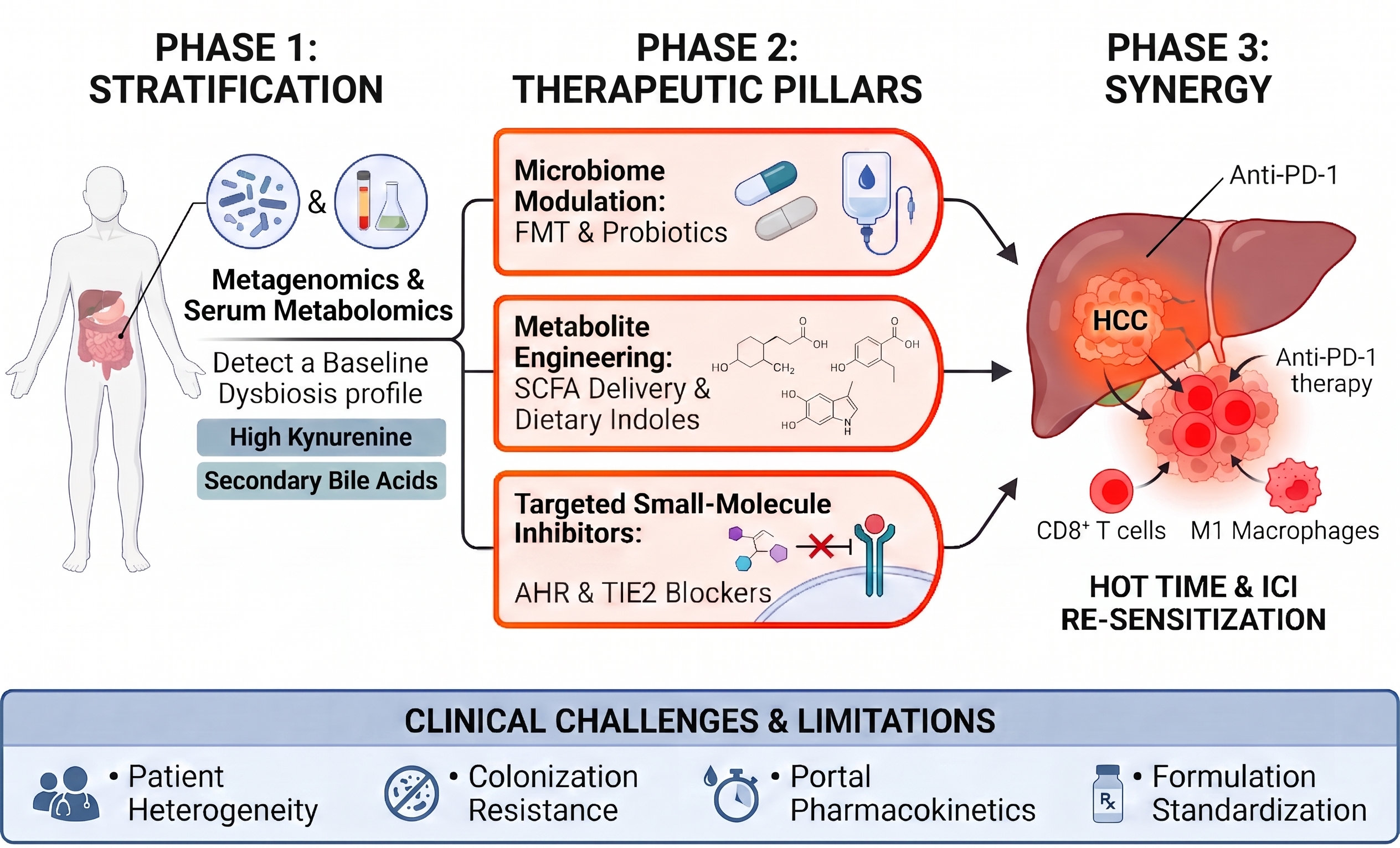
Beyond conventional FMT and probiotic supplementation, next‑generation strategies are likely to rely on engineered probiotics and synthetic biology platforms that deliver defined metabolites directly to the gut-liver axis, such as butyrate, specific primary BAs, or I3C‑like indole derivatives, in a controlled and sustained manner. Rational combination approaches may further enhance efficacy, for example by pairing FMT with high‑fiber or resistant‑starch diets to boost endogenous SCFA production, or by combining metabolite‑targeted agents with PD‑1/PD‑L1 blockade to concurrently reprogram both the microbiota and the tumor immune microenvironment. Future trials will need to integrate longitudinal multi‑omics profiling and adaptive designs to identify which patients benefit most from specific microbiota‑modulating strategies and to mitigate the risks of off‑target ecological effects.
Conclusion
Gut microbiota metabolites are critical determinants of immunotherapy efficacy in HCC, serving as key messengers along the gut-liver axis that shape hepatic antitumor immunity. SCFAs, BAs, LPS, and Trp metabolites modulate ICI responses through distinct mechanisms, including immune cell recruitment, epigenetic reprogramming, and regulation of signaling pathways. Clinical evidence identifies fecal acetate as a promising pan-etiological biomarker for patient stratification, while ongoing trials are evaluating fecal microbiota transplantation and probiotic supplementation to overcome treatment resistance. Despite challenges, including biological complexity and inter-individual variability, microbiota-targeted approaches represent a transformative frontier in HCC immunotherapy. Future research must prioritize prospective biomarker validation and rational combination strategies to translate these mechanistic insights into personalized therapeutic interventions that improve outcomes for patients with this devastating malignancy.
References
1. Bray F, Laversanne M, Sung H, Ferlay J, Siegel RL, Soerjomataram I, et al. Global cancer statistics 2022: GLOBOCAN estimates of incidence and mortality worldwide for 36 cancers in 185 countries. CA Cancer J Clin. 2024;74(3):229–263. https://doi.org/10.3322/caac.21834
2. Vogel A, Meyer T, Sapisochin G, Salem R, Saborowski A. Hepatocellular carcinoma. Lancet. 2022;400(10360):1345–1362. https://doi.org/10.1016/S0140-6736(22)01200-4
3. Singal AG, Kanwal F, Llovet JM. Global trends in hepatocellular carcinoma epidemiology: implications for screening, prevention and therapy. Nat Rev Clin Oncol. 2023;20(12):864–884. https://doi.org/10.1038/s41571-023-00825-3
4. Mak LY, Liu K, Chirapongsathorn S, Yew KC, Tamaki N, Rajaram RB, et al. Liver diseases and hepatocellular carcinoma in the Asia-Pacific region: burden, trends, challenges and future directions. Nat Rev Gastroenterol Hepatol. 2024;21(12):834–851. https://doi.org/10.1038/s41575-024-00967-4
5. de Martel C, Georges D, Bray F, Ferlay J, Clifford GM. Global burden of cancer attributable to infections in 2018: a worldwide incidence analysis. Lancet Glob Health. 2020;8(2):e180–e190. https://doi.org/10.1016/S2214-109X(19)30488-7
6. Hwang SY, Danpanichkul P, Agopian V, Mehta N, Parikh ND, Abou-Alfa GK, et al. Hepatocellular carcinoma: updates on epidemiology, surveillance, diagnosis and treatment. Clin Mol Hepatol. 2025;31(Suppl):S228–S254. https://doi.org/10.3350/cmh.2024.0824
7. Estes C, Anstee QM, Arias-Loste MT, Bantel H, Bellentani S, Caballeria J, et al. Modeling NAFLD disease burden in China, France, Germany, Italy, Japan, Spain, United Kingdom, and United States for the period 2016-2030. J Hepatol. 2018;69(4):896–904. https://doi.org/10.1016/j.jhep.2018.05.036
8. Cui T, Guan C, Song K, Yu J. Global trends and forecasts of nonalcoholic steatohepatitis causing liver cancer incidence and deaths. Front Oncol. 2026;15:1623789. https://doi.org/10.3389/fonc.2025.1623789
9. El-Khoueiry AB, Trojan J, Meyer T, Yau T, Melero I, Kudo M, et al. Nivolumab in sorafenib-naive and sorafenib-experienced patients with advanced hepatocellular carcinoma: 5-year follow-up from CheckMate 040. Ann Oncol. 2024;35(4):381–391. https://doi.org/10.1016/j.annonc.2023.12.008
10. Rimassa L, Chan SL, Sangro B, Lau G, Kudo M, Reig M, et al. Five-year overall survival update from the HIMALAYA study of tremelimumab plus durvalumab in unresectable HCC. J Hepatol. 2025;83(4):899–908. https://doi.org/10.1016/j.jhep.2025.03.033
11. Wang Y, Li Y, Lin Y, Cao C, Chen D, Huang X, et al. Roles of the gut microbiota in hepatocellular carcinoma: from the gut dysbiosis to the intratumoral microbiota. Cell Death Discov. 2025;11(1):140. https://doi.org/10.1038/s41420-025-02413-z
12. Llovet JM, Castet F, Heikenwalder M, Maini MK, Mazzaferro V, Pinato DJ, et al. Immunotherapies for hepatocellular carcinoma. Nat Rev Clin Oncol. 2022;19(3):151–172. https://doi.org/10.1038/s41571-021-00573-2
13. Yau T, Park JW, Finn RS, Cheng AL, Mathurin P, Edeline J, et al. Nivolumab versus sorafenib in advanced hepatocellular carcinoma (CheckMate 459): a randomised, multicentre, open-label, phase 3 trial. Lancet Oncol. 2022;23(1):77–90. https://doi.org/10.1016/S1470-2045(21)00604-5
14. De Martin E, Fulgenzi CAM, Celsa C, Laurent-Bellue A, Torkpour A, Lombardi P, et al. Immune checkpoint inhibitors and the liver: balancing therapeutic benefit and adverse events. Gut. 2025;74(7):1165–1177. https://doi.org/10.1136/gutjnl-2024-332125
15. Cheng AL, Qin S, Ikeda M, Galle PR, Ducreux M, Kim TY, et al. Updated efficacy and safety data from IMbrave150: Atezolizumab plus bevacizumab vs. sorafenib for unresectable hepatocellular carcinoma. J Hepatol. 2022;76(4):862–873. https://doi.org/10.1016/j.jhep.2021.11.030
16. Ohara M, Suda G, Kohya R, Yasui Y, Tsuchiya K, Kurosaki M, et al. Three-year overall survival in unresectable hepatocellular carcinoma treated with atezolizumab plus bevacizumab. Hepatol Int. 2025;19(6):1371–1381. https://doi.org/10.1007/s12072-025-10875-7
17. Ladd AD, Duarte S, Sahin I, Zarrinpar A. Mechanisms of drug resistance in HCC. Hepatology. 2024;79(4):926–940. https://doi.org/10.1097/HEP.0000000000000237
18. Wang Z, Wang Y, Gao P, Ding J. Immune checkpoint inhibitor resistance in hepatocellular carcinoma. Cancer Lett. 2023;555:216038. https://doi.org/10.1016/j.canlet.2022.216038
19. Chan YT, Zhang C, Wu J, Lu P, Xu L, Yuan H, et al. Biomarkers for diagnosis and therapeutic options in hepatocellular carcinoma. Mol Cancer. 2024;23(1):189. https://doi.org/10.1186/s12943-024-02101-z
20. Davar D, Zarour HM. Facts and Hopes for Gut Microbiota Interventions in Cancer Immunotherapy. Clin Cancer Res. 2022;28(20):4370–4384. https://doi.org/10.1158/1078-0432.CCR-21-1129
21. Xu J, Song J, Fu Z, Zhou H, Zhang Y, Shi C. Unraveling gut microbiome interferences in cancer immunotherapy: a meta-analysis of diverse drug effects. BMC Cancer. 2025;25(1):1776. https://doi.org/10.1186/s12885-025-15094-9
22. Tilg H, Adolph TE, Trauner M. Gut-liver axis: Pathophysiological concepts and clinical implications. Cell Metab. 2022;34(11):1700–1718. https://doi.org/10.1016/j.cmet.2022.09.017
23. Brandl K, Kumar V, Eckmann L. Gut-liver axis at the frontier of host-microbial interactions. Am J Physiol Gastrointest Liver Physiol. 2017;312(5):G413–G419. https://doi.org/10.1152/ajpgi.00361.2016
24. Albillos A, de Gottardi A, Rescigno M. The gut-liver axis in liver disease: Pathophysiological basis for therapy. J Hepatol. 2020;72(3):558–577. https://doi.org/10.1016/j.jhep.2019.10.003
25. Chopyk DM, Grakoui A. Contribution of the Intestinal Microbiome and Gut Barrier to Hepatic Disorders. Gastroenterology. 2020;159(3):849–863. https://doi.org/10.1053/j.gastro.2020.04.077
26. Zhang Y, Liu Y, Liang X, Wen Y, Zhao J, He Y, et al. Intestinal barrier in chronic gut and liver diseases: Pathogenesis and therapeutic targets. Acta Pharm Sin B. 2025;15(11):5515–5536. https://doi.org/10.1016/j.apsb.2025.08.028
27. Yu J, Yang YN, Chen W, Hu J, Jin Z, Wu C, et al. Role of gut microbiota and derived metabolites in cardiovascular diseases. iScience. 2025;28(9):113247. https://doi.org/10.1016/j.isci.2025.113247
28. Foley NM, Wang J, Redmond HP, Wang JH. Current knowledge and future directions of TLR and NOD signaling in sepsis. Mil Med Res. 2015;2:1. https://doi.org/10.1186/s40779-014-0029-7
29. Żeromski J, Kierepa A, Brzezicha B, Kowala-Piaskowska A, Mozer-Lisewska I. Pattern Recognition Receptors: Significance of Expression in the Liver. Arch Immunol Ther Exp (Warsz). 2020;68(5):29. https://doi.org/10.1007/s00005-020-00595-1
30. Min L, Wang H, Qi H. Astragaloside IV inhibits the progression of liver cancer by modulating macrophage polarization through the TLR4/NF-κB/STAT3 signaling pathway. Am J Transl Res. 2022;14(3):1551–1566. PMID: 35422920
31. Ma N, Xie X, Wang J, Zheng Z, Jin H, Chen X, et al. Enterococcus faecalis extracellular vesicles deliver the bacterial GTPase Obg to hijack mTOR signalling in hepatocellular carcinoma. J Extracell Vesicles. 2026;15(6):e70323. https://doi.org/10.1002/jev2.70323
32. Goyal H, Fatima K, Kaur J. Understanding crosstalk between the gut and liver microbiome: pathogenesis to therapeutic approaches in liver cancer. Cancer Cell Int. 2025;25(1):291. https://doi.org/10.1186/s12935-025-03840-9
33. Ma C, Han M, Heinrich B, Fu Q, Zhang Q, Sandhu M, et al. Gut microbiome-mediated bile acid metabolism regulates liver cancer via NKT cells. Science. 2018;360(6391):eaan5931. https://doi.org/10.1126/science.aan5931
34. Kim Y, Kim G, Kim S, Cho B, Kim SY, Do EJ, et al. Fecal microbiota transplantation improves anti-PD-1 inhibitor efficacy in unresectable or metastatic solid cancers refractory to anti-PD-1 inhibitor. Cell Host Microbe. 2024;32(8):1380–1393.e9. https://doi.org/10.1016/j.chom.2024.06.010
35. Lee PC, Wu CJ, Hung YW, Lee CJ, Mon HC, Chi CT, et al. Distinct gut microbiota but common metabolomic signatures between viral and MASLD HCC contribute to outcomes of combination immunotherapy. Hepatology. 2026;84(2):411-423. https://doi.org/10.1097/HEP.0000000000001446
36. Yu Y, Liu H, Liu K, Zhao M, Zhang Y, Jiang R, et al. Multi-omics identification of a polyamine metabolism related signature for hepatocellular carcinoma and revealing tumor microenvironment characteristics. Front Immunol. 2025;16:1570378. https://doi.org/10.3389/fimmu.2025.1570378
37. Fujisaka S, Watanabe Y, Tobe K. The gut microbiome: a core regulator of metabolism. J Endocrinol. 2023;256(3):e220111. https://doi.org/10.1530/JOE-22-0111
38. Zhuang M, Shang W, Ma Q, Strappe P, Zhou Z. Abundance of Probiotics and Butyrate-Production Microbiome Manages Constipation via Short-Chain Fatty Acids Production and Hormones Secretion. Mol Nutr Food Res. 2019;63(23):e1801187. https://doi.org/10.1002/mnfr.201801187
39. Caetano MAF, Castelucci P. Role of short chain fatty acids in gut health and possible therapeutic approaches in inflammatory bowel diseases. World J Clin Cases. 2022;10(28):9985–10003. https://doi.org/10.12998/wjcc.v10.i28.9985
40. Mayorga-Ramos A, Barba-Ostria C, Simancas-Racines D, Guamán LP. Protective role of butyrate in obesity and diabetes: New insights. Front Nutr. 2022;9:1067647. https://doi.org/10.3389/fnut.2022.1067647
41. Zhang CY, Liu S, Sui YX, Yang M. Roles of short-chain fatty acids in metabolic dysfunction-associated steatotic liver disease and metabolic dysfunction-associated steatohepatitis. World J Hepatol. 2025;17(11):113756. https://doi.org/10.4254/wjh.v17.i11.113756
42. Donkin SS, Armentano LE. Regulation of gluconeogenesis by insulin and glucagon in the neonatal bovine. Am J Physiol. 1994;266(4 Pt 2):R1229–R1237. https://doi.org/10.1152/ajpregu.1994.266.4.R1229
43. Carroll KK. Acetate incorporation into cholesterol and fatty acids by liver slices from rats fed commercial or semisynthetic diets: The effect of dietary fats. Can J Biochem. 1964;42(1):79–86. https://doi.org/10.1139/o64-007
44. Zhang M, Huang X, Zhang Y, Yu M, Yuan X, Xu Y, et al. Gut microbial metabolite butyrate suppresses hepatocellular carcinoma growth via CXCL11-dependent enhancement of natural killer cell infiltration. Gut Microbes. 2025;17(1):2519706. https://doi.org/10.1080/19490976.2025.2519706
45. Kumar M, Kaur R, Kanthaje S, Dhiman RK, Chakraborti A. Bacterial metabolite butyrate in modulating sorafenib-targeted microRNAs to curtail its resistance in hepatocellular carcinoma. J Cancer Res Clin Oncol. 2023;149(9):5823–5839. https://doi.org/10.1007/s00432-022-04544-7
46. Ren S, Zhang Y, Wang X, Su J, Wang X, Yuan Z, et al. Emerging insights into the gut microbiota as a key regulator of immunity and response to immunotherapy in hepatocellular carcinoma. Front Immunol. 2025;16:1526967. https://doi.org/10.3389/fimmu.2025.1526967
47. Maslowski KM, Vieira AT, Ng A, Kranich J, Sierro F, Yu D, et al. Regulation of inflammatory responses by gut microbiota and chemoattractant receptor GPR43. Nature. 2009;461(7268):1282–1286. https://doi.org/10.1038/nature08530
48. Arpaia N, Campbell C, Fan X, Dikiy S, van der Veeken J, deRoos P, et al. Metabolites produced by commensal bacteria promote peripheral regulatory T-cell generation. Nature. 2013;504(7480):451–455. https://doi.org/10.1038/nature12726
49. Mann ER, Lam YK, Uhlig HH. Short-chain fatty acids: linking diet, the microbiome and immunity. Nat Rev Immunol. 2024;24(8):577–595. https://doi.org/10.1038/s41577-024-01014-8
50. Danne C, Sokol H. Butyrate, a new microbiota-dependent player in CD8+ T cells immunity and cancer therapy? Cell Rep Med. 2021;2(7):100328. https://doi.org/10.1016/j.xcrm.2021.100328
51. Macia L, Tan J, Vieira AT, Leach K, Stanley D, Luong S, et al. Metabolite-sensing receptors GPR43 and GPR109A facilitate dietary fibre-induced gut homeostasis through regulation of the inflammasome. Nat Commun. 2015;6(1):6734. https://doi.org/10.1038/ncomms7734
52. Li C, Zhang H, Liu Y, Zhang T, Gu F. Gpr109A in TAMs promoted hepatocellular carcinoma via increasing PKA/PPARγ/MerTK/IL-10/TGFβ induced M2c polarization. Sci Rep. 2025;15(1):18820. https://doi.org/10.1038/s41598-025-02447-4
53. Ma H, Yang L, Liang Y, Liu F, Hu J, Zhang R, et al. B. thetaiotaomicron-derived acetic acid modulate immune microenvironment and tumor growth in hepatocellular carcinoma. Gut Microbes. 2024;16(1):2297846. https://doi.org/10.1080/19490976.2023.2297846
54. Cook KJ, Coulter A, Keenan M, Greenway F, Losso JN. Sodium propionate or sodium butyrate promotes fatty acid oxidation in HepG2 cells under oxidative stress. J Med Food. 2023;26(1):74–79. https://doi.org/10.1089/jmf.2021.0120
55. McBrearty N, Arzumanyan A, Bichenkov E, Merali S, Merali C, Feitelson M. Short chain fatty acids delay the development of hepatocellular carcinoma in HBx transgenic mice. Neoplasia. 2021;23(5):529–538. https://doi.org/10.1016/j.neo.2021.04.004
56. Luo W, Li R, Pan C, Luo C. Gut microbiota-derived metabolites in immunomodulation and gastrointestinal cancer immunotherapy. Front Immunol. 2025;16:1710880. https://doi.org/10.3389/fimmu.2025.1710880
57. Zhang X, Shi L, Lu X, Zheng W, Shi J, Yu S, et al. Bile Acids and Liver Cancer: Molecular Mechanism and Therapeutic Prospects. Pharmaceuticals (Basel). 2024;17(9):1142. https://doi.org/10.3390/ph17091142
58. Yang J, Dai Y, Li J. Gut microbiota-immunity cascade in hepatocellular carcinoma: mechanisms and therapeutic opportunities. Oncol Rev. 2025;19:1687901. https://doi.org/10.3389/or.2025.1687901
59. Varanasi SK, Chen D, Liu Y, Johnson MA, Miller CM, Ganguly S, et al. Bile acid synthesis impedes tumor-specific T cell responses during liver cancer. Science. 2025;387(6730):192–201. https://doi.org/10.1126/science.adl4100
60. Thomas CE, Luu HN, Wang R, Xie G, Adams-Haduch J, Jin A, et al. Association between pre-diagnostic serum bile acids and hepatocellular carcinoma: The Singapore Chinese Health Study. Cancers (Basel). 2021;13(11):2648. https://doi.org/10.3390/cancers13112648
61. Nguyen PT, Kanno K, Pham QT, Kikuchi Y, Kakimoto M, Kobayashi T, et al. Senescent hepatic stellate cells caused by deoxycholic acid modulates malignant behavior of hepatocellular carcinoma. J Cancer Res Clin Oncol. 2020;146(12):3255–3268. https://doi.org/10.1007/s00432-020-03374-9
62. Xu K, Dong X, Qu H, Hu J, Jiao J, Chen R, et al. AF6 knockout-induced upregulation of bile acid production promotes CXCL14-mediated antitumor immunity in HCC. J Hepatol. 2026;84(5):976–992. https://doi.org/10.1016/j.jhep.2025.12.029
63. Tong Y, Lou X. Interplay between bile acids, gut microbiota, and the tumor immune microenvironment: mechanistic insights and therapeutic strategies. Front Immunol. 2025;16:1638352. https://doi.org/10.3389/fimmu.2025.1638352
64. Deng Z, Ouyang Z, Mei S, Zhang X, Li Q, Meng F, et al. Enhancing NKT cell-mediated immunity against hepatocellular carcinoma: Role of XYXD in promoting primary bile acid synthesis and improving gut microbiota. J Ethnopharmacol. 2024;318(Pt B):116945. https://doi.org/10.1016/j.jep.2023.116945
65. Wu J, Zheng W, Ding XZ, Jin QP, Ding MX. Deoxycholic acid and lipoteichoic acid cooperatively drive macrophage M2/M1 polarization via TGR5/STAT3 and TLR2/NF-κB to fuel HCC progression in obesity. Metabol Open. 2025;28:100420. https://doi.org/10.1016/j.metop.2025.100420
66. Chu H, Shan Y, Jiang C, Zhong Y, Liu Z, Fang X, et al. PNMA1 is a novel immune modulator and therapeutic target in hepatocellular carcinoma linked to bile acid metabolism. Sci Rep. 2025;15(1):738. https://doi.org/10.1038/s41598-024-84368-2
67. Jing G, Xu W, Ma W, Yu Q, Zhu H, Liu C, et al. Echinacea purpurea polysaccharide intervene in hepatocellular carcinoma via modulation of gut microbiota to inhibit TLR4/NF-κB pathway. Int J Biol Macromol. 2024;261(Pt 2):129917. https://doi.org/10.1016/j.ijbiomac.2024.129917
68. Gupta MK, Vadde R. TLR-based therapeutic strategies for hepatocellular carcinoma. Cytokine Growth Factor Rev. 2025;85:179–189. https://doi.org/10.1016/j.cytogfr.2025.08.002
69. Yang J, Zhang JX, Wang H, Wang GL, Hu QG, Zheng QC. Hepatocellular carcinoma and macrophage interaction induced tumor immunosuppression via Treg requires TLR4 signaling. World J Gastroenterol. 2012;18(23):2938–2947. https://doi.org/10.3748/wjg.v18.i23.2938
70. Song M, Yang T, He M, Cao G. Lipopolysaccharide-induced M1-type macrophages enhance T cell activity and promote the apoptosis of hepatocellular carcinoma cells. Curr Top Med Chem. 2026;26(3):270–281. https://doi.org/10.2174/0115680266394539250707102011
71. Yu M, Yu H, Wang H, Xu X, Sun Z, Chen W, et al. Tumor-associated macrophages activated in the tumor environment of hepatocellular carcinoma: characterization and treatment. Int J Oncol. 2024;65(4):100. https://doi.org/10.3892/ijo.2024.5688
72. Sardar P, Beresford-Jones BS, Xia W, Shabana O, Suyama S, Ramos RJF, et al. Gut microbiota-derived hexa-acylated lipopolysaccharides enhance cancer immunotherapy responses. Nat Microbiol. 2025;10(3):795–807. https://doi.org/10.1038/s41564-025-01930-y
73. Perl M, Fante MA, Herfeld K, Scherer JN, Poeck H, Orberg ET. Microbiota-derived metabolites: Key modulators of cancer immunotherapies. Med. 2025;6(8):100773. https://doi.org/10.1016/j.medj.2025.100773
74. Venkateswaran N, Garcia R, Lafita-Navarro MC, Hao YH, Perez-Castro L, Nogueira PAS, et al. Tryptophan fuels MYC-dependent liver tumorigenesis through indole 3-pyruvate synthesis. Nat Commun. 2024;15(1):4266. https://doi.org/10.1038/s41467-024-47868-3
75. Yu C, Rao D, Zhu H, Liu Q, Huang W, Zhang L, et al. TDO2 was downregulated in hepatocellular carcinoma and inhibited cell proliferation by upregulating the expression of p21 and p27. Biomed Res Int. 2021;2021:4708439. https://doi.org/10.1155/2021/4708439
76. Opitz CA, Litzenburger UM, Sahm F, Ott M, Tritschler I, Trump S, et al. An endogenous tumour-promoting ligand of the human aryl hydrocarbon receptor. Nature. 2011;478(7368):197–203. https://doi.org/10.1038/nature10491
77. Park JH, Lee JM, Lee EJ, Kim DJ, Hwang WB. Kynurenine promotes the goblet cell differentiation of HT-29 colon carcinoma cells by modulating Wnt, Notch and AhR signals. Oncol Rep. 2018;39(4):1930–1938. https://doi.org/10.3892/or.2018.6266
78. Shi Z, Gan G, Xu X, Zhang J, Yuan Y, Bi B, et al. Kynurenine derivative 3-HAA is an agonist ligand for transcription factor YY1. J Hematol Oncol. 2021;14(1):153. https://doi.org/10.1186/s13045-021-01165-4
79. Zuo X, Chen Z, Cai J, Gao W, Zhang Y, Han G, et al. 5-Hydroxytryptamine receptor 1D aggravates hepatocellular carcinoma progression through FoxO6 in AKT-dependent and independent manners. Hepatology. 2019;69(5):2031–2047. https://doi.org/10.1002/hep.30430
80. Wu Y, Tao Q, Xie J, Liu X, Zhou Y, Wei C, et al. Indole-3-carbinol inhibits PD-L1-mediated immune evasion in hepatocellular carcinoma via suppressing NF-κB p105 ubiquitination. Phytomedicine. 2025;141:156692. https://doi.org/10.1016/j.phymed.2025.156692
81. Jia D, Kuang Z, Wang L. The role of microbial indole metabolites in tumor. Gut Microbes. 2024;16(1):2409209. https://doi.org/10.1080/19490976.2024.2409209
82. Wu M, Zhang Y, Yu J. How the gut microbiome affects the immunotherapy response in hepatocellular carcinoma. Cancer Biol Med. 2026;23(5):561-579. https://doi.org/10.20892/j.issn.2095-3941.2025.0761
83. Sadik A, Somarribas Patterson LF, Öztürk S, Mohapatra SR, Panitz V, Secker PF, et al. IL4I1 is a metabolic immune checkpoint that activates the AHR and promotes tumor progression. Cell. 2020;182(5):1252–1270.e34. https://doi.org/10.1016/j.cell.2020.07.038
84. Ye G, Zhang H, Feng Q, Xiao J, Wang J, Liu J. Important role of bacterial metabolites in development and adjuvant therapy for hepatocellular carcinoma. Curr Oncol. 2025;32(12):673. https://doi.org/10.3390/curroncol32120673
85. Xiao K, Li K, Xiao K, Yang J, Zhou L. Gut microbiota and hepatocellular carcinoma: metabolic products and immunotherapy modulation. Cancer Med. 2025;14(9):e70914. https://doi.org/10.1002/cam4.70914
86. Postler TS, Ghosh S. Understanding the holobiont: how microbial metabolites affect human health and shape the immune system. Cell Metab. 2017;26(1):110–130. https://doi.org/10.1016/j.cmet.2017.05.008
87. Levy M, Thaiss CA, Zeevi D, Dohnalová L, Zilberman-Schapira G, Mahdi JA, et al. Microbiota-modulated metabolites shape the intestinal microenvironment by regulating NLRP6 inflammasome signaling. Cell. 2015;163(6):1428–1443. https://doi.org/10.1016/j.cell.2015.10.048
88. Ide Y, Zhang H, Hamajima H, Kawaguchi Y, Eguchi Y, Mizuta T, et al. Inhibition of matrix metalloproteinase expression by menatetrenone, a vitamin K2 analogue. Oncol Rep. 2009;22(3):599–604. https://doi.org/10.3892/or_00000478
89. Fang ZK, Xiao YT, Feng X, Shi ZJ, Liu SY, Yu Y, et al. Early PIVKA-II response associated with treatment efficacy and survival outcomes for patients with advanced hepatocellular carcinoma receiving immune checkpoint inhibitors and targeted therapy. J Hepatocell Carcinoma. 2025;12:2235–2246. https://doi.org/10.2147/JHC.S552528
90. Cheng SB, Lin PT, Liu HT, Peng YS, Huang SC, Huang YC. Vitamin B-6 supplementation could mediate antioxidant capacity by reducing plasma homocysteine concentration in patients with hepatocellular carcinoma after tumor resection. Biomed Res Int. 2016;2016:7658981. https://doi.org/10.1155/2016/7658981
91. Ezzine C, Loison L, Montbrion N, Bôle-Feysot C, Déchelotte P, Coëffier M, et al. Fatty acids produced by the gut microbiota dampen host inflammatory responses by modulating intestinal SUMOylation. Gut Microbes. 2022;14(1):2108280. https://doi.org/10.1080/19490976.2022.2108280
92. Wen B, Huang Y, Deng G, Yan Q, Jia L. Gut microbiota analysis and LC-MS-based metabolomics to investigate AMPK/NF-κB regulated by Clostridium butyricum in the treatment of acute pancreatitis. J Transl Med. 2024;22(1):1072. https://doi.org/10.1186/s12967-024-05764-w
93. Zanoni I, Ostuni R, Marek LR, Barresi S, Barbalat R, Barton GM, et al. CD14 controls the LPS-induced endocytosis of Toll-like receptor 4. Cell. 2011;147(4):868–880. https://doi.org/10.1016/j.cell.2011.09.051
94. Mao H, Zhao X, Sun SC. NF-κB in inflammation and cancer. Cell Mol Immunol. 2025;22(8):811–839. https://doi.org/10.1038/s41423-025-01310-w
95. Fruman DA, Chiu H, Hopkins BD, Bagrodia S, Cantley LC, Abraham RT. The PI3K pathway in human disease. Cell. 2017;170(4):605–635. https://doi.org/10.1016/j.cell.2017.07.029
96. Zhang Y, Liu W, Wong CC, Song Q, Zhang X, Zhou Q, et al. Catenibacterium mitsuokai promotes hepatocellular carcinogenesis by binding to hepatocytes and generating quinolinic acid. Cell Metab. 2025;37(10):1998–2013.e7. https://doi.org/10.1016/j.cmet.2025.09.001
97. He J, Zhang P, Shen L, Niu L, Tan Y, Chen L, et al. Short-chain fatty acids and their association with signalling pathways in inflammation, glucose and lipid metabolism. Int J Mol Sci. 2020;21(17):6356. https://doi.org/10.3390/ijms21176356
98. Xue M, Yu W, Zhang K, Chen Y, Zhang L, Zhang H, et al. GCDCA promotes hepatocellular carcinoma progression through S1PR2/PI3K/AKT-mediated polarization of M2-type macrophages. Front Immunol. 2026;17:1640450. https://doi.org/10.3389/fimmu.2026.1640450
99. Zhu Z, Zuo S, Zhu Z, Wang C, Du Y, Chen F. THSWD upregulates the LTF/AMPK/mTOR/Becn1 axis and promotes lysosomal autophagy in hepatocellular carcinoma cells by regulating gut flora and metabolic reprogramming. Int Immunopharmacol. 2025;148:114091. https://doi.org/10.1016/j.intimp.2025.114091
100. Xiang H, Xiang H, Wang S, Wu P, Luo Z, Zhang J. The role of bile acid-activated receptor TGR5 in inflammation and liver diseases. Front Physiol. 2026;17:1747341. https://doi.org/10.3389/fphys.2026.1747341
101. Pathak P, Liu H, Boehme S, Xie C, Krausz KW, Gonzalez F, et al. Farnesoid X receptor induces Takeda G-protein receptor 5 cross-talk to regulate bile acid synthesis and hepatic metabolism. J Biol Chem. 2017;292(26):11055–11069. https://doi.org/10.1074/jbc.M117.784322
102. Xu Y, Zhu Y, Wu X, Peng W, Zhong Y, Cai Y, et al. Gut microbiota-derived acetate ameliorates endometriosis via JAK1/STAT3-mediated M1 macrophage polarisation. Microb Biotechnol. 2025;18(8):e70202. https://doi.org/10.1111/1751-7915.70202
103. Huo R, Xu QG, You YQ, Chen YL, Su GJ, Yang KR, et al. Bifidobacterium boosts anti-PD-1 effectiveness through JAK pathway in hepatocellular carcinoma. NPJ Precis Oncol. 2025;9(1):251. https://doi.org/10.1038/s41698-025-00960-3
104. Ding M, Li B, Chen H, Ross RP, Stanton C, Jiang S, et al. Bifidobacterium longum subsp. infantis regulates Th1/Th2 balance through the JAK-STAT pathway in growing mice. Microbiome Res Rep. 2024;3(2):16. https://doi.org/10.20517/mrr.2023.64
105. Lee PC, Wu CJ, Hung YW, Lee CJ, Chi CT, Lee IC, et al. Gut microbiota and metabolites associate with outcomes of immune checkpoint inhibitor-treated unresectable hepatocellular carcinoma. J Immunother Cancer. 2022;10(6):e004779. https://doi.org/10.1136/jitc-2022-004779
106. Lyu S, Wang E, Lyu J, Xu H, Zhang D, Fang Z, et al. CDCA3 Regulates Tumor-Associated Macrophages Polarize to Promote the Malignant Progression of Hepatocellular Carcinoma. J Hepatocell Carcinoma. 2025;12:3029–3047. https://doi.org/10.2147/JHC.S559772
107. Wu D, Fan Y, Zhang M, Wang X, He X, Guo X, et al. Tumor-suppressing multi-enterobacteria and PD-1/PD-L1 immune checkpoint inhibitor combination improves the outcome of hepatocellular carcinoma therapy. Front Immunol. 2025;16:1598436. https://doi.org/10.3389/fimmu.2025.1598436
108. Ai J, Du Y, Xue Q, Peng W, Zhou Q. Metabolic Checkpoints in CD8+ T Cells within the Tumor Microenvironment: A Comprehensive Review and Emerging Insights. Int J Biol Sci. 2026;22(4):1950–1973. https://doi.org/10.7150/ijbs.125206
109. Zhang Q, Ma C, Duan Y, Heinrich B, Rosato U, Diggs LP, et al. Gut Microbiome Directs Hepatocytes to Recruit MDSCs and Promote Cholangiocarcinoma. Cancer Discov. 2021;11(5):1248–1267. https://doi.org/10.1158/2159-8290.CD-20-0304
110. Chen PJ, Devkota S, Shiao S, Hendifar A, Yang JD. Gut microbiome, a novel precision medicine biomarker for hepatocellular carcinoma. Front Immunol. 2025;16:1568962. https://doi.org/10.3389/fimmu.2025.1568962
111. Huang M, Ji Q, Huang H, Wang X, Wang L. Gut microbiota in hepatocellular carcinoma immunotherapy: immune microenvironment remodeling and gut microbiota modification. Gut Microbes. 2025;17(1):2486519. https://doi.org/10.1080/19490976.2025.2486519
112. Liu X, Lu B, Tang H, Jia X, Zhou Q, Zeng Y, et al. Gut microbiome metabolites, molecular mimicry, and species-level variation drive long-term efficacy and adverse event outcomes in lung cancer survivors. EBioMedicine. 2024;109:105427. https://doi.org/10.1016/j.ebiom.2024.105427
113. Zhu C, Zhang C, Wang S, Xun Z, Zhang D, Lan Z, et al. Characterizations of multi-kingdom gut microbiota in immune checkpoint inhibitor-treated hepatocellular carcinoma. J Immunother Cancer. 2024;12(6):e008686. https://doi.org/10.1136/jitc-2023-008686
114. Ma J, Cheng M, Jin L, Wang Y, Feng Z, Shen Y, et al. Comprehensive multi-omics analysis of bile acid metabolism in hepatocellular carcinoma: implications for prognosis, immune microenvironment, and therapeutic resistance. Clin Transl Oncol. 2025;27(12):4430–4449. https://doi.org/10.1007/s12094-025-03963-5
115. Rauber C, Roberti MP, Vehreschild MJGT, Tsakmaklis A, Springfeld C, Teufel A, et al. Protocol: Faecal microbiota transfer in liver cancer to overcome resistance to atezolizumab/bevacizumab - a multicentre, randomised, placebo-controlled, double-blind phase II trial (the FLORA trial). BMJ Open. 2025;15(9):e097802. https://doi.org/10.1136/bmjopen-2024-097802
116. Pomej K, Frick A, Scheiner B, Balcar L, Pajancic L, Klotz A, et al. Study protocol: Fecal Microbiota Transplant combined with Atezolizumab/Bevacizumab in Patients with Hepatocellular Carcinoma who failed to achieve or maintain objective response to Atezolizumab/Bevacizumab - the FAB-HCC pilot study. PLoS One. 2025;20(4):e0321189. https://doi.org/10.1371/journal.pone.0321189
117. Yu J, Zhu P, Shi L, Gao N, Li Y, Shu C, et al. Bifidobacterium longum promotes postoperative liver function recovery in patients with hepatocellular carcinoma. Cell Host Microbe. 2024;32(1):131–144.e6. https://doi.org/10.1016/j.chom.2023.11.011
118. Zhou Y, Yang L, Nan Y, Liver Related Digestive Diseases Group, Chinese Society of Hepatology, Chinese Medical Association. Expert Consensus on Clinical Applications of Fecal Microbiota Transplantation for Chronic Liver Disease (2025 edition). J Clin Transl Hepatol. 2025;13(12):1107–1116. https://doi.org/10.14218/JCTH.2025.00456
Declarations
Funding Statement
This work is financially supported by the Shenzhen Natural Science Foundation (Project Code: JCYJ20240813113007011), the Young Qihuang Scholar of the "Tens of Millions" Talent Project of China (Ning Wang), the University Research Committee of The University of Hong Kong (Project Code: 109000349), the Research Grant Committee of Hong Kong (Project Code: 17119621, 17111424, 17120225), the Health and Medical Research Fund (Project Code: 21222151, 19201591), and the Innovation and Technology Fund (Project Code: PRP/028/22FX, PRP/029/24FX).
Competing Interest
The authors declare no financial or personal relationships with other individuals or organizations that could inappropriately influence or bias the content of this work. All authors have read the final version of the manuscript and confirm that there are no competing interests.
Consent for Publication
Consent for publication: All authors have approved the final version of the manuscript.
Use of Artificial Intelligence Disclosure
This article was written by human contributors. Artificial intelligence-based tools were used to improve grammar, language, and readability without affecting the article's scientific content, data interpretation, or conclusions. The authors reviewed and verified all content to ensure its accuracy and integrity.
Data Availability Statement
“No datasets were generated or analyzed in the current study.”
Ethics approval and consent to participate
Not applicable, as this study did not involve the conduct of research.
Authors’ affiliations
1. School of Chinese Medicine, LKS Faculty of Medicine, The University of Hong Kong, 3 Sassoon Road, Pokfulam, Hong Kong
2. Department of Chinese Medicine, the University of Hong Kong-Shenzhen Hospital (HKU-SZH), Shenzhen, China.
CRediT authorship contribution statement
Yau-Tuen Chan: Writing – review & editing. Pengde Lu: Writing – review & editing. Jiaqi Zou: Writing – review & editing. Zhuofeng Jiang: Writing – review & editing. Ning Wang: Conceptualization, Funding acquisition, Writing – review & editing. All authors contributed to the work and approved the final version of the manuscript.
ORCID ID
Yau-Tuen Chan: https://orcid.org/0000-0002-1592-2492
Ning Wang: https://orcid.org/0000-0003-1410-329X