Microbiota-Immune Crosstalk in Colorectal Cancer: Mechanisms, Metabolism, and Therapeutic Opportunities
1 Tomas Lindahl Nobel Laureate Laboratory, The Seventh Affiliated Hospital, Sun Yat-sen University, Shenzhen, China
2 School of Medicine, Sun Yat-Sen University, Shenzhen, China
3 Digestive Diseases Center, Guangdong Provincial Key Laboratory of Digestive Cancer Research, The Seventh Affiliated Hospital, Sun Yat-sen University, Shenzhen, China
4 Future Medical Center, Shenzhen University of Advanced Technology, Shenzhen, China.
Correspondence: Zhiqiang Zhang (zhangzhq79@mail2.sysu.edu.cn); Huaixiang Zhou (zhouhx37@mail2.sysu.edu.cn)
* Equal contributors to this work.
Received: January 27, 2026
Accepted: March 13, 2026
Published: May 12, 2026
© 2026 The Author(s). Published by GCINC Press, Spokane, Washington, United States. Open Access licensed under a Creative Commons Attribution 4.0 International License (CC BY 4.0), which permits unrestricted use, distribution, and reproduction in any medium, provided the original author(s) and source are credited. To view a copy of this license, visit: Creative Commons Attribution 4.0 International License (CC BY 4.0)
Abstract
Microsatellite-stable (MSS) colorectal cancer (CRC) makes up most CRC cases but remains mostly resistant to immune checkpoint blockade (ICB), unlike the durable responses seen in microsatellite instability–high tumors. Increasing evidence indicates that resistance in MSS CRC stems not only from a lack of immune cells but also from complex constraints imposed by the tumor environment. Here, we review recent progress and introduce an immunometabolic barrier model in which tumor metabolic reprogramming, gut microbial imbalance, and immune suppression create mutually reinforcing resistance circuits. Processes like aerobic glycolysis, hypoxia, and nutrient competition produce lactate-rich, glucose-poor areas that impair cytotoxic T and NK cell activity while promoting regulatory T cells and suppressive myeloid cells. Meanwhile, CRC-related microbial imbalance damages barrier integrity and alters metabolite signaling, thereby enhancing inflammatory and inhibitory pathways, including adenosine and kynurenine–aryl hydrocarbon receptor signaling. These combined mechanisms lead to a metabolically restricted and immunologically “cold” tumor microenvironment, reducing the effectiveness of ICB alone. We believe that overcoming MSS resistance will require reprogramming the entire ecosystem by combining immunotherapy with metabolic and microbiome-targeted strategies guided by spatial and multi-omic biomarkers. Therefore, MSS CRC should be seen not just as a genetic subtype but as a dynamic state of immune resistance within the tumor ecosystem.
Keywords
microsatellite-stable colorectal cancer; immune checkpoint blockade; tumor microenvironment; immunometabolism; gut microbiome; metabolic reprogramming; microbial metabolites; immunotherapy resistance.
1. Introduction
Immune checkpoint blockade (ICB) has transformed the treatment landscape of several malignancies. However, its success in colorectal cancer (CRC) has been largely restricted to the minority of tumors exhibiting microsatellite instability–high (MSI-H) status. In contrast, the vast majority of CRC cases, classified as microsatellite stable (MSS), remain refractory to checkpoint inhibition, representing one of the most persistent challenges in cancer immunotherapy. Historically, resistance of MSS CRC to immunotherapy has been attributed primarily to low tumor mutational burden and limited neoantigen availability. However, emerging evidence indicates that the failure of immune checkpoint blockade in MSS tumors cannot be explained solely by genomic features. Instead, immune resistance appears to arise from complex interactions within the tumor microenvironment (TME), where metabolic constraints, microbial signals, and immunosuppressive cell populations cooperate to prevent effective anti-tumor immunity. In this review, we propose that resistance in MSS colorectal cancer reflects an ecosystem-level resistance state that we term the immunometabolic barrier. In this model, tumor metabolic reprogramming, microbial dysbiosis, and suppressive immune circuits function as interconnected regulatory axes that collectively prevent effective anti-tumor immunity and limit the efficacy of immune checkpoint blockade.
MSI-MSS dichotomy as the core translational problem
Colorectal cancer (CRC) is a leading global cause of cancer morbidity and mortality (1-3). Although screening has reduced incidence and mortality in older adults, CRC incidence is increasing in individuals younger than 50 years, consistent with changing etiologic exposures and host susceptibility (3, 4). CRC is genetically heterogeneous: ~10% hereditary, ~20% familial clustering, and ~70% sporadic. Major pathways include APC-driven tumorigenesis and mismatch repair (MMR) defects involving MLH1, MSH2, MSH6, and PMS2 (5). MMR deficiency produces microsatellite instability (MSI) and a hypermutable phenotype through the accumulation of replication errors (5). The MSI-high (MSI-H) versus microsatellite stable (MSS) distinction is clinically decisive. MSI-H tumors tend to respond to immune checkpoint blockade (ICB) due to neoantigen enrichment (6), whereas MSS tumors constitute the majority of CRC cases and are largely resistant to ICB monotherapy (7, 8). Therefore, the primary challenge in CRC immunotherapy lies in converting the MSS 'cold' tumor microenvironment (TME) into an immune-active 'hot' phenotype, necessitating the identification of biological constraints and the design of rational combination strategies.
The TME as the proximal determinant of MSS ICB resistance
CRC outcome correlates with the “immune contexture” (cell type, density, and spatial distribution), which can rival TNM staging as a prognostic factor (9, 10). This implies that checkpoint receptor blockade alone is insufficient unless the TME supports priming, infiltration, and effector function. The TME includes immune cells, cancer-associated fibroblasts (CAFs), endothelial cells, pericytes, and extracellular matrix (ECM), which collectively regulate progression, metastasis, and treatment response (11, 12). In MSS CRC, particularly the mesenchymal subtype (CMS4), TGF-β-driven stromal density promotes immune exclusion, preventing T cells from infiltrating the tumor parenchyma (13). This dense fibrotic architecture also compromises vascular perfusion, creating a hostile metabolic niche. Solid tumors frequently develop metabolic stress characterized by hypoxia, nutrient depletion, and lactate accumulation resulting from rapid growth and abnormal vascularization (14). These physical and chemical constraints directly suppress immune effector programs and favor suppressive states, positioning metabolic reprogramming as a critical adjunct for MSS conversion.
The immunometabolic barrier model
We propose that MSS colorectal cancer can be understood through an integrated ecosystem framework that we refer to as the immunometabolic barrier model. In this model, three interdependent regulatory axes, tumor metabolic reprogramming, microbial dysbiosis, and suppressive immune circuits, cooperate to prevent effective anti-tumor immunity. Tumor metabolic programs generate nutrient competition, hypoxia, and inhibitory metabolites that impair effector lymphocyte function (14–16). In parallel, dysbiotic microbial communities influence inflammatory signaling, epithelial barrier integrity, and metabolite availability within the intestinal tumor microenvironment (17, 21). Together, these forces reshape the tumor microenvironment into a state that restricts immune priming, limits cytotoxic lymphocyte function, and promotes regulatory immune populations that sustain immune suppression (18–20, 22).
Consequently, the resistance of MSS tumors likely stems from a 'conspiracy' between metabolic exclusion and microbial dysbiosis. In this setting, the deficiency of immunostimulatory microbial metabolites, such as inosine and butyrate, coincides with an inherently hostile tumor metabolic environment, collectively forming a formidable barrier to ICB efficacy (21, 22). The integrated relationship between host metabolism, microbial dysbiosis, and immune suppression within this tripartite TME framework is illustrated in Figure 1.
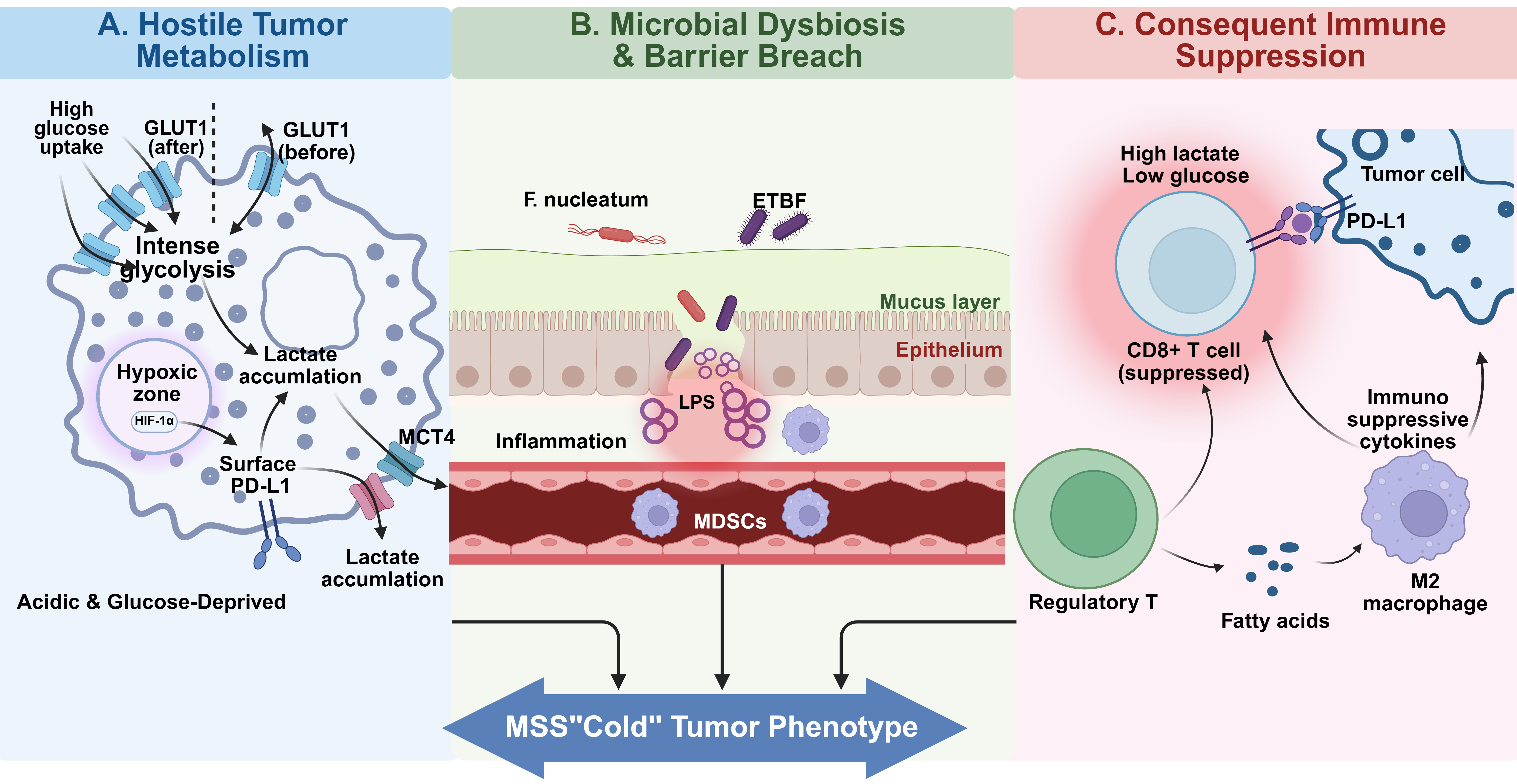
Here, we define the immunometabolic barrier ecosystem model of MSS immune resistance, in which tumor metabolism, microbial dysbiosis, and immune suppression function as interdependent regulatory axes that collectively prevent effective anti-tumor immunity. Immune infiltration in CRC is heterogeneous and plastic. In MSS CRC, immune failure often reflects impaired priming, inadequate infiltration, and intratumoral suppression rather than complete immune absence.
2. Immune architecture in CRC: constraints relevant to MSS conversion
Cytotoxic and helper T cell dysfunction
High intratumoral CD8+ CTL density associates with improved survival in CRC (10, 16). However, chronic antigen exposure and suppressive cues drive exhaustion, characterized by reduced IFN-γ and TNF-α production, impaired proliferation, and increased expression of inhibitory receptors (PD-1, CTLA-4, TIM-3, LAG-3) (23, 24). In MSS CRC, exhaustion is commonly reinforced by metabolic insufficiency and suppressive myeloid/stromal programs, limiting the restorative capacity of ICB alone. Th1 responses support CTL function via IFN-γ and inflammatory macrophage activity (25, 26). Th17 biology is context dependent; persistent Th17-driven inflammation, particularly in dysbiosis-associated settings, has been implicated in CRC initiation and progression (27, 28). The cytokine and metabolite environment shapes this balance, linking helper polarization to tissue state.
Regulatory immune circuits
Foxp3+ Tregs suppress anti-tumor responses via IL-10/TGF-β production and IL-2 consumption (29-31). Their accumulation and suppressive potency can be supported by high lactate and microbiota-derived signals (20, 32), providing a mechanistic bridge between metabolism/microbiome and immune suppression. B cells promote anti-tumor immunity through antibody production, antigen presentation, and cytokine secretion (33). Crucially, their organization into tertiary lymphoid structures (TLS) strongly correlates with a favorable prognosis and improved response to immunotherapy, serving as local sites for T cell priming (34-36). However, B cell effects are heterogeneous: Bregs can suppress immunity via IL-10 (37), and metabolic crosstalk within the TME can further constrain their effector differentiation (38).
Myeloid dominance in MSS tumors: Impaired immune priming and innate cytotoxicity
Macrophages adopt a spectrum of states; M2-like TAM programs support angiogenesis, matrix remodeling, metastasis, and T cell suppression (30). In MSS CRC, myeloid dominance can establish a suppressive baseline that is not reversed by ICB alone. DCs are required for priming tumor-reactive T cells (39). Tumor-derived factors and metabolic stress inhibit DC maturation and antigen presentation, reducing co-stimulation and T cell-activating cytokines and promoting tolerance/anergy (40, 41). When priming is limited, ICB has reduced substrate to act upon. MDSCs suppress T and NK cells through amino acid depletion, reactive oxygen/nitrogen species, and promotion of Treg expansion (42). Their accumulation provides a plausible mechanism for “immune-present but ineffective” MSS phenotypes.
NK cells kill targets with reduced MHC class I (43) but are inhibited by hypoxia, nutrient restriction, and mediators such as TGF-β, adenosine, and lactate (44). Reduced NK activity weakens innate cytotoxic pressure and adaptive cross-talk. TANs can adopt divergent programs; pro-tumor phenotypes promote angiogenesis, metastasis, immunosuppression, and neutrophil extracellular traps (45). Their state is strongly conditioned by TME signals.
Although immune cell composition strongly influences clinical outcomes in colorectal cancer, immune architecture alone does not fully explain the profound resistance of MSS tumors to immunotherapy. Increasing evidence indicates that immune dysfunction in MSS CRC is tightly coupled to metabolic conditions within the tumor microenvironment. Rapid tumor growth, inefficient vascularization, and oncogenic metabolic reprogramming generate hypoxic, nutrient-restricted, and lactate-rich environments that selectively impair effector immune cells while favoring suppressive populations. Understanding these metabolic constraints is therefore essential for interpreting immune failure in MSS CRC.
3. Metabolic reprogramming of the MSS tumor ecosystem
Tumor metabolic reprogramming is increasingly recognized as a central determinant of immune competence in the tumor microenvironment. In MSS colorectal cancer, oncogenic metabolic programs generate conditions of nutrient depletion, hypoxia, and metabolite accumulation that selectively disadvantage cytotoxic immune cells while supporting suppressive populations. These metabolic alterations therefore function not only as drivers of tumor growth but also as regulators of immune exclusion.
Aerobic glycolysis and PPP: growth and TME conditioning
CRC cells frequently increase aerobic glycolysis (Warburg effect) (46). Glycolysis supports rapid ATP generation and diversion of intermediates to anabolic pathways (47), and upregulation can occur early in tumorigenesis (48). The pentose phosphate pathway (PPP) generates ribose-5-phosphate and NADPH for nucleotide synthesis, lipogenesis, and redox control; enzymes such as G6PD and transketolase are often upregulated (49, 50). These programs contribute to significant extracellular lactate accumulation, which functions as a key immunomodulatory signaling molecule rather than a mere metabolic byproduct (51). Specifically, high lactate levels in the TME impair the effector functions of CD8+ T cells and NK cells by inhibiting their lactate efflux via MCT1, thereby reducing IFN-γ production and cytolytic activity (52). Simultaneously, lactate promotes the M2-like polarization of TAMs through activation of the GPR81 and HIF-1 signaling pathways and via histone lactylation (53, 54), thereby further enhancing an immunosuppressive environment. Moreover, Tregs can metabolically adapt to use lactate as a fuel source, thereby maintaining their suppressive capacity even in the nutrient-depleted TME (51). Together, these mechanisms collectively reshape the TIME to favor tumor immune evasion.
Amino acid metabolism: shared nutrient bottlenecks
Glutamine supports anaplerosis and nucleotide synthesis in CRC cells (14, 46), creating a metabolic bottleneck that constrains T cell activation. Targeting glutamine metabolism has been shown to unleash anti-tumor immunity by differentially affecting CRC cells versus infiltrating CD8+ T cells through intense competition for extracellular glutamine (55). The serine/glycine synthesis pathway supports one-carbon metabolism via PHGDH/PSAT1/PSPH and SHMT, thereby contributing to nucleotide synthesis and methylation capacity; elevated expression is associated with poor prognosis (56, 57). These pathways further constrain effector T cell programs through direct competition for serine, an indispensable substrate for maintaining redox balance and supporting rapid proliferation. Additionally, the depletion of arginine, driven by tumor uptake and the secretion of Arginase 1 (ARG1) by suppressive myeloid cells, directly impairs T cell proliferation and T cell receptor (TCR) expression (58), further tightening the metabolic constraint within the TIME.
Lipid metabolism: synthesis, uptake, and stress adaptation
CRC increases de novo fatty acid synthesis via ACLY/FASN (47) and may enhance uptake via CD36, linked to metastatic potential (59). Under nutrient stress, fatty acid oxidation can supply ATP and acetyl-CoA (60). Because specific immunosuppressive populations-primarily Tregs, M2-like TAMs, and MDSCs-can utilize fatty acids and lactate more flexibly than glycolysis-dependent effectors (14, 15, 19, 61), lipid programs may bias immune composition; for instance, M2-like TAMs actively rely on FAO to sustain their pro-tumorigenic programs and suppressive functions (30). Moreover, intratumoral Tregs upregulate CD36 to adapt to the lipid-enriched TME, sustaining their suppressive function (62). Collectively, these lipid-driven adaptations reinforce the metabolic dominance of inhibitory cells within the MSS CRC ecosystem.
Hypoxia and HIF-1α: coupling vascular inefficiency to immune dysfunction
Hypoxia stabilizes HIF-1α, upregulating glycolysis (GLUT1, HK2, LDHA), angiogenesis (VEGF), and epithelial-mesenchymal transition (EMT) programs (14, 63, 64). Crucially, hypoxia directly drives immune checkpoints: HIF-1α binds the PD-L1 promoter, inducing its expression in tumor cells (65). Beyond its effects on tumor cells, hypoxia orchestrates a multi-layered suppressive landscape across the TIME. For instance, hypoxic stress impairs the maturation and antigen-presenting competency of DCs, thereby limiting the initial priming of tumor-reactive T cells (40, 41). Within the cytotoxic compartment, hypoxia directly blunts the effector activity of CD8+ T cells (14) and NK cells (44), while favoring the recruitment of MDSCs (65) and promoting the M2-like polarization of TAMs (30). Additionally, hypoxia promotes extracellular accumulation of adenosine, which signals through A2A receptors to inhibit T and NK cell function (66), creating a multi-layered barrier to effector function.
Oncogenic control of metabolic baseline
MYC regulates broad metabolic gene networks, including glycolysis and glutaminolysis (67). KRAS/BRAF activation increases glucose uptake and glycolytic flux by upregulating GLUT1, thereby providing a survival advantage in low-glucose environments (68). APC loss activates Wnt/β-catenin signaling, which not only drives proliferation but also induces the expression of metabolic genes (47). p53 normally restrains glycolysis and PPP flux (e.g., via TIGAR); loss of p53 removes this restraint (69). Genetics and microenvironment, therefore, jointly set metabolic conditions that can limit immune effector competence in MSS CRC.
While tumor-intrinsic metabolic programs reshape the biochemical landscape of the tumor microenvironment, colorectal cancer develops within a unique ecological context shaped by the intestinal microbiome. Microbial communities interact continuously with host metabolism and mucosal immunity, influencing inflammatory tone, epithelial barrier integrity, and metabolite availability. Consequently, microbial dysbiosis represents a second major regulatory layer that can reinforce the immunosuppressive metabolic landscape established by tumor cells.
4. The gut microbiome as an ecological regulator of tumor immunity
Microbial metabolites influence tumor immunity through three principal mechanisms: epigenetic regulation, immune receptor signaling, and modulation of host metabolic pathways.
Dysbiosis and epithelial barrier disruption
CRC-associated dysbiosis often involves a reduction in fiber-fermenting commensals and an enrichment of pro-inflammatory taxa (70). Barrier disruption permits the translocation of microbial products, such as LPS, thereby sustaining low-grade inflammation and a milieu that supports tumorigenesis through growth factors, angiogenic mediators, and reactive oxygen species (27). Chronic inflammation can coexist with weak anti-tumor immunity by promoting myeloid suppression and T cell dysfunction (71). In MSS colorectal cancer, the gut microbiome functions as an ecological regulator of tumor immunity by shaping inflammatory tone, epithelial barrier integrity, and metabolite-driven immune signaling pathways.
Pro-tumor and protective microbial species
F. nucleatum is enriched in CRC tissue, can activate Wnt/β-catenin via FadA-E-cadherin interactions, and promotes immunosuppression by recruiting MDSCs and inhibiting T and NK cells (17, 72). Colibactin-producing pks+ E. coli induces DNA double-strand breaks and genomic instability (17, 73). Enterotoxigenic Bacteroides fragilis (ETBF) secretes BFT, disrupts barrier function, activates Wnt/β-catenin signaling, and induces STAT3-dependent inflammation with Th17 skewing (28). In contrast, beneficial taxa (e.g., Lactobacillus, Bifidobacterium, and butyrate producers such as Faecalibacterium prausnitzii and Roseburia) support barrier integrity and generate SCFAs (70, 74).
Microbial metabolites shaping tumor immunity
Beyond direct cellular interactions, the gut microbiota and the host communicate through a complex network of metabolites that function as critical signaling molecules. These metabolites originate from both microbial fermentation of dietary components and host-driven metabolic pathways, collectively modulating the inflammatory and immune landscape of the TME. The primary microbial and host-derived metabolites involved in this crosstalk, along with their specific receptors and biological effects, are summarized in Table 1.
Table 1. Microbial and host-derived metabolites shaping tumor immunity
| Metabolite | Primary Source | Primary Host Receptor(s) /Target | Key Downstream Signaling Pathways | Primary Effect on TME | Ref |
|---|---|---|---|---|---|
| Butyrate | Microbial fermentation of fiber | GPR43, GPR109A, HDACs | Treg differentiation; IL-10 production; inhibits HDACs, | Anti-inflammatory, anti-proliferative (tumor cells) | (1, 75-77) |
| Deoxycholic Acid (DCA) | Microbial modification of primary bile acids | FXR, TGR5 | NF-κB, STAT3 | Pro-inflammatory, pro-tumorigenic | (74, 78, 79) |
| Trimethylamine N-oxide (TMAO) | Microbial metabolism of choline/carnitine + host oxidation | PERK (Direct binding ligand) | NF-κB, AKT/mTOR, VEGFA | Pro-inflammatory, pro-angiogenic | (74, 78, 79) |
| Kynurenine | Host/tumor cell metabolism of tryptophan (IDO1) | Aryl Hydrocarbon Receptor (AhR) | AhR activation induces Tregs, CTLs | Immunosuppressive | (82-85) |
| Indole derivatives (IPA, IAA, I3A, ILA) | Gut microbiota | AhR, PXR | Myelopoiesis rewiring; IL-35 axis; Epigenetic remodeling | Anti-tumor; "warms up" MSS TME; inhibits metastasis | (86-89) |
| Lactate | Tumor cell glycolysis (Warburg effect) | GPR81, MCT1/4 | cAMP, MHC-II, intracellular acidification | Immunosuppressive, acidification | (18, 48, 51, 52, 90, 91) |
SCFAs
SCFAs (e. g., acetate, propionate, and butyrate), produced through microbial fermentation of dietary fiber, function as critical bridges between diet and host immunity. Beyond signaling through GPCRs like GPR43/FFAR2 and GPR109A/HCAR2 to support anti-inflammatory homeostasis and IL-10 production (92), recent research highlights their role as potent HDAC inhibitors (77). In the TME, butyrate acts as an epigenetic modulator, promoting Foxp3 expression and the differentiation of colonic Tregs. Furthermore, the "butyrate paradox" remains a key research frontier: in glycolytic CRC cells, butyrate accumulates in the nucleus, inducing cell-cycle arrest and apoptosis (77), whereas in healthy colonocytes, it is rapidly oxidized as a primary fuel source. These lipid-derived signals collectively shape the metabolic and epigenetic landscape of the CRC microenvironment.
Secondary bile acids
Secondary bile acids, such as DCA and lithocholic acid (LCA), are generated through microbial modification of primary bile acids and are significant metabolic risk factors in CRC (74). The latest evidence indicates that elevated DCA levels drive tumorigenesis by inducing DNA damage and oxidative stress and activating the EGFR and Wnt/β-catenin pathways (78). From an immunological perspective, secondary bile acids subvert the TIME by signaling through TGR5 to activate NF-κB and STAT3 in myeloid cells, promoting an M2-like suppressive phenotype (79). Conversely, the silencing of the FXR (Farnesoid X receptor)-a primary host receptor for bile acids-is a common feature in MSS CRC that correlates with barrier breach and increased inflammatory cell infiltration. Notably, microbial-driven bile acid metabolism can even modulate colorectal liver metastasis (CRLM) by shaping the pre-metastatic niche (79, 93), positioning secondary bile acids as potential therapeutic targets for ecosystem reconditioning.
TMAO
TMAO is generated from dietary choline/L-carnitine via microbial conversion to TMA and hepatic oxidation by FMO3 (80). Elevated TMAO has been linked to CRC risk and may promote NF-κB activation, ILK/AKT/mTOR signaling, and VEGFA production (80, 81). Mechanistically, TMAO has been identified as a direct ligand for the endoplasmic reticulum stress kinase PERK (EIF2AK3). By binding to PERK, TMAO selectively activates the unfolded protein response (UPR) and upregulates the metabolic transcription factor FoxO1, thereby linking gut microbial metabolism directly to host metabolic dysfunction (94).
IDO1-kynurenine-AhR axis
Metabolic crosstalk involving host-driven IDO1/TDO-mediated tryptophan catabolism depletes local tryptophan and generates kynurenines (82, 83). Kynurenine activates AhR, an endogenous ligand-receptor interaction that drives Treg generation and suppresses antitumor immunity (84), promotes Treg differentiation and suppresses CTL function (85), thereby providing a receptor-defined immunosuppressive pathway.
Microbial indole metabolites and AhR-centered immune regulation
Microbiota-derived indole metabolites, such as indole-3-propionic acid (IPA), indole-3-acetic acid (IAA), indole-3-aldehyde (I3A), and indole-3-lactic acid (ILA), serve as potent AhR ligands, promoting tumor immunity and counteracting the immunosuppressive effects of the host Kyn-AhR axis. Recent evidence indicates that chemotherapy-induced intestinal dysbiosis can elevate IPA levels, which subsequently reprogram host myelopoiesis by redirecting common myeloid progenitor differentiation toward anti-tumor macrophages rather than immunosuppressive monocytes, thereby effectively curbing colorectal liver metastasis (86).
During the early stages of colorectal cancer, IAA activates the pregnane X receptor (PXR), thereby inducing the generation of IL-35+ immune cells that ameliorate the precancerous inflammatory milieu and inhibit tumorigenesis (87). Concurrently, ILA has been shown to enhance the fitness and cytotoxicity of tumor-infiltrating CD8+ T cells through epigenetic reprogramming, specifically by upregulating H3K27ac modification at IL-12a enhancers and transcriptionally suppressing the cholesterol metabolism-associated gene Saa3(88). Furthermore, I3A released by intratumoral Lactobacillus reuteri directly activates AhR signaling in CD8+ T cells, promoting IFN-γ production and significantly augmenting the efficacy of ICB (89). These metabolites, particularly when delivered via engineered probiotic systems, represent a promising therapeutic strategy for "warming up" the cold tumor microenvironment of MSS CRC.
5. The immunometabolic barrier in MSS colorectal cancer
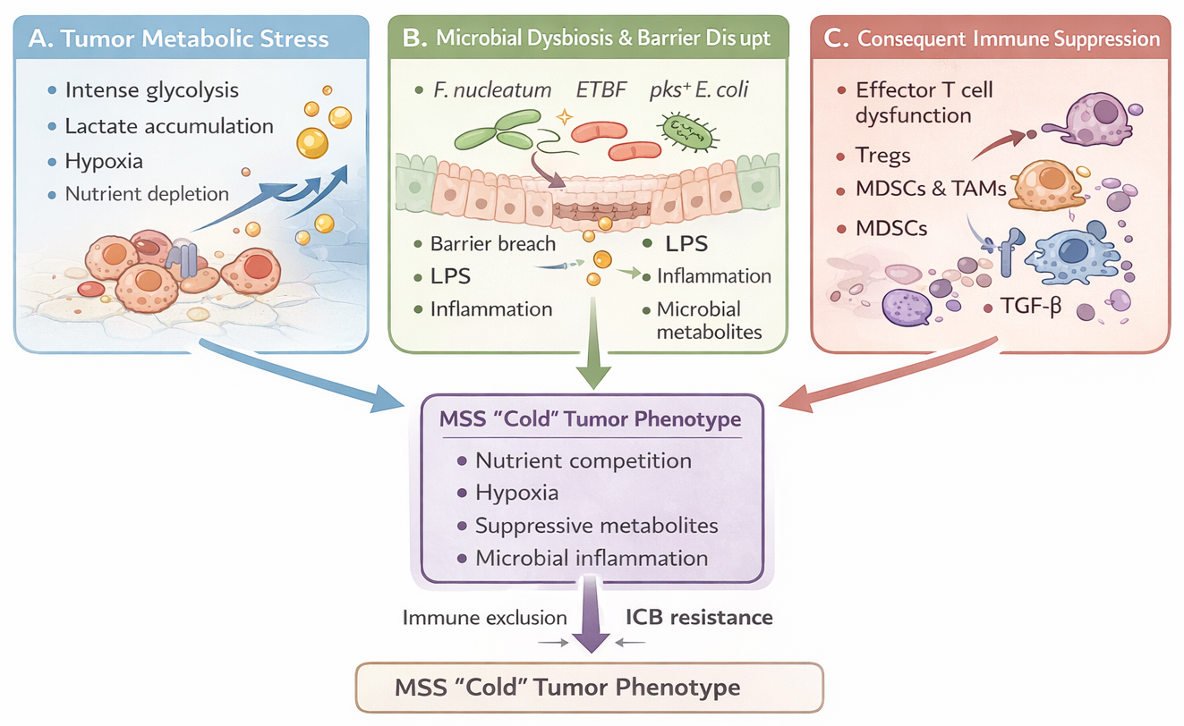
The metabolic, microbial, and immunological processes described above converge to create an integrated ecosystem of immune resistance in MSS colorectal cancer. Within this ecosystem, tumor metabolic reprogramming, microbial dysbiosis, and suppressive immune circuits reinforce one another through multiple feedback loops. These interactions collectively establish what we refer to as the immunometabolic barrier, a tumor microenvironment characterized by nutrient competition, inhibitory metabolites, and chronic inflammatory signaling that limit effective anti-tumor immunity. The resulting state, characterized by nutrient competition, accumulation of inhibitory metabolites, and chronic inflammatory conditioning, creates an immunometabolic barrier to immune checkpoint blockade. This section synthesizes the metabolic and microbial mechanisms described above into a unified framework explaining immune exclusion in MSS colorectal cancer. In this model, metabolic competition, suppressive metabolites, and microbiome-derived signals interact to form an immunometabolic barrier that constrains effective anti-tumor immunity (Figure 2).
Metabolic constraints shaping immune suppression
Tumor metabolic reprogramming creates multiple constraints that limit effective immune responses within the MSS tumor microenvironment. As described in Section 3, tumor glycolytic metabolism generates nutrient competition that constrains the metabolic fitness of cytotoxic lymphocytes (16, 18). Competition for glutamine and arginine further limits effector T-cell activity while supporting suppressive myeloid programs (22, 55, 58). In parallel, lactate accumulation and extracellular acidosis inhibit CTL and NK cell cytotoxicity, reduce T-cell proliferation and cytokine production, and impair dendritic cell maturation and antigen presentation (51, 52). Hypoxia further intensifies these metabolic constraints by limiting cellular bioenergetics and promoting suppressive immune programs (15, 95).
Beyond simple metabolic deprivation, several metabolites function as active immunoregulatory signals. Lactate suppresses effector lymphocytes while supporting regulatory T cells and M2-like macrophages that can utilize lactate as an alternative fuel source (20, 22). Lactate signaling through GPR81 and histone lactylation can further stabilize suppressive macrophage programs (51, 90). Hypoxia also promotes extracellular adenosine accumulation through CD39/CD73 activity, and adenosine signaling through A2A receptors inhibits T-cell and NK-cell function (44, 66). Similarly, kynurenine generated through tryptophan metabolism activates AhR signaling, promoting Treg differentiation and suppressing cytotoxic T-cell activity (83, 84). These metabolic constraints are reinforced by immune checkpoint signaling. PD-1 signaling limits glycolysis and promotes fatty acid oxidation in T cells, restricting effector output during anti-tumor responses (22, 96). At the same time, nutrient deprivation and hypoxia suppress PI3K-Akt-mTOR and c-Myc metabolic programs required for immune activation (16, 22). Together, these mechanisms create a metabolic environment that strongly limits the efficacy of immune checkpoint blockade unless the tumor ecosystem is reconditioned.
Microbial-metabolic feedback loops in the MSS ecosystem
Tumor metabolism, microbial activity, and immune suppression interact through reciprocal feedback mechanisms that stabilize the immunometabolic barrier in MSS colorectal cancer. For example, Fusobacterium nucleatum can modulate host glucose metabolism to support bacterial survival while simultaneously promoting immune evasion. This pathogen recruits myeloid-derived suppressor cells by inducing inflammatory chemokines such as CCL20 and CXCL1 (97) and directly inhibits T- and NK-cell cytotoxicity through the Fap2-TIGIT interaction (98). Conversely, tumor metabolic conditions can influence microbial ecology. Lactate-rich and acidic tumor environments may favor acid-tolerant pro-inflammatory taxa, reinforcing local immune suppression. Increasing evidence suggests that gut microbes condition the colorectal tumor microenvironment through at least three non-mutually exclusive mechanisms: microbial metabolites, bacterial proteins or toxins, and direct host-cell interactions (99, 100).
Microbial metabolites can directly affect immune surveillance. For example, Fusobacterium nucleatum-derived succinic acid suppresses cGAS-IFN-β signaling and limits CD8+ T-cell trafficking, promoting resistance to immunotherapy (101). Bacterial proteins may also engage inhibitory immune receptors; the Fap2 adhesin of F. nucleatum binds TIGIT and suppresses NK- and T-cell cytotoxicity (102). In addition, pathobiont-driven host-cell reprogramming occurs in infections with enterotoxigenic Bacteroides fragilis, where BFT- and IL-17-dependent signaling promotes the accumulation of Arg1+Nos2+ monocytic MDSCs while reinforcing epithelial inflammatory oncogenic programs (103). Together, these mechanisms illustrate how microbial activity can mechanistically reinforce immune exclusion and resistance to immune checkpoint blockade in MSS colorectal cancer (99, 104).
MSI-H versus MSS: contrasting tumor ecosystems
Divergent responses to immune checkpoint blockade in MSI-H and MSS colorectal cancers reflect fundamentally different tumor ecosystem architectures. MSI-H tumors are typically immune-inflamed, characterized by high tumor mutational burden and dense infiltration of activated CD8+ T cells (8, 105). In contrast, MSS CRC frequently presents as an immune-excluded or immune-desert phenotype dominated by suppressive myeloid populations, regulatory T cells, and M2-polarized macrophages (106). Metabolic differences further distinguish these tumor types. MSS tumors often exhibit asynchronous cholesterol biosynthesis, generating distal cholesterol precursors that activate RORγt signaling and promote Th17 polarization, thereby reinforcing suppressive immune environments (107). Microbial composition also differs between tumor types: MSI-H tumors are enriched for immunostimulatory taxa such as Akkermansia (108), whereas MSS CRC more commonly harbors pathobionts, including Fusobacterium nucleatum, that recruit suppressive myeloid cells and inhibit T-cell activity (109).
Together, these integrated metabolic, microbial, and immune constraints establish the primary resistance observed in MSS tumors. This coordinated state represents the immunometabolic barrier, a systems-level resistance mechanism in which metabolic stress, microbial dysbiosis, and immune suppression collectively limit the efficacy of immune checkpoint blockade.
Conceptual Model: The Immunometabolic Barrier in MSS Colorectal Cancer
MSS colorectal cancer develops within a tumor ecosystem shaped by metabolic stress, microbial dysbiosis, and suppressive immune circuits. Tumor glycolysis and hypoxia lead to lactate accumulation, extracellular acidosis, and nutrient depletion. These conditions impair the metabolic fitness of cytotoxic lymphocytes. In parallel, dysbiotic microbial communities and epithelial barrier disruption expose the tumor microenvironment to microbial products and metabolites that promote inflammatory signaling and myeloid recruitment. These metabolic and microbial cues converge to expand regulatory immune populations, including Tregs, MDSCs, and M2 macrophages, while suppressing CD8+ T-cell activity.
Collectively, these processes establish an immunometabolic barrier characterized by nutrient competition, suppressive metabolites, and pathogen-associated inflammatory signaling. Within this environment, checkpoint inhibition alone is often insufficient to restore effective immune responses because immune priming remains weak and metabolic constraints persist. Converting MSS tumors from “cold” to “hot” states will therefore likely require coordinated interventions targeting tumor metabolism, microbial ecology, and immune activation.
6. MSS/ICB conversion: failure mechanisms, matched combinations, and measurable readouts
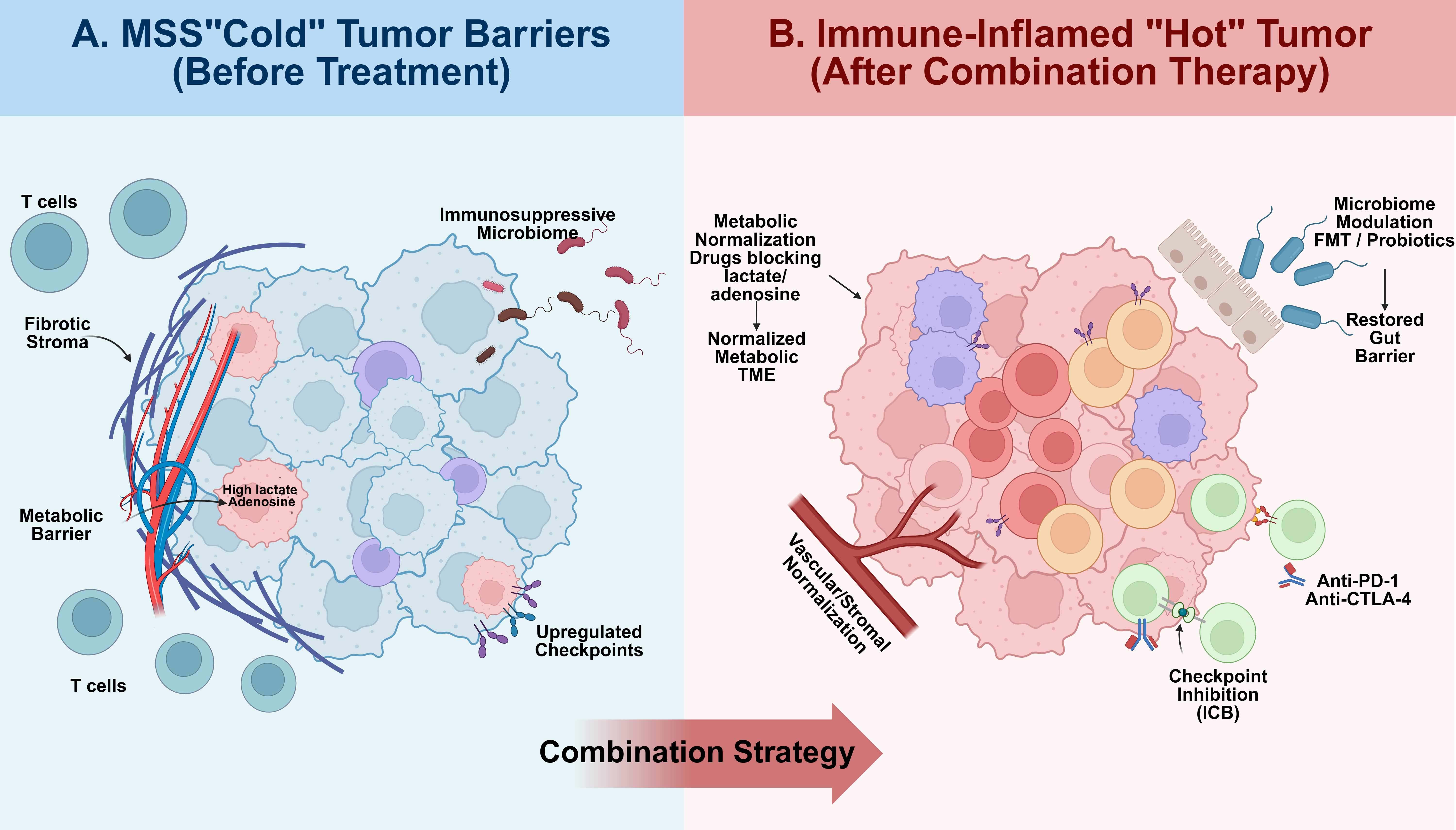
Effective immunotherapy for MSS CRC requires a transition from understanding biological constraints to implementing rational, multimodal rescue strategies. This conceptual framework for converting the MSS 'cold' TME into an immune-inflamed 'hot' phenotype is summarized in Figure 3, which contrasts the multi-layered barriers present before treatment with the normalized ecosystem achieved through combination therapies.
Failure mechanism 1: insufficient priming and antigen presentation
Mechanism. DC maturation and antigen presentation are impaired by tumor-derived factors and metabolic stress, reducing co-stimulation and T cell-activating cytokines and promoting tolerance (40, 41). Matched combination strategy. Use interventions that increase priming capacity and local immune organization before/with ICB, including microbiome modulation as a priming amplifier (1, 110) and strategies that favor TLS-like organization, given TLS associations with better outcomes and immunotherapy responsiveness (34-36). Readouts (biomarker/PD). DC maturation/activation markers and antigen-presentation competency (40, 41); TLS presence/density and spatial immune organization (34, 36); immune contexture metrics linked to prognosis (9, 10).
Failure mechanism 2: suppressive myeloid dominance and Treg accumulation
Mechanism. M2-like TAM programs and MDSCs suppress effector immunity and promote Treg expansion (42, 43). Tregs suppress via IL-10/TGF-β and IL-2 consumption (31), and are supported by lactate and microbiota-derived signals (20, 30, 32). Matched combination strategy. Combine ICB with approaches that reduce suppressive cell dominance or weaken their metabolic support: metabolic normalization to reduce lactate burden (111-113) and microbiome modulation to reduce pro-inflammatory/suppressive conditioning (1, 17, 22, 70).
Readouts (biomarker/PD). Frequencies/phenotypes of TAMs/MDSCs/Tregs (30-32, 42, 43); cytokine milieu reflecting suppressive signaling (30, 31); lactate-associated signatures relevant to suppressive conditioning (51, 91).
Failure mechanism 3: metabolic suppression (nutrient scarcity, acidosis, hypoxia)
Mechanism. Warburg metabolism and tumor growth lead to glucose depletion, lactate accumulation, acidosis, and hypoxia, which impair CTL/NK function and DC priming (14, 15, 18, 51). Matched combination strategy. Pair ICB with metabolic interventions that aim at TME normalization: glycolysis inhibition (46, 112), glutaminolysis inhibition (55, 113), lipid metabolism targeting (60, 113), and broader metabolic targeting strategies to reduce stressors and improve immune metabolic fitness (111). Readouts (biomarker/PD). Lactate-related gene-expression scores and metabolomic readouts proposed as prognostic/predictive indicators (114, 115); immune functional recovery measures in relation to metabolic stress (15, 22, 51); activity of hypoxia-driven pathways (inferred from HIF-1α regulatory networks and targets) (14, 63, 64).
Failure mechanism 4: suppressive metabolite signaling (adenosine, kynurenine/AhR)
Mechanism. Hypoxia-induced adenosine suppresses T/NK function via A2A receptor-cAMP signaling (44, 66, 116). IDO1/TDO-mediated kynurenine production depletes tryptophan and activates AhR, promoting Tregs and inhibiting CTLs (82, 83, 85). Matched combination strategy. Combine ICB with interventions that reduce production or signaling of these metabolites, guided by dominant-axis selection; note that IDO1-inhibitor trial failures highlight redundancy and biomarker dependence (82). Metabolic reconditioning that reduces hypoxia/lactate may also reduce adenosine pressure indirectly (15, 51, 95). Readouts (biomarker/PD). CD39/CD73/A2A axis activity and adenosine-associated suppression markers (44, 116); kynurenine/AhR pathway activity and tryptophan-catabolism signatures (82, 83, 85); Treg expansion linked to these axes (32, 83-85).
Failure mechanism 5: adverse microbiome ecology and pro-tumor metabolites
Mechanism. Dysbiosis promotes barrier dysfunction and chronic inflammation (70), enriches pro-tumor taxa (F. nucleatum, pks+ E. coli, ETBF) that reinforce oncogenic signaling and immunosuppression (70), and alters metabolite outputs (SCFAs, SBAs, TMAO) that modulate immune tone and inflammatory signaling (78-80). Matched combination strategy. Use microbiome modulation (probiotics/prebiotics/synbiotics/FMT) to shift community structure and metabolite output as an adjunct to ICB and TME normalization (117). The therapeutic potential and specific mechanisms of these microbiota-based interventions are summarized in Table 2.
Table 2. Summary of microbiota-related interventions and their impact on CRC progression and therapy.
| Research type | Sample Type | Total (N) | Group Distribution (n values) | Microbiota intervention | Duration/ Follow-Up | Cancer Therapy | Outcome | Ref |
|---|---|---|---|---|---|---|---|---|
| Clinical Trial | Human (CRC) | 52 | Probiotic (n=27) vs. Placebo (n=25) | Probiotic mixture | 6 months | Surgery | Modulates gut microenvironment, reduces pro-inflammatory cytokines, and lowers postoperative infection risk. | (118) |
| Case Report | Human (Case Report) | 1 | N/A (Single-arm case study) | FMT | 4 months | anti PD-1 + anti-VEGF | Downstages tumors to resectable levels; enables complete pathological remission (pCR). | (119) |
| Preclinical (In vivo) | Mouse (MC38) | 32* | 4 Groups: Control, JP, CTX, JP+CTX (n=8 each) | Prebiotic (Jujube powder) |
3 weeks | CTX | Enhances CTX efficacy by increasing CD8+ T-cell infiltration and reducing eosinophilia. | (120) |
| Preclinical (In vivo) | Rat (Liver Met) |
20 | Probiotic (n=10) vs. Control (n=10) | Probiotic mixture | 34 days | N/A | Reduces angiogenesis and inhibits CRLM. | (121) |
| Preclinical (In vivo) | Mouse (CRC) | 20* | Control (n=10) vs. CB-treated (n=10) | Probiotics (oral C. butyricum) | N/A | 5-FU + anti-PD-1 | Reduces 5-FU resistance, enhances anti-PD-1 efficacy, and inhibits CRC proliferation and metastasis. | (122) |
| Preclinical (In vivo) | Mouse (Metastasis) | 12 | Control (n=6) vs. Minocycline (n=6) | Antibiotics (Minocycline) | 6 weeks | N/A | Impedes the EMT process in CRC cells to inhibit metastasis. | (123) |
| Preclinical (In vivo) | Mouse (Liver Met) | 12 | Control (n=6) vs. NABs (n=6) | Antibiotics (Gentamicin/ Amikacin) | N/A | N/A | Successfully inhibits CRLM in a mouse model | (93) |
| Preclinical (In vivo) | Mouse (Peritoneal) | 10 | Normal Diet (n=5) vs. High-Fat Diet (n=5) | HFD | 3 weeks | Oxaliplatin + 5-FU | Short-term HFD enhances chemotherapeutic efficacy in CRC peritoneal metastasis. | (124) |
| Preclinical (In vivo) | Mouse (AOM/DSS) | 30 | 3 Groups: Control, Model, YYC-3 (n=10 each) | HFD + YYC-3 | 7 weeks | N/A | Prevents early-stage CRC by modulating the colonic microenvironment and reducing inflammation. | (125) |
Readouts (biomarker/PD)
Baseline microbiome signatures associated with immunotherapy response across cancers, including CRC (32); metabolite-oriented profiling (e.g., lactate-related scores, broader metabolomics) as functional correlates (114, 115); pathway-level readouts reflecting SBA/TMAO-associated inflammatory and angiogenic signaling (79, 81). The translational transition of these microbiome-based strategies from theory to bedside is evident in the current clinical landscape. Table 3 summarizes key ongoing trials investigating the synergy of dietary, microbial, and metabolic interventions in CRC.
Table 3. Ongoing clinical trials exploring microbiome and metabolic interventions in CRC.
| NCT number | Status | Interventions | Phases | Purposes |
|---|---|---|---|---|
| NCT04729322 | Active | Procedure: Biopsy Procedure: Fecal Microbiota Transplantation Drug: Fecal Microbiota Transplantation Capsule Drug: Metronidazole Drug: Neomycin Biological: Nivolumab |
Phase 2 | Evaluate ORR in dMMR/MSI-H or MSS patients who are Anti-PD-1 non-responders. |
| NCT04707365 | Active | Biological: Tumor samples | N/A | Explore tripartite links between molecular subtypes, TME, and host metabolism/microbiota |
| NCT03941080 | Recruiting | Diagnostic Test: fecal sample Behavioral: questionnaire Diagnostic Test: Blood sample |
N/A | Investigate the correlation between gut microbiome and chemotherapy response |
| NCT06347198 | Recruiting | Drug: Fruquintinib Drug: Sintilimab Drug: Inulin |
Early Phase 1 | Explore the synergy of prebiotic (Inulin) with TKI and ICB in metastatic CRC |
| NCT06049901 | Recruiting | Drug: Nitazoxanide | Phase 3 | Evaluate the efficacy and safety of Nitazoxanide as an adjuvant treatment |
| NCT06349590 | Recruiting | Dietary Supplement: High fiber/low-fat meals | Phase 1 / Phase 2 | Assess preoperative microbiome modulation to prevent recurrence and metastasis |
| NCT05655780 | Recruiting | Biomarker Analysis | N/A | Identify biomarkers to predict clinical response and toxicity during irinotecan treatment |
Abbreviations: NCT, National Clinical Trial; FMT, Fecal Microbiota Transplantation; ICB, Immune Checkpoint Blockade (e.g., Nivolumab, Pembrolizumab, Sintilimab); TKI, Tyrosine Kinase Inhibitor (e.g., Fruquintinib); TME, Tumor Microenvironment; CRC, Colorectal Cancer.
5. Discussion
Microsatellite-stable colorectal cancer (MSS CRC) is a clinically important example of resistance to immune checkpoint blockade (ICB). Unlike MSI-H tumors, where high mutational burden and robust neoantigen generation enable immune recognition, MSS CRC typically lacks effective immune priming and develops within a tumor microenvironment (TME) that actively suppresses cytotoxic immunity. In our view, resistance in MSS CRC is best understood as the convergence of metabolic, microbial, and immunological processes rather than a single dominant mechanism.
MSS tumors develop within a metabolically hostile tumor microenvironment. Rapid tumor glycolysis and hypoxia lead to lactate accumulation, extracellular acidosis, and nutrient depletion. These conditions impair the metabolic fitness of cytotoxic lymphocytes while favoring regulatory immune populations (14, 18, 51). At the same time, the intestinal location of colorectal cancer exposes the tumor to continuous microbial signaling. Dysbiotic microbial communities and epithelial barrier disruption promote inflammatory signaling and generate metabolites that influence immune cell differentiation and activation (1, 74, 79). In parallel, tumor- and microbiome-derived immunosuppressive metabolites, including kynurenine and other AhR ligands, further reinforce tolerogenic immune circuits (82, 83, 85).
Together, tumor metabolism, microbial dysbiosis, and suppressive immune circuits create an immunometabolic barrier to effective antitumor immunity. Within this ecosystem, nutrient competition, lactate-driven acidosis, and pathogen-associated inflammatory signals bias immune responses toward regulatory states and limit the expansion and persistence of cytotoxic effector cells. This model also explains why checkpoint inhibition alone rarely produces durable responses in MSS CRC. Releasing inhibitory signaling cannot restore immunity if immune priming is weak and the surrounding metabolic environment remains hostile. Consequently, durable therapeutic responses will likely require ecosystem-level reconditioning rather than checkpoint release alone (15, 16, 22).
Emerging Technologies to Decode the MSS Tumor Ecosystem
Understanding this ecosystem requires approaches that resolve the spatial and cellular heterogeneity of the tumor microenvironment. Bulk genomic and transcriptomic analyses obscure the localized metabolic niches and immune states that define the MSS tumor landscape. Advances in single-cell and spatial multi-omics technologies now enable the simultaneous mapping of transcriptional programs, immune cell phenotypes, and metabolic gradients within intact tumor tissues. Integration of these datasets with machine-learning approaches can identify therapy-relevant cellular interactions (126). In parallel, spatial metabolomics, including matrix-assisted laser desorption ionization mass spectrometry imaging (MALDI-MSI), provides direct visualization of metabolic gradients across tumor regions. Recent studies applying MALDI-MSI to colorectal tumors have identified spatially distinct patterns of fatty-acid and amino-acid metabolism that correlate with immune infiltration and stromal organization (127), highlighting the importance of metabolic geography in shaping immune responses.
Targeting Microbial and Metabolic Drivers of Immune Suppression
The growing recognition that microbial metabolism contributes to immune suppression also creates new therapeutic opportunities. Current microbiome interventions, such as fecal microbiota transplantation or probiotic supplementation, primarily aim to restore community diversity. Future strategies may therefore move beyond simple community replacement toward engineering specific microbial metabolic functions. Advances in synthetic biology may enable the design of microbial consortia capable of producing beneficial metabolites or degrading immunosuppressive compounds, thereby allowing controlled modulation of host-microbe metabolic signaling (117). Such approaches could complement metabolic targeting strategies aimed at limiting lactate production, restoring nutrient availability, or inhibiting pathways such as IDO-kynurenine signaling that reinforce immunosuppression.
Toward Personalized Immunometabolic Therapy
Because MSS tumors differ widely in their metabolic, microbial, and immune characteristics, integrative computational approaches combining tumor genomics, microbiome composition, and metabolomic states are beginning to enable the identification of dominant suppressive pathways within individual tumors. Artificial intelligence-based models may help guide rational therapeutic combinations, distinguishing, for example, tumors characterized by lactate-dominant metabolic suppression from those driven by kynurenine/AhR signaling or microbiome-induced inflammation (126, 128). Such stratification could help avoid dilution of therapeutic effects in unselected MSS populations and accelerate the development of targeted combination strategies.
Conclusions
The immune resistance of MSS colorectal cancer cannot be explained solely by low tumor immunogenicity. Instead, it reflects a dynamic ecosystem involving tumor metabolism, microbial dysbiosis, and immune suppression. Nutrient competition, lactate-mediated acidosis, and pathogen-associated inflammatory signaling together create an immunometabolic barrier that prevents effective activation and persistence of cytotoxic immune responses. Overcoming this barrier will likely require coordinated therapeutic strategies that simultaneously restore immune priming, normalize tumor metabolism, and reshape microbial ecology. Future integration of spatial multi-omics technologies with AI-driven patient stratification may provide the framework needed to identify dominant suppressive pathways within individual tumors and guide personalized immunometabolic therapies. From this ecosystem perspective, successful conversion of MSS tumors from “cold” to “hot” states will depend not on checkpoint inhibition alone but on multimodal reprogramming of the tumor microenvironment.
References
1. Karam F, El Deghel Y, Iratni R, Dakroub AH, Eid AH. The gut microbiome and colorectal cancer: an integrative review of the underlying mechanisms. Cell Biochem Biophys. 2025;83(3):2637–2650. https://doi.org/10.1007/s12013-025-01683-9
2. Sung H, Ferlay J, Siegel RL, Laversanne M, Soerjomataram I, Jemal A, et al. Global cancer statistics 2020: GLOBOCAN estimates of incidence and mortality worldwide for 36 cancers in 185 countries. CA Cancer J Clin. 2021;71(3):209–249. https://doi.org/10.3322/caac.21660
3. Siegel RL, Wagle NS, Cercek A, Smith RA, Jemal A. Colorectal cancer statistics, 2023. CA Cancer J Clin. 2023;73(3):233–254. https://doi.org/10.3322/caac.21772
4. Hofseth LJ, Hebert JR, Chanda A, Chen H, Love BL, Pena MM, et al. Early-onset colorectal cancer: initial clues and current views. Nat Rev Gastroenterol Hepatol. 2020;17(6):352–364. https://doi.org/10.1038/s41575-019-0253-4
5. The Cancer Genome Atlas Network. Comprehensive molecular characterization of human colon and rectal cancer. Nature. 2012;487(7407):330–337. https://doi.org/10.1038/nature11252
6. Roudko V, Bozkus CC, Orfanelli T, McClain CB, Carr C, O'Donnell T, et al. Shared immunogenic poly-epitope frameshift mutations in microsatellite unstable tumors. Cell. 2020;183(6):1634–1649.e17. https://doi.org/10.1016/j.cell.2020.11.004
7. Yan S, Wang W, Feng Z, Xue J, Liang W, Wu X, et al. Immune checkpoint inhibitors in colorectal cancer: limitation and challenges. Front Immunol. 2024;15:1403533. https://doi.org/10.3389/fimmu.2024.1403533
8. Le DT, Uram JN, Wang H, Bartlett BR, Kemberling H, Eyring AD, et al. PD-1 blockade in tumors with mismatch-repair deficiency. N Engl J Med. 2015;372(26):2509–2520. https://doi.org/10.1056/nejmoa1500596
9. Rahimi A, Baghernejadan Z, Hazrati A, Malekpour K, Samimi LN, Najafi A, et al. Combination therapy with immune checkpoint inhibitors in colorectal cancer: Challenges, resistance mechanisms, and the role of microbiota. Biomed Pharmacother. 2025;186:118014. https://doi.org/10.1016/j.biopha.2025.118014
10. Galon J, Costes A, Sanchez-Cabo F, Kirilovsky A, Mlecnik B, Lagorce-Pages C, et al. Type, density, and location of immune cells within human colorectal tumors predict clinical outcome. Science. 2006;313(5795):1960–1964. https://doi.org/10.1126/science.1129139
11. Almquist DR, Ahn DH, Bekaii-Saab TS. The role of immune checkpoint inhibitors in colorectal adenocarcinoma. BioDrugs. 2020;34(3):349–362. https://doi.org/10.1007/s40259-020-00420-3
12. Sahin IH, Saridogan T, Kim R. Predictive biomarkers for immune checkpoint inhibition for patients with colorectal cancer: a comprehensive review. Immunotherapy. 2025;17(10):727–734. https://doi.org/10.1080/1750743x.2025.2530853
13. Tauriello DVF, Palomo-Ponce S, Stork D, Berenguer-Llergo A, Badia-Ramentol J, Iglesias M, et al. TGFβ drives immune evasion in genetically reconstituted colon cancer metastasis. Nature. 2018;554(7693):538–543. https://doi.org/10.1038/nature25492
14. Nenkov M, Ma Y, Gassler N, Chen Y. Metabolic Reprogramming of Colorectal Cancer Cells and the Microenvironment: Implication for Therapy. Int J Mol Sci. 2021;22(12):6262. https://doi.org/10.3390/ijms22126262
15. Zhang Z, Hu Y, Chen Y, Chen Z, Zhu Y, Chen M, et al. Immunometabolism in the tumor microenvironment and its related research progress. Front Oncol. 2022;12:1024789. https://doi.org/10.3389/fonc.2022.1024789
16. Chang CH, Qiu J, O'Sullivan D, Buck MD, Noguchi T, Curtis JD, et al. Metabolic competition in the tumor microenvironment is a driver of cancer progression. Cell. 2015;162(6):1229–1241. https://doi.org/10.1016/j.cell.2015.08.016
17. Hanus M, Parada-Venegas D, Landskron G, Wielandt AM, Hurtado C, Alvarez K, et al. Immune system, microbiota, and microbial metabolites: the unresolved triad in colorectal cancer microenvironment. Front Immunol. 2021;12:612826. https://doi.org/10.3389/fimmu.2021.612826
18. Roy DG, Kaymak I, Williams KS, Ma EH, Jones RG. Immunometabolism in the tumor microenvironment. Annu Rev Cancer Biol. 2021;5:137-159. https://doi.org/10.1146/annurev-cancerbio-030518-055817
19. Guo C, Chen S, Liu W, Ma Y, Li J, Fisher PB, et al. Immunometabolism: a new target for improving cancer immunotherapy. Adv Cancer Res. 2019;143:195–253. https://doi.org/10.1016/bs.acr.2019.03.004
20. Angelin A, Gil-de-Gomez L, Dahiya S, Jiao J, Guo L, Levine MH, et al. Foxp3 reprograms T cell metabolism to function in low-glucose, high-lactate environments. Cell Metab. 2017;25(6):1282–1293.e7. https://doi.org/10.1016/j.cmet.2016.12.018
21. Mager LF, Burkhard R, Pett N, Cooke NCA, Brown K, Ramay H, et al. Microbiome-derived inosine modulates response to checkpoint inhibitor immunotherapy. Science. 2020;369(6510):1481–1489. https://doi.org/10.1126/science.abc3421
22. Kaymak I, Williams KS, Cantor JR, Jones RG. Immunometabolic interplay in the tumor microenvironment. Cancer Cell. 2021;39(1):28–37. https://doi.org/10.1016/j.ccell.2020.09.004
23. Wherry EJ, Kurachi M. Molecular and cellular insights into T cell exhaustion. Nat Rev Immunol. 2015;15(8):486–499. https://doi.org/10.1038/nri3862
24. Zhang L, Yu X, Zheng L, Zhang Y, Li Y, Fang Q, et al. Lineage tracking reveals dynamic relationships of T cells in colorectal cancer. Nature. 2018;564(7735):268–272. https://doi.org/10.1038/s41586-018-0694-x
25. Zhou Y, Han W, Feng Y, Wang Y, Sun T, Xu J. Microbial metabolites affect tumor progression, immunity and therapy prediction by reshaping the tumor microenvironment (Review). Int J Oncol. 2024;65(1):73. https://doi.org/10.3892/ijo.2024.5661
26. Li X, Jia Y, Li Y, Hei H, Zhang S, Qin J. Crosstalk between metabolic reprogramming and microbiota: implications for cancer progression and novel therapeutic opportunities. Front Immunol. 2025;16:1582166. https://doi.org/10.3389/fimmu.2025.1582166
27. Grivennikov SI, Wang K, Mucida D, Stewart CA, Schnabl B, Jauch D, et al. Adenoma-linked barrier defects and microbial products drive IL-23/IL-17-mediated tumour growth. Nature. 2012;491(7423):254–258. https://doi.org/10.1038/nature11465
28. Wu S, Rhee KJ, Albesiano E, Rabizadeh S, Wu X, Yen HR, et al. A human colonic commensal promotes colon tumorigenesis via activation of T helper type 17 T cell responses. Nat Med. 2009;15(9):1016–1022. https://doi.org/10.1038/nm.2015
29. Yang H, Zhang Z, Li J, Wang K, Zhu W, Zeng Y. The dual role of B cells in the tumor microenvironment: implications for cancer immunology and therapy. Int J Mol Sci. 2024;25(21):11825. https://doi.org/10.3390/ijms252111825
30. Karimova AF, Khalitova AR, Suezov R, Markov N, Mukhamedshina Y, Rizvanov AA, et al. Immunometabolism of tumor-associated macrophages: A therapeutic perspective. Eur J Cancer. 2025;220:115332. https://doi.org/10.1016/j.ejca.2025.115332
31. Tanaka A, Sakaguchi S. Regulatory T cells in cancer immunotherapy. Cell Res. 2017;27(1):109–118. https://doi.org/10.1038/cr.2016.151
32. Zhang C, Wang Y, Cheng L, Cao X, Liu C. Gut microbiota in colorectal cancer: a review of its influence on tumor immune surveillance and therapeutic response. Front Oncol. 2025;15:1557959. https://doi.org/10.3389/fonc.2025.1557959
33. Edin S, Kaprio T, Hagstrom J, Larsson P, Mustonen H, Bockelman C, et al. The prognostic importance of CD20+ B lymphocytes in colorectal cancer and the relation to other immune cell subsets. Sci Rep. 2019;9(1):19997. https://doi.org/10.1038/s41598-019-56441-8
34. Helmink BA, Reddy SM, Gao J, Zhang S, Basar R, Thakur R, et al. B cells and tertiary lymphoid structures promote immunotherapy response. Nature. 2020;577(7791):549–555. https://doi.org/10.1038/s41586-019-1922-8
35. Cabrita R, Lauss M, Sanna A, Donia M, Skaarup Larsen M, Mitra S, et al. Tertiary lymphoid structures improve immunotherapy and survival in melanoma. Nature. 2020;577(7791):561–565. https://doi.org/10.1038/s41586-019-1914-8
36. Posch F, Silina K, Leibl S, Mundlein A, Moch H, Siebenhuner A, et al. Maturation of tertiary lymphoid structures and recurrence of stage II and III colorectal cancer. Oncoimmunology. 2018;7(2):e1378844. https://doi.org/10.1080/2162402x.2017.1378844
37. Rosser EC, Mauri C. Regulatory B cells: origin, phenotype, and function. Immunity. 2015;42(4):607–612. https://doi.org/10.1016/j.immuni.2015.04.005
38. Jiang S, Feng R, Tian Z, Zhou J, Zhang W. Metabolic dialogs between B cells and the tumor microenvironment: Implications for anticancer immunity. Cancer Lett. 2023;556:216076. https://doi.org/10.1016/j.canlet.2023.216076
39. Xiao Z, Wang R, Wang X, Yang H, Dong J, He X, et al. Impaired function of dendritic cells within the tumor microenvironment. Front Immunol. 2023;14:1213629. https://doi.org/10.3389/fimmu.2023.1213629
40. Miao Y, Liu Y, Tang H, Zhang Z. Dendritic cell maturation in the tumor microenvironment. Natl Sci Open. 2023;2(6):20220053. https://doi.org/10.1360/nso/20220053
41. Peng X, He Y, Huang J, Tao Y, Liu S. Metabolism of dendritic cells in tumor microenvironment: for immunotherapy. Front Immunol. 2021;12:613492. https://doi.org/10.3389/fimmu.2021.613492
42. Sieminska I, Baran J. Myeloid-derived suppressor cells in colorectal cancer. Front Immunol. 2020;11:1526. https://doi.org/10.3389/fimmu.2020.01526
43. Singer K, Cheng WC, Kreutz M, Ho PC, Siska PJ. Immunometabolism in cancer at a glance. Dis Model Mech. 2018;11(8):dmm034272. https://doi.org/10.1242/dmm.034272
44. Chambers AM, Lupo KB, Matosevic S. Tumor microenvironment-induced immunometabolic reprogramming of natural killer cells. Front Immunol. 2018;9:2517. https://doi.org/10.3389/fimmu.2018.02517
45. Bodac A, Meylan E. Neutrophil metabolism in the cancer context. Semin Immunol. 2021;57:101583. https://doi.org/10.1016/j.smim.2021.101583
46. Wang M, Qu L, Du X, Song P, Ng JPL, Wong VKW, et al. Natural products and derivatives targeting metabolic reprogramming in colorectal cancer: a comprehensive review. Metabolites. 2024;14(9):490. https://doi.org/10.3390/metabo14090490
47. Zhang J, Zou S, Fang L. Metabolic reprogramming in colorectal cancer: regulatory networks and therapy. Cell Biosci. 2023;13(1):25. https://doi.org/10.1186/s13578-023-00977-w
48. Satoh K, Yachida S, Sugimoto M, Oshima M, Nakagawa T, Akamoto S, et al. Global metabolic reprogramming of colorectal cancer occurs at adenoma stage and is induced by MYC. Proc Natl Acad Sci U S A. 2017;114(37):E7697–E7706. https://doi.org/10.1073/pnas.1710366114
49. Patra KC, Hay N. The pentose phosphate pathway and cancer. Trends Biochem Sci. 2014;39(8):347–354. https://doi.org/10.1016/j.tibs.2014.06.005
50. Jin L, Zhou Y. Crucial role of the pentose phosphate pathway in malignant tumors. Oncol Lett. 2019;17(5):4213–4221. https://doi.org/10.3892/ol.2019.10112
51. Gu XY, Yang JL, Lai R, Zhou ZJ, Tang D, Hu L, et al. Impact of lactate on immune cell function in the tumor microenvironment: mechanisms and therapeutic perspectives. Front Immunol. 2025;16:1563303. https://doi.org/10.3389/fimmu.2025.1563303
52. Brand A, Singer K, Koehl GE, Kolitzus M, Schoenhammer G, Thiel A, et al. LDHA-associated lactic acid production blunts tumor immunosurveillance by T and NK cells. Cell Metab. 2016;24(5):657–671. https://doi.org/10.1016/j.cmet.2016.08.011
53. Li C, Jiang X, Yuan Y, Wang Q. Lactylation and tumor immune regulation: insights from recent studies. Front Immunol. 2025;16:1699314. https://doi.org/10.3389/fimmu.2025.1699314
54. Wei J, Ding Q, Wang H, Liu Y. Lactylation in digestive system tumors: from mechanisms to therapeutic target. Front Oncol. 2025;15:1607249. https://doi.org/10.3389/fonc.2025.1607249
55. Leone RD, Zhao L, Englert JM, Sun IM, Oh MH, Sun IH, et al. Glutamine blockade induces divergent metabolic programs to overcome tumor immune evasion. Science. 2019;366(6468):1013–1021. https://doi.org/10.1126/science.aav2588
56. Antonov A, Agostini M, Morello M, Minieri M, Melino G, Amelio I. Bioinformatics analysis of the serine and glycine pathway in cancer cells. Oncotarget. 2014;5(22):11004–1113. https://doi.org/10.18632/oncotarget.2668
57. Pan S, Fan M, Liu Z, Li X, Wang H. Serine, glycine and one-carbon metabolism in cancer (Review). Int J Oncol. 2021;58(2):158–170. https://doi.org/10.3892/ijo.2020.5158
58. Geiger R, Rieckmann JC, Wolf T, Basso C, Feng Y, Fuhrer T, et al. L-arginine modulates T cell metabolism and enhances survival and anti-tumor activity. Cell. 2016;167(3):829–842.e13. https://doi.org/10.1016/j.cell.2016.09.031
59. Zha J, Cang P, Zhong Y, Liu R, Fan G. The role and mechanism of abnormal lipid metabolism in colorectal cancer. Sci Prog. 2025;108(3):368504251368237. https://doi.org/10.1177/00368504251368237
60. Zhuang Z, Chen Y, Yao Y, Zhu X. Metabolic reprogramming in colorectal cancer: the impact of fatty acid metabolism. Hum Mutat. 2025;2025:9567214. https://doi.org/10.1155/humu/9567214
61. Michalek RD, Gerriets VA, Jacobs SR, Macintyre AN, MacIver NJ, Mason EF, et al. Cutting edge: distinct glycolytic and lipid oxidative metabolic programs are essential for effector and regulatory CD4+ T cell subsets. J Immunol. 2011;186(6):3299–3303. https://doi.org/10.4049/jimmunol.1003613
62. Wang H, Franco F, Tsui YC, Xie X, Trefny MP, Zappasodi R, et al. CD36-mediated metabolic adaptation supports regulatory T cell survival and function in tumors. Nat Immunol. 2020;21(3):298–308. https://doi.org/10.1038/s41590-019-0589-5
63. Pandey P, Lakhanpal S, Mahmood D, Baldaniya L, Kang HN, Hwang S, et al. Recent update of natural compounds as HIF-1alpha inhibitors in colorectal carcinoma. Drug Des Devel Ther. 2025;19:2017–2034. https://doi.org/10.2147/dddt.s511406
64. Gong L, Zhang H, Liu Y, Wang X, Xia R. Interactions between non-coding RNAs and HIF-1alpha in the context of colorectal cancer. Biomolecules. 2025;15(4):510. https://doi.org/10.3390/biom15040510
65. Noman MZ, Desantis G, Janji B, Hasmim M, Karray S, Dessen P, et al. PD-L1 is a novel direct target of HIF-1α, and its blockade under hypoxia enhanced MDSC-mediated T cell activation. J Exp Med. 2014;211(5):781–790. https://doi.org/10.1084/jem.20131916
66. Ohta A, Gorelik E, Prasad SJ, Ronchese F, Lukashev D, Wong MK, et al. A2A adenosine receptor protects tumors from antitumor T cells. Proc Natl Acad Sci U S A. 2006;103(35):13132–13137. https://doi.org/10.1073/pnas.0605251103
67. Stine ZE, Walton ZE, Altman BJ, Hsieh AL, Dang CV. MYC, metabolism, and cancer. Cancer Discov. 2015;5(10):1024–1039. https://doi.org/10.1158/2159-8290.cd-15-0507
68. Yun J, Rago C, Cheong I, Pagliarini R, Angenendt P, Rajagopalan H, et al. Glucose deprivation contributes to the development of KRAS pathway mutations in tumor cells. Science. 2009;325(5947):1555–1559. https://doi.org/10.1126/science.1174229
69. Bensaad K, Tsuruta A, Selak MA, Vidal MN, Nakano K, Bartrons R, et al. TIGAR, a p53-inducible regulator of glycolysis and apoptosis. Cell. 2006;126(1):107–120. https://doi.org/10.1016/j.cell.2006.05.036
70. Liu Y, Lau HC, Cheng WY, Yu J. Gut microbiome in colorectal cancer: clinical diagnosis and treatment. Genomics Proteomics Bioinformatics. 2023;21(1):84–96. https://doi.org/10.1016/j.gpb.2022.07.002
71. Greten FR, Grivennikov SI. Inflammation and cancer: triggers, mechanisms, and consequences. Immunity. 2019;51(1):27–41. https://doi.org/10.1016/j.immuni.2019.06.025
72. Rubinstein MR, Wang X, Liu W, Hao Y, Cai G, Han YW. Fusobacterium nucleatum promotes colorectal carcinogenesis by modulating E-cadherin/beta-catenin signaling via its FadA adhesin. Cell Host Microbe. 2013;14(2):195–206. https://doi.org/10.1016/j.chom.2013.07.012
73. Pleguezuelos-Manzano C, Puschhof J, Rosendahl Huber A, van Hoeck A, Wood HM, Nomburg J, et al. Mutational signature in colorectal cancer caused by genotoxic pks+ E. coli. Nature. 2020;580(7802):269–273. https://doi.org/10.1038/s41586-020-2080-8
74. Ionescu VA, Diaconu CC, Gheorghe G, Mihai MM, Diaconu CC, Bostan M, et al. Gut microbiota and colorectal cancer: a balance between risk and protection. Int J Mol Sci. 2025;26(8):3733. https://doi.org/10.3390/ijms26083733
75. Carretta MD, Quiroga J, Lopez R, Hidalgo MA, Burgos RA. Participation of short-chain fatty acids and their receptors in gut inflammation and colon cancer. Front Physiol. 2021;12:662739. https://doi.org/10.3389/fphys.2021.662739
76. Tang Y, Chen Y, Jiang H, Robbins GT, Nie D. G-protein-coupled receptor for short-chain fatty acids suppresses colon cancer. Int J Cancer. 2011;128(4):847–856. https://doi.org/10.1002/ijc.25638
77. Donohoe DR, Collins LB, Wali A, Bigler R, Sun W, Bultman SJ. The Warburg effect dictates the mechanism of butyrate-mediated histone acetylation and cell proliferation. Mol Cell. 2012;48(4):612–626. https://doi.org/10.1016/j.molcel.2012.08.033
78. Louis P, Hold GL, Flint HJ. The gut microbiota, bacterial metabolites and colorectal cancer. Nat Rev Microbiol. 2014;12(10):661–672. https://doi.org/10.1038/nrmicro3344
79. Li Z, Deng L, Cheng M, Ye X, Yang N, Fan Z, et al. Emerging role of bile acids in colorectal liver metastasis: From molecular mechanism to clinical significance. Int J Oncol. 2025;66(3):65. https://doi.org/10.3892/ijo.2025.5730
80. Saha B, Banerjee A, Pathak R, Duttaroy AK, Pathak S. Trimethylamine N-oxide (TMAO) and cancer risk: Insights into a possible link. Biomed Pharmacother. 2025;192:118592. https://doi.org/10.1016/j.biopha.2025.118592
81. Yang S, Dai H, Lu Y, Li R, Gao C, Pan S. Trimethylamine N-oxide promotes cell proliferation and angiogenesis in colorectal cancer. J Immunol Res. 2022;2022:7043856. https://doi.org/10.1155/2022/7043856
82. Kang I, Theodoropoulos G, Wangpaichitr M. Targeting the kynurenine pathway: another therapeutic opportunity in the metabolic crosstalk between cancer and immune cells. Front Oncol. 2024;14:1524651. https://doi.org/10.3389/fonc.2024.1524651
83. Leon-Letelier RA, Dou R, Vykoukal J, Sater AHA, Ostrin E, Hanash S, et al. The kynurenine pathway presents multi-faceted metabolic vulnerabilities in cancer. Front Oncol. 2023;13:1256769. https://doi.org/10.3389/fonc.2023.1256769
84. Opitz CA, Litzenburger UM, Sahm F, Ott M, Tritschler I, Trump S, et al. An endogenous tumour-promoting ligand of the human aryl hydrocarbon receptor. Nature. 2011;478(7368):197–203. https://doi.org/10.1038/nature10491
85. Gouasmi R, Ferraro-Peyret C, Nancey S, Coste I, Renno T, Chaveroux C, et al. The kynurenine pathway and cancer: why keep it simple when you can make it complicated. Cancers (Basel). 2022;14(11):2793. https://doi.org/10.3390/cancers14112793
86. Bersier L, Lorenzo-Martin LF, Chiang YH, Durot S, Czauderna A, Yarahmadov T, et al. Chemotherapy-driven intestinal dysbiosis and indole-3-propionic acid rewire myelopoiesis to promote a metastasis-refractory state. Nat Commun. 2025;17:832. https://doi.org/10.1038/s41467-025-67169-7
87. Wang J, Hao Y, Yang Y, Zhang Y, Xu C, Yang R. Gut microbiota derived indole-3-acetic acid ameliorates precancerous inflammatory intestinal milieu to inhibit tumorigenesis through IL-35. J Immunother Cancer. 2025;13(4):e011155. https://doi.org/10.1136/jitc-2024-011155
88. Zhang Q, Zhao Q, Li T, Lu L, Wang F, Zhang H, et al. Lactobacillus plantarum-derived indole-3-lactic acid ameliorates colorectal tumorigenesis via epigenetic regulation of CD8+ T cell immunity. Cell Metab. 2023;35(6):943–960.e9. https://doi.org/10.1016/j.cmet.2023.04.015
89. Bender MJ, McPherson AC, Phelps CM, Pandey SP, Laughlin CR, Shapira JH, et al. Dietary tryptophan metabolite released by intratumoral Lactobacillus reuteri facilitates immune checkpoint inhibitor treatment. Cell. 2023;186(9):1846–1862.e26. https://doi.org/10.1016/j.cell.2023.03.011
90. Zhang D, Tang Z, Huang H, Zhou G, Cui C, Weng Y, et al. Metabolic regulation of gene expression by histone lactylation. Nature. 2019;574(7779):575–580. https://doi.org/10.1038/s41586-019-1678-1
91. Sun Q, Wu J, Zhu G, Li T, Zhu X, Ni B, et al. Lactate-related metabolic reprogramming and immune regulation in colorectal cancer. Front Endocrinol (Lausanne). 2022;13:1089918. https://doi.org/10.3389/fendo.2022.1089918
92. Tan J, McKenzie C, Potamitis M, Thorburn AN, Mackay CR, Macia L. The role of short chain fatty acids in health and disease. Nat Rev Immunol. 2014;14(11):733–745. https://doi.org/10.1038/nri3736
93. Deng J, Yuan W, Tan Q, Wei X, Ma J. Non-absorbable antibiotic treatment inhibits colorectal cancer liver metastasis by modulating deoxycholic acid metabolism by intestinal microbes. J Cancer. 2022;13(3):764–774. https://doi.org/10.7150/jca.63490
94. Chen S, Henderson A, Petriello MC, Romano KA, Gearing M, Miao J, et al. Trimethylamine N-oxide binds and activates PERK to promote metabolic dysfunction. Cell Metab. 2019;30(6):1141–1151.e5. https://doi.org/10.1016/j.cmet.2019.08.021
95. Hatfield SM, Kjaergaard J, Lukashev D, Schreiber TH, Belikoff B, Abbott R, et al. Immunological mechanisms of the antitumor effects of supplemental oxygenation. Sci Transl Med. 2015;7(277):277ra30. https://doi.org/10.1126/scitranslmed.aaa1260
96. Patsoukis N, Bardhan K, Chatterjee P, Sari D, Liu B, Bell LN, et al. PD-1 alters T-cell metabolic reprogramming by inhibiting glycolysis and promoting lipolysis and fatty acid oxidation. Nat Commun. 2015;6:6692. https://doi.org/10.1038/ncomms7692
97. Kostic AD, Chun E, Robertson L, Glickman JN, Gallini CA, Michaud M, et al. Fusobacterium nucleatum potentiates intestinal tumorigenesis and modulates the tumor-immune microenvironment. Cell Host Microbe. 2013;14(2):207–215. https://doi.org/10.1016/j.chom.2013.07.007
98. Gur C, Ibrahim Y, Isaacson B, Yamin R, Abed J, Gamliel M, et al. Binding of the Fap2 protein of Fusobacterium nucleatum to human inhibitory receptor TIGIT protects tumors from immune cell attack. Immunity. 2015;42(2):344–355. https://doi.org/10.1016/j.immuni.2015.01.010
99. Galasso L, Termite F, Mignini I, Esposto G, Borriello R, Vitale F, et al. Unraveling the role of Fusobacterium nucleatum in colorectal cancer: molecular mechanisms and pathogenic insights. Cancers (Basel). 2025;17(3):368. https://doi.org/10.3390/cancers17030368
100. Mignini I, Piccirilli G, Galasso L, Termite F, Esposto G, Ainora ME, et al. From the colon to the liver: how gut microbiota may influence colorectal cancer metastatic potential. J Clin Med. 2024;13(2):420. https://doi.org/10.3390/jcm13020420
101. Jiang SS, Xie YL, Xiao XY, Kang ZR, Lin XL, Zhang L, et al. Fusobacterium nucleatum-derived succinic acid induces tumor resistance to immunotherapy in colorectal cancer. Cell Host Microbe. 2023;31(5):781–797.e9. https://doi.org/10.1016/j.chom.2023.04.010
102. Gur C, Maalouf N, Shhadeh A, Berhani O, Singer BB, Bachrach G, et al. Fusobacterium nucleatum suppresses anti-tumor immunity by activating CEACAM1. Oncoimmunology. 2019;8(6):e1581531. https://doi.org/10.1080/2162402X.2019.1581531
103. Thiele Orberg E, Fan H, Tam AJ, Dejea CM, Destefano Shields CE, Wu S, et al. The myeloid immune signature of enterotoxigenic Bacteroides fragilis-induced murine colon tumorigenesis. Mucosal Immunol. 2017;10(2):421–433. https://doi.org/10.1038/mi.2016.53
104. Kang X, Lau HC, Yu J. Modulating gut microbiome in cancer immunotherapy: harnessing microbes to enhance treatment efficacy. Cell Rep Med. 2024;5(4):101478. https://doi.org/10.1016/j.xcrm.2024.101478
105. Mlecnik B, Bindea G, Angell HK, Maby P, Angelova M, Tougeron D, et al. Integrative analyses of colorectal cancer show immunoscore is a stronger predictor of patient survival than microsatellite instability. Immunity. 2016;44(3):698–711. https://doi.org/10.1016/j.immuni.2016.02.025
106. Giannakis M, Mu XJ, Shukla SA, Qian ZR, Cohen O, Nishihara R, et al. Genomic correlates of immune-cell infiltrates in colorectal carcinoma. Cell Rep. 2016;15(4):857–865. https://doi.org/10.1016/j.celrep.2016.03.075
107. Bai Y, Li T, Wang Q, You W, Yang H, Xu X, et al. Shaping immune landscape of colorectal cancer by cholesterol metabolites. EMBO Mol Med. 2024;16:334–360. https://doi.org/10.1038/s44321-023-00015-9
108. Routy B, Le Chatelier E, Derosa L, Duong CPM, Alou MT, Daillère R, et al. Gut microbiome influences efficacy of PD-1-based immunotherapy against epithelial tumors. Science. 2018;359(6371):91–97. https://doi.org/10.1126/science.aan3706
109. Borowsky J, Haruki K, Lau MC, Dias Costa A, Väyrynen JP, Ugai T, et al. Association of Fusobacterium nucleatum with specific T-cell subsets in the colorectal carcinoma microenvironment. Clin Cancer Res. 2021;27(10):2816–2826. https://doi.org/10.1158/1078-0432.ccr-20-4009
110. Ye M, Yuan G, Jin Y, Zeng X. Role of crosstalk between gut microbiota and immune cells in colorectal cancer: a narrative review. Infect Microbes Dis. 2025;7(1):18–26. https://doi.org/10.1097/im9.0000000000000172
111. Zhang J, Chen C, Yan W, Fu Y. New sights of immunometabolism and agent progress in colitis associated colorectal cancer. Front Pharmacol. 2023;14:1303913. https://doi.org/10.3389/fphar.2023.1303913
112. Lee B, Lee C, Moon HM, Jo SY, Jang SJ, Suh YA. Repurposing metabolic inhibitors in the treatment of colon adenocarcinoma patient-derived models. Cells. 2023;12(24):2859. https://doi.org/10.3390/cells12242859
113. Schcolnik-Cabrera A, Dominguez-Gomez G, Chavez-Blanco A, Ramirez-Yautentzi M, Morales-Barcenas R, Chavez-Diaz J, et al. A combination of inhibitors of glycolysis, glutaminolysis and de novo fatty acid synthesis decrease the expression of chemokines in human colon cancer cells. Oncol Lett. 2019;18(6):6909–6916. https://doi.org/10.3892/ol.2019.11008
114. Zhu D, Jiang Y, Cao H, Yang J, Shu Y, Feng H, et al. Lactate: a regulator of immune microenvironment and a clinical prognosis indicator in colorectal cancer. Front Immunol. 2022;13:876195. https://doi.org/10.3389/fimmu.2022.876195
115. Muc-Wierzgon M, Nowakowska-Zajdel E, Dziegielewska-Gesiak S, Kokot T, Klakla K, Fatyga E, et al. Specific metabolic biomarkers as risk and prognostic factors in colorectal cancer. World J Gastroenterol. 2014;20(29):9759–9774. https://doi.org/10.3748/wjg.v20.i29.9759
116. Terren I, Orrantia A, Vitalle J, Zenarruzabeitia O, Borrego F. NK cell metabolism and tumor microenvironment. Front Immunol. 2019;10:2278. https://doi.org/10.3389/fimmu.2019.02278
117. Hu Y, Zhou P, Deng K, Zhou Y, Hu K. Targeting the gut microbiota: a new strategy for colorectal cancer treatment. J Transl Med. 2024;22(1):915. https://doi.org/10.1186/s12967-024-05671-0
118. Zaharuddin L, Mokhtar NM, Muhammad Nawawi KN, Raja Ali RA. A randomized double-blind placebo-controlled trial of probiotics in post-surgical colorectal cancer. BMC Gastroenterol. 2019;19(1):131. https://doi.org/10.1186/s12876-019-1047-4
119. Cheng X, Li X, Yang X, Fang S, Wang Z, Liu T, et al. Successful treatment of pMMR MSS IVB colorectal cancer using anti-VEGF and anti-PD-1 therapy in combination with gut microbiota transplantation: a case report. Cureus. 2023;15(7):e42347. https://doi.org/10.7759/cureus.42347
120. Zhuang H, Jing N, Wang L, Jiang G, Liu Z. Jujube powder enhances cyclophosphamide efficiency against murine colon cancer by enriching CD8+ T cells while inhibiting eosinophilia. Nutrients. 2021;13(8):2700. https://doi.org/10.3390/nu13082700
121. Jakubauskas M, Jakubauskiene L, Leber B, Horvath A, Strupas K, Stiegler P, et al. Probiotic supplementation suppresses tumor growth in an experimental colorectal cancer liver metastasis model. Int J Mol Sci. 2022;23(14):7674. https://doi.org/10.3390/ijms23147674
122. Xu H, Luo H, Zhang J, Li K, Lee MH. Therapeutic potential of Clostridium butyricum anticancer effects in colorectal cancer. Gut Microbes. 2023;15(1):2186114. https://doi.org/10.1080/19490976.2023.2186114
123. Yang L, Yang J, Liu H, Lan J, Xu Y, Wu X, et al. Minocycline binds and inhibits LYN activity to prevent STAT3-meditated metastasis of colorectal cancer. Int J Biol Sci. 2022;18(6):2540–2552. https://doi.org/10.7150/ijbs.70708
124. Xiang W, Shi R, Zhang D, Kang X, Zhang L, Yuan J, et al. Dietary fats suppress the peritoneal seeding of colorectal cancer cells through the TLR4/Cxcl10 axis in adipose tissue macrophages. Signal Transduct Target Ther. 2020;5(1):239. https://doi.org/10.1038/s41392-020-00327-z
125. Yue Y, Ye K, Lu J, Wang X, Zhang S, Liu L, et al. Probiotic strain Lactobacillus plantarum YYC-3 prevents colon cancer in mice by regulating the tumour microenvironment. Biomed Pharmacother. 2020;127:110159. https://doi.org/10.1016/j.biopha.2020.110159
126. Du Y, Miao Z, Li P, Feng D, Liu M, Ji A, et al. Machine learning integration of bulk and single-cell RNA-seq data reveals glycolytic heterogeneity in colorectal cancer. Med Oncol. 2025;42(10):458. https://doi.org/10.1007/s12032-025-03007-6
127. Zhang Y, Chen Q, Wang L, Geng H, Zhu Z, Lv C, et al. Spatially-resolved characterization of the metabolic and N-glycan alterations in colorectal cancer using matrix-assisted laser desorption/ionization mass spectrometry imaging. RSC Adv. 2025;15(3):1838–1845. https://doi.org/10.1039/d4ra08100e
128. Addissouky TA, El Sayed IET, Ali MM, Alubiady MHS, Wang Y. Precision medicine and immunotherapy advances transforming colorectal cancer treatment. J Cancer Biol. 2024;5(2):38–43. https://doi.org/10.46439/cancerbiology.5.063
Declarations
Funding Statement
This work was supported by the Shenzhen Clinical Research Center for Gastroenterology (Gastrointestinal Surgery) (Grant No. LCYSSQ20220823091203008) and Hohhot Science and Technology Bureau Applied Research and Development Funds (2023–SHE-14).
Competing Interest
The authors declare no financial or personal relationships with other individuals or organizations that could inappropriately influence or bias the content of this work. All authors have read the final version of the manuscript and confirm that there are no competing interests.
Consent for Publication
Consent for publication: All authors have approved the final version of the manuscript.
Use of Artificial Intelligence Disclosure
This article was written by human contributors. Artificial intelligence-based tools were used to improve grammar, language, and readability without affecting the article's scientific content, data interpretation, or conclusions. The authors reviewed and verified all content to ensure its accuracy and integrity.
Data Availability Statement
“No datasets were generated or analyzed in the current study.”
Ethics approval and consent to participate
Not applicable, as this study did not involve the conduct of research.
Authors’ affiliations
1. Tomas Lindahl Nobel Laureate Laboratory, The Seventh Affiliated Hospital, Sun Yat-sen University, Shenzhen, China
2. School of Medicine, Sun Yat-Sen University, Shenzhen, China
3. Digestive Diseases Center, Guangdong Provincial Key Laboratory of Digestive Cancer Research, The Seventh Affiliated Hospital, Sun Yat-sen University, Shenzhen, China
4. Future Medical Center, Shenzhen University of Advanced Technology, Shenzhen, China.
CRediT authorship contribution statement
Yu Mi: Writing - original draft, Writing - review & editing, Conceptualization, Software, Validation, Visualization. Zerong Lin: Writing - original draft, Writing - review & editing, Visualization, Methodology. Qingyuan Zhang: Writing - original draft, Writing - review & editing, Supervision, Methodology, Software, Funding acquisition. Ningning Li: Writing - original draft, Writing - review & editing, Supervision, Methodology, Software, Funding acquisition. Zhiqiang Zhang: Writing - original draft, Writing - review & editing, Supervision, Methodology, Software, Funding acquisition. Huaixiang Zhou: Writing - original draft, Writing - review & editing, Supervision, Methodology, Software, Funding acquisition.
ORCID ID
Huaixiang Zhou: https://orcid.org/0009-0005-1779-8674