Reprogramming the Tumor Immune Microenvironment to Overcome Immunotherapy Resistance
1 Department of Radiation Oncology, The First Affiliated Hospital, Air Force Medical University, Xi'an, China
Correspondence: Shigao Huang (huangshigao2010@aliyun.com)
Received: January 27, 2026
Accepted: March 28, 2026
Published: May 12, 2026
© 2026 The Author(s). Published by GCINC Press, Spokane, Washington, United States. Open Access licensed under a Creative Commons Attribution 4.0 International License (CC BY 4.0), which permits unrestricted use, distribution, and reproduction in any medium, provided the original author(s) and source are credited. To view a copy of this license, visit: Creative Commons Attribution 4.0 International License (CC BY 4.0)
Abstract
Immunotherapy has improved outcomes in selected cancers, but most patients either fail to respond or eventually develop resistance, highlighting a persistent gap between clinical efficacy and biological promise. Current evidence supports a role for the tumor immune microenvironment (TIME) in shaping therapeutic response, although its relative contribution varies across tumor types and remains incompletely resolved in clinical settings. This review integrates selected mechanistic and clinical observations of TIME organization, emphasizing how immunosuppressive cell populations, stromal remodeling, and cytokine-chemokine networks converge to establish immune “hot,” “cold,” and immune-excluded tumor states. We further examine how these features constrain the efficacy of immune checkpoint inhibitors (ICIs) and adoptive cell therapies (ACT), highlighting the roles of intratumoral heterogeneity, immune exclusion, and therapy-induced dysfunction. Notably, many proposed resistance mechanisms derive from preclinical or correlative studies, and their relative contribution in clinical settings remains incompletely defined. We propose a simplified framework in which immunotherapy resistance may arise from the interplay of three partially overlapping axes: immune suppression, stromal exclusion, and metabolic constraint. This framework provides a basis for rational combination strategies, including targeting immunosuppressive networks, remodeling tumor stroma, and integrating multi-omics-guided patient stratification. However, this model does not capture the full diversity of resistance mechanisms and may not be universally applicable across tumor contexts. A more precise understanding of TIME heterogeneity will be essential to convert immunologically refractory tumors into responsive states.
Keywords
tumor immune microenvironment; TIME; immunotherapy resistance; immune checkpoint blockade; ICI; T-cell exhaustion; adoptive cell therapy; ACT.
1. Introduction
Cancer remains a significant global health challenge. Epidemiological estimates indicate that approximately 18 million new cancer cases are diagnosed annually, with this number expected to increase to 35 million by 2050 (1). This alarming rise not only emphasizes the widespread prevalence of cancer but also highlights the urgent need to bolster healthcare systems and improve early prevention and intervention strategies (2). Conventional cancer treatments have improved survival in several settings. However, these therapies are often limited by tumor recurrence, drug resistance, and non-specific cytotoxicity (3).
Immunotherapy has emerged as an important therapeutic modality (4). The core principle of cancer immunotherapy is to activate and enhance the host immune system to recognize and destroy malignant cells. With rapid progress in molecular biology and immunology, immunotherapeutic strategies have advanced significantly and have yielded durable responses across various tumor types (5). Notably, immune checkpoint inhibitors (ICIs) and adoptive cell therapies (ACTs) have achieved notable clinical success. Nevertheless, their effectiveness varies greatly among patients, and both primary and acquired resistance remain significant barriers to sustained benefit. For instance, about 80-85% of patients with non-small cell lung cancer (NSCLC) show primary resistance to ICIs (6). The mechanisms underlying immunotherapy resistance are complex and involve tumor-intrinsic changes, immune evasion strategies, the immunosuppressive tumor microenvironment, and the host's overall immune health (7, 8). These observations suggest that immunotherapy outcomes are influenced by both tumor-intrinsic features and tumor-host interactions. However, the relative contribution of these factors remains highly context-dependent across tumor types and clinical settings.
However, disentangling these contributions remains challenging due to overlapping biological pathways and limited longitudinal clinical data. Beyond tumor-local interactions, emerging evidence suggests that the gut microbiota influences antitumor immunity through systemic metabolic and immunoregulatory pathways, although these effects are highly context-dependent and not uniformly observed across tumor types. These interactions are conceptually summarized in Figure 6, which links microbiota-derived metabolites to immune cell function and tumor microenvironment dynamics. The composition and functional diversity of the gut microbiome have been associated with variability in immunotherapy response, but these associations are often cohort-specific and not consistently reproducible across studies (9, 10).
In this context, the current review focuses on TIME as a key factor influencing responses to and resistance to cancer immunotherapy. To build a conceptual foundation, two major immune phenotypes are commonly described: “hot tumors," which are marked by dense CD8⁺ T-cell infiltration, high levels of pro-inflammatory cytokines, and generally positive responses to immunotherapy; and “cold tumors," which feature sparse T-cell presence, an abundance of immunosuppressive cells (such as regulatory T cells and myeloid-derived suppressor cells), and are usually resistant to immune checkpoint blockade. “Immune-excluded tumors” are a distinct category where T cells are present but confined to the stroma, unable to penetrate the tumor tissue. We begin by describing the cellular and molecular makeup, heterogeneity, and functional states of the TIME, with particular focus on immunosuppressive cell populations, cytokine and chemokine networks, and barriers to effective T-cell infiltration. Next, we review the significant advances and limitations of ICIs and ACT across various cancers, highlighting how the features of the TIME influence clinical outcomes. Finally, we synthesize current understanding of the mechanisms behind immune resistance from tumor-intrinsic, stromal, and microenvironmental perspectives, and explore how these insights can inform the development of personalized immunotherapies and rational combination treatments, while also considering systemic factors such as the gut microbiota.
Across the literature, variability in study design, patient selection, and analytical approaches contributes to inconsistent findings, underscoring the need for standardized and prospective validation frameworks. This gap contributes to inconsistencies between experimental findings and clinical outcomes, underscoring the need for more integrative and translationally grounded approaches.
2. Tumor Immune Microenvironment: Composition and Functional States
A key reason immunotherapy benefits only a subset of patients lies in the tumor immune microenvironment itself. Rather than a passive backdrop, the TIME functions as an active, evolving system in which cellular composition, stromal structure, and signaling networks collectively determine whether antitumor immunity is engaged or suppressed.
Overview of the Tumor Immune Microenvironment
TIME contributes to tumor initiation, progression, and therapeutic response. The TIME is characterized by remarkable complexity and heterogeneity, comprising not only tumor cells but also a diverse array of immune cells, non-immune stromal cells, cytokines, and intricate signaling networks. The interactions among these components can establish a dynamic and often immunosuppressive milieu that drives tumor immune evasion and contributes to therapeutic resistance. ICIs, among the most significant breakthroughs in modern oncology, have markedly improved clinical outcomes across several malignancies. Nevertheless, a considerable proportion of patients exhibit either primary or acquired resistance to immunotherapy. Available evidence suggests that heterogeneity and immunosuppressive mechanisms within the TIME are among the principal causes of therapeutic failure (11, 12). However, most supporting evidence derives from preclinical or correlative studies, and direct causal relationships in patients remain incompletely defined.
To integrate the complex mechanisms behind immunotherapy resistance, we propose a conceptual framework where tumor-intrinsic changes, such as programmed death-ligand 1 (PD-L1) overexpression and defects in antigen presentation, along with stromal remodeling, like cancer-associated fibroblasts (CAFs)-driven fibrosis and immune-exclusion barriers, and metabolic reprogramming, including hypoxia and lactate build-up, all come together to influence the TIME. These factors collectively determine whether a tumor appears as “hot” (T-cell-inflamed), “cold” (immune-desert), or “immune-excluded” (T-cells trapped in stroma), which affects the chance of responding to immunotherapy. To operationalize these interactions, we outline a conceptual model integrating tumor-intrinsic, stromal, and metabolic determinants of immune resistance (Figure 1). This framework is intended to organize heterogeneous observations rather than imply a fixed hierarchy, as these processes frequently co-occur and dynamically evolve during tumor progression and treatment.
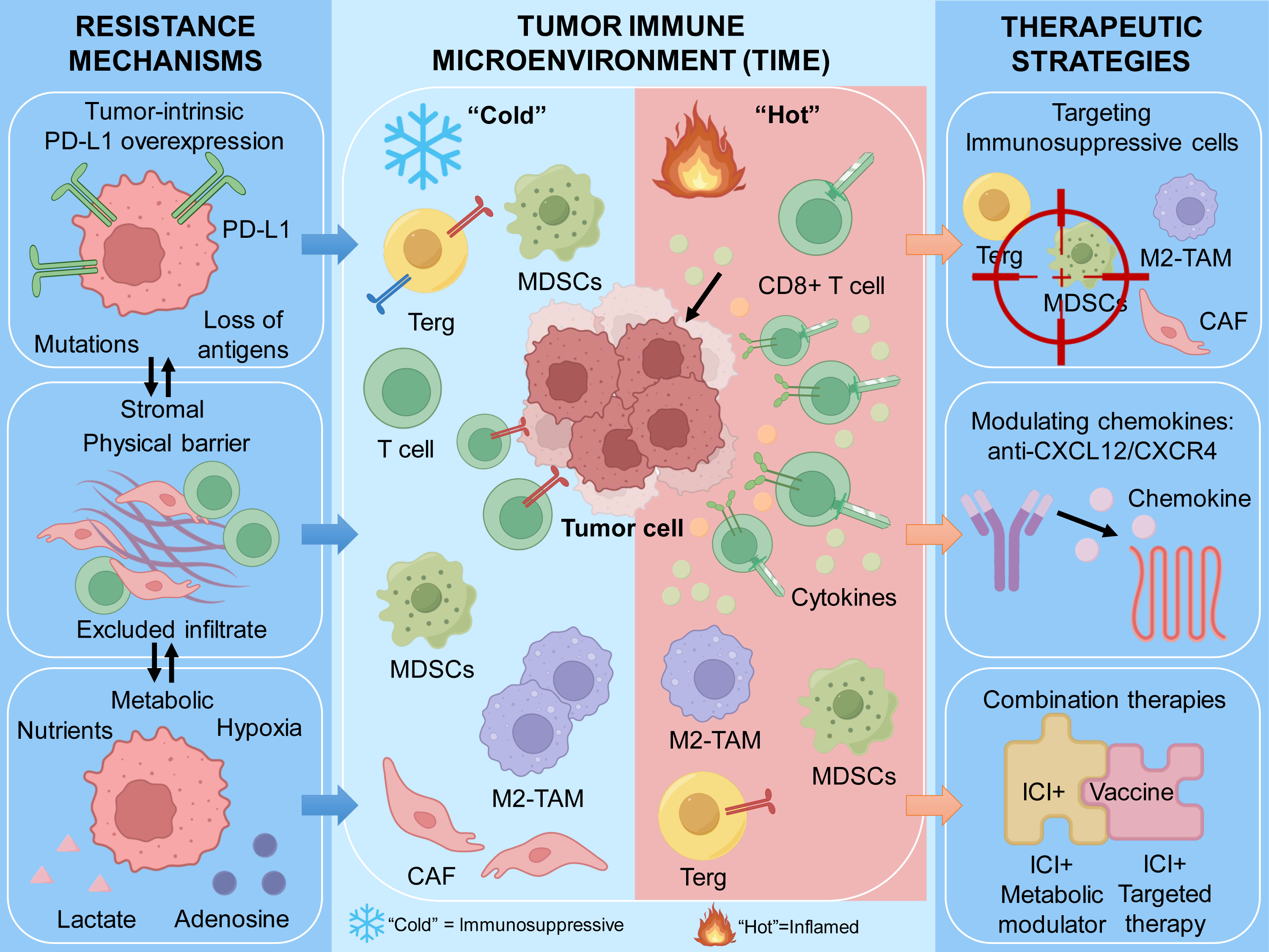
Head and neck squamous cell carcinoma (HNSCC) is often cited as an example of “cold tumor”, as its immune microenvironment is marked by strong immunosuppression and immune escape, leading to limited treatment response and poor overall survival. Importantly, Figure 1 should be interpreted as a dynamic rather than static model: tumors may transition between ‘hot,’ ‘cold,’ and ‘excluded’ states under therapeutic pressure, which may partially explain variable responses to immunotherapy over time. Preclinical and translational studies suggest that tumor cells in HNSCC evade immune surveillance through multiple mechanisms; however, the relative contribution of each pathway to clinical resistance remains unclear (13). Additionally, tumor-derived extracellular vesicles (EVs) have been implicated in reshaping the immune microenvironment by modulating the differentiation, proliferation, and activation of both innate and adaptive immune cells, as well as myeloid cell populations, thereby promoting immunosuppression and facilitating tumor escape from immune detection (13).
Among the various cellular components of the TIME, immunosuppressive populations such as M2-polarized tumor-associated macrophages (TAMs) are frequently implicated. M2-type TAMs not only suppress antitumor immune responses directly but also promote angiogenesis and tissue remodeling, making them attractive but clinically challenging therapeutic targets with limited success in clinical trials to date. While preclinical studies indicate that modifying macrophage activation states may enhance responses to ICIs, clinical translation has been inconsistent, highlighting unresolved challenges in targeting TAM plasticity (14, 15). Other important immunosuppressive groups include regulatory T cells (Tregs) and myeloid-derived suppressor cells (MDSCs), which produce various cytokines and inhibitory mediators, forming complex immunoregulatory networks that limit effector T-cell infiltration and activity, thereby contributing to immune resistance (16, 17).
Cytotoxic CD8⁺ T cells are key effectors of antitumor immunity, and their infiltration levels and functional state are associated with, but do not reliably predict, responses to immunotherapy. Persistent antigen stimulation and ongoing exposure to immunosuppressive signals within the TIME lead to T-cell exhaustion, characterized by loss of effector functions and increased expression of inhibitory checkpoint molecules, a key mechanism of immune resistance. Experimental studies indicate that metabolic reprogramming of CD8⁺ T cells, blocking exhaustion-related pathways, or combined targeting of checkpoint and metabolic signaling pathways can restore T-cell activity in preclinical models, with uncertain translation into durable clinical benefit (18–20). Notably, most of these findings are derived from controlled experimental systems, and their durability and safety in clinical settings remain uncertain.
Interferon-γ (IFN-γ) is a pleiotropic cytokine with context-dependent roles in antitumor immunity and immunosuppression. It enhances the cytotoxic activity of CD8⁺ T cells and natural killer (NK) cells, promotes antigen presentation, and stimulates the secretion of chemokines that help recruit T cells. However, chronic IFN-γ signaling can increase PD-L1 expression on tumor cells and lead to the production of immunosuppressive mediators, such as indoleamine 2,3-dioxygenase (IDO), which contribute to T-cell exhaustion and immune evasion. This dual nature of IFN-γ highlights the complexity of cytokine networks within the TIME and emphasizes the importance of careful therapeutic modulation. Importantly, the context-dependent effects of IFN-γ signaling are not uniformly observed across tumor types, underscoring the need for tumor-specific therapeutic strategies. Notably, IFN-γ-driven immune activation and immune suppression may coexist within the same tumor, complicating therapeutic targeting strategies.
Dendritic cells (DCs) are essential mediators between innate and adaptive immunity by cross-presenting antigens and activating T cells; their dysfunction has been identified as another major factor in tumor immune escape (21). Similarly, CAFs and other stromal components shape the immunosuppressive environment by creating physical barriers and secreting immunomodulatory factors that limit immune-cell infiltration and impair effector functions. Transcriptomic analyses have identified associations between CAF-related gene signatures and immune evasion; however, these correlations do not consistently translate into robust predictive biomarkers across independent clinical cohorts, but these findings are largely correlative and often fail to demonstrate causal relationships in vivo (22).
Furthermore, tumor metabolic reprogramming, hypoxia, and abnormal glycosylation have been proposed to contribute to the immunosuppressive features of the TIME and decrease the effectiveness of immunotherapy (23–25). Collectively, these observations support a multifactorial model of immune resistance; however, the relative contribution of each pathway remains incompletely defined.
Cellular and Stromal Composition of the TIME
Building on this framework, it is important to examine the cellular and stromal components that make up the TIME. These elements form the immediate environment in which immune responses are either supported or actively restrained, ultimately influencing therapeutic outcomes.
Immunosuppressive Cells in the TIME
TIME contains a diverse range of immunosuppressive cell populations that work together through various mechanisms to create an immunosuppressive environment, thus aiding tumor immune evasion and progression. Among these, Tregs, M2-polarized tumor-associated macrophages (M2-TAMs), and MDSCs are the main cellular groups that contribute to immune resistance.
Regulatory T cells (Tregs)
Tregs are a critical immunosuppressive subset that helps maintain immune self-tolerance and regulate immune homeostasis. In the tumor microenvironment, Tregs suppress antitumor immune responses primarily by secreting inhibitory cytokines such as interleukin-10 (IL-10) and transforming growth factor-β (TGF-β). IL-10 reduces the production of pro-inflammatory cytokines and diminishes the cytotoxic activity of effector T cells and NK cells, while TGF-β suppresses cell proliferation and promotes immune tolerance, aiding tumor immune escape. Additionally, low-dose recombinant IL-2 has been shown to encourage Treg expansion and survival, further boosting their immunosuppressive function and offering a potential therapy for autoimmune diseases and tumor immune tolerance (26, 27). Importantly, in tumor tissues, Tregs not only increase in number but also undergo functional and phenotypic reprogramming, thereby strengthening immune evasion and contributing to treatment resistance.
M2-polarized tumor-associated macrophages (M2-TAMs)
TAMs are among the most abundant immune cells in the TIME and demonstrate significant phenotypic and functional diversity. The M2 phenotype, which is predominant in most tumors, has protumor and immunosuppressive effects. M2-TAMs promote tumor growth, angiogenesis, and metastasis by secreting various immunosuppressive mediators, including IL-10, TGF-β, and vascular endothelial growth factor (VEGF). Additionally, they inhibit effector T-cell activity and recruit Tregs, strengthening the immunosuppressive environment (28, 29). The interaction between M2-TAMs and NK cells also plays a crucial role in controlling antitumor immunity by affecting NK-cell cytotoxicity (30). The polarization and functions of M2-TAMs are heavily influenced by the tumor’s metabolic environment; tumor-produced metabolites can direct macrophage polarization toward the M2 phenotype, thereby further facilitating immune escape (31).
Myeloid-derived suppressor cells (MDSCs)
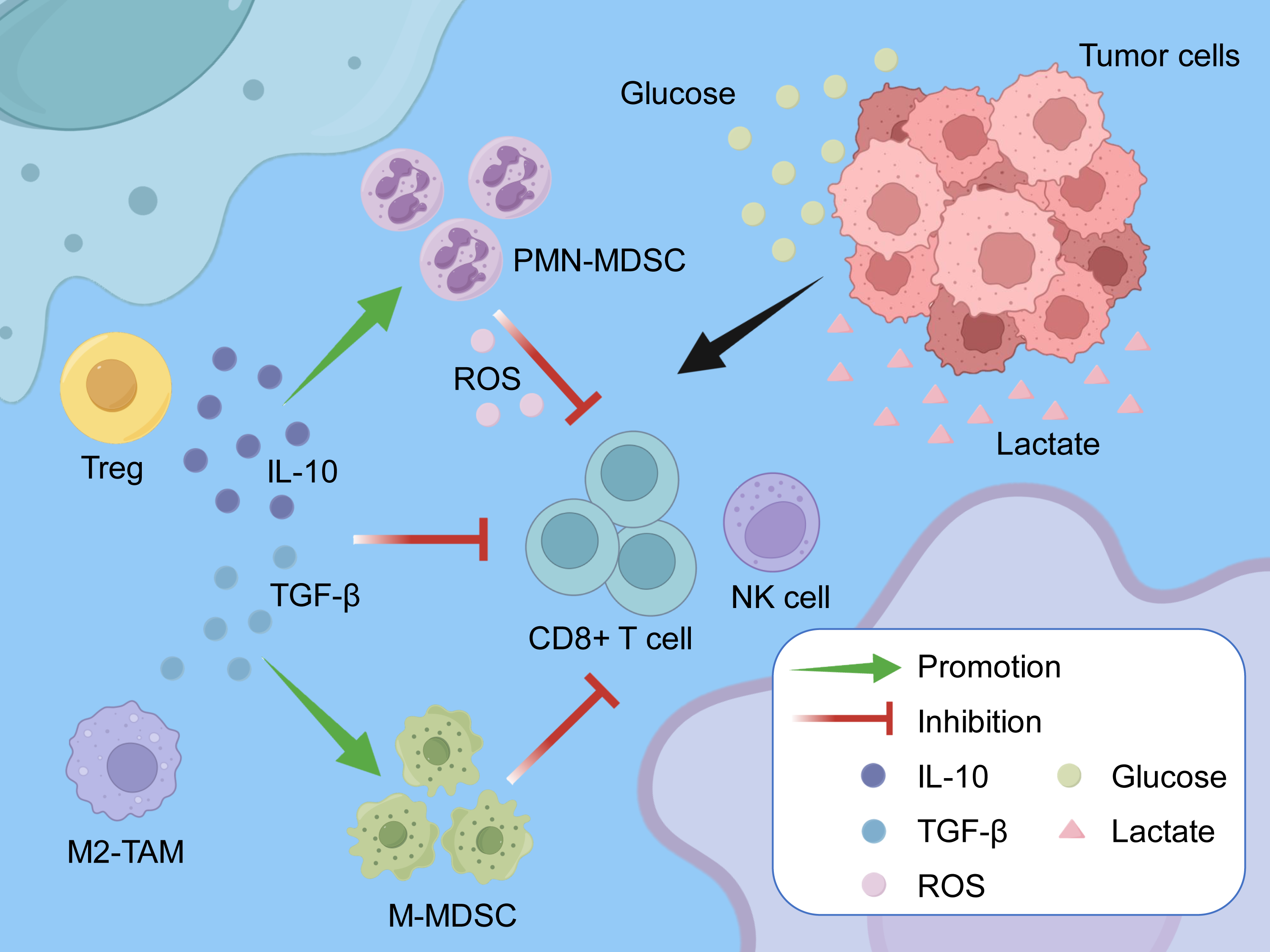
MDSCs are a heterogeneous population of immature myeloid cells originating from the bone marrow. They accumulate in the TIME and are closely linked to tumor-induced immune tolerance. These cells suppress effector T-cell responses through various mechanisms, including the production of reactive oxygen species (ROS), induction of inducible nitric oxide synthase (iNOS), expression of PD-L1, and secretion of inhibitory cytokines. MDSCs not only directly inhibit T-cell proliferation and cytotoxicity but also promote Treg expansion and sustain their suppressive activity through metabolic reprogramming (32–34). They are mainly classified into two subsets: monocytic MDSCs (M-MDSCs) and polymorphonuclear MDSCs (PMN-MDSCs). Although both subsets share immunosuppressive functions, PMN-MDSCs are more common across most tumor types and primarily exert their effects through ROS production, whereas M-MDSCs rely more on iNOS and arginase-1. Targeting MDSCs represents a promising therapeutic strategy; however, clinical efficacy has thus far been limited, reflecting the complexity and redundancy of myeloid suppressive networks.
Recent findings show that the deubiquitinase USP12 enhances the immunosuppressive function of monocytic MDSCs (M-MDSCs) by stabilizing the NF-κB signaling molecule p65, identifying USP12 as a potential therapeutic target (33). In mouse models, the depletion of polymorphonuclear MDSCs (PMN-MDSCs) significantly restores CD8⁺ T-cell function and boosts antitumor immunity, suggesting that targeting MDSCs could improve the effectiveness of immune checkpoint blockade (35). Additionally, metabolic reprogramming, including changes in glucose, lipid, and amino acid metabolism, is a key mechanism underlying MDSC immunosuppressive function (34, 36), offering a potential point of therapeutic intervention; however, the redundancy of these pathways may limit the efficacy of single-target approaches. These interactions are summarized in Figure 2. Figure 2 highlights that immunosuppression within the TIME arises from interdependent cellular and metabolic circuits rather than isolated pathways. This interconnectedness may underlie the limited success of therapies targeting single immune populations in clinical settings.
Non-Immune Stromal Components
Within TIME, non-immune stromal components, mainly CAFs and the extracellular matrix (ECM), play crucial roles in regulating immune cell infiltration, function, and overall tumor behavior. These elements not only provide structural support but also actively influence the immunological and biochemical environment that affects therapeutic response.
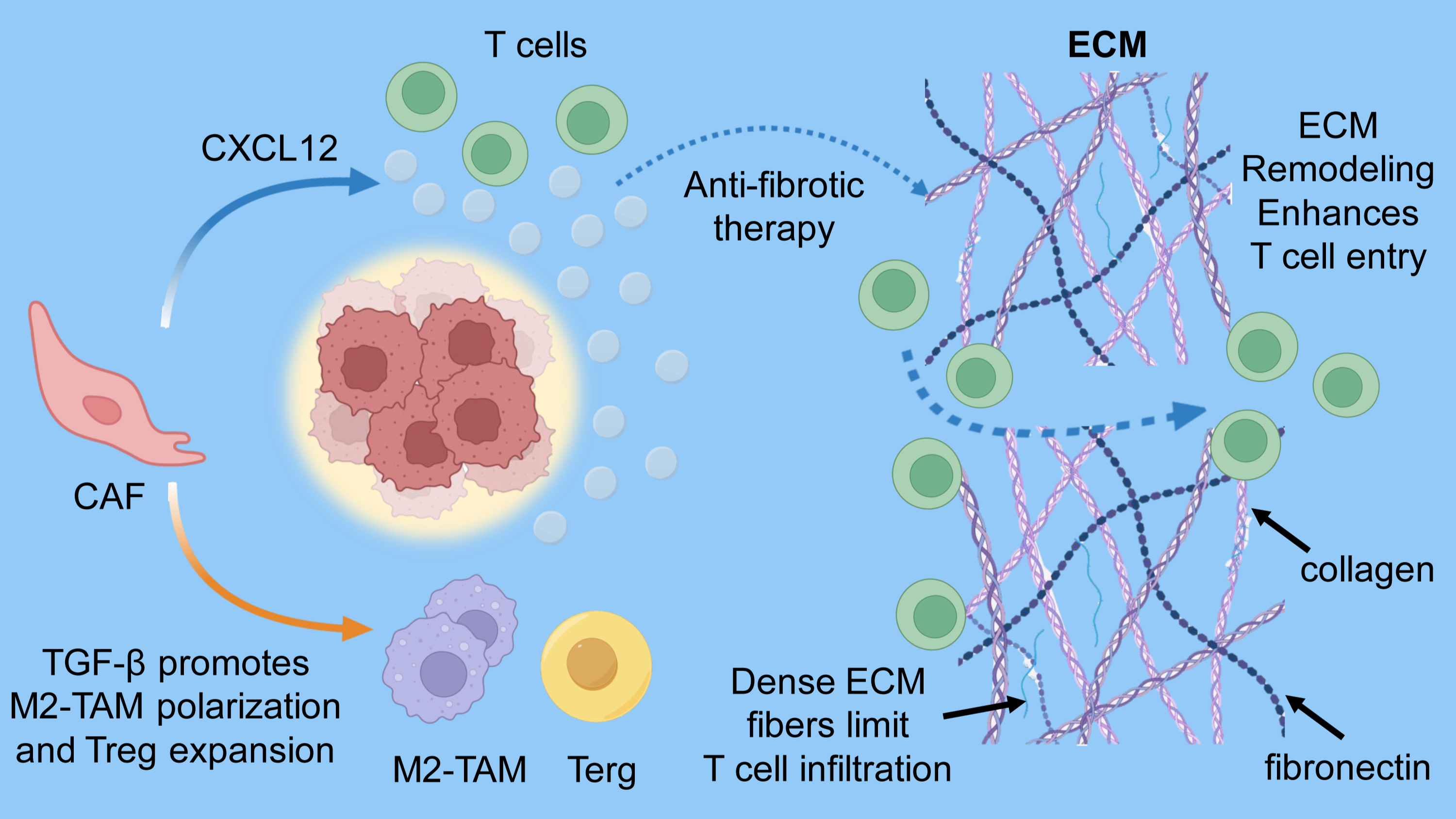
CAFs are the most common non-immune cells in the tumor stroma and show significant heterogeneity and plasticity. They modify the tumor microenvironment by releasing cytokines, chemokines, and enzymes, as well as by remodeling the ECM, thereby affecting immune cell recruitment and activity. For example, CAF-derived CXCL12 binds to CXCR4, creating an immune-exclusion barrier that restricts effector T-cell infiltration and reduces the effectiveness of immune checkpoint inhibitors. Additionally, CAFs produce immunosuppressive cytokines such as TGF-β, which promote the recruitment and polarization of Tregs and M2-TAMs, thereby further helping the tumor evade immune responses (36–38). In tumor types such as pancreatic ductal adenocarcinoma (PDAC) and triple-negative breast cancer (TNBC), CAF-mediated modulation of the stromal composition and immune environment has been shown to directly influence therapeutic resistance and clinical outcomes (38, 39). Emerging therapeutic strategies, such as nanoparticle-based co-targeting of CAFs, have demonstrated potential to remodel the tumor stroma, increase CD8⁺ T-cell infiltration, and enhance the effectiveness of immunotherapy. Additionally, CAFs communicate with tumor and immune cells via extracellular vesicles and long noncoding RNAs (lncRNAs), thereby creating complex signaling networks that further modulate immune regulation and tumor progression (40, 41). As a result, CAFs serve not only as structural “scaffolds” within the tumor but also as vital regulators of immune dynamics, making them important targets in modern cancer immunotherapy research.
The ECM is the main non-cellular component of the tumor stroma, creating a physical and biochemical barrier that prevents immune cells from entering the tumor core. Made mostly of collagen, fibronectin, and proteoglycans, the ECM’s density and makeup significantly influence tumor mechanics and the paths immune cells take (42, 43). In cancers like pancreatic carcinoma, a thick ECM limits blood vessel growth and immune cell infiltration, resulting in an immunologically “cold” tumor microenvironment that responds poorly to immunotherapy (39, 44). Some ECM components, including fibronectin and collagen, can bind immunosuppressive cytokines, thereby amplifying inhibitory signals and exacerbating immune dysfunction (42, 45). Recent spatial omics studies have shown that ECM changes at the tumor-stroma boundary are closely linked to the efficacy of immune checkpoint blockade. Changing the spatial arrangement and mechanical qualities of the ECM could thus reprogram the immune environment and improve the effectiveness of immunotherapy (46).
Collectively, non-immune stromal components exert multilayered, multifactorial effects on the TIME. CAFs actively shape an immunosuppressive niche through cytokine release, ECM remodeling, and communication with tumor and immune cells, while the ECM functions both as a physical barrier and as an enhancer of immunosuppression through molecular interactions with regulatory cytokines. Therefore, targeting CAFs and ECM remodeling, especially when combined with immunotherapy, shows promise for overcoming tumor immune resistance and improving clinical outcomes (46). Future research clarifying the heterogeneity and molecular mechanisms of these non-immune stromal components across tumor types will be crucial for designing precise strategies to reprogram the TIME. These stromal mechanisms converge to create both physical and biochemical barriers to immune infiltration, as illustrated in Figure 3.
Cytokine and Chemokine Networks
Cytokines and chemokines create a complex regulatory signaling network within TIME, significantly influencing its immune activity and thereby affecting tumor growth, metastasis, and immune evasion. Based on recent research, this section emphasizes the roles and mechanisms of IL-10, TGF-β, CXCL12, and the overall cytokine-chemokine network in shaping the TIME and regulating immune responses. IL-10 and TGF-β are major immunosuppressive mediators within the TIME, as described above. Their elevated expression is associated with tumor progression and sustained immune evasion; for example, persistent signaling in cervical cancer reinforces an immunosuppressive microenvironment (47). TGF-β pathway activation is further linked to CAF-associated stromal remodeling, supporting tumor survival and spread (47). Collectively, these cytokines coordinate immune suppression by limiting effector cell function and promoting suppressive cellular and stromal programs.
The CXCL12-CXCR4 signaling axis regulates immune cell trafficking and tumor cell migration within the TIME, contributing to invasion, metastasis, and immune exclusion (48, 49). CXCL12 produced by tumor and stromal cells establishes chemotactic gradients that recruit immunosuppressive populations, including MDSCs and regulatory T cells, thereby shaping an immune-evasive microenvironment (48). Elevated CXCL12 expression is associated with reduced lymphocyte infiltration and increased immune resistance, while therapeutic blockade of this pathway can enhance antitumor immune responses (48, 49). The dynamic balance of cytokine and chemokine networks determines immune activity in the TIME. Cytokine and chemokine networks collectively regulate immune activity within the TIME by balancing pro- and anti-inflammatory signals (50, 51). Proinflammatory cytokines, including IFN-γ, IL-12, and IL-15, promote cytotoxic T-cell and NK-cell function, whereas immunosuppressive cytokines, such as IL-10 and TGF-β, reinforce immune evasion (52–54). Chemokine signaling further shapes immune cell recruitment and spatial organization, with the CXCL9/10/11-CXCR3 axis supporting effector T-cell trafficking and CCL2 and CCL5 promoting infiltration of suppressive immune populations (55–57). These signaling networks are dynamically regulated through tumor-derived feedback mechanisms, and their disruption can shift the TIME toward immune-cold or immune-excluded states, reducing immunotherapy efficacy (58–61).
T-Cell Infiltration and Exhaustion in the TIME
One of the most direct consequences of this immunosuppressive environment is its impact on effector T cells. The extent to which T cells can enter the tumor and retain their function is a critical determinant of response, making T-cell infiltration and exhaustion key features of TIME-driven resistance.
T-Cell Infiltration and Clinical Prognosis
CD8⁺ T cells, the primary effector immune cells within TIME, show infiltration levels closely linked to patient survival and response to therapy. Numerous clinical studies suggest that high CD8⁺ T-cell infiltration generally predicts better prognosis and increased immunotherapy success. For example, in patients with HNSCC, dense infiltration of CD8⁺ T cells in the tumor stroma is strongly associated with improved survival, and the presence of CD8⁺ T cells has been reported as an independent prognostic factor in some cohorts (62). Nevertheless, this relationship is not universal, and exceptions across tumor types highlight the limitations of CD8⁺ T-cell density as a standalone biomarker. In NSCLC, while total tumor-infiltrating lymphocyte (TIL) numbers alone do not correlate significantly with survival, high-dimensional flow cytometry analyses reveal distinct immune subtypes. Among these, subtypes enriched in highly functional CD8⁺ T cells are associated with prolonged recurrence-free survival (63). Similarly, in colorectal cancer (CRC), cytotoxic CD8⁺ T-cell infiltration positively correlates with favorable survival outcomes (64). Conversely, insufficient or dysfunctional T-cell infiltration promotes tumor immune escape and therapeutic failure. For example, in osteosarcoma, high BMPR2 expression is associated with reduced CD8⁺ T-cell infiltration and a poor prognosis, highlighting a strong association between low CD8⁺ T-cell infiltration and malignant progression (65). In NSCLC, although CD36⁺CD8⁺ T-cell infiltration can be high, these cells often show signs of functional exhaustion, including decreased cytotoxic molecules and increased inhibitory receptors such as PD-1 and TIGIT, which are associated with poor prognosis and inadequate chemotherapy response (66). Immunosuppressive cells within the TIME, including Tregs, further suppress CD8⁺ T-cell antitumor activity, aiding immune escape (67). The heterogeneity of CD8⁺ T-cell infiltration across different tumor types and molecular subtypes highlights the complexity of the TIME. For example, in medulloblastoma, patients with the WNT subtype, which is characterized by high CD8⁺ T-cell infiltration, tend to have a better prognosis. CD8⁺ T-cell chemotaxis in this context may be driven by the CXCL11-CXCR3 axis. In hepatocellular carcinoma (HCC), higher LPAR6 expression is associated with increased CD8⁺ T-cell activation and infiltration, suggesting a potential role in boosting antitumor immunity and reducing T-cell exhaustion, ultimately leading to improved patient outcomes (68). Spatial distribution of immune cells also impacts prognosis. In CRC, intratumoral CD8⁺ T-cell density is the most significant independent prognostic factor, whereas macrophage and Treg levels indirectly influence prognosis by modulating CD8⁺ T-cell antitumor activity (69). Additionally, the expression of immune checkpoint molecules, such as PD-L1, is associated with CD8⁺ T-cell infiltration levels, suggesting that immune escape mechanisms may operate by modulating effector T-cell activity (70, 71).
In summary, high CD8⁺ T-cell infiltration is often associated with improved outcomes, whereas inadequate or dysfunctional infiltration leads to immune escape and treatment failure. The intricate interactions among immune cells within the TIME influence T-cell function and clinical outcomes. Future immunotherapy approaches should thoroughly evaluate the quantity, functional status, and spatial arrangement of T cells to improve treatment success.
Mechanisms of T-Cell Exhaustion and Checkpoint Expression
T-cell exhaustion is a dysfunctional state commonly seen in chronic infections and the tumor microenvironment, marked by a gradual loss of effector function due to ongoing antigen stimulation. Exhausted T cells display a series of distinctive molecular markers, especially the high expression of immune checkpoint molecules, which not only indicate the functional status of T cells but also serve as key targets for immunotherapy.
Programmed cell death protein 1 (PD-1) is the most extensively studied immune checkpoint molecule and is highly expressed on exhausted T cells, serving as a key marker of their impaired function. Increased PD-1 expression is commonly associated with T-cell dysfunction; however, its predictive value for therapeutic response varies with tumor context and the co-expression of additional inhibitory receptors (72, 73). Additionally, T-cell immunoglobulin and mucin domain-containing protein 3 (TIM-3), another inhibitory receptor often co-expressed with PD-1, indicates a more advanced stage of exhaustion. High TIM-3 expression is associated with significantly reduced T-cell effector activity and signals terminal exhaustion in various tumors and chronic infections (74). Beyond PD-1 and TIM-3, other immune checkpoint molecules, including LAG-3, TIGIT, and CTLA-4, are upregulated in exhausted T cells, contributing to an interconnected inhibitory signaling network that collectively regulates T-cell function. For example, LAG-3 and TIGIT levels positively correlate with the extent of CD8⁺ T-cell exhaustion, and these molecules are expressed across various tumor microenvironments, suggesting a widely conserved role in exhaustion mechanisms (72, 75). The co-expression of these checkpoints reflects both the diversity of exhausted T cells and the complexity of their molecular regulation. Functionally, exhausted T cells show not only reduced proliferation but also impaired effector activity, including lower secretion of cytotoxic molecules (perforin, granzymes) and key cytokines (IFN-γ, TNF-α) (73, 76). Their metabolic state is also altered, with defects in energy metabolism that further limit antitumor capacity (77). Additionally, the transcriptional regulation of exhausted T cells has become increasingly understood, with key transcription factors such as TOX and Eomesodermin (EOMES) involved in establishing and maintaining the exhaustion phenotype. Notably, high TOX expression is strongly linked to terminally exhausted T cells (78, 79).
In summary, the molecular markers of T-cell exhaustion mainly include the increased expression of immune checkpoint molecules such as PD-1, TIM-3, LAG-3, and TIGIT. These markers not only signify the exhausted state but also serve as targets for immunotherapy. Exhausted T cells show reduced proliferative capacity and impaired effector functions, with underlying mechanisms involving immune checkpoint signaling, metabolic reprogramming, and transcription factor regulation. A deeper understanding of these molecular markers and their roles will help develop more effective immunomodulatory strategies to improve clinical outcomes in patients with tumors and chronic infections.
CAF-mediated suppression of T-cell infiltration and function
CAFs can inhibit T-cell-mediated antitumor activity through coordinated immunosuppressive signaling and ECM remodeling, thereby promoting tumor immune escape. By releasing immunoregulatory factors and restructuring the tumor stroma, CAFs limit effector T-cell infiltration and reduce immune surveillance. CAF-derived cytokines and chemokines suppress CD4⁺ and CD8⁺ T-cell function and promote recruitment of immunosuppressive cell populations (80, 81). These effects are mediated by activation of signaling pathways such as STAT3 and NF-κB, and by the induction of immunosuppressive molecules, including PD-L1 and IDO, which impair T-cell proliferation and activation (82, 83). Additional CAF-associated signaling mechanisms further disrupt dendritic cell function and T-cell activity, reinforcing immunosuppressive conditions within the TIME (84, 85). CAF-mediated ECM remodeling contributes to immune exclusion by limiting T-cell infiltration and promoting tumor immune escape (86–88). This process is reinforced by CAF-driven signaling interactions that further sustain immunosuppressive conditions within the TIME (89). Experimental evidence indicates that disrupting CAF activity or ECM integrity can enhance T-cell infiltration and restore antitumor immunity (86, 90, 91).
Collectively, CAFs promote tumor immune escape through integrated cytokine signaling and ECM remodeling, providing a strong rationale for CAF-targeted strategies to restore immune infiltration and improve immunotherapy efficacy. Tumor-intrinsic immune evasion mechanisms, including impaired antigen presentation and checkpoint ligand expression, are conceptually summarized in Figure 4 and interact with microenvironmental factors to shape immune resistance.
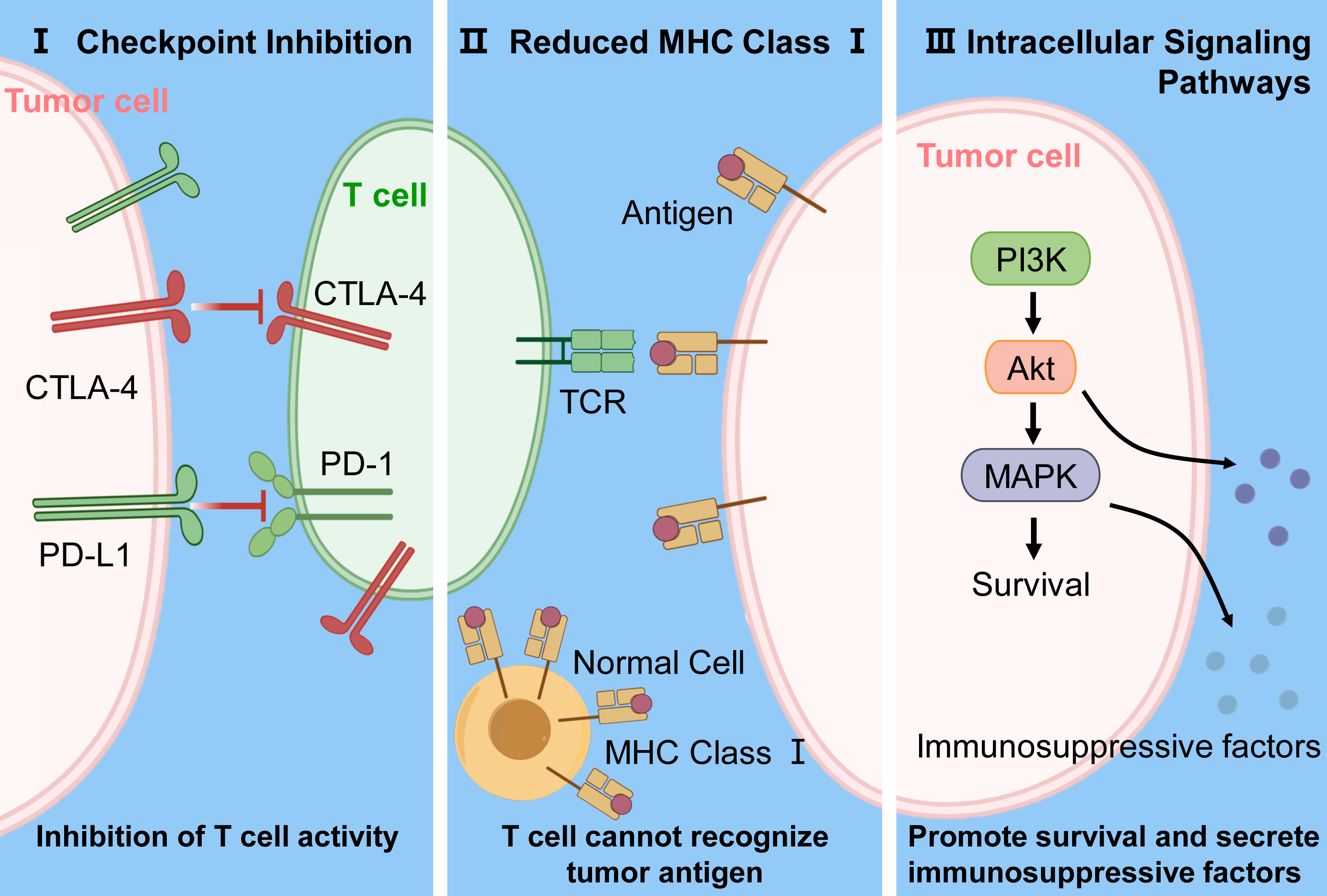
However, these intrinsic mechanisms rarely act in isolation and are often modulated by the surrounding microenvironment, limiting the predictive value of tumor-intrinsic features alone.
Monocyte-macrophage axis in the TIME
T-cell dysfunction does not occur in isolation but is shaped by surrounding immune populations. In particular, the monocyte-macrophage axis plays a central role in establishing and maintaining the suppressive conditions that limit effective T-cell responses.
Chemokine-driven monocyte recruitment in the tumor microenvironment
Chemokines are crucial for recruiting monocytes within TIME, with CCL2 and VEGF being two key chemotactic factors. CCL2, also known as monocyte chemoattractant protein-1 (MCP-1), binds to its receptor, CCR2, to facilitate monocyte movement from the bone marrow into the bloodstream and guide their migration toward tumor sites. Studies have shown that tumor cells and tumor-associated stromal cells secrete CCL2, creating a concentration gradient that draws monocytes to the tumor microenvironment, thereby aiding TAM formation and tumor progression (92, 93). Similarly, vascular endothelial growth factor (VEGF), a major angiogenic factor, has chemotactic activity for monocytes, encouraging their migration toward sites of new blood vessel formation in tumors and providing both immune support and vascular nourishment (94). CCL2 is highly expressed in multiple tumor types, and its expression level closely correlates with tumor invasiveness and patient prognosis. Through the CCR2 axis, CCL2 not only recruits monocytes but also activates them to release pro-inflammatory cytokines, creating a positive feedback loop that further enhances inflammatory and immunoregulatory networks (95). Other chemokine receptors, including CCR1, also contribute to monocyte recruitment, with CCR1 and CCR2 playing distinct roles in guiding monocytes to metastatic sites, underscoring the complexity of the chemokine network within the tumor microenvironment (96). For example, CCL16 mediates chemotaxis of monocytes and M2-polarized macrophages via CCR1 and CCR5, thereby promoting the progression of hepatocellular carcinoma (93).
Monocyte recruitment is regulated not only by chemokine expression but also by intercellular interactions, activation of signaling pathways such as NF-κB, and other molecules present in the tumor microenvironment. For example, osteopontin (OPN) promotes cholangiocytes to release CCL2, CCL5, and CXCL1, thereby promoting hepatic fibrosis and the accumulation of inflammatory monocytes (94). Furthermore, monocyte chemotaxis can be affected by viral infections and immunotherapy; for instance, antiviral monoclonal antibody treatment can trigger monocyte recruitment through FcγR-mediated mechanisms (97). Within the tumor microenvironment, recruited monocytes further differentiate into M2-polarized macrophages, a process regulated by chemokines and tumor-derived cytokines, including IL-6 and IL-10. M2 macrophages display immunosuppressive and tumor-promoting phenotypes, and their accumulation is a key mechanism of tumor immune escape (98). The differentiation of monocytes into M2 macrophages is mediated not only by cytokine signaling but also by tumor-derived exosomes and metabolites (99, 100). These M2 macrophages then release additional chemokines and immunosuppressive factors, further sustaining and amplifying the immunosuppressive state of the TIME (101).
In summary, chemokines such as CCL2 and VEGF coordinate monocyte recruitment and functional regulation through multiple receptor-mediated pathways, promoting their migration into the tumor microenvironment and M2 polarization, thereby creating a pro-tumorigenic immune landscape. These findings provide a theoretical basis for targeting chemokine axes to block monocyte recruitment and macrophage polarization. Therapeutic approaches, including CCR2 or CCL2 neutralization, have been shown to slow tumor growth and improve treatment outcomes (93, 95, 102). Future research should further explore the interactions among chemokines and their receptors, as well as the specific mechanisms behind tumor immune resistance, to develop more precise immunoregulatory therapies.
JAK/STAT, PI3K/Akt and NF-κB pathways driving M2 macrophage polarization
Within TIME, M2 macrophage polarization is a crucial process that promotes tumor growth, immune evasion, and therapeutic resistance. The regulation of M2 polarization involves multiple signaling pathways, with the JAK/STAT, PI3K/Akt, and NF-κB pathways being among the most significant. This section focuses on the mechanisms and regulatory features of these pathways in M2 macrophage polarization.
JAK/STAT Signaling Pathway
The JAK/STAT pathway is a key regulator of M2 polarization, with the activation of STAT3 and STAT6 widely recognized as hallmark signaling events in M2 macrophages. Tumor-derived cytokines, including IL-4, IL-10, and TGF-β, induce M2 polarization by activating the JAK/STAT pathway. For example, in breast cancer, the YAP/STAT3 signaling axis promotes tumor-associated macrophage (TAM) M2 polarization, suppresses CD8+ T cell activity, and facilitates tumor progression (103). In pancreatic cancer, sphingomyelin synthase 2 enhances M2 polarization by modulating the CSF1R-STAT3 pathway (104). Additionally, tumor-secreted CXCL1 activates the Cxcr2-JAK-STAT3 pathway in macrophages, inducing M2 polarization and establishing an immunosuppressive microenvironment (105). Therapeutically, targeting STAT6 with antisense oligonucleotides combined with radiotherapy in solid tumors such as lung cancer significantly suppresses M2 polarization and enhances antitumor immunity (106). Recent studies also indicate that miR-373 inhibits TAM M2 polarization in CRC by modulating the JAK2/STAT6 pathway, thereby suppressing tumor growth and metastasis (107). Overall, the JAK/STAT pathway is a major driver of cytokine-driven M2 polarization and a critical target in tumor immunotherapy.
PI3K/Akt Signaling Pathway
The PI3K/Akt pathway controls macrophage metabolic reprogramming and survival, acting as a crucial regulator of M2 functional polarization. Various signals in the tumor microenvironment activate PI3K/Akt to promote M2 differentiation in macrophages. For example, TAM M2 polarization in pancreatic cancer is closely linked to PI3K/Akt signaling (108). In chronic hepatitis B-related hepatocellular carcinoma, SIRT1 inhibitors delivered via nanoparticles partially suppress M2 polarization through the PI3K/Akt pathway (109). Tumor-secreted lactate also influences PI3K/Akt signaling through mitochondrial metabolism to encourage M2 polarization (110). In lung cancer therapy, dipeptidyl peptidase 4 inhibitors enhance immune responses, partly by inhibiting the PI3K/Akt pathway and preventing M2 polarization (111). Therefore, activation of the PI3K/Akt pathway supports immunosuppressive M2 macrophage polarization, contributing to immune evasion mediated by the tumor microenvironment, and interacts with multiple metabolic pathways to sustain M2 survival and function, establishing it as a key regulatory center in tumor-associated M2 polarization.
NF-κB Signaling Pathway
As a key pathway in inflammation, NF-κB plays complex roles in macrophage polarization. While traditional views link NF-κB activation to M1 polarization, it also promotes M2 polarization within the tumor microenvironment. Inhibiting autophagy in TAMs increases ubiquitination-mediated degradation of TAB3, reducing NF-κB activity and encouraging M2 polarization (112). In bladder cancer, Pedunculoside blocks M2 polarization and reduces malignant tumor traits by targeting the TRAF6/NF-κB pathway (113). Some tumor-secreted factors or pathogens can also affect NF-κB signaling to control macrophage polarization; for example, Treponema pallidum TpF1 encourages M2 polarization through METTL14-mediated NF-κB activation (114). NF-κB regulation interacts with JAK/STAT and PI3K/Akt pathways, creating a complex network that influences macrophage polarization and function. Targeting NF-κB and its downstream partners offers significant potential in tumor immunotherapy (115). However, systemic inhibition of these pathways raises concerns regarding toxicity and off-target immune effects, limiting clinical applicability.
In summary, the JAK/STAT, PI3K/Akt, and NF-κB pathways are vital in controlling TAM M2 polarization. The JAK/STAT pathway drives cytokine-induced expression of M2 phenotype genes; the PI3K/Akt pathway promotes M2 functions by regulating macrophage metabolism and survival; and NF-κB acts as an inflammatory and immunoregulatory center that affects polarization under various conditions. These signaling pathways form a partially redundant regulatory network governing M2 polarization, as summarized in Figure 5, which may explain the limited efficacy of targeting individual pathways.
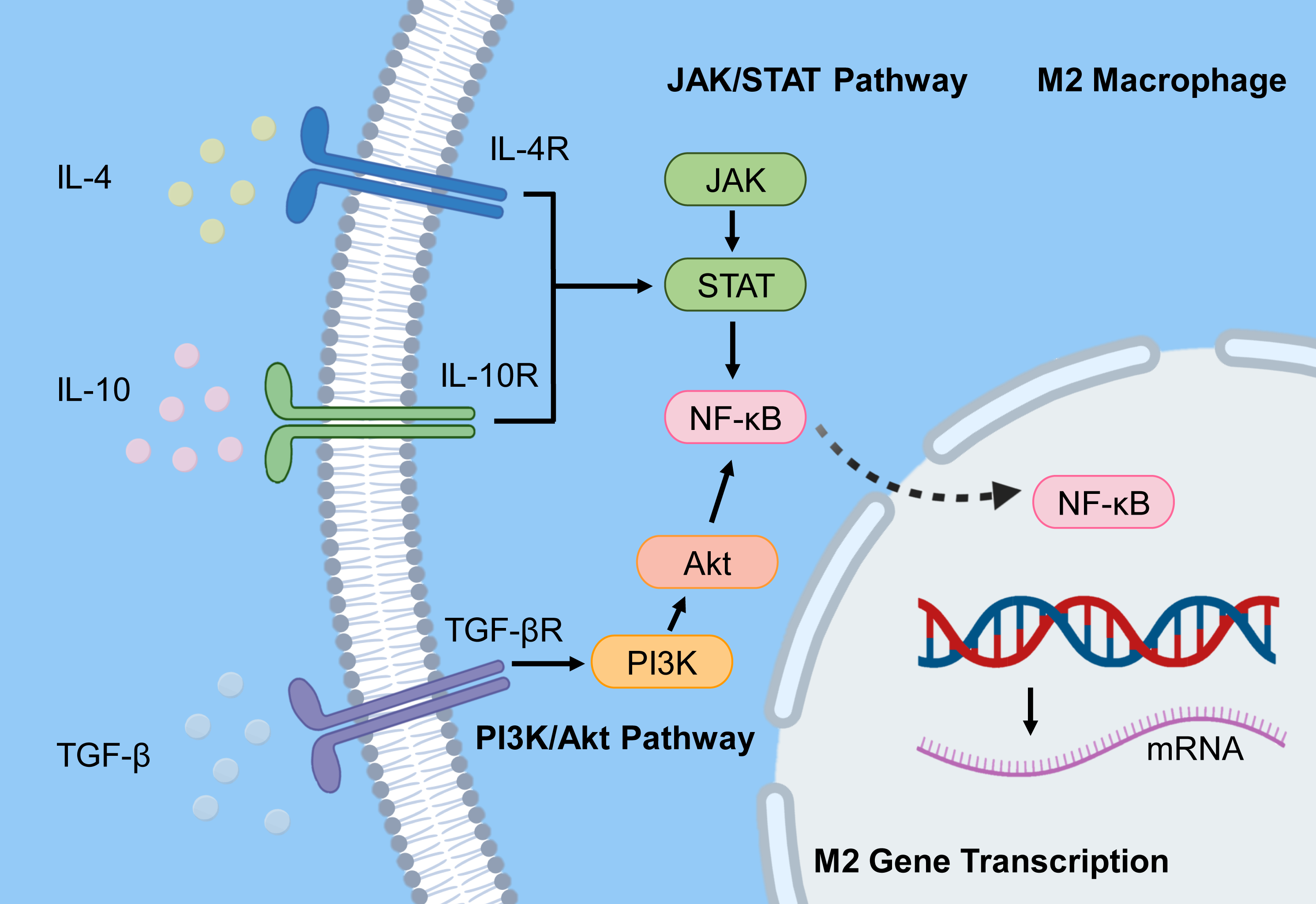
Figure 5 also illustrates the convergence of inflammatory and immunosuppressive signaling, underscoring the challenge of selectively modulating macrophage polarization without disrupting broader immune homeostasis.
Pro-tumor and immunosuppressive functions of M2 tumor-associated macrophages
TAMs, especially the M2 subtype, have well-described dual biological roles: they mediate immunosuppression while also encouraging tumor angiogenesis, supporting tumor growth and metastasis. These functions make M2-TAMs key regulators within the TIME and major contributors to tumor immune resistance.
First, M2-TAMs suppress the antitumor activity of effector immune cells by secreting immunosuppressive cytokines, including IL-10 and TGF-β. IL-10 and TGF-β inhibit cytotoxic T lymphocytes (CTLs) and NK cells while promoting Treg growth, thereby collectively reducing antitumor immune responses and creating an immunosuppressive microenvironment (116, 117). Additionally, M2-TAMs express immune checkpoint molecules, including PD-L1 and arginase-1, which further block the recognition and destruction of tumor cells by the immune system (116, 118). These mechanisms work together to enable tumor immune escape and resistance to immunotherapy, posing a major challenge in current cancer treatment (119, 120).
Second, M2-TAMs actively promote tumor angiogenesis by secreting vascular endothelial growth factor (VEGF), matrix metalloproteinases (MMPs), and other pro-angiogenic factors, thereby supporting rapid tumor growth by providing oxygen and nutrients and creating routes for tumor cell dissemination. This pro-angiogenic activity facilitates both local tumor expansion and distal metastasis, negatively impacting patient prognosis (121, 122). Moreover, M2-TAMs contribute to tumor matrix remodeling and enhance epithelial-mesenchymal transition (EMT), further increasing tumor cell migration and invasiveness (123, 124).
Collectively, the dual biological functions of M2-TAMs position them as key contributors in the TIME, both blocking antitumor immunity and promoting tumor progression through angiogenesis and matrix remodeling. Recently, various therapeutic strategies targeting M2-TAMs have been developed, including nanomaterial-mediated reprogramming of M2-TAMs to M1 macrophages, inhibition of M2-polarizing signaling pathways, and blockade of pro-angiogenic factor secretion, all aimed at remodeling the TIME and improving the effectiveness of immunotherapy (125-127). For example, blocking the CSF-1R signaling pathway to decrease M2-TAM numbers or delivering drugs via targeted nanocarriers to reprogram M2-TAMs has shown promise in boosting antitumor immune responses. Furthermore, M2-TAMs secrete exosomes containing non-coding RNAs and other molecules that influence tumor cell stemness and immune evasion, worsening malignant progression (128, 129). A deeper understanding of the immunosuppressive mechanisms and pro-angiogenic pathways of M2-TAMs will help identify new therapeutic targets and advance tumor immunotherapy in the clinic. Future research should explore the link between M2-TAM metabolic reprogramming and functional polarization, combining nanotechnology and molecular-targeted strategies to precisely control M2-TAMs, overcome immune resistance, and sustain tumor suppression (130, 131).
TIME heterogeneity and immune checkpoint resistance
These interconnected stromal and immune interactions are not uniform across tumors. Instead, they vary widely between patients and tumor types, providing a biological basis for the marked heterogeneity observed in immunotherapy response and resistance.
Tumor immune microenvironment heterogeneity and therapy response
Heterogeneity of TIME is a key factor affecting the effectiveness of immunotherapy. There is significant variation in TIME composition across different tumor types and patients, which directly impacts treatment responses. Despite this recognition, current clinical strategies remain ill-equipped to stratify patients by TIME composition, limiting the translation of these insights into precision therapy.
The biological features of the tumor type influence the composition and proportion of infiltrating immune cells. For instance, in breast cancer, especially HER2-positive and basal-like subtypes, higher levels of tumor-infiltrating lymphocytes (TILs) are usually seen, which are associated with a better prognosis and treatment response (132, 133). However, breast cancer is generally considered an “immune-cold” tumor, with low T cell infiltration and tumor mutation burden, which limits the effectiveness of immunotherapy (133). In contrast, tumors such as melanoma and NSCLC typically exhibit greater immune infiltration and higher response rates to immunotherapy (134, 135). Additionally, the proportion and functional state of immunosuppressive cells within the TIME are crucial factors. Immunosuppressive populations, including Tregs, MDSCs, and TAMs, foster an immune-evasive microenvironment by secreting inhibitory factors and altering immune cell function, thereby decreasing the effectiveness of immunotherapy (136, 137). For example, the heterogeneity of TAMs and their dynamic changes across various tumor microenvironments have been shown to significantly affect tumor progression and immunotherapy resistance (137). In gliomas, the infiltration of immunosuppressive cells correlates with poor immunotherapy outcomes (138, 139). Tumor stromal components, such as CAFs and collagen, further worsen immunosuppression by forming physical barriers and modulating immune cell activity, which limits effector T cell infiltration and function (140).
Spatial and temporal heterogeneity of the TIME also influences treatment responses. Single-cell RNA sequencing and spatial transcriptomics uncover significant regional differences in immune cell composition and function, with region-specific interactions among immune, tumor, and stromal cells (141, 142). This spatial heterogeneity leads to uneven immunotherapy effectiveness across tumor regions, with some regions being immune “hot” zones and others immune “cold,” thereby affecting overall treatment outcomes (143, 144). Additionally, dynamic changes in TIME, such as redistribution of immune cells and changes in functional states before and after treatment, determine the success or failure of immunotherapy (145, 146). Patient-specific factors, including genetic background and tumor cell genomic heterogeneity, also influence TIME composition and function, impacting immunotherapy response. Certain genetic variants affect immune cell infiltration and activity, thereby regulating tumor sensitivity to immunotherapy (136, 147). Differences in tumor driver mutations can lead to distinct immune microenvironments that show either pro- or anti-tumor immune activity. For example, in lung cancer, KRAS mutations are associated with an inflammatory immune response, whereas non-KRAS-mutant models display distinct immune characteristics (135).
In summary, TIME heterogeneity is a key factor influencing variability in tumor immunotherapy responses. Differences in TIME composition among tumor types and patients, particularly the proportions and functional states of immunosuppressive cells, affect immune cell infiltration and effector functions, ultimately shaping therapeutic outcomes. Future strategies that utilize multimodal approaches, including single-cell sequencing, spatial omics, and molecular imaging, to precisely characterize TIME heterogeneity and guide personalized immunotherapy will be vital for enhancing treatment effectiveness (148).
Tumor and immune-intrinsic mechanisms shaping resistance to immune checkpoint inhibitors
Immune checkpoint inhibitors (ICIs) have significantly improved outcomes across multiple cancer types; however, primary and acquired resistance remain major clinical challenges. These resistance mechanisms arise from interconnected tumor-intrinsic and immune-intrinsic processes within the TIME, which collectively constrain effective antitumor immunity.
A central immune-intrinsic driver of resistance is T-cell exhaustion, characterized by reduced effector function, impaired proliferative capacity, and sustained expression of inhibitory receptors, including PD-1, LAG-3, and TIM-3, ultimately limiting ICI efficacy (149, 150). This dysfunctional state is reinforced by persistent immunosuppressive signaling within the TIME, including cytokine-mediated effects such as IL-10 and TGF-β, as well as increased expression of immune checkpoint ligands, including PD-L1, which together stabilize inhibitory signaling circuits and maintain T-cell dysfunction (151, 152).
Beyond receptor-mediated inhibition, epigenetic regulation contributes to the persistence of exhausted T-cell states and tumor-associated immune evasion. Epigenetic remodeling alters both tumor and immune cell phenotypes and sustains exhaustion-associated transcriptional programs, thereby limiting the reversibility of dysfunctional immune states even under checkpoint blockade (153, 154). Tumor-intrinsic mechanisms further reinforce resistance by impairing immune recognition. Defects in antigen presentation, including reduced expression of major histocompatibility complex class I molecules and disruptions in antigen-processing machinery such as β2-microglobulin, limit T-cell recognition and diminish therapeutic efficacy (155, 156). In parallel, tumor and stromal compartments produce immunosuppressive factors and metabolic signals that inhibit effector lymphocytes while promoting the expansion of suppressive immune populations, thereby reinforcing a non-permissive immune contexture (152, 157). Additional innate immune evasion pathways, including CD47- and CD24-mediated “don’t eat me” signaling, further attenuate antitumor responses by impairing phagocytic clearance (158). Importantly, tumor heterogeneity and oncogenic signaling pathways introduce an additional layer of complexity. Alterations such as EGFR and KRAS mutations modulate tumor immunogenicity, antigenicity, and immune cell infiltration, thereby shaping the overall responsiveness to immunotherapy (159, 160). These tumor-intrinsic features operate in concert with the coordinated activity of immunosuppressive cell populations within the TIME, which collectively sustain immune resistance (161).
Taken together, resistance to ICIs reflects the convergence of stable tumor-intrinsic alterations and reinforced immune dysfunction programs within the TIME. These mechanisms define the biological constraints on therapeutic response and provide a mechanistic framework for understanding variability in ICI efficacy across tumor types and patients (150, 152, 162). Importantly, most proposed resistance mechanisms are derived from static or retrospective analyses, limiting insight into their temporal evolution during therapy.
Cytokine and chemokine networks driving immunotherapy resistance
Cytokine networks play a key role in TIME, especially in controlling immune resistance mechanisms. Immunosuppressive cytokines such as IL-10 and TGF-β help tumors evade the immune system and resist therapy through various signaling pathways. These cytokines inhibit antitumor immune responses, reduce the cytotoxic activity of effector immune cells, and promote the recruitment and activation of immunosuppressive cells, thereby creating an immunosuppressive microenvironment that supports tumor survival. For example, in chronic cytokine dysregulation in cervical cancer, sustained expression of IL-10 and TGF-β effectively suppresses antitumor immune responses, promoting immune evasion and disease progression (47). Furthermore, IL-10 and TGF-β regulate downstream pathways, including Smad and STAT3, modulating the function of TAMs and Tregs and further increasing immune resistance (161). Excessive IL-10 secretion not only directly inhibits effector T cell function but also encourages the expansion of immunosuppressive cells, creating a vicious cycle that strengthens immune resistance (163).
Chemokines, as essential parts of the cytokine network, critically shape immune activity within the tumor microenvironment by controlling immune cell infiltration and spatial distribution, ultimately influencing treatment outcomes. Chemokines such as CCL1, CCL8, CXCR1, and CXCR2, along with their receptors, are extensively involved in attracting and directing immune cells within the tumor environment. For example, research on gastrointestinal helminth infections has shown that CXCR1 and CXCR2 family members are key in guiding immune cell movement, offering insights into how chemokines regulate immune cell infiltration in tumors (164). In HER2-positive breast cancer patients, chemokine levels closely correlate with macrophage infiltration, and impaired macrophage chemotaxis is a key factor contributing to trastuzumab resistance (165). Moreover, chemokines affect the recruitment of TAMs and MDSCs, thereby creating an immunosuppressive microenvironment that diminishes the effectiveness of therapies, including immune checkpoint inhibitors (166).
In summary, immunosuppressive cytokines such as IL-10 and TGF-β promote immune resistance through multiple signaling pathways, creating an inhibitory microenvironment, while chemokines regulate immune cell infiltration and function, thereby influencing tumor responses to immunotherapy and chemotherapy. Targeting these cytokines and their signaling pathways has become a key strategy for overcoming tumor immune resistance and improving therapeutic outcomes. Current research aims to block immunosuppressive cytokine signaling and modify chemokine-driven immune cell infiltration to reverse immune resistance and achieve more effective cancer immunotherapy. Collectively, Figures 1–5 illustrate that immunotherapy resistance arises from multilayered interactions among tumor-intrinsic programs, immune cell dysfunction, stromal exclusion, and metabolic constraints. Additional systemic regulators, including the gut microbiota, further modulate these processes through metabolic and immunological pathways.
Precision strategies targeting the TIME
Recognizing that immunotherapy resistance arises from distinct and dynamically evolving TIME states raises a central therapeutic challenge: how can these context-specific mechanisms be effectively targeted? Addressing this requires strategies that are not only mechanistically grounded but also adaptable to the dominant resistance programs within individual tumors. Rather than relying on uniform intensification of treatment, emerging approaches emphasize rational combination therapies that simultaneously target tumor-intrinsic defects, stromal barriers, and immune dysfunction. In this context, the microbiota-TIME axis introduces an additional systemic layer of regulation, suggesting that interventions such as microbiome modulation or metabolite-directed therapies may complement tumor-directed strategies. However, the clinical translation of these approaches remains limited, underscoring the need for integrative frameworks that align mechanistic insight with patient-specific therapeutic design.
Therapeutic targeting of immunosuppressive cell populations
A major challenge in current antitumor immunotherapy is the presence of immunosuppressive cells within the TIME, including Tregs, MDSCs, and M2-TAMs. These cells suppress effector immune cell activity through multiple mechanisms, promoting tumor immune evasion and resistance to immunotherapy. Therefore, developing strategies to specifically target these immunosuppressive cells remains an important goal for enhancing the effectiveness of cancer immunotherapy. Clinical strategies aimed at Tregs focus on inhibiting their recruitment, proliferation, or function, or selectively depleting them via surface markers. For instance, high expression of TUBA1C promotes infiltration of Tregs and MDSCs into tumors, establishing an immunosuppressive microenvironment and conferring resistance to immune checkpoint inhibitors (ICIs). Targeting TUBA1C and its downstream PI3K/AKT signaling pathway has been reported to partially reverse this immunosuppressive state, improving responses to immunotherapy (167). Additionally, the STAT3 signaling pathway is a key regulator of immunosuppressive cell function; STAT3 activation enhances the immunosuppressive activity of MDSCs and TAMs, whereas STAT3 inhibition reduces their suppressive effects and boosts antitumor immunity (168, 169).
MDSCs, a diverse group of immunosuppressive cells, greatly inhibit T-cell activity and promote tumor growth within the TIME. Tumor cells release cytokines and chemokines, such as IL-6, IL-10, and TGF-β, to attract and activate MDSCs, thereby strengthening the immunosuppressive environment (170, 171). Therapeutic approaches targeting MDSCs include preventing their formation, recruitment, or function, or encouraging their differentiation into mature immune cells that attack tumors. Strategies targeting immune checkpoint molecules or signaling pathways in MDSCs show promise for reducing their suppressive effects.
M2-TAMs promote tumor progression by secreting immunosuppressive factors and supporting tumor growth and metastasis. Their regulation involves signaling pathways such as Notch and molecules like IL-34. Targeting these pathways can reprogram TAMs into a pro-inflammatory M1 phenotype, thereby restoring antitumor immunity (172, 173). Strategies for modulating TAMs include pharmacological polarization, combination therapy with ICIs, and advanced nanotechnology-based drug-delivery systems (174, 175).
In recent years, nanotechnology and smart drug delivery systems have provided novel approaches for targeting immunosuppressive cells. Nanoparticles can precisely deliver therapeutic agents to tumors, regulate MDSC and TAM activity, improve the immunosuppressive microenvironment, and boost the effectiveness of immunotherapy (176, 177). For example, nanomedicines that promote TAM polarization and inhibit IDO-1 activity have successfully converted immunologically “cold” tumors into “hot” tumors, with potential to enhance therapeutic outcomes (177). Combining ICIs with strategies targeting immunosuppressive cells is a rational approach to overcome immune resistance. ICIs enhance effector T cell function, while targeting immunosuppressive cells removes inhibitory signals from the microenvironment, working together to boost antitumor immunity. For example, combining STAT3 inhibitors with anti-PD-1 antibodies effectively reduces MDSC and TAM-mediated immunosuppression, promotes CD8⁺ T cell infiltration, and slows BRAF inhibitor-resistant melanoma progression (178). Similarly, blocking tumor-expressed CD47 “don’t eat me” signals along with M2-to-M1 macrophage polarization improves the immunosuppressive environment and increases macrophage-mediated tumor phagocytosis (179).
In summary, targeting strategies for Tregs, MDSCs, and M2-TAMs, including the development of specific inhibitors and their combination with ICIs, are a key focus of cancer immunotherapy research. By precisely controlling the function and number of immunosuppressive cells while simultaneously activating effector immune cells, these methods have the potential to greatly enhance the effectiveness of immunotherapy and overcome tumor immune resistance.
Modulating cytokine and chemokine networks in the tumor immune microenvironment
The regulation of cytokine and chemokine networks within the TIME plays a crucial role in the development of immune resistance. Tumors release various immunoregulatory cytokines, such as IL-10 and TGF-β, as well as chemokines like CXCL12, which remodel the immune microenvironment, promote immunosuppressive states, and thus facilitate immune evasion and resistance to therapy. Targeting key signaling pathways, especially IL-10, TGF-β, and CXCL12, offers the potential to reverse immunosuppression and restore antitumor immune functions.
IL-10 and TGF-β are classic immunosuppressive cytokines commonly found in the tumor microenvironment. They regulate immune cell activation and effector function, promote the recruitment and activity of Tregs and MDSCs, and thus inhibit the antitumor activity of effector T cells. For example, in gastric cancer, mesenchymal stem cells secrete IL-6, TGF-β, and IL-10, creating an immunosuppressive environment that supports tumor growth and enables immune escape (163). High IL-10 expression often correlates with suppressed antitumor immunity and is a key factor in immunotherapy resistance (180). The chemokine CXCL12 and its receptor CXCR4 play crucial roles in the TIME, affecting immune cell migration and localization while directly supporting tumor cell growth, invasion, and metastasis. Abnormal CXCL12 expression recruits immunosuppressive cells and activates tumor-associated fibroblasts, creating a microenvironment conducive to tumor progression. In colorectal cancer, the CXCL12-CXCR4 axis is seen as a key pathway driving tumor growth, metastasis, and immune escape. Small-molecule inhibitors and antibodies targeting this axis are emerging as promising treatment options (181, 182).
Several pharmacologic interventions have been developed to target these cytokine and chemokine pathways. TGF-β inhibitors can block their immunosuppressive effects, enhance the infiltration of tumor-infiltrating lymphocytes, and improve the efficacy of immune checkpoint inhibitors (54). Similarly, antibody-based blockade of IL-10 or its receptor can relieve immunosuppression and restore effector T cell function (177). CXCL12-CXCR4 inhibitors, such as Plerixafor, have entered clinical trials, demonstrating the potential to prevent the establishment of an immunosuppressive tumor microenvironment (181, 183). It is important to recognize that cytokine and chemokine networks in tumors are highly complex and can have bidirectional effects. For example, IL-15 can promote cytotoxic T cell expansion and infiltration, contributing to antitumor immunity (52, 53). Therefore, precise modulation of these signaling pathways requires careful consideration of the tumor’s immune context and microenvironment to prevent adverse effects or immune dysregulation (51, 59).
In summary, targeting key cytokine and chemokine signaling pathways, including IL-10, TGF-β, and CXCL12, with small-molecule or antibody-based interventions offers a promising strategy for remodeling the TIME and overcoming immune resistance. This approach not only opens new avenues for cancer immunotherapy but also lays the foundation for personalized treatment strategies. Future research should further clarify the specific functions and therapeutic potential of these factors across various tumor types and immune settings.
Restoring T-cell function and promoting tumor infiltration
Restoring T cell functionality and enhancing their infiltration into tumors are essential for effective antitumor immune responses. During tumor progression, T cells often become exhausted, characterized by reduced effector function, decreased proliferation, and reduced cytokine secretion, which can lead to immune evasion and resistance to therapy. Therefore, strategies that reverse T cell exhaustion, restore their effector functions, and improve tumor tissue architecture to support T cell infiltration have become a key focus in current cancer immunotherapy research.
Firstly, reversing T cell exhaustion remains an important goal for restoring antitumor immune responses. Tregs infiltrate damaged tissues and exert immunomodulatory effects by secreting inhibitory factors that reduce local inflammation and support T cell recovery. For example, in a model of cerebral ischemia, Tregs infiltrate brain tissue and secrete osteopontin, which interacts with integrin receptors on microglia to promote microglial repair activity, ultimately aiding white matter recovery and functional restoration (182). Likewise, after spinal cord injury, Tregs reduce microglial inflammation by inhibiting the STAT3 signaling pathway, promoting neural functional recovery (184). These findings demonstrate that Tregs not only provide immediate immune protection but also modulate the immune environment to reverse exhaustion and support long-term functional recovery. Secondly, promoting T cell infiltration is another essential aspect of boosting immune responses. Physical and biological barriers in the tumor microenvironment, such as fibrosis, abnormal blood vessels, and clusters of immunosuppressive cells, hinder effector T cell infiltration and function. Studies have shown that tumor-intrinsic pathways, such as the ZEB1 transcription factor, suppress CXCL10 secretion, thereby reducing CD8⁺ T cell infiltration and facilitating immune evasion (185). CAFs can also further restrict T-cell infiltration through Endo180-mediated fibrosis and immune exclusion (186). Targeting these molecules can reshape tumor structure, break down physical barriers, and promote T cell entry.
Additionally, modulation of tumor immune metabolism greatly influences T cell function. For instance, tumor-expressed deubiquitinase USP14 regulates immune cell infiltration and activation; USP14 deficiency increases intratumoral immune cell infiltration and cytotoxicity, thereby enhancing antitumor responses (187). Tumor cells can also compete for resources by stabilizing GLUT1 via USP14, thereby sequestering glucose and limiting CD8⁺ T cell activity, thereby impairing immune surveillance (188). These findings suggest that metabolic modulation can help restore T cell effector activity. ICIs, such as PD-1/PD-L1 antibodies, restore T-cell cytotoxicity but are associated with resistance. Emerging targets like CD38 and TIM-3 can reverse T cell exhaustion and improve therapeutic outcomes (189, 190). IL-2-based strategies, especially CD8-targeted IL-2 fusion proteins, effectively increase the number and function of intratumoral T cells (191). Combining these methods with intratumoral viral therapies can further activate and expand T cells, offering additional ways to restore T cell effector function (192).
In summary, strategies that modulate Treg-mediated immune environments, target immunosuppressive factors and cells in the tumor microenvironment, and correct metabolic competition can effectively reverse T cell exhaustion and restore antitumor effector function. Enhancing tumor tissue structure to alleviate physical and biological barriers further improves T cell infiltration. Comprehensive research and application of these mechanisms provide a strong theoretical foundation and clinical potential for advancing cancer immunotherapy.
Multi-omics Analysis of TIME Heterogeneity to Guide Immunotherapy
Heterogeneity of the TIME is a key factor influencing tumor initiation, progression, and differential treatment responses. Traditional bulk tissue-based omics analyses are limited in their ability to resolve the spatial distribution and functional diversity of distinct cellular populations within tumors. In recent years, the emergence of single-cell sequencing and spatial omics technologies has provided powerful tools to dynamically explore the complexity of the TIME.
Firstly, single-cell sequencing, such as single-cell RNA sequencing (scRNA-seq), enables transcriptomic profiling at the individual cell level, revealing cellular identities, states, and regulatory networks. For example, in pancreatic ductal adenocarcinoma (PDAC), single-cell and multi-omics approaches have identified system-level interactions among tumor, immune, and stromal cells, elucidating biomarkers associated with immune resistance and therapeutic response (193). Similarly, in breast cancer, single-cell multi-omics has allowed detailed characterization of T cells, B cells, macrophages, and stromal cells, including their spatial localization, providing insights for clinical classification, diagnosis, and therapy (194). Moreover, multi-omics analyses have revealed differential expression and epigenetic regulation of neuro-related genes between immunologically “hot” and “cold” tumors, thereby enhancing understanding of immune exclusion (195).
Concrete examples of how multi-omics approaches have advanced our understanding of TIME heterogeneity include: (i) spatial transcriptomics studies in CRC revealing that the tumor-stroma boundary organization predicts ICI response (196); (ii) single-cell proteogenomic analyses in NSCLC identifying distinct macrophage and fibroblast subsets associated with resistance to anti-PD-1 therapy (197); and (iii) integrated metabolomics and transcriptomics in melanoma demonstrating that metabolic reprogramming of tumor-associated myeloid cells drives immune evasion (198). These studies exemplify how multi-omics can uncover actionable targets and guide the design of rational combination therapy.
Spatial omics complements single-cell techniques by capturing gene expression while preserving spatial context within tissue sections. In lung cancer, spatial multi-omics has revealed the organization of tumor and immune cells, their interactions with histological features, and helped identify predictive biomarkers (199). Advances in spatial transcriptomics enable analysis at single-cell or even subcellular levels, combining morphological and metabolic data to deepen understanding of tumor heterogeneity and mechanisms of therapeutic resistance (200, 201). By combining single-cell and spatial multi-omics, researchers can dynamically monitor cellular composition, functional states, and the spatiotemporal evolution of TIME. For example, studies of malignant gliomas have uncovered transcriptional and epigenetic diversity among tumor cells and their complex interactions with immune cells, using combined single-cell and spatial techniques (202). Multi-omics subclone analyses in multiple myeloma have identified mechanisms of treatment resistance, including genetic subclones, epigenetic signatures, and tumor-microenvironment cell interactions (203). Deep learning models that integrate multi-omics data in CRC have successfully created predictive frameworks based on spatial immune cell interactions, providing new strategies for predicting individual therapy responses (204).
Multi-omics technologies also enable accurate identification of immune resistance mechanisms. They uncover genetic, transcriptomic, and metabolic changes in tumor cells, as well as immune cell activity, exhaustion states, and immunosuppressive pathways. For example, in triple-negative breast cancer, specific dendritic cell subsets have been linked to improved anti-PD-1 responses, aiding in patient stratification (205). The diversity of tumor-associated fibroblasts and macrophages, key contributors to immunosuppression and therapy resistance, has also been clarified by multi-omics analyses (206). Integrative analysis of tumor and immune cells helps discover new therapeutic targets and immune regulatory pathways, supporting personalized treatment strategies (207, 208).
In summary, combining single-cell sequencing with spatial omics offers unparalleled advantages for dissecting heterogeneity in TIME. These methods deliver high-resolution insights into cellular diversity, dynamic changes, and spatial organization, enabling a deeper understanding of immune resistance and tumor immune evasion. Future integration with artificial intelligence and advanced computational techniques will further enable comprehensive analysis of TIME heterogeneity, providing a solid foundation for precision immunotherapy and personalized clinical management (209–211).
Immunotherapy modalities and combination strategies targeting the TIME
Given the complexity of immune resistance, combination strategies that target multiple components of the TIME simultaneously are increasingly being developed. This section emphasizes emerging therapeutic modalities and their rational integration to overcome resistance. In recent years, cancer immunotherapy has seen the rise of innovative treatment strategies aimed at achieving multi-dimensional, multi-target interventions to reshape TIME, overcome resistance and immune evasion, and improve overall therapeutic effectiveness.
First, immune cell therapies have rapidly advanced as innovative cancer treatments. By expanding and modifying patient-derived or donor immune cells outside the body, these therapies enhance tumor recognition and cytotoxicity. Commonly studied methods include chimeric antigen receptor T cells (CAR-T), tumor-infiltrating lymphocytes (TILs), and adoptive cell transfer therapies. CAR-T therapy has achieved significant success in blood cancers; however, challenges persist in solid tumors due to the immunosuppressive microenvironment and tumor heterogeneity (212). Beyond CAR-T cells, new cell-based platforms like chimeric antigen receptor macrophages (CAR-M) and chimeric antigen receptor dendritic cells (CAR-DC) are being developed to overcome the limitations of T-cell therapies in solid tumors. CAR-M can directly engulf tumor cells and modulate the tumor immune microenvironment, whereas CAR-DCs are designed to enhance antigen presentation and T-cell activation. Additionally, growing evidence suggests that B cells play a role in immunotherapy responses, with tertiary lymphoid structures and B-cell signatures associated with better outcomes across various tumor types. To address these challenges, researchers are combining immune cell therapies with nanotechnology, using nanocarriers to deliver immune activators or gene-editing tools with precision, thereby optimizing cell function and improving survival and effectiveness within the tumor microenvironment (213).
Secondly, tumor vaccines aim to activate the host immune system to specifically recognize and destroy tumor cells. Personalized vaccines based on patient-specific neoantigens, combined with precise immunoadjuvants and delivery systems, have the potential to overcome barriers to immune evasion and induce strong, lasting anti-tumor immunity (214). Additionally, combining vaccines with ICIs can synergistically stimulate the immune system, transform immunologically “cold” tumors into inflamed “hot” tumors, and thus enhance response rates (215). Nanotechnology has been widely used in cancer immunotherapy due to its excellent biocompatibility and tunable physicochemical properties. Nanomaterials enable targeted delivery of immunomodulatory agents, enhance immune cell infiltration, and restore effector functions. For instance, nanoparticles can co-deliver photosensitizers and immune activators, combining photodynamic therapy (PDT) with immunotherapy to boost immune cell activity and overcome immunosuppressive conditions (216). Smart drug delivery systems (SDDSs) facilitate the co-delivery of multiple immunomodulatory agents, creating synergistic effects and overcoming the limitations of single-drug therapies and resistance (217).
Multi-target combination strategies are crucial for overcoming immune resistance. The complexity and heterogeneity of the tumor microenvironment often limit the effectiveness of single-target therapies. By combining ICIs, tumor vaccines, immune cell therapies, and nanotechnology, these strategies can simultaneously target tumor cells, immunosuppressive cells (such as tumor-associated macrophages and myeloid-derived suppressor cells), and immune regulatory pathways. This comprehensive approach effectively reshapes the immune microenvironment and boosts anti-tumor immunity synergistically (218, 219). For example, nanoenzyme composite materials can alter the redox state of the tumor microenvironment and, when used with ICIs, trigger anti-tumor immune responses to overcome immune suppression (220). Moreover, precisely regulating tumor metabolic pathways to reprogram the immunosuppressive microenvironment is another important strategy in combination therapies (221).
Several combination strategies are currently being tested in clinical trials. For instance, the combination of anti-PD-1 antibodies with anti-TGF-β bispecific antibodies (called BiTP) has shown activity in patients with checkpoint-refractory tumors (222). The addition of CXCR4 antagonists to ICIs is being explored in pancreatic cancer to overcome immune exclusion (223). Additionally, the combination of ICIs with oncolytic viruses or metabolic modulators (such as IDO inhibitors) is under active investigation (224, 225). These examples highlight the growing momentum toward rationally designed combination regimens that address the complexity of TIME-mediated resistance. In terms of clinical translation, systemic immune activation strategies, such as intravenous BCG vaccination, have been shown to more effectively shape the anti-tumor immune microenvironment than local administration, suggesting that combined and systemic immunomodulation holds promise for treating immunotherapy-resistant tumors (226). At the same time, screening multi-gene immune-related biomarkers and establishing risk-scoring models enable the design of personalized immunotherapeutic regimens, thereby guiding the precise implementation of multi-targeted combination therapies (227, 228).
Collectively, the integration of immune cell therapies, tumor vaccines, and nanotechnology, along with multi-targeted combination strategies, offers emerging strategies with potential to overcome tumor immune resistance. With a deeper understanding of TIME complexity and resistance mechanisms, and advances in nanomaterials and immunological technologies, these new immunotherapeutic methods may expand their clinical applicability, although substantial validation in controlled trials is still required (229, 230). The complexity and heterogeneity of TIME have repeatedly been identified as key factors influencing immunotherapy outcomes. As illustrated in Figures 1-5, the complexity of the TIME reflects overlapping, context-dependent resistance mechanisms, indicating that future progress will depend on integrating mechanistic insights with clinically validated, multidimensional therapeutic strategies.
TIME is composed of a highly intricate network in which immunosuppressive cells, such as Tregs, MDSCs, and M2-polarized macrophages, interact with non-immune stromal components and cytokine networks to create a highly immunosuppressive environment. This setting not only hampers effector immune cell function but also encourages tumor growth and metastasis through various signaling pathways. In particular, M2-polarized macrophages secrete immunosuppressive factors and promote angiogenesis, facilitating immune evasion. These observations argue against single-target approaches and support the need for multi-dimensional therapeutic strategies; however, the optimal combination and sequencing of such interventions remain unresolved in clinical practice. Effector T cell infiltration and functionality are widely recognized as important predictors of patient prognosis and response to immunotherapy. While high levels of effector T-cell infiltration are usually associated with better outcomes, T cells often become functionally exhausted in immunosuppressive environments, which limits their ability to kill tumors. Therefore, restoring and maintaining T cell activity remains a major focus of current research.
Given the complex regulation of immune resistance, combination therapy strategies are increasingly emphasized. Although single-agent immune checkpoint inhibitors have achieved breakthroughs in some patients, the intricate regulation of TIME leads to primary or acquired resistance in others. Multi-targeted approaches that balance immune activation with suppression are expected to produce more durable and widespread therapeutic effects. For example, combination strategies that simultaneously inhibit immunosuppressive cell signaling and boost effector T cell function are becoming a key focus of clinical research. Looking ahead, dynamic modulation and precise intervention with TIME will be pivotal to improving outcomes in cancer immunotherapy. The tumor microenvironment is not static; its composition and function evolve with treatment and disease progression. Dynamic monitoring of TIME, coupled with advanced technologies such as single-cell sequencing and spatial omics, allows high-resolution characterization of the spatiotemporal features of the immune microenvironment, providing a scientific basis for individualized therapy. Furthermore, the development of intelligent therapeutic strategies capable of concurrently modulating multiple immunosuppressive mechanisms will greatly facilitate clinical translation.
In summary, the complexity of TIME underpins both the challenges and opportunities of cancer immunotherapy. By integrating multidisciplinary research findings and balancing diverse perspectives, future studies should focus on multi-dimensional modulation of TIME and developing combination therapy strategies. These approaches will not only clarify the mechanisms behind immune resistance but also establish a strong foundation for precise and personalized cancer immunotherapy, ultimately helping patients achieve greater and more durable clinical benefits. Immune cells within the tumor microenvironment play vital roles, with different cell types showing either anti-tumor or pro-tumor activities. For example, TAMs can polarize into M1 or M2 phenotypes in response to microenvironmental cues, with M1 generally displaying anti-tumor activity and M2 promoting tumor progression and immunosuppression (231). Additionally, intratumoral Tregs and MDSCs help maintain an immunosuppressive environment (232).
The characteristics of TIME extend beyond cellular composition to include intercellular interactions and metabolic status. Tumor cells can release various cytokines and chemokines to shape the microenvironment, recruiting immunosuppressive cells and further suppressing anti-tumor immune responses (233). Hypoxia and nutrient deprivation in the tumor microenvironment can lead to the accumulation of metabolic byproducts, such as adenosine and kynurenine, which impair immune cell function and promote tumor immune evasion (234). Interactions between tumor cells and immune cells are a key part of TIME. Tumor cells influence immune cell activity by secreting cytokines and expressing immune checkpoint molecules such as PD-L1, which help them evade immune attack (235). For example, PD-L1 reduces T cell activity, allowing tumor cells to survive despite the immune system's efforts. The composition of immune cells also changes: TILs, DCs, TAMs, and Tregs each play distinct roles, either stimulating or suppressing immune responses within TIME (236). Usually, a high TIL density is associated with a better prognosis; however, as tumors progress, TILs may become less effective, potentially leading to increased immune escape (237). Furthermore, immune cell function is affected by tumor-derived metabolites and hypoxic microenvironments, thereby reducing effector activity. Tumor cells can alter local metabolic conditions to suppress immune cell function, creating an immunosuppressive niche that promotes both immune escape and tumor progression (235).
In conclusion, TIME displays multidimensional features involving cellular composition, functional states, and intercellular interactions. Understanding these traits is essential for developing new cancer treatments and improving the effectiveness of immunotherapy (238).
3. Clinical Advances in Cancer Immunotherapy
Immune Checkpoint Inhibitors (ICIs): Mechanisms and Clinical Applications
ICIs boost the immune system’s ability to attack tumor cells by blocking inhibitory signaling pathways. The primary targets of ICIs are the PD-1/PD-L1 and CTLA-4 pathways. PD-1 is an inhibitory receptor expressed on activated T cells, while its ligand, PD-L1, is expressed on various tumor cells. The interaction between PD-1 and PD-L1 suppresses T cell activation, helping tumor cells evade immune responses (239). Similarly, CTLA-4 serves as an inhibitory receptor, mainly by limiting the initial activation of T cells. By blocking these signals, ICIs can restore or enhance T cell-mediated antitumor effects.
Clinically, monoclonal antibodies targeting PD-1 and PD-L1, such as nivolumab and pembrolizumab, have demonstrated durable responses in subsets of patients across multiple cancers, including melanoma and non-small cell lung cancer, especially in patients who do not respond to standard treatments (240). However, despite their transformative impact in some patients, a large number still exhibit primary resistance to ICIs. This resistance is closely linked to factors such as the patient’s immune status, tumor mutational burden, and PD-L1 expression levels (241). Recent clinical trials indicate that combination ICI strategies, particularly those that include additional immunotherapies or chemotherapy, may improve response rates in selected settings, although durability and generalizability remain under investigation (242).
Adoptive Cell Therapy (ACT): Principles and Clinical Applications
Adoptive cell therapy (ACT) involves infusing immune cells, such as T cells, that have been expanded outside the body or genetically modified back into patients to boost antitumor responses. The main types of ACT include chimeric antigen receptor T cells (CAR-T cells), tumor-infiltrating lymphocytes (TILs), and T cell receptor-engineered (TCR) T cells. CAR-T cell therapy has demonstrated significant efficacy in hematologic malignancies such as acute lymphoblastic leukemia (ALL), and its use in solid tumors is actively being explored (243). For example, TIL therapy targeting melanoma has shown positive clinical results; however, its effectiveness in other solid tumors is limited by challenges such as the absence of specific tumor-associated targets and the immunosuppressive tumor microenvironment.
The ACT manufacturing process generally involves collecting patient-derived T cells, expanding and activating them outside the body, then reinfusing the activated cells. Major technical challenges include efficiently expanding tumor-specific T cells and overcoming the inhibitory effects of the tumor microenvironment (244). Recently, combined strategies integrating immune checkpoint inhibitors with ACT are being explored to overcome the current limitations of ACT in solid tumors (242).
Limitations and Challenges of ICIs and ACT
Despite the significant potential of ICIs and adoptive cell therapy (ACT) in cancer treatment, their clinical use faces several challenges. First, the overall response rate to ICIs remains relatively low, and many patients do not benefit, underscoring the urgent need for improved patient selection strategies (242), which are currently insufficiently defined for routine clinical use. Second, immune-related side effects, such as rash and diarrhea, may require stopping treatment, making the management of these toxicities a vital issue in clinical practice (245).
Furthermore, the high costs and complex procedures associated with ICIs and ACT limit their availability, especially in resource-limited settings, contributing to disparities in access to immunotherapy across healthcare systems. Additionally, immunotherapy for solid tumors faces further barriers posed by the tumor microenvironment, including immunosuppressive factors and mechanisms of tumor immune evasion, which can greatly reduce therapeutic effectiveness (246). Addressing these issues requires ongoing basic research and clinical trials to refine treatment strategies and improve patient response rates and overall survival (242). While Figure 1 focuses on tumor-local determinants of immune resistance, systemic factors, particularly microbiota-derived metabolites, can modulate these same axes, influencing immune suppression, metabolic constraints, and T-cell function (Figure 6). Figure 6 illustrates that microbiota-derived metabolites, including short-chain fatty acids, bile acid derivatives, and tryptophan metabolites, may influence immune cell differentiation, cytokine production, and checkpoint signaling within the TIME. However, these pathways are supported primarily by preclinical and associative human studies, and their causal contribution to immunotherapy response remains incompletely defined. Notably, microbiome signatures associated with immunotherapy response are often not reproducible across cohorts, likely reflecting variability in diet, host genetics, tumor type, and analytical methods. This variability limits the immediate clinical applicability of microbiome-based stratification strategies.
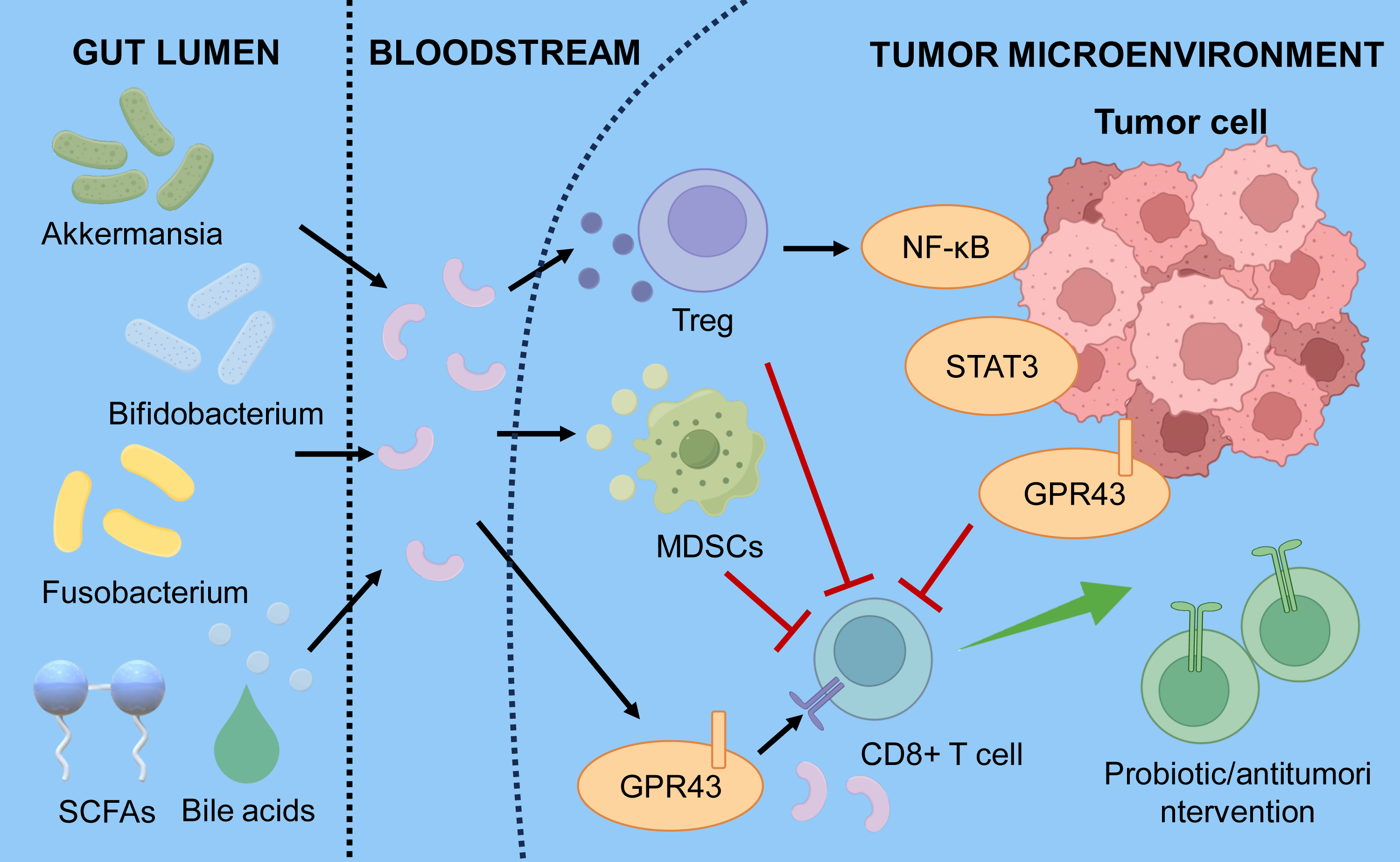
4. Cancer Immunotherapy Resistance and Precision Strategies
Tumor-Intrinsic Mechanisms of Resistance
Tumor cells can evade host immune surveillance through multiple mechanisms, including PD-1 and CTLA-4, which typically help regulate immune responses in healthy cells, but their expression is often increased in tumor cells, boosting immunosuppressive effects. For example, tumor cells may overexpress PD-L1 to suppress T-cell activity, aiding immune evasion (247). Additionally, in the tumor microenvironment, the regulation of immune checkpoint molecules is affected by various cytokines and metabolic products, which can further influence immune escape by activating or blocking related signaling pathways (248).
Defects in antigen presentation further weaken immune recognition and targeting of tumor cells. Tumor cells may lack essential major histocompatibility complex (MHC) molecules, leading to impaired antigen presentation. Additionally, the heterogeneity of tumor antigens results in diverse antigenic profiles among different tumor cells within the same lesion, challenging the immune system’s ability to develop effective memory responses against specific antigens (249). This heterogeneity not only increases the tumor’s ability to escape but also contributes to highly variable therapeutic responses.
Tumor cells also promote immune evasion by altering intracellular signaling pathways. For example, certain tumor cells activate PI3K/Akt or MAPK pathways to boost survival and proliferation, which not only drives tumor growth but may also suppress T cell function (250), although these effects are context-dependent and vary across tumor models. These pathway changes can also modify the tumor microenvironment, leading to the buildup of immunosuppressive cells and further strengthening the immune-evasive abilities of tumor cells.
Microenvironmental and Stromal Mechanisms of Resistance
CAFs play a crucial role within TIME. They hinder immune cell infiltration and function by releasing immunosuppressive factors and remodeling the ECM (251). For example, CAFs can promote tumor cell growth while suppressing T cell activity by secreting factors like TGF-β, thereby supporting tumor initiation and progression (252). The physical barrier created by the ECM further restricts immune cell migration, affecting immune surveillance and the success of immunotherapy. The composition and structure of the ECM directly shape immune cell behavior within the tumor microenvironment. Tumors often show high levels of fibronectin and other ECM components, which not only provide structural support for tumor growth but also help sustain the immunosuppressive environment of tumor-associated immune cells (253). Signaling interactions between stromal and immune cells control the immune status of TIME, influencing the tumor's immune response. Therefore, targeting CAFs or their secreted factors is a promising approach to improve immunotherapy results (254).
Systemic Factors and Precision Strategies
Immunosuppressive cells within the tumor microenvironment, such as Tregs and MDSCs, play a crucial role in tumor immune evasion. These cells inhibit effector T-cell activity by releasing immunosuppressive cytokines, including IL-10 and TGF-β, thereby weakening the immune system’s ability to attack tumor cells (255). Additionally, the accumulation of Tregs and MDSCs is closely linked to tumor progression, underscoring how the microenvironmental composition of immune cells influences tumor immune escape. Immunosuppressive cytokines and metabolic products in the tumor microenvironment further influence immune responses. Tumor cells produce metabolic byproducts such as lactate, which lower the local pH and suppress T cell activity. Furthermore, immunosuppressive cytokines produced by tumor cells can impair immune cell function through various mechanisms, creating conditions that favor tumor survival and growth (256). Abnormal blood vessels and hypoxic conditions in the tumor microenvironment significantly affect immune cell infiltration and function. The irregular structure of tumor blood vessels hampers effective immune cell trafficking to tumor sites, while hypoxia can reduce immune cell activity and proliferation, impairing the overall immune response (247). Additionally, hypoxia can stimulate tumor cells to release pro-tumorigenic factors, further promoting immune evasion.
5. Discussion
The TIME is not a static entity but a dynamically evolving system in which tumor-intrinsic programs, stromal architecture, immune composition, and metabolic constraints interact to determine therapeutic response. Rather than functioning as independent layers, these components form interconnected regulatory circuits that collectively govern immune activation, suppression, or exclusion. This systems-level organization helps explain why tumors with superficially similar histology can exhibit markedly different responses to immunotherapy.
A key insight emerging from recent studies is that immune resistance is rarely driven by a single dominant mechanism. Instead, resistance reflects the convergence of multiple processes, including impaired antigen presentation, sustained T-cell dysfunction, stromal-mediated immune exclusion, and the accumulation of immunosuppressive cytokine and metabolic networks. These features do not act in isolation; for example, tumor-intrinsic alterations that reduce antigenicity can synergize with CAF-driven extracellular matrix remodeling and myeloid cell recruitment to establish immune-excluded states that are refractory to checkpoint blockade. Similarly, chronic inflammatory signaling can simultaneously promote T-cell exhaustion and reinforce suppressive cellular circuits, creating self-sustaining feedback loops within the TIME. This integrated view has important therapeutic implications. It suggests that simply augmenting existing immunotherapies, such as increasing the intensity of checkpoint blockade, is unlikely to overcome resistance in most patients. Instead, effective intervention will require coordinated strategies that reprogram multiple dimensions of the TIME. These may include restoring antigen presentation, disrupting stromal barriers, targeting immunosuppressive cell populations, and modulating metabolic and cytokine networks that constrain immune function. Notably, the success of such approaches will depend on aligning therapeutic strategies with the dominant resistance mechanisms present in each tumor.
The increasing availability of multi-omics technologies, including single-cell transcriptomics, spatial profiling, and integrative genomic analyses, provides an opportunity to redefine TIME classification beyond the traditional “hot” and “cold” dichotomy. Emerging evidence supports the existence of distinct, clinically relevant TIME states, such as immune-inflamed, immune-excluded, and immune-desert phenotypes, each associated with specific mechanistic vulnerabilities. Incorporating these classifications into clinical decision-making could enable more precise patient stratification and guide the rational design of combination therapies.
Future progress will depend on translating mechanistic insights into clinically actionable strategies. This will require carefully designed clinical trials that incorporate biomarker-driven patient selection, longitudinal monitoring of TIME dynamics, and adaptive therapeutic approaches. In particular, combination regimens targeting complementary pathways, such as checkpoint inhibition with stromal modulation, metabolic reprogramming, or microbiome-based interventions, represent a promising avenue for overcoming resistance.
In summary, resistance to immunotherapy reflects the coordinated behavior of tumor, immune, and stromal components within a dynamically regulated microenvironment. A deeper mechanistic understanding of these interactions, coupled with advances in molecular profiling and therapeutic targeting, may enable more precise immunotherapy strategies; however, their ability to deliver durable clinical benefit will depend on context-specific patient selection and remains to be fully established. Immunotherapy resistance reflects the coordinated interplay of tumor-intrinsic alterations, stromal barriers, and immune dysfunction within a dynamically evolving TIME. Rather than acting independently, these processes form interconnected networks that constrain effective antitumor immunity and limit the efficacy of single-agent strategies. While current classifications, such as “hot,” “cold,” and immune-excluded tumors, provide a useful framework, they fail to capture the temporal plasticity of the TIME under therapeutic pressure. Emerging insights, including contributions from systemic factors such as the gut microbiota, further highlight the need to consider tumor-extrinsic regulation of immune responses. Moving forward, integrating multi-omics profiling with mechanism-based combination therapies will be essential for identifying dominant resistance pathways and guiding precision immunotherapy. A systems-level, dynamically informed approach will be critical to convert immunologically refractory tumors into durable responders.
References
1. Zhao Y, Tan F, Zhao J, Zhou S, Luo Y, Gong C. Targeting the enhanced sensitivity of radiotherapy in cancer: mechanisms, applications, and challenges. MedComm. 2025;6(6):e70202. https://doi.org/10.1002/mco2.70202
2. Tudor C. A novel approach to modeling and forecasting cancer incidence and mortality rates through web queries and automated forecasting algorithms: evidence from Romania. Biology (Basel). 2022;11(6):857. https://doi.org/10.3390/biology11060857
3. Pak J, Le N, Metwally EM, Wang J, Planey AM, Lowman AM, et al. Evaluating the intersection of climate vulnerability and cancer burden in North Carolina. JNCI Cancer Spectr. 2025;9(1):pkae124. https://doi.org/10.1093/jncics/pkae124
4. Bergman PJ. Cancer immunotherapy. Vet Clin North Am Small Anim Pract. 2024;54(3):441–468. https://doi.org/10.1016/j.cvsm.2023.12.002
5. Zeiser R. Immunotherapy in cancer. Semin Arthritis Rheum. 2025;72(Suppl):152666. https://doi.org/10.1016/j.semarthrit.2025.152666
6. Rangamuwa K, Aloe C, Christie M, Asselin-Labat ML, Batey D, Irving L, et al. Methods for assessment of the tumour microenvironment and immune interactions in non-small cell lung cancer. a narrative review. Front Oncol. 2023;13:1129195. https://doi.org/10.3389/fonc.2023.1129195
7. Huang Q, Lei Y, Li X, Guo F, Liu M. A highlight of the mechanisms of immune checkpoint blocker resistance. Front Cell Dev Biol. 2020;8:580140. https://doi.org/10.3389/fcell.2020.580140
8. Li L, Zhao L, Zhou D, Yu Y, Zhang P, Zheng J, et al. Targeting pyroptosis reverses KIAA1199-mediated immunotherapy resistance in colorectal cancer. J Immunother Cancer. 2025;13(2):e010000. https://doi.org/10.1136/jitc-2024-010000
9. Guven DC, Aktas BY, Simsek C, Aksoy S. Gut microbiota and cancer immunotherapy: prognostic and therapeutic implications. Future Oncol. 2020;16(9):497–506. https://doi.org/10.2217/fon-2019-0783
10. Sardar P, Beresford-Jones BS, Xia W, Shabana O, Suyama S, Ramos RJF, et al. Gut microbiota-derived hexa-acylated lipopolysaccharides enhance cancer immunotherapy responses. Nat Microbiol. 2025;10:795–807. https://doi.org/10.1038/s41564-025-01930-y
11. Seliger B, Massa C, Yang B, Bethmann D, Kappler M, Eckert AW, et al. Immune escape mechanisms and their clinical relevance in head and neck squamous cell carcinoma. Int J Mol Sci. 2020;21(19):7032. https://doi.org/10.3390/ijms21197032
12. Liu S, Zhang Y. Challenges and interventions of chimeric antigen receptor-T cell therapy in solid tumors. Chin J Cancer Res. 2023;35(3):239–244. https://doi.org/10.21147/j.issn.1000-9604.2023.03.03
13. Xu Q, Li X. Tumor-derived extracellular vesicles in the immune microenvironment of head and neck squamous cell carcinoma: Foe or future? J Stomatol Oral Maxillofac Surg. 2024;125(4):101738. https://doi.org/10.1016/j.jormas.2023.101738
14. Sadri M, Heidari S, Faridzadeh A, Roozbehani M, Toosi S, Mahmoudian RA, et al. Potential applications of macrophages in cancer immunotherapy. Biomed Pharmacother. 2024;178:117161. https://doi.org/10.1016/j.biopha.2024.117161
15. Carnevalli LS, Taylor MA, King M, Coenen-Stass AML, Hughes AM, Bell S, et al. Macrophage activation status rather than repolarization is associated with enhanced checkpoint activity in combination with PI3Kγ inhibition. Mol Cancer Ther. 2021;20(6):1080–1091. https://doi.org/10.1158/1535-7163.mct-20-0961
16. Hou A, Hou K, Huang Q, Lei Y, Chen W. Targeting myeloid-derived suppressor cell, a promising strategy to overcome resistance to immune checkpoint inhibitors. Front Immunol. 2020;11:783. https://doi.org/10.3389/fimmu.2020.00783
17. Ozbay Kurt FG, Lasser S, Arkhypov I, Utikal J, Umansky V. Enhancing immunotherapy response in melanoma: myeloid-derived suppressor cells as a therapeutic target. J Clin Invest. 2023;133(13):e170762. https://doi.org/10.1172/jci170762
18. Sun F, Ma F, Wang T, Zhou Y, Xu D, Mo H, et al. STS2 deficiency revives CD8+ T cells from exhaustion and augments checkpoint blockade efficacy in cancer immunotherapy. J Immunother Cancer. 2025;13(6):e010735. https://doi.org/10.1136/jitc-2024-010735
19. Yang MQ, Zhang SL, Sun L, Huang LT, Yu J, Zhang JH, et al. Targeting mitochondria: restoring the antitumor efficacy of exhausted T cells. Mol Cancer. 2024;23(1):260. https://doi.org/10.1186/s12943-024-02175-9
20. Mortezaee K, Majidpoor J. Mechanisms of CD8+ T cell exclusion and dysfunction in cancer resistance to anti-PD-(L)1. Biomed Pharmacother. 2023;163:114824. https://doi.org/10.1016/j.biopha.2023.114824
21. Diamond MS, Lin JH, Vonderheide RH. Site-dependent immune escape due to impaired dendritic cell cross-priming. Cancer Immunol Res. 2021;9(8):877–890. https://doi.org/10.1158/2326-6066.cir-20-0785
22. Zhu X, Li B, Qin L, Liang T, Hu W, Li J, et al. Development and validation of a prognostic model for lung adenocarcinoma based on CAF-related genes: unveiling the role of COX6A1 in cancer progression and CAF infiltration. Int J Mol Sci. 2025;26(8):3478. https://doi.org/10.3390/ijms26083478
23. Rodriguez E. Tumor glycosylation: a main player in the modulation of immune responses. Eur J Immunol. 2025;55(3):e202451318. https://doi.org/10.1002/eji.202451318
24. Wen Q, Han T, Wang Z, Jiang S. Role and mechanism of programmed death-ligand 1 in hypoxia-induced liver cancer immune escape. Oncol Lett. 2020;19(4):2595–2601. https://doi.org/10.3892/ol.2020.11369
25. Jiao J, Wu Y, Wu S, Jiang J. Enhancing colorectal cancer treatment through VEGF/VEGFR inhibitors and immunotherapy. Curr Treat Options Oncol. 2025;26(3):213–225. https://doi.org/10.1007/s11864-025-01306-8
26. Czaja AJ. Exploring the pathogenic role and therapeutic implications of interleukin 2 in autoimmune hepatitis. Dig Dis Sci. 2021;66(8):2493–2512. https://doi.org/10.1007/s10620-020-06562-2
27. Shi P, Yu Y, Xie H, Yin X, Chen X, Zhao Y, et al. Recent advances in regulatory immune cells: exploring the world beyond Tregs. Front Immunol. 2025;16:1530301. https://doi.org/10.3389/fimmu.2025.1530301
28. Zhang W, Zhang W, Wu H, Han X. Harnessing innate immunity against glioblastoma microenvironment. Front Immunol. 2025;16:1648601. https://doi.org/10.3389/fimmu.2025.1648601
29. Xiong Z, Huang Y, Cao S, Huang X, Zhang H. A new strategy for the treatment of advanced ovarian cancer: utilizing nanotechnology to regulate the tumor microenvironment. Front Immunol. 2025;16:1542326. https://doi.org/10.3389/fimmu.2025.1542326
30. Palmer N, Khakoo S, Sanchez-Elsner T, Vallejo AF. Enhancing natural killer cell anti-tumour activity through macrophage manipulation. Front Immunol. 2025;16:1656925. https://doi.org/10.3389/fimmu.2025.1656925
31. Ramel E, Lillo S, Daher B, Fioleau M, Daubon T, Saleh M. The metabolic control of myeloid cells in the tumor microenvironment. Cells. 2021;10(11):2960. https://doi.org/10.3390/cells10112960
32. Xiong X, Zhang Y, Wen Y. Diverse functions of myeloid-derived suppressor cells in autoimmune diseases. Immunol Res. 2024;72(1):34–49. https://doi.org/10.1007/s12026-023-09421-0
33. Zhan X, He Q, Sheng J, Jiang X, Lin L, Huang Y, et al. USP12 positively regulates M-MDSC function to inhibit antitumour immunity through deubiquitinating and stabilizing p65. Immunology. 2022;167(4):544–557. https://doi.org/10.1111/imm.13552
34. Wang H, Zhou F, Qin W, Yang Y, Li X, Liu R. Metabolic regulation of myeloid-derived suppressor cells in tumor immune microenvironment: targets and therapeutic strategies. Theranostics. 2025;15(6):2159–2184. https://doi.org/10.7150/thno.105276
35. Cheng B, Yu Q, Wang W. Intimate communications within the tumor microenvironment: stromal factors function as an orchestra. J Biomed Sci. 2023;30(1):1. https://doi.org/10.1186/s12929-022-00894-z
36. Wang J, He Y, Hu F, Hu C, Sun Y, Yang K, et al. Metabolic reprogramming of immune cells in the tumor microenvironment. Int J Mol Sci. 2024;25(22):12223. https://doi.org/10.3390/ijms252212223
37. Çakal S, Er Urgancı B, Şimşek S. Key players in the breast cancer microenvironment: From fibroblasts to immune cells. World J Clin Oncol. 2025;16(7):107339. https://doi.org/10.5306/wjco.v16.i7.107339
38. Zhang Y, Han X, Wang K, Liu D, Ding X, Hu Z, et al. Co-delivery nanomicelles for potentiating TNBC immunotherapy by synergetically reshaping CAFs-mediated tumor stroma and reprogramming immunosuppressive microenvironment. Int J Nanomedicine. 2023;18:4329–4346. https://doi.org/10.2147/ijn.S418100
39. Du W, Pasca di Magliano M, Zhang Y. Therapeutic potential of targeting stromal crosstalk-mediated immune suppression in pancreatic cancer. Front Oncol. 2021;11:682217. https://doi.org/10.3389/fonc.2021.682217
40. Uddin MN, Wang X. The landscape of long non-coding RNAs in tumor stroma. Life Sci. 2021;264:118725. https://doi.org/10.1016/j.lfs.2020.118725
41. Marima R, Basera A, Miya T, Damane BP, Kandhavelu J, Mirza S, et al. Exosomal long non-coding RNAs in cancer: Interplay, modulation, and therapeutic avenues. Noncoding RNA Res. 2024;9(3):887–900. https://doi.org/10.1016/j.ncrna.2024.03.014
42. Faisal SM, Comba A, Varela ML, Argento AE, Brumley E, Abel C II, et al. The complex interactions between the cellular and non-cellular components of the brain tumor microenvironmental landscape and their therapeutic implications. Front Oncol. 2022;12:1005069. https://doi.org/10.3389/fonc.2022.1005069
43. Gamradt P, De La Fouchardière C, Hennino A. Stromal protein-mediated immune regulation in digestive cancers. Cancers (Basel). 2021;13(1):146. https://doi.org/10.3390/cancers13010146
44. Khurana A, Ansell SM. Role of microenvironment in non-hodgkin lymphoma: understanding the composition and biology. Cancer J. 2020;26(3):206–216. https://doi.org/10.1097/ppo.0000000000000446
45. Hu J, Mao F, Li L, Wang X, Cai D, He L, et al. Analysis of the heterogeneity of the tumor microenvironment and the prognosis and immunotherapy response of different immune subtypes in hepatocellular carcinoma. J Oncol. 2022;2022:1087399. https://doi.org/10.1155/2022/1087399
46. Feng Y, Ma W, Zang Y, Guo Y, Li Y, Zhang Y, et al. Spatially organized tumor-stroma boundary determines the efficacy of immunotherapy in colorectal cancer patients. Nat Commun. 2024;15(1):10259. https://doi.org/10.1038/s41467-024-54710-3
47. Obeagu EI. Modulation of chronic cytokine dysregulation in cervical cancer: potential biomarkers and therapeutic targets. Cancer Manag Res. 2025;17:1113–1126. https://doi.org/10.2147/cmar.S527913
48. Hjazi A, Nasir F, Noor R, Alsalamy A, Zabibah RS, Romero-Parra RM, et al. The pathological role of C-X-C chemokine receptor type 4 (CXCR4) in colorectal cancer (CRC) progression; special focus on molecular mechanisms and possible therapeutics. Pathol Res Pract. 2023;248:154616. https://doi.org/10.1016/j.prp.2023.154616
49. D'Uonnolo G, Isci D, Nosirov B, Kuppens A, Wantz M, Nazarov PV, et al. Patient-based multilevel transcriptome exploration highlights relevant chemokines and chemokine receptor axes in glioblastoma. Comput Biol Med. 2024;182:109197. https://doi.org/10.1016/j.compbiomed.2024.109197
50. Ozga AJ, Chow MT, Luster AD. Chemokines and the immune response to cancer. Immunity. 2021;54(5):859–874. https://doi.org/10.1016/j.immuni.2021.01.012
51. Yi M, Li T, Niu M, Zhang H, Wu Y, Wu K, et al. Targeting cytokine and chemokine signaling pathways for cancer therapy. Signal Transduct Target Ther. 2024;9(1):176. https://doi.org/10.1038/s41392-024-01868-3
52. Reschke R, Enk AH, Hassel JC. Chemokines and cytokines in immunotherapy of melanoma and other tumors: from biomarkers to therapeutic targets. Int J Mol Sci. 2024;25(12):6532. https://doi.org/10.3390/ijms25126532
53. Bindea G, Mlecnik B, Galon J. Expand to shield: IL-15 and in situ lymphocytic proliferation. Oncoimmunology. 2021;10(1):1886726. https://doi.org/10.1080/2162402x.2021.1886726
54. Dai NN, Hu MY, Wang JP, Dai ZH, Xu L, Ye TW. Tertiary lymphoid structures in the microenvironment of colorectal cancer: exploring new therapeutic targets. Cancer Immunol Immunother. 2025;74(8):245. https://doi.org/10.1007/s00262-025-04108-x
55. Pan M, Wei X, Xiang X, Liu Y, Zhou Q, Yang W. Targeting CXCL9/10/11-CXCR3 axis: an important component of tumor-promoting and antitumor immunity. Clin Transl Oncol. 2023;25(8):2306–2320. https://doi.org/10.1007/s12094-023-03126-4
56. Kasimanickam R, Ferreira J, Kasimanickam V. Integrative analysis of differentially expressed miRNAs and noncoding RNA networks reveals molecular mechanisms underlying metritis in postpartum dairy cows. Curr Issues Mol Biol. 2025;47(8):643. https://doi.org/10.3390/cimb47080643
57. Xavier H, Gireesh AGM, Thomas JA, Suboj P, Suresh A, Biju E, et al. Chemokines: humble yet mighty players in the tumour microenvironment. Front Immunol. 2025;16:1601756. https://doi.org/10.3389/fimmu.2025.1601756
58. Lam KC, Goldszmid RS. Single-cell RNA flow cytometry to assess intratumoral production of cytokines/chemokines. Methods Cell Biol. 2025;191:221–246. https://doi.org/10.1016/bs.mcb.2024.10.013
59. Wang Y, Luo S, Dong H, Huang RP. Unraveling tumor cell-tumor microenvironment crosstalk through antibody array technologies (Review). Int J Oncol. 2025;67(4):81. https://doi.org/10.3892/ijo.2025.5787
60. Yi M, Niu M, Wu Y, Ge H, Jiao D, Zhu S, et al. Combination of oral STING agonist MSA-2 and anti-TGF-β/PD-L1 bispecific antibody YM101: a novel immune cocktail therapy for non-inflamed tumors. J Hematol Oncol. 2022;15(1):142. https://doi.org/10.1186/s13045-022-01363-8
61. Zhang Y, Guan XY, Jiang P. Cytokine and chemokine signals of T-cell exclusion in tumors. Front Immunol. 2020;11:594609. https://doi.org/10.3389/fimmu.2020.594609
62. Zhang Y, Li L, Zheng W, Zhang L, Yao N. CD8+ T-cell exhaustion in the tumor microenvironment of head and neck squamous cell carcinoma determines poor prognosis. Ann Transl Med. 2022;10(6):273. https://doi.org/10.21037/atm-22-867
63. Federico L, McGrail DJ, Bentebibel SE, Haymaker C, Ravelli A, Forget MA, et al. Distinct tumor-infiltrating lymphocyte landscapes are associated with clinical outcomes in localized non-small-cell lung cancer. Ann Oncol. 2022;33(1):42–56. https://doi.org/10.1016/j.annonc.2021.09.021
64. Edin S, Gylling B, Li X, Stenberg Å, Löfgren-Burström A, Zingmark C, et al. Opposing roles by KRAS and BRAF mutation on immune cell infiltration in colorectal cancer - possible implications for immunotherapy. Br J Cancer. 2024;130(1):143–150. https://doi.org/10.1038/s41416-023-02483-9
65. Cao H, Quan S, Zhang L, Chen Y, Jiao G. BMPR2 expression level is correlated with low immune infiltration and predicts metastasis and poor survival in osteosarcoma. Oncol Lett. 2021;21(5):391. https://doi.org/10.3892/ol.2021.12652
66. Ao YQ, Gao J, Zhang LX, Deng J, Wang S, Lin M, et al. Tumor-infiltrating CD36+ CD8+ T cells determine exhausted tumor microenvironment and correlate with inferior response to chemotherapy in non-small cell lung cancer. BMC Cancer. 2023;23(1):367. https://doi.org/10.1186/s12885-023-10836-z
67. Scott EN, Gocher AM, Workman CJ, Vignali DAA. Regulatory T cells: barriers of immune infiltration into the tumor microenvironment. Front Immunol. 2021;12:702726. https://doi.org/10.3389/fimmu.2021.702726
68. He J, Meng M, Wang H. A novel prognostic biomarker LPAR6 in hepatocellular carcinoma via associating with immune infiltrates. J Clin Transl Hepatol. 2022;10(1):90–103. https://doi.org/10.14218/jcth.2021.00047
69. Nonnast E, Fernández-Aceñero MJ, Pons T, Díaz Suárez MP, Sorzano CÓ S, Mira E, et al. A laminin α4-CD8+ T cell axis shapes the prognostic impact of macrophages and regulatory T cells in early-stage colorectal cancer. Oncoimmunology. 2025;14(1):2546181. https://doi.org/10.1080/2162402x.2025.2546181
70. Kang X, Zhao S, Lin S, Li J, Wang S. Synergistic upregulation of PD‑L1 in tumor cells and CD39 in tumor-infiltrating CD8+ T cells leads to poor prognosis in patients with hepatocellular carcinoma. Oncol Lett. 2024;28(2):368. https://doi.org/10.3892/ol.2024.14501
71. Shadbad MA, Asadzadeh Z, Hosseinkhani N, Derakhshani A, Alizadeh N, Brunetti O, et al. A systematic review of the tumor-infiltrating CD8+ T-cells/PD-L1 axis in high-grade glial tumors: toward personalized immuno-oncology. Front Immunol. 2021;12:734956. https://doi.org/10.3389/fimmu.2021.734956
72. Juthi RT, Sazed SA, Mareboina M, Zaravinos A, Georgakopoulos-Soares I. Characterization of exhausted T cell signatures in pan-cancer settings. Int J Mol Sci. 2025;26(5):2311. https://doi.org/10.3390/ijms26052311
73. Zhang MX, Jing LY, Tan HT, Dai ZR, Long DZ, Liu HC, et al. MIAT promotes tumor-infiltrating CD8+ T-cell exhaustion and malignant progression of renal cell carcinoma via activating JAK3/STAT3 pathway. J Immunother Cancer. 2025;13(7):e011162. https://doi.org/10.1136/jitc-2024-011162
74. Rachayon M, Jirakran K, Sodsai P, Tunvirachaisakul C, Sughondhabirom A, Li J, et al. Immune cell exhaustion and apoptotic markers in major depressive disorder: effects of in vitro cannabidiol administration. Brain Behav Immun Health. 2025;48:101066. https://doi.org/10.1016/j.bbih.2025.101066
75. Sorrentino C, D'Antonio L, Fieni C, Ciummo SL, Di Carlo E. Colorectal cancer-associated immune exhaustion involves T and B lymphocytes and conventional NK cells and correlates with a shorter overall survival. Front Immunol. 2021;12:778329. https://doi.org/10.3389/fimmu.2021.778329
76. Studstill CJ, Hahm B. Chronic LCMV infection is fortified with versatile tactics to suppress host T cell immunity and establish viral persistence. Viruses. 2021;13(10):1951. https://doi.org/10.3390/v13101951
77. Lv LL, Zhai JW, Wu JJ, Fan GQ, Zhang YX, Shen Y, et al. High CD38 expression defines a mitochondrial function-adapted CD8+ T cell subset with implications for lung cancer immunotherapy. Cancer Immunol Immunother. 2025;74(2):49. https://doi.org/10.1007/s00262-024-03881-5
78. Wildner NH, Walker A, Brauneck F, Ditt V, Peine S, Huber S, et al. Transcriptional pattern analysis of virus-specific CD8+ T cells in hepatitis C infection: increased expression of TOX and eomesodermin during and after persistent antigen recognition. Front Immunol. 2022;13:886646. https://doi.org/10.3389/fimmu.2022.886646
79. Jenkins E, Whitehead T, Fellermeyer M, Davis SJ, Sharma S. The current state and future of T-cell exhaustion research. Oxf Open Immunol. 2023;4(1):iqad006. https://doi.org/10.1093/oxfimm/iqad006
80. Freag MS, Mohammed MT, Kulkarni A, Emam HE, Maremanda KP, Elzoghby AO. Modulating tumoral exosomes and fibroblast phenotype using nanoliposomes augments cancer immunotherapy. Sci Adv. 2024;10(9):eadk3074. https://doi.org/10.1126/sciadv.adk3074
81. Lee H, Hwang M, Jang S, Um SW. Immune regulatory function of cancer-associated fibroblasts in non-small cell lung cancer. Tuberc Respir Dis (Seoul). 2023;86(4):304–318. https://doi.org/10.4046/trd.2022.0129
82. Li R, O'Connor DJ, Digby B, Broin P, Hu X, Ge N, et al. Aurantio-obtusin modulates Wilms Tumour 1 within the breast tumour microenvironment reducing immunosuppression and tumour growth. Cell Commun Signal. 2025;23(1):309. https://doi.org/10.1186/s12964-025-02292-y
83. Wei R, Song J, Liu C, Zhao Z, Liu X, Yamamoto M, et al. FAP upregulates PD-L1 expression in cancer-associated fibroblasts to exacerbate T cells dysfunction and suppress anti-tumor immunity. Cancer Lett. 2025;612:217475. https://doi.org/10.1016/j.canlet.2025.217475
84. Huang TX, Tan XY, Huang HS, Li YT, Liu BL, Liu KS, et al. Targeting cancer-associated fibroblast-secreted WNT2 restores dendritic cell-mediated antitumour immunity. Gut. 2022;71(2):333–344. https://doi.org/10.1136/gutjnl-2020-322924
85. Ramil CP, Xiang H, Zhang P, Cronin A, Cabral L, Yin Z, et al. Extracellular vesicles released by cancer-associated fibroblast-induced myeloid-derived suppressor cells inhibit T-cell function. Oncoimmunology. 2024;13(1):2300882. https://doi.org/10.1080/2162402x.2023.2300882
86. Hu J, Lazar AJ, Ingram D, Wang WL, Zhang W, Jia Z, et al. Cell membrane-anchored and tumor-targeted IL-12 T-cell therapy destroys cancer-associated fibroblasts and disrupts extracellular matrix in heterogenous osteosarcoma xenograft models. J Immunother Cancer. 2024;12(1):e006991. https://doi.org/10.1136/jitc-2023-006991
87. Milosevic V, Östman A. Interactions between cancer-associated fibroblasts and T-cells: functional crosstalk with targeting and biomarker potential. Ups J Med Sci. 2024;129:10710. https://doi.org/10.48101/ujms.v129.10710
88. Freeman P, Mielgo A. Cancer-associated fibroblast mediated inhibition of CD8+ cytotoxic T cell accumulation in tumours: mechanisms and therapeutic opportunities. Cancers (Basel). 2020;12(9):2687. https://doi.org/10.3390/cancers12092687
89. McAndrews KM, Vázquez-Arreguín K, Kwak C, Sugimoto H, Zheng X, Li B, et al. αSMA+ fibroblasts suppress Lgr5+ cancer stem cells and restrain colorectal cancer progression. Oncogene. 2021;40(26):4440–4452. https://doi.org/10.1038/s41388-021-01866-7
90. Lu L, Gao Y, Huang D, Liu H, Yin D, Li M, et al. Targeting integrin α5 in fibroblasts potentiates colorectal cancer response to PD-L1 blockade by affecting extracellular-matrix deposition. J Immunother Cancer. 2023;11(12):e007447. https://doi.org/10.1136/jitc-2023-007447
91. Ford K, Hanley CJ, Mellone M, Szyndralewiez C, Heitz F, Wiesel P, et al. NOX4 inhibition potentiates immunotherapy by overcoming cancer-associated fibroblast-mediated CD8 T-cell exclusion from tumors. Cancer Res. 2020;80(9):1846–1860. https://doi.org/10.1158/0008-5472.can-19-3158
92. Tripathi DK, Nagar N, Kumar V, Joshi N, Roy P, Poluri KM. Gallate moiety of catechin is essential for inhibiting CCL2 chemokine-mediated monocyte recruitment. J Agric Food Chem. 2023;71(12):4990–5005. https://doi.org/10.1021/acs.jafc.3c01283
93. Chi HC, Lin YH, Wu YH, Chang CC, Wu CH, Yeh CT, et al. CCL16 is a pro-tumor chemokine that recruits monocytes and macrophages to promote hepatocellular carcinoma progression. Am J Cancer Res. 2024;14(7):3600–3613. https://doi.org/10.62347/vctw6889
94. Coombes JD, Manka PP, Swiderska-Syn M, Vannan DT, Riva A, Claridge LC, et al. Osteopontin promotes cholangiocyte secretion of chemokines to support macrophage recruitment and fibrosis in MASH. Liver Int. 2025;45(4):e16131. https://doi.org/10.1111/liv.16131
95. Akbarzadeh R, Czyz C, Thomsen SY, Schilf P, Murthy S, Sadik CD, et al. Monocyte populations are involved in the pathogenesis of experimental epidermolysis bullosa acquisita. Front Immunol. 2023;14:1241461. https://doi.org/10.3389/fimmu.2023.1241461
96. Liner AG, van Gogh M, Roblek M, Heikenwalder M, Borsig L. Non-redundant roles of the CCR1 and CCR2 chemokine axes in monocyte recruitment during lung metastasis. Neoplasia. 2025;59:101089. https://doi.org/10.1016/j.neo.2024.101089
97. Lambour J, Naranjo-Gomez M, Boyer-Clavel M, Pelegrin M. Differential and sequential immunomodulatory role of neutrophils and Ly6Chi inflammatory monocytes during antiviral antibody therapy. Emerg Microbes Infect. 2021;10(1):964–981. https://doi.org/10.1080/22221751.2021.1913068
98. Hannan CJ, Lewis D, O'Leary C, Waqar M, Brough D, Couper KN, et al. Increased circulating chemokines and macrophage recruitment in growing vestibular schwannomas. Neurosurgery. 2023;92(3):581–589. https://doi.org/10.1227/neu.0000000000002252
99. Almeida PP, Moraes JA, Barja-Fidalgo TC, Renovato-Martins M. Extracellular vesicles as modulators of monocyte and macrophage function in tumors. An Acad Bras Cienc. 2024;96(2):e20231212. https://doi.org/10.1590/0001-3765202420231212
100. Kim SW, Kim CW, Moon YA, Kim HS. Reprogramming of tumor-associated macrophages by metabolites generated from tumor microenvironment. Anim Cells Syst (Seoul). 2024;28(1):123–136. https://doi.org/10.1080/19768354.2024.2336249
101. Shiuan E, Wang S, Brantley-Sieders DM. Tumor-specific EphA2 receptor tyrosine kinase inhibits anti-tumor immunity by recruiting suppressive myeloid populations in murine models of non-small cell lung cancer. Cancers (Basel). 2025;17(16):2693. https://doi.org/10.3390/cancers17162693
102. Wen J, Guan Y, Niu H, Dang Y, Guan J. Targeting cardiac resident CCR2+ macrophage-secreted MCP-1 to attenuate inflammation after myocardial infarction. Acta Biomater. 2026;211:18–32. https://doi.org/10.1016/j.actbio.2024.08.025
103. Wang C, Shen N, Guo Q, Tan X, He S. YAP/STAT3 inhibited CD8+ T cells activity in the breast cancer immune microenvironment by inducing M2 polarization of tumor-associated macrophages. Cancer Med. 2023;12(15):16295–16309. https://doi.org/10.1002/cam4.6242
104. He S, Gu X, Yang J, Xu F, Hu J, Wang W, et al. Sphingomyelin synthase 2 is a positive regulator of the CSF1R-STAT3 pathway in pancreatic cancer-associated macrophage. Front Pharmacol. 2022;13:902016. https://doi.org/10.3389/fphar.2022.902016
105. Ou Y, Jiang HM, Wang YJ, Shuai QY, Cao LX, Guo M, et al. The Zeb1-Cxcl1 axis impairs the antitumor immune response by inducing M2 macrophage polarization in breast cancer. Am J Cancer Res. 2024;14(9):4378–4397. https://doi.org/10.62347/uais7070
106. He K, Barsoumian HB, Puebla-Osorio N, Hu Y, Sezen D, Wasley MD, et al. Inhibition of STAT6 with antisense oligonucleotides enhances the systemic antitumor effects of radiotherapy and anti-PD-1 in metastatic non-small cell lung cancer. Cancer Immunol Res. 2023;11(4):486–500. https://doi.org/10.1158/2326-6066.cir-22-0547
107. Butkute A, Baltramonaitis M, Malmige S, Darinskas A, Pasukoniene V, Mlynska A. Targeting stemness pathways modulates macrophage polarization and reprograms the tumor microenvironment. Front Immunol. 2025;16:1513404. https://doi.org/10.3389/fimmu.2025.1513404
108. Yang D, Yang L, Cai J, Li H, Xing Z, Hou Y. Phosphoinositide 3-kinase/Akt and its related signaling pathways in the regulation of tumor-associated macrophages polarization. Mol Cell Biochem. 2022;477(10):2469–2480. https://doi.org/10.1007/s11010-022-04461-w
109. Zhang H, Yuan F, Zhao N, Tang W, Zhao P, Liu C, et al. Nanoparticle-mediated SIRT1 inhibition suppresses M2 macrophage polarization and hepatocarcinogenesis in chronic hepatitis B. J Nanobiotechnology. 2025;23(1):385. https://doi.org/10.1186/s12951-025-03447-2
110. Noe JT, Rendon BE, Geller AE, Conroy LR, Morrissey SM, Young LEA, et al. Lactate supports a metabolic-epigenetic link in macrophage polarization. Sci Adv. 2021;7(46):eabi8602. https://doi.org/10.1126/sciadv.abi8602
111. Zuo B, Li T, Liu X, Wang S, Cheng J, Liu X, et al. Dipeptidyl peptidase 4 inhibitor reduces tumor-associated macrophages and enhances anti-PD-L1-mediated tumor suppression in non-small cell lung cancer. Clin Transl Oncol. 2023;25(11):3188–3202. https://doi.org/10.1007/s12094-023-03187-5
112. Gao Z, Li XG, Feng SR, Chen JF, Song K, Shi YH, et al. Autophagy suppression facilitates macrophage M2 polarization via increased instability of NF-κB pathway in hepatocellular carcinoma. Int Immunopharmacol. 2023;123:110685. https://doi.org/10.1016/j.intimp.2023.110685
113. Xu J, Wang G, Wang M, Xu X, Xu Y, Zhang C, et al. Pedunculoside regulates tumor immune microenvironment and malignant characteristics of bladder cancer by TRAF6 mediated M2 macrophage polarization. Int Immunopharmacol. 2025;164:115301. https://doi.org/10.1016/j.intimp.2025.115301
114. Cao Q, Zhou X, Zou F, He R, He L, Chen J, et al. Treponema pallidum lipoprotein TpF1 regulates METTL14-mediated NF-κB signaling pathway to promote macrophage M2 polarization. J Leukoc Biol. 2025;117(8):qiaf099. https://doi.org/10.1093/jleuko/qiaf099
115. He R, He Y, Du R, Liu C, Chen Z, Zeng A, et al. Revisiting of TAMs in tumor immune microenvironment: Insight from NF-κB signaling pathway. Biomed Pharmacother. 2023;165:115090. https://doi.org/10.1016/j.biopha.2023.115090
116. Xu J, Ding L, Mei J, Hu Y, Kong X, Dai S, et al. Dual roles and therapeutic targeting of tumor-associated macrophages in tumor microenvironments. Signal Transduct Target Ther. 2025;10(1):268. https://doi.org/10.1038/s41392-025-02325-5
117. Ye Z, Yi J, Jiang X, Shi W, Xu H, Cao H, et al. Gastric cancer-derived exosomal let-7g-5p mediated by SERPINE1 promotes macrophage M2 polarization and gastric cancer progression. J Exp Clin Cancer Res. 2025;44(1):2. https://doi.org/10.1186/s13046-024-03269-4
118. Wang L, Fan J, Wu S, Cheng S, Zhao J, Fan F, et al. LTBR acts as a novel immune checkpoint of tumor-associated macrophages for cancer immunotherapy. iMeta. 2024;3(5):e233. https://doi.org/10.1002/imt2.233
119. Jung M, Bonavida B. Immune evasion in cancer is regulated by tumor-asociated macrophages (TAMs): targeting TAMs. Crit Rev Oncog. 2024;29(4):1–17. https://doi.org/10.1615/critrevoncog.2024053096
120. Liu F, Li X, Zhang Y, Ge S, Shi Z, Liu Q, et al. Targeting tumor-associated macrophages to overcome immune checkpoint inhibitor resistance in hepatocellular carcinoma. J Exp Clin Cancer Res. 2025;44(1):227. https://doi.org/10.1186/s13046-025-03490-9
121. Mo W, Liu S, Zhao X, Wei F, Li Y, Sheng X, et al. ROS scavenging nanozyme modulates immunosuppression for sensitized cancer immunotherapy. Adv Healthc Mater. 2023;12(21):e2300191. https://doi.org/10.1002/adhm.202300191
122. Wang Y, Ma C, Li X, Yang F, Wang N, Ji G, et al. Unraveling the role of M2 TAMs in ovarian cancer dynamics: a systematic review. J Transl Med. 2025;23(1):623. https://doi.org/10.1186/s12967-025-06643-8
123. Feng L, Wang Y, Wang X, Li B, Liu Y, Zhang W, et al. Exosomal lncRNA MIR210HG derived from hypoxic TAMs promotes the metastasis of triple negative breast cancer. Int Immunopharmacol. 2025;165:115416. https://doi.org/10.1016/j.intimp.2025.115416
124. Zhang X, Sun Y, Ma Y, Gao C, Zhang Y, Yang X, et al. Tumor-associated M2 macrophages in the immune microenvironment influence the progression of renal clear cell carcinoma by regulating M2 macrophage-associated genes. Front Oncol. 2023;13:1157861. https://doi.org/10.3389/fonc.2023.1157861
125. Cao X, Wan S, Wu B, Liu Z, Xu L, Ding Y, et al. Antitumor research based on drug delivery carriers: reversing the polarization of tumor-associated macrophages. Mol Pharm. 2025;22(3):1174–1197. https://doi.org/10.1021/acs.molpharmaceut.4c01277
126. Liang C, Zhang Y, Wang S, Jiao W, Guo J, Zhang N, et al. Nanomaterials in modulating tumor-associated macrophages and enhancing immunotherapy. J Mater Chem B. 2024;12(20):4809–4823. https://doi.org/10.1039/d4tb00230j
127. Peng H, Wang JH, Guo F, Zhu FF, Wen ZJ, Zhong HJ, et al. Legumain protease-activated tuftsin-functionalized nanoparticles for dual-targeting TAMs and cancer chemotherapy. Colloids Surf B Biointerfaces. 2021;197:111442. https://doi.org/10.1016/j.colsurfb.2020.111442
128. Hu X, Li Y, Wang X, Xue X. Role of M2 macrophage-derived exosomes in cancer drug resistance via noncoding RNAs. Discov Oncol. 2025;16(1):741. https://doi.org/10.1007/s12672-025-02195-x
129. Zhao G, Ding L, Yu H, Wang W, Wang H, Hu Y, et al. M2-like tumor-associated macrophages transmit exosomal miR-27b-3p and maintain glioblastoma stem-like cell properties. Cell Death Discov. 2022;8(1):350. https://doi.org/10.1038/s41420-022-01081-7
130. Li L, Tian Y. The role of metabolic reprogramming of tumor-associated macrophages in shaping the immunosuppressive tumor microenvironment. Biomed Pharmacother. 2023;161:114504. https://doi.org/10.1016/j.biopha.2023.114504
131. Yang L, Shao Y, Zhang Z, Li Y, Wang F, Yu H. Metabolic insights into TAMs and the tumor immune microenvironment: regulatory mechanisms and therapeutic interventions. Biochim Biophys Acta Rev Cancer. 2025;1880(5):189411. https://doi.org/10.1016/j.bbcan.2025.189411
132. Kawaguchi K, Maeshima Y, Toi M. Tumor immune microenvironment and systemic response in breast cancer. Med Oncol. 2022;39(12):208. https://doi.org/10.1007/s12032-022-01782-0
133. Perrone M, Talarico G, Chiodoni C, Sangaletti S. Impact of immune cell heterogeneity on HER2+ breast cancer prognosis and response to therapy. Cancers (Basel). 2021;13(24):6352. https://doi.org/10.3390/cancers13246352
134. Liu W, Puri A, Fu D, Chen L, Wang C, Kellis M, et al. Dissecting the tumor microenvironment in response to immune checkpoint inhibitors via single-cell and spatial transcriptomics. Clin Exp Metastasis. 2024;41(4):313–332. https://doi.org/10.1007/s10585-023-10246-2
135. Armstrong D, Chang CY, Lazarus DR, Corry D, Kheradmand F. Lung cancer heterogeneity in modulation of Th17/IL17A responses. Front Oncol. 2019;9:1384. https://doi.org/10.3389/fonc.2019.01384
136. Pagadala M, Sears TJ, Wu VH, Pérez-Guijarro E, Kim H, Castro A, et al. Germline modifiers of the tumor immune microenvironment implicate drivers of cancer risk and immunotherapy response. Nat Commun. 2023;14(1):2744. https://doi.org/10.1038/s41467-023-38271-5
137. Yang S, Wang M, Hua Y, Li J, Zheng H, Cui M, et al. Advanced insights on tumor-associated macrophages revealed by single-cell RNA sequencing: The intratumor heterogeneity, functional phenotypes, and cellular interactions. Cancer Lett. 2024;584:216610. https://doi.org/10.1016/j.canlet.2024.216610
138. Wu Y, Mao M, Wang LJ. Integrated clustering signature of genomic heterogeneity, stemness and tumor microenvironment predicts glioma prognosis and immunotherapy response. Aging (Albany NY). 2023;15(17):9086–9104. https://doi.org/10.18632/aging.205018
139. Tzeng HT, Huang YJ. Tumor vasculature as an emerging pharmacological target to promote anti-tumor immunity. Int J Mol Sci. 2023;24(5):4422. https://doi.org/10.3390/ijms24054422
140. Barrett R, Puré E. Cancer-associated fibroblasts: key determinants of tumor immunity and immunotherapy. Curr Opin Immunol. 2020;64:80–87. https://doi.org/10.1016/j.coi.2020.03.004
141. Launonen IM, Vähärautio A, Färkkilä A. The emerging role of the single-cell and spatial tumor microenvironment in high-grade serous ovarian cancer. Cold Spring Harb Perspect Med. 2023;13(10):a041314. https://doi.org/10.1101/cshperspect.a041314
142. Song W, Wang Y, Zhou M, Guo F, Liu Y. Spatial transcriptomics and scRNA-seq: decoding tumor complexity and constructing prognostic models in colorectal cancer. Hum Genomics. 2025;19(1):92. https://doi.org/10.1186/s40246-025-00805-x
143. Sahu A, Kose K, Kraehenbuehl L, Byers C, Holland A, Tembo T, et al. In vivo tumor immune microenvironment phenotypes correlate with inflammation and vasculature to predict immunotherapy response. Nat Commun. 2022;13(1):5312. https://doi.org/10.1038/s41467-022-32738-7
144. Xu Z, Zhao Y, Zhang Y, Liu X, Song L, Chen M, et al. Prediction of immunotherapy response of bladder cancer with a pyroptosis-related signature indicating tumor immune microenvironment. Front Pharmacol. 2024;15:1387647. https://doi.org/10.3389/fphar.2024.1387647
145. Thomas N, Foukakis T, Willard-Gallo K. The interplay between the immune response and neoadjuvant therapy in breast cancer. Front Oncol. 2025;15:1469982. https://doi.org/10.3389/fonc.2025.1469982
146. Turpin R, Peltonen K, Rannikko JH, Liu R, Kumari AN, Nicorici D, et al. Patient-derived tumor explant models of tumor immune microenvironment reveal distinct and reproducible immunotherapy responses. Oncoimmunology. 2025;14(1):2466305. https://doi.org/10.1080/2162402x.2025.2466305
147. Jin Z, Zhou Q, Cheng JN, Jia Q, Zhu B. Heterogeneity of the tumor immune microenvironment and clinical interventions. Front Med (Lausanne). 2023;17(4):617–648. https://doi.org/10.1007/s11684-023-1015-9
148. Glazer SE, Kummar S, Mittra E. Illuminating immunotherapy response via precision T cell-targeted PET imaging. Front Med (Lausanne). 2024;11:1233913. https://doi.org/10.3389/fmed.2024.1233913
149. Kawashima S, Togashi Y. Resistance to immune checkpoint inhibitors and the tumor microenvironment. Exp Dermatol. 2023;32(3):240–249. https://doi.org/10.1111/exd.14716
150. Schoenfeld AJ, Hellmann MD. Acquired resistance to immune checkpoint inhibitors. Cancer Cell. 2020;37(4):443–455. https://doi.org/10.1016/j.ccell.2020.03.017
151. Alsaafeen BH, Ali BR, Elkord E. Resistance mechanisms to immune checkpoint inhibitors: updated insights. Mol Cancer. 2025;24(1):20. https://doi.org/10.1186/s12943-024-02212-7
152. Gill GS, Kharb S, Goyal G, Das P, Kurdia KC, Dhar R, et al. Immune checkpoint inhibitors and immunosuppressive tumor microenvironment: current challenges and strategies to overcome resistance. Immunopharmacol Immunotoxicol. 2025;47(4):485–507. https://doi.org/10.1080/08923973.2025.2504906
153. Perrier A, Didelot A, Laurent-Puig P, Blons H, Garinet S. Epigenetic mechanisms of resistance to immune checkpoint inhibitors. Biomolecules. 2020;10(7):1061. https://doi.org/10.3390/biom10071061
154. Chen JS, Hsieh YC, Chou CH, Wu YH, Yang MH, Chu SH, et al. Chidamide plus tyrosine kinase inhibitor remodel the tumor immune microenvironment and reduce tumor progression when combined with immune checkpoint inhibitor in naïve and anti-PD-1 resistant CT26-bearing mice. Int J Mol Sci. 2022;23(18):10677. https://doi.org/10.3390/ijms231810677
155. Nagasaki J, Ishino T, Togashi Y. Mechanisms of resistance to immune checkpoint inhibitors. Cancer Sci. 2022;113(10):3303–3312. https://doi.org/10.1111/cas.15497
156. Wang Z, Wang Y, Gao P, Ding J. Immune checkpoint inhibitor resistance in hepatocellular carcinoma. Cancer Lett. 2023;555:216038. https://doi.org/10.1016/j.canlet.2022.216038
157. Basnet A, Landreth KM, Nohoesu R, Santiago SP, Geldenhuys WJ, Boone BA, et al. Targeting myeloperoxidase limits myeloid cell immunosuppression enhancing immune checkpoint therapy for pancreatic cancer. Cancer Immunol Immunother. 2024;73(3):57. https://doi.org/10.1007/s00262-024-03647-z
158. Hirade K, Tanaka N, Kajino T, Adachi Y, Kimura R, Kasuya H, et al. Inhibiting KRAS with CD47 and immune checkpoint overcomes intrinsic resistance to combined KRAS and immune checkpoint inhibitor therapy. Cell Rep Med. 2025;6(9):102317. https://doi.org/10.1016/j.xcrm.2025.102317
159. Alsaafeen BH, Ali BR, Elkord E. Combinational therapeutic strategies to overcome resistance to immune checkpoint inhibitors. Front Immunol. 2025;16:1546717. https://doi.org/10.3389/fimmu.2025.1546717
160. Zhu P, Li Z, Sun Y, Liu T, Yin R. Persist or resist: Immune checkpoint inhibitors in EGFR-mutated NSCLC. Cancer Sci. 2025;116(3):581–591. https://doi.org/10.1111/cas.16428
161. Kalkusova K, Smite S, Darras E, Taborska P, Stakheev D, Vannucci L, et al. Mast cells and dendritic cells as cellular immune checkpoints in immunotherapy of solid tumors. Int J Mol Sci. 2022;23(19):11080. https://doi.org/10.3390/ijms231911080
162. Cervera-Carrascon V, Quixabeira DCA, Santos JM, Havunen R, Milenova I, Verhoeff J, et al. Adenovirus armed with TNFa and IL2 added to aPD-1 regimen mediates antitumor efficacy in tumors refractory to aPD-1. Front Immunol. 2021;12:706517. https://doi.org/10.3389/fimmu.2021.706517
163. Shaibu Z, Danbala IA, Chen Z, Zhu W. Role of mesenchymal stem cells in modulating cytokine networks and immune checkpoints in gastric cancer therapy. Biochim Biophys Acta Rev Cancer. 2025;1880(5):189433. https://doi.org/10.1016/j.bbcan.2025.189433
164. Rafeie F, Abdoli R, Hossein-Zadeh NG, Talebi R, Szmatoła T. Interaction networks and pathway analysis of genetic resistance to gastrointestinal nematodes in sheep. Trop Anim Health Prod. 2023;55(1):34. https://doi.org/10.1007/s11250-022-03448-5
165. Song Y, Geng QC, An WJ, Zhang FC, Jiang R, Zhao RS, et al. Hypofunction of macrophage chemotaxis contributes to defective efficacy of herceptin in HER2-positive breast cancer patients. Mol Cell Oncol. 2024;11(1):2309715. https://doi.org/10.1080/23723556.2024.2309715
166. Farhadi Rad H, Tahmasebi H, Javani S, Hemati M, Zakerhamidi D, Hosseini M, et al. Microbiota and cytokine modulation: innovations in enhancing anticancer immunity and personalized cancer therapies. Biomedicines. 2024;12(12):2776. https://doi.org/10.3390/biomedicines12122776
167. Li J, Chen M, Tong M, Cao Q. TUBA1C orchestrates the immunosuppressive tumor microenvironment and resistance to immune checkpoint blockade in clear cell renal cell carcinoma. Front Immunol. 2024;15:1457691. https://doi.org/10.3389/fimmu.2024.1457691
168. Dong Y, Chen J, Chen Y, Liu S. Targeting the STAT3 oncogenic pathway: Cancer immunotherapy and drug repurposing. Biomed Pharmacother. 2023;167:115513. https://doi.org/10.1016/j.biopha.2023.115513
169. Martincuks A, Song J, Kohut A, Zhang C, Li YJ, Zhao Q, et al. PARP inhibition activates STAT3 in both tumor and immune cells underlying therapy resistance and immunosuppression in ovarian cancer. Front Oncol. 2021;11:724104. https://doi.org/10.3389/fonc.2021.724104
170. Li L, Yu R, Cai T, Chen Z, Lan M, Zou T, et al. Effects of immune cells and cytokines on inflammation and immunosuppression in the tumor microenvironment. Int Immunopharmacol. 2020;88:106939. https://doi.org/10.1016/j.intimp.2020.106939
171. Pramanik A, Bhattacharyya S. Myeloid derived suppressor cells and innate immune system interaction in tumor microenvironment. Life Sci. 2022;305:120755. https://doi.org/10.1016/j.lfs.2022.120755
172. Monteleone G, Franzè E, Maresca C, Colella M, Pacifico T, Stolfi C. Targeted therapy of interleukin-34 as a promising approach to overcome cancer therapy resistance. Cancers (Basel). 2023;15(3):971. https://doi.org/10.3390/cancers15030971
173. Yan W, Menjivar RE, Bonilla ME, Steele NG, Kemp SB, Du W, et al. Notch signaling regulates immunosuppressive tumor-associated macrophage function in pancreatic cancer. Cancer Immunol Res. 2024;12(1):91–106. https://doi.org/10.1158/2326-6066.cir-23-0037
174. Molgora M, Colonna M. Turning enemies into allies-reprogramming tumor-associated macrophages for cancer therapy. Med. 2021;2(6):666–681. https://doi.org/10.1016/j.medj.2021.05.001
175. Shao Y, Han S, Hou Z, Yang C, Zhao Y. Tumor-associated macrophages within the immunological milieu: An emerging focal point for therapeutic intervention. Heliyon. 2024;10(17):e36839. https://doi.org/10.1016/j.heliyon.2024.e36839
176. Qi FL, Wang MF, Li BZ, Lu ZF, Nie GJ, Li SP. Reversal of the immunosuppressive tumor microenvironment by nanoparticle-based activation of immune-associated cells. Acta Pharmacol Sin. 2020;41(7):895–901. https://doi.org/10.1038/s41401-020-0423-5
177. Yang Z, Gao D, Guo X, Jin L, Zheng J, Wang Y, et al. Fighting immune cold and reprogramming immunosuppressive tumor microenvironment with red blood cell membrane-camouflaged nanobullets. ACS Nano. 2020;14(12):17442–17457. https://doi.org/10.1021/acsnano.0c07721
178. Kim TW, Kim Y, Keum H, Jung W, Kang M, Jon S. Combination of a STAT3 inhibitor with anti-PD-1 immunotherapy is an effective treatment regimen for a vemurafenib-resistant melanoma. Mol Ther Oncolytics. 2022;26:1–14. https://doi.org/10.1016/j.omto.2022.06.001
179. Dong Q, Han D, Li B, Yang Y, Ren L, Xiao T, et al. Bionic lipoprotein loaded with chloroquine-mediated blocking immune escape improves antitumor immunotherapy. Int J Biol Macromol. 2023;240:124342. https://doi.org/10.1016/j.ijbiomac.2023.124342
180. Ifeanyi Obeagu E. The role of cytokines in pediatric hematologic malignancies: mechanisms of tumor progression and therapeutic implications - a narrative review. Ann Med Surg (Lond). 2025;87(6):3551–3555. https://doi.org/10.1097/ms9.0000000000003295
181. Wang Y, Ren S, Wang Z, Wang Z, Zhu N, Cai D, et al. Chemokines in bone-metastatic breast cancer: Therapeutic opportunities. Int Immunopharmacol. 2020;87:106815. https://doi.org/10.1016/j.intimp.2020.106815
182. Shi L, Sun Z, Su W, Xu F, Xie D, Zhang Q, et al. Treg cell-derived osteopontin promotes microglia-mediated white matter repair after ischemic stroke. Immunity. 2021;54(7):1527–1542.e8. https://doi.org/10.1016/j.immuni.2021.04.022
183. Bule P, Aguiar SI, Aires-Da-Silva F, Dias JNR. Chemokine-directed tumor microenvironment modulation in cancer immunotherapy. Int J Mol Sci. 2021;22(18):9804. https://doi.org/10.3390/ijms22189804
184. Liu R, Li Y, Wang Z, Chen P, Xie Y, Qu W, et al. Regulatory T cells promote functional recovery after spinal cord injury by alleviating microglia inflammation via STAT3 inhibition. CNS Neurosci Ther. 2023;29(8):2129–2144. https://doi.org/10.1111/cns.14161
185. Plaschka M, Benboubker V, Grimont M, Berthet J, Tonon L, Lopez J, et al. ZEB1 transcription factor promotes immune escape in melanoma. J Immunother Cancer. 2022;10(3):e003484. https://doi.org/10.1136/jitc-2021-003484
186. Jenkins L, Jungwirth U, Avgustinova A, Iravani M, Mills A, Haider S, et al. Cancer-associated fibroblasts suppress CD8+ T-cell infiltration and confer resistance to immune-checkpoint blockade. Cancer Res. 2022;82(16):2904–2917. https://doi.org/10.1158/0008-5472.can-21-4141
187. Wang QL, Lu SY, Xu DD, Ma JX, Guo R, Zhang L, et al. USP47 deficiency in mice modulates tumor infiltrating immune cells and enhances antitumor immune responses in prostate cancer. Cancer Immunol Immunother. 2024;73(8):143. https://doi.org/10.1007/s00262-024-03730-5
188. Yuan Y, Niu Y, Huang Z, Shi Y, Lin Z, Zuo D, et al. USP14-mediated metabolic competition impairs CD8+ T cell immunosurveillance in hepatocellular carcinoma. Proc Natl Acad Sci U S A. 2025;122(38):e2510576122. https://doi.org/10.1073/pnas.2510576122
189. Chaudhary A, Yadav P, Chaudhary P, Sargam S, Manna PP. Immunomodulatory role of T cell immunoglobulin and mucin domain-3 in cancer. Discov Med. 2025;37(195):631–646. https://doi.org/10.24976/discov.med.202537195.55
190. Revach O, Cicerchia AM, Shorer O, Sade-Feldman M, Hacohen N, Jenkins RW. Overcoming resistance to immunotherapy by targeting CD38 in human tumor explants. Cell Rep Med. 2025;6(7):102210. https://doi.org/10.1016/j.xcrm.2025.102210
191. Kaptein P, Slingerland N, Metoikidou C, Prinz F, Brokamp S, Machuca-Ostos M, et al. CD8-targeted IL2 unleashes tumor-specific immunity in human cancer tissue by reviving the dysfunctional T-cell pool. Cancer Discov. 2024;14(7):1226–1251. https://doi.org/10.1158/2159-8290.cd-23-1263
192. Giehl E, Kosaka H, Liu Z, Feist M, Kammula US, Lotze MT, et al. In vivo priming of peritoneal tumor-reactive lymphocytes with a potent oncolytic virus for adoptive cell therapy. Front Immunol. 2021;12:610042. https://doi.org/10.3389/fimmu.2021.610042
193. Montagne JM, Jaffee EM, Fertig EJ. Multiomics empowers predictive pancreatic cancer immunotherapy. J Immunol. 2023;210(7):859–868. https://doi.org/10.4049/jimmunol.2200660
194. Tan Z, Kan C, Sun M, Yang F, Wong M, Wang S, et al. Mapping breast cancer microenvironment through single-cell omics. Front Immunol. 2022;13:868813. https://doi.org/10.3389/fimmu.2022.868813
195. Wang B, Mou H, Liu M, Ran Z, Li X, Li J, et al. Multiomics characteristics of neurogenesis-related gene are dysregulated in tumor immune microenvironment. NPJ Genom Med. 2021;6(1):37. https://doi.org/10.1038/s41525-021-00202-y
196. Ke H, Li P, Li Z, Zeng X, Zhang C, Luo S, et al. Immune profiling of the macroenvironment in colorectal cancer unveils systemic dysfunction and plasticity of immune cells. Clin Transl Med. 2025;15(2):e70175. https://doi.org/10.1002/ctm2.70175
197. Ke H, Li Z, Li P, Ye S, Huang J, Hu T, et al. Dynamic heterogeneity of colorectal cancer during progression revealed clinical risk-associated cell types and regulations in single-cell resolution and spatial context. Gastroenterol Rep (Oxf). 2023;11:goad034. https://doi.org/10.1093/gastro/goad034
198. Liu J, Su Y, Zhang C, Dong H, Yu R, Yang X, et al. NCOA3 impairs the efficacy of anti-PD-L1 therapy via HSP90α/EZH2/CXCL9 axis in colon cancer. Int Immunopharmacol. 2025;155:114579. https://doi.org/10.1016/j.intimp.2025.114579
199. Lubo I, Hernandez S, Wistuba II, Solis Soto LM. Novel spatial approaches to dissect the lung cancer immune microenvironment. Cancers (Basel). 2024;16(24):4145. https://doi.org/10.3390/cancers16244145
200. Du J, An ZJ, Huang ZF, Yang YC, Zhang MH, Fu XH, et al. Novel insights from spatial transcriptome analysis in solid tumors. Int J Biol Sci. 2023;19(15):4778–4792. https://doi.org/10.7150/ijbs.83098
201. Nagasawa S, Zenkoh J, Suzuki Y, Suzuki A. Spatial omics technologies for understanding molecular status associated with cancer progression. Cancer Sci. 2024;115(10):3208–3217. https://doi.org/10.1111/cas.16283
202. Sussman JH, Xu J, Amankulor N, Tan K. Dissecting the tumor microenvironment of epigenetically driven gliomas: Opportunities for single-cell and spatial multiomics. Neurooncol Adv. 2023;5(1):vdad101. https://doi.org/10.1093/noajnl/vdad101
203. Poos AM, Prokoph N, Przybilla MJ, Mallm JP, Steiger S, Seufert I, et al. Resolving therapy resistance mechanisms in multiple myeloma by multiomics subclone analysis. Blood. 2023;142(19):1633–1646. https://doi.org/10.1182/blood.2023019758
204. Bao X, Li Q, Chen D, Dai X, Liu C, Tian W, et al. A multiomics analysis-assisted deep learning model identifies a macrophage-oriented module as a potential therapeutic target in colorectal cancer. Cell Rep Med. 2024;5(2):101399. https://doi.org/10.1016/j.xcrm.2024.101399
205. Wu SY, Zhang SW, Ma D, Xiao Y, Liu Y, Chen L, et al. CCL19+ dendritic cells potentiate clinical benefit of anti-PD-(L)1 immunotherapy in triple-negative breast cancer. Med. 2023;4(6):373–393.e8. https://doi.org/10.1016/j.medj.2023.04.008
206. Shinkawa T, Ohuchida K, Nakamura M. Heterogeneity of cancer-associated fibroblasts and the tumor immune microenvironment in pancreatic cancer. Cancers (Basel). 2022;14(16):3994. https://doi.org/10.3390/cancers14163994
207. Chao NN, Zhang L. Elucidating cellular origins and TME dynamic evolution in NSCLC through multi-omics technologies. Biochim Biophys Acta Rev Cancer. 2025;1880(5):189425. https://doi.org/10.1016/j.bbcan.2025.189425
208. Le J, Dian Y, Zhao D, Guo Z, Luo Z, Chen X, et al. Single-cell multi-omics in cancer immunotherapy: from tumor heterogeneity to personalized precision treatment. Mol Cancer. 2025;24(1):221. https://doi.org/10.1186/s12943-025-02426-3
209. Guan A, Quek C. Single-cell multi-omics: insights into therapeutic innovations to advance treatment in cancer. Int J Mol Sci. 2025;26(6):2447. https://doi.org/10.3390/ijms26062447
210. Sabit H, Arneth B, Pawlik TM, Abdel-Ghany S, Ghazy A, Abdelazeem RM, et al. Leveraging single-cell multi-omics to decode tumor microenvironment diversity and therapeutic resistance. Pharmaceuticals (Basel). 2025;18(1):75. https://doi.org/10.3390/ph18010075
211. Walsh LA, Quail DF. Decoding the tumor microenvironment with spatial technologies. Nat Immunol. 2023;24(12):1982–1993. https://doi.org/10.1038/s41590-023-01678-9
212. Choi YS. Immuno-oncology for B-cell lymphomas. Blood Res. 2021;56(S1):S70–S74. https://doi.org/10.5045/br.2021.2021032
213. Zhang J, Liu S, Chen X, Xu X, Xu F. Non-immune cell components in tumor microenvironment influencing lung cancer immunotherapy. Biomed Pharmacother. 2023;166:115336. https://doi.org/10.1016/j.biopha.2023.115336
214. Mishchenko TA, Turubanova VD, Gorshkova EN, Krysko O, Vedunova MV, Krysko DV. Glioma: bridging the tumor microenvironment, patient immune profiles and novel personalized immunotherapy. Front Immunol. 2023;14:1299064. https://doi.org/10.3389/fimmu.2023.1299064
215. Mei T, Ye T, Huang D, Xie Y, Xue Y, Zhou D, et al. Triggering immunogenic death of cancer cells by nanoparticles overcomes immunotherapy resistance. Cell Oncol (Dordr). 2024;47(6):2049–2071. https://doi.org/10.1007/s13402-024-01009-6
216. Chen Q, Xu L, Liang C, Wang C, Peng R, Liu Z. Photothermal therapy with immune-adjuvant nanoparticles together with checkpoint blockade for effective cancer immunotherapy. Nat Commun. 2016;7:13193. https://doi.org/10.1038/ncomms13193
217. Yi W, Yan D, Wang D, Li Y. Smart drug delivery systems to overcome drug resistance in cancer immunotherapy. Cancer Biol Med. 2023;20(4):248–267. https://doi.org/10.20892/j.issn.2095-3941.2023.0009
218. Chen L, Yin J, Xu K, Cui Y, Zhu S, Li T, et al. Novel bioengineered drugs with immunotherapies for malignant pleural effusion: Remodulate tumor immune microenvironment and activate immune system. Crit Rev Oncol Hematol. 2025;211:104717. https://doi.org/10.1016/j.critrevonc.2025.104717
219. Jing H, Gao Y, Sun Z, Liu S. Recent advances in novel tumor immunotherapy strategies based on regulating the tumor microenvironment and immune checkpoints. Front Immunol. 2025;16:1529403. https://doi.org/10.3389/fimmu.2025.1529403
220. Xie Y, Wang M, Qian Y, Li L, Sun Q, Gao M, et al. Novel PdPtCu nanozymes for reprogramming tumor microenvironment to boost immunotherapy through endoplasmic reticulum stress and blocking IDO-mediated immune escape. Small. 2023;19(44):e2303596. https://doi.org/10.1002/smll.202303596
221. Lin Y, Zhou X, Ni Y, Zhao X, Liang X. Metabolic reprogramming of the tumor immune microenvironment in ovarian cancer: A novel orientation for immunotherapy. Front Immunol. 2022;13:1030831. https://doi.org/10.3389/fimmu.2022.1030831
222. Cheng B, Ding K, Chen P, Ji J, Luo T, Guo X, et al. Anti-PD-L1/TGF-βR fusion protein (SHR-1701) overcomes disrupted lymphocyte recovery-induced resistance to PD-1/PD-L1 inhibitors in lung cancer. Cancer Commun (Lond). 2022;42(1):17–36. https://doi.org/10.1002/cac2.12244
223. Muhetarijiang M, Zhu P, Zhang H, Sun X, Wu Y, Xu Q, et al. A pan-cancer single-cell analysis reveals the effect of PD-1 blockades on tumor angiogenesis by inhibiting the endothelial CXCL12-CXCR4 axis. Cancer Immunol Immunother. 2025;74(11):330. https://doi.org/10.1007/s00262-025-04188-9
224. Li W, Xu M, Cheng M, Wei J, Zhu L, Deng Y, et al. Current advances and future directions for sensitizing gastric cancer to immune checkpoint inhibitors. Cancer Med. 2025;14(14):e71065. https://doi.org/10.1002/cam4.71065
225. Ziogas DC, Theocharopoulos C, Koutouratsas T, Haanen J, Gogas H. Mechanisms of resistance to immune checkpoint inhibitors in melanoma: What we have to overcome? Cancer Treat Rev. 2023;113:102499. https://doi.org/10.1016/j.ctrv.2022.102499
226. Atallah A, Grossman A, Nauman RW, Paré JF, Khan A, Siemens DR, et al. Systemic versus localized Bacillus Calmette Guérin immunotherapy of bladder cancer promotes an anti-tumoral microenvironment: Novel role of trained immunity. Int J Cancer. 2024;155(2):352–364. https://doi.org/10.1002/ijc.34897
227. Wang Q, Zhao Y, Wang F, Tan G. A novel immune signature predicts immunotherapy responsiveness and reveals the landscape of the tumor immune microenvironment in head and neck squamous cell carcinoma. Front Genet. 2022;13:1051051. https://doi.org/10.3389/fgene.2022.1051051
228. Zheng X, Zhou X, Xu H, Jin D, Yang L, Shen B, et al. A novel immune-gene pair signature revealing the tumor microenvironment features and immunotherapy prognosis of muscle-invasive bladder cancer. Front Genet. 2021;12:764184. https://doi.org/10.3389/fgene.2021.764184
229. Zhang M, Wang H, Wang H, Du Y, Yang M, Wang L, et al. Cancer vaccines and immunotherapy combinations: emerging strategies for reshaping the tumor immune microenvironment. Front Immunol. 2024;15:1418820. https://doi.org/10.3389/fimmu.2024.1418820
230. Li MSC, Chan ALS, Mok KKS, Chan LL, Mok TSK. Next-generation immunotherapy: igniting new hope for lung cancer. Ther Adv Med Oncol. 2024;16:17588359241302021. https://doi.org/10.1177/17588359241302021
231. Li M, Jiang P, Wei S, Wang J, Li C. The role of macrophages-mediated communications among cell compositions of tumor microenvironment in cancer progression. Front Immunol. 2023;14:1113312. https://doi.org/10.3389/fimmu.2023.1113312
232. Zhang Y, Xiang G, Jiang AY, Lynch A, Zeng Z, Wang C, et al. MetaTiME integrates single-cell gene expression to characterize the meta-components of the tumor immune microenvironment. Nat Commun. 2023;14(1):2634. https://doi.org/10.1038/s41467-023-38333-8
233. Jung JG, Le A. Metabolism of immune cells in the tumor microenvironment. Adv Exp Med Biol. 2021;1311:173–185. https://doi.org/10.1007/978-3-030-65768-0_13
234. Moffett AS, Deng Y, Levine H. Modeling the role of immune cell conversion in the tumor-immune microenvironment. Bull Math Biol. 2023;85(10):93. https://doi.org/10.1007/s11538-023-01201-z
235. Park EG, Pyo SJ, Cui Y, Yoon SH, Nam JW. Tumor immune microenvironment lncRNAs. Brief Bioinform. 2022;23(1):bbab504. https://doi.org/10.1093/bib/bbab504
236. Cao H, Gao S, Jogani R, Sugimura R. The tumor microenvironment reprograms immune cells. Cell Reprogram. 2022;24(6):343–352. https://doi.org/10.1089/cell.2022.0047
237. Rajbhandary S, Dhakal H, Shrestha S. Tumor immune microenvironment (TIME) to enhance antitumor immunity. Eur J Med Res. 2023;28(1):169. https://doi.org/10.1186/s40001-023-01125-3
238. Cui K, Hu S, Mei X, Cheng M. Innate immune cells in the esophageal tumor microenvironment. Front Immunol. 2021;12:654731. https://doi.org/10.3389/fimmu.2021.654731
239. Aden D, Zaheer S, Sureka N, Trisal M, Chaurasia JK, Zaheer S. Exploring immune checkpoint inhibitors: Focus on PD-1/PD-L1 axis and beyond. Pathol Res Pract. 2025;269:155864. https://doi.org/10.1016/j.prp.2025.155864
240. Friedlaender A, Liu SV, Addeo A. Tracking the tail. J Immunother Cancer. 2020;8(2):e000971. https://doi.org/10.1136/jitc-2020-000971
241. Rai V, Mukherjee S. Targets of immunotherapy for hepatocellular carcinoma: An update. World J Hepatol. 2022;14(1):140–157. https://doi.org/10.4254/wjh.v14.i1.140
242. Tulsian K, Thakker D, Vyas VK. Overcoming chimeric antigen receptor-T (CAR-T) resistance with checkpoint inhibitors: existing methods, challenges, clinical success, and future prospects: a comprehensive review. Int J Biol Macromol. 2025;306(Pt 3):141364. https://doi.org/10.1016/j.ijbiomac.2025.141364
243. Hirai I, Funakoshi T, Kamijuku H, Fukuda K, Mori M, Sakurai M, et al. Adoptive cell therapy using tumor-infiltrating lymphocytes for melanoma refractory to immune-checkpoint inhibitors. Cancer Sci. 2021;112(8):3163–3172. https://doi.org/10.1111/cas.15009
244. Gao L, Zhang A, Yang F, Du W. Immunotherapeutic strategies for head and neck squamous cell carcinoma (HNSCC): current perspectives and future prospects. Vaccines (Basel). 2022;10(8):1272. https://doi.org/10.3390/vaccines10081272
245. Kennedy LB, Salama AKS. A review of cancer immunotherapy toxicity. CA Cancer J Clin. 2020;70(2):86–104. https://doi.org/10.3322/caac.21596
246. Fayyaz F, Yazdanpanah N, Rezaei N. Cytokine-induced killer cells mediated pathways in the treatment of colorectal cancer. Cell Commun Signal. 2022;20(1):41. https://doi.org/10.1186/s12964-022-00836-0
247. Seliger B, Massa C. Immune therapy resistance and immune escape of tumors. Cancers (Basel). 2021;13(3):551. https://doi.org/10.3390/cancers13030551
248. Spella M, Stathopoulos GT. Immune resistance in lung adenocarcinoma. Cancers (Basel). 2021;13(3):384. https://doi.org/10.3390/cancers13030384
249. Wang Y, Hays E, Rama M, Bonavida B. Cell-mediated immune resistance in cancer. Cancer Drug Resist. 2020;3(2):232–251. https://doi.org/10.20517/cdr.2019.98
250. Haibe Y, El Husseini Z, El Sayed R, Shamseddine A. Resisting resistance to immune checkpoint therapy: a systematic review. Int J Mol Sci. 2020;21(17):6176. https://doi.org/10.3390/ijms21176176
251. Jiang Z, Hsu JL, Li Y, Hortobagyi GN, Hung MC. Cancer cell metabolism bolsters immunotherapy resistance by promoting an immunosuppressive tumor microenvironment. Front Oncol. 2020;10:1197. https://doi.org/10.3389/fonc.2020.01197
252. Zhang S, Regan K, Najera J, Grinstaff MW, Datta M, Nia HT. The peritumor microenvironment: physics and immunity. Trends Cancer. 2023;9(8):609–623. https://doi.org/10.1016/j.trecan.2023.04.004
253. Zhou C, Liu Q, Xiang Y, Gou X, Li W. Role of the tumor immune microenvironment in tumor immunotherapy. Oncol Lett. 2022;23(2):53. https://doi.org/10.3892/ol.2021.13171
254. Koukourakis MI, Giatromanolaki A. Tumor microenvironment, immune response and post-radiotherapy tumor clearance. Clin Transl Oncol. 2020;22(12):2196–2205. https://doi.org/10.1007/s12094-020-02378-8
255. Ajina R, Malchiodi ZX, Fitzgerald AA, Zuo A, Wang S, Moussa M, et al. Antitumor T-cell immunity contributes to pancreatic cancer immune resistance. Cancer Immunol Res. 2021;9(4):386–400. https://doi.org/10.1158/2326-6066.cir-20-0272
256. Ababneh E, Velez S, Zhao J. Immune evasion and resistance in breast cancer. Am J Cancer Res. 2025;15(4):1517–1539. https://doi.org/10.62347/pngt6996
Declarations
Funding Statement
This work was supported by the First Affiliated Hospital, Air Force Medical University Talent Program (AL202407), and the Shaanxi Province Traditional Chinese Medicine Research and Innovation Talents Program (2025-CXRC-08).
Competing Interest
The authors declare no financial or personal relationships with other individuals or organizations that could inappropriately influence or bias the content of this work. All authors have read the final version of the manuscript and confirm that there are no competing interests.
Consent for Publication
Consent for publication: All authors have approved the final version of the manuscript.
Use of Artificial Intelligence Disclosure
This article was written by human contributors. Artificial intelligence-based tools were used to improve grammar, language, and readability without affecting the article's scientific content, data interpretation, or conclusions. The authors reviewed and verified all content to ensure its accuracy and integrity.
Data Availability Statement
“No datasets were generated or analyzed in the current study.”
Ethics approval and consent to participate
Not applicable, as this study did not involve the conduct of research.
Authors’ affiliations
1. Department of Radiation Oncology, The First Affiliated Hospital, Air Force Medical University, Xi'an, China
CRediT authorship contribution statement
Shigao Huang designed and supervised the review and approved it for publication. Jindong Mao collected the references, wrote, and revised the manuscript. All authors have read and agreed to the published version of the manuscript.
ORCID ID
Shigao Huang: https://orcid.org/0000-0001-7365-4441