Human pluripotent stem cell-derived organoids as translational platforms for drug discovery and regenerative medicine
1 Department of Strategic Outreach, Hangzhou AimingMed Technologies Co., Ltd., Hangzhou, China
2 College of Biotechnology & Bioengineering, Zhejiang University of Technology, Hangzhou, China
3 School of Basic Medical Sciences, Shandong University, Jinan, China
4 School of Basic Medical Sciences, Chengdu University of Traditional Chinese Medicine, Chengdu, China
Correspondence: Mingliang You (youml@aimingmed.com); Chongyang Shen (shenchongyang@cdutcm.edu.cn); Yawei Wang (wangyw@aimingmed.com)
Received: January 26, 2026
Accepted: March 12, 2026
Published: May 12, 2026
© 2026 The Author(s). Published by GCINC Press, Spokane, Washington, United States. Open Access licensed under a Creative Commons Attribution 4.0 International License (CC BY 4.0), which permits unrestricted use, distribution, and reproduction in any medium, provided the original author(s) and source are credited. To view a copy of this license, visit: Creative Commons Attribution 4.0 International License (CC BY 4.0)
Abstract
Organoid technologies have quickly become vital experimental systems for modeling human tissues in vitro. These three-dimensional structures can reproduce aspects of tissue architecture, cellular diversity, and functional organization that are difficult to capture in traditional two-dimensional cultures. Advances in lineage-specification protocols and synthetic matrix engineering have substantially improved the reproducibility and maturity of hPSC-derived organoids. As a result, hPSC-derived organoids are being used more widely across biomedical research, including developmental studies, disease modeling, drug testing, and regenerative medicine. In this review, we analyze major hPSC-derived organoid systems across various organ types and discuss their growing importance in translational research. We focus on three emerging areas. First, organoids are increasingly used as human-relevant platforms for drug discovery, enabling assessment of therapeutic efficacy, mechanisms of action, and potential toxicity in physiologically relevant settings. Second, these systems are being studied as experimental foundations for regenerative medicine, supporting research on tissue repair, gene correction, and cell-based therapies. Third, advances in engineering, such as organoid-on-chip technologies, vascularization strategies, and multi-organoid culture systems, are enhancing the reproducibility, scalability, and functional maturity of organoid models. Despite rapid progress, several challenges remain. Organoid cultures can display variability, incomplete maturation, and limited representation of the full in vivo microenvironment. Continued integration of stem cell biology, biomaterials engineering, and computational analysis will be important for overcoming these limitations. With these advancements, hPSC-derived organoids are poised to become increasingly valuable platforms linking basic developmental biology with predictive pharmacology and regenerative medicine.
Keywords
human pluripotent stem cells; organoids; disease modeling; drug development; regenerative medicine; 3D culture; organ-on-chip; stem cell engineering.
1. Introduction
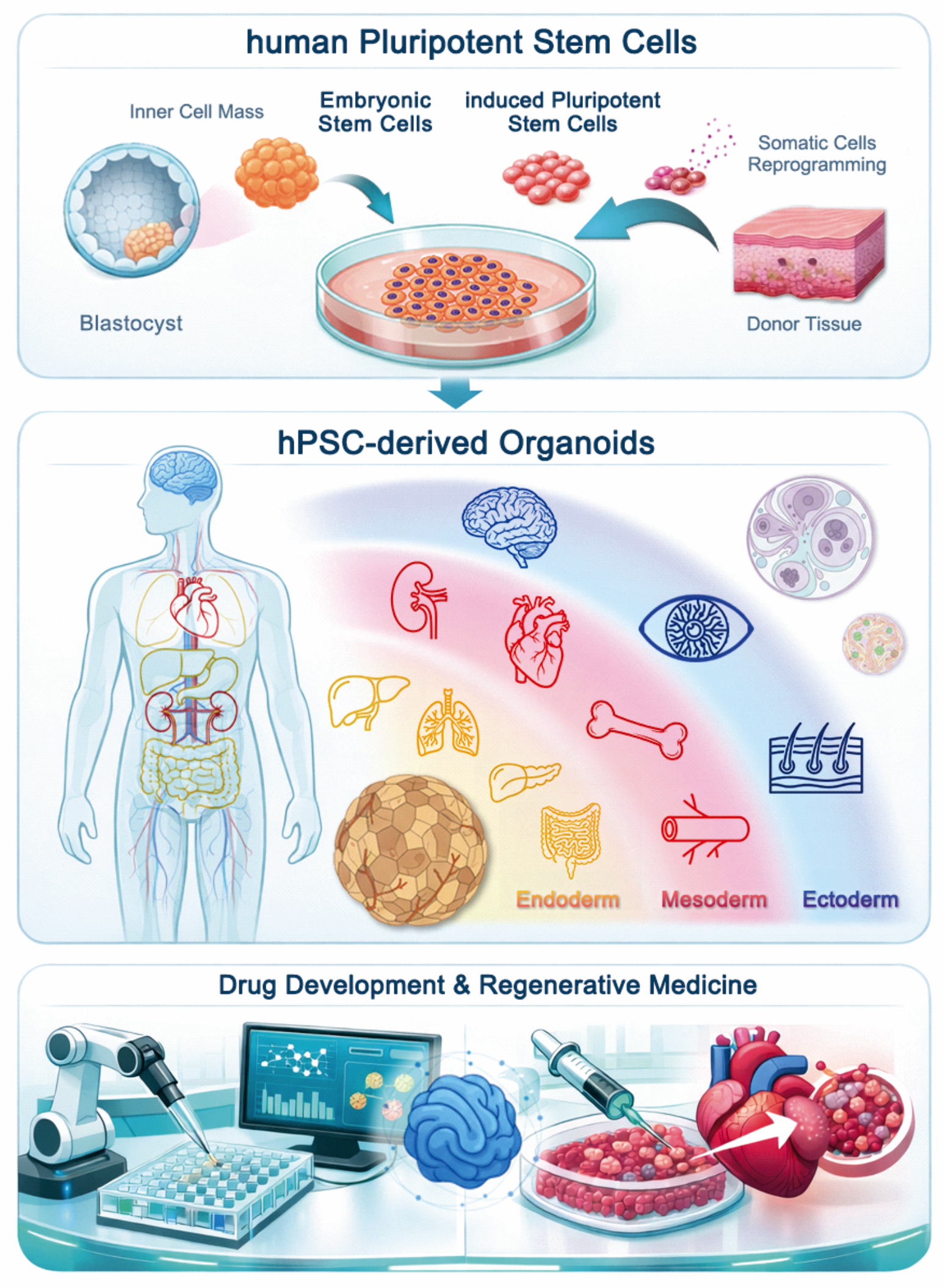
Organoids are three-dimensional (3D) in vitro cellular assemblies derived from tissue-resident stem or progenitor cells, embryonic stem cells (ESCs), or induced pluripotent stem cells (iPSCs). Through intrinsic self-organization, these systems can partially recapitulate the cellular composition and functional characteristics of the tissues from which they originate (1, 2). Unlike traditional two-dimensional monolayer cultures, 3D systems more faithfully reproduce aspects of the spatial organization and extracellular microenvironment found in vivo, supporting more physiologically relevant tissue structure, signaling interactions, and functional phenotypes (3). Reflecting the rapid growth and significance of this field, organoids were named Method of the Year 2017 by Nature Methods (4). Advances in stem cell biology, targeted differentiation strategies, and biomaterial-based 3D culture systems have enabled the creation of organoids from hPSCs. These developments reduce reliance on embryonic or adult tissue samples while allowing the in vitro reconstruction of key structural and functional features of human organs (1–4).
The translational potential of hPSC-derived organoids has expanded rapidly across many organ systems. By combining lineage specification with self-organizing developmental processes, hPSCs can produce organoid models representing all three germ layers. These include endoderm-derived organoids such as lung, liver, islet, and intestine (5–13); mesoderm-derived systems including bone marrow, vascular, and cardiac tissues (14–21); and ectoderm-derived models such as retina, brain, and skin (22–27). Overall, these platforms now support diverse applications, from developmental biology and disease modeling to therapeutic testing and regenerative research.
Instead of seeing organoids mainly as models specific to each organ, a new view is to consider them as modular experimental systems that can be customized along several translational axes, including biological accuracy, screening capacity, and engineering integration. In this review, we therefore analyze major hPSC-derived organoid systems using this design-focused framework, showing how different tissues demonstrate unique solutions to common translational challenges.
In this review, we examine hPSC-derived organoid systems within a translational framework that highlights three emerging roles of these models. Organoid systems have quickly progressed from proof-of-concept developmental models to widely used experimental platforms in translational biology (28, 29). Second, organoids are being investigated as regenerative building blocks, providing experimental systems for disease modeling, gene correction, and the development of cell-based therapies (30). Third, advances in bioengineering, including microfluidic platforms, organoid-on-chip technologies, vascularization methods, and quantitative functional readouts, are enhancing the reproducibility, scalability, and physiological relevance needed for translational applications (31). Collectively, these studies show that hPSC-derived organoids now serve as an intermediate experimental scale between simplified cell cultures and whole-organism physiology (Table 1) (Figure 1).
Table 1. Translational Applications of hPSC-Derived Organoids
| Organoid | Key Applications | Major Advantages | Current Limitations |
|---|---|---|---|
| Lung | infection modeling, inhalation toxicology | multicellular airway architecture | incomplete immune components |
| Liver | drug metabolism, hepatotoxicity | CYP450 activity | metabolic maturation |
| Islet | diabetes therapy | glucose-responsive β-cells | vascularization |
| Brain | neurodevelopmental disorders | human-specific neuronal programs | variability |
| Cardiac | cardiotoxicity screening | contractile physiology | immature cardiomyocytes |
| Skin | wound repair, grafting | epithelial-mesenchymal architecture | vascular integration |
| Intestinal | host-pathogen studies | crypt-villus structure | microbiome absence |
2. Lung Organoids
Lung organoids are a clear example of how hPSC-derived systems can bridge developmental biology with translational respiratory research (32–41). The design and assessment of these systems depend on their specific use: drug discovery focuses on scalability and consistency, while regenerative medicine emphasizes structural accuracy, functional development, and engraftment potential. hPSC-derived lung organoids have emerged as sophisticated 3D systems that recapitulate essential components of the human respiratory system, including airway and alveolar epithelial cells, and offer greater physiological relevance than 2D cultures or animal models for studying respiratory diseases and developing drugs. These different needs highlight a general concept in organoid engineering: the best level of biological complexity depends on the specific translational goal. From a translational perspective, lung organoids have been particularly informative in three settings: genetic disease modeling, infectious disease research, and inhalation toxicology, and they also support emerging regenerative applications.
Genetic Disease Modeling
One of the most well-established uses of lung organoids is modeling inherited respiratory disorders. Directed differentiation protocols allow the creation of both proximal and distal lung structures, making these systems versatile platforms for studying conditions such as cystic fibrosis (CF) and idiopathic pulmonary fibrosis (IPF) (31–33). In CF research, a key breakthrough was the development of functional organoid assays that measure CFTR activity. Forskolin-induced swelling assays have been used to evaluate CFTR modulators like Ivacaftor and Lumacaftor and to support personalized treatment testing (33, 34). Beyond profiling pharmacologic responses, CRISPR/Cas9 correction of CFTR mutations in hPSC lines, followed by organoid differentiation, has demonstrated the potential for ex vivo gene-correction modeling in the human respiratory system (35).
Infectious Disease Modeling
Lung organoids have become important platforms for studying respiratory viral infections in human tissue-like environments. During the COVID-19 pandemic, these systems were quickly adapted to model SARS-CoV-2 infection and antiviral responses in vitro, supporting both mechanistic research and therapeutic screening (36–38). Similar organoid models have also been utilized to examine influenza and respiratory syncytial virus, offering insights into host-pathogen interactions within multicellular epithelial environments that are challenging to replicate in standard monolayer systems (36–38). Because lung organoids preserve key epithelial lineages and tissue architecture, they provide a more human-relevant framework for investigating viral tropism, epithelial damage, and antiviral efficacy.
Inhalation Toxicology
A third major application of lung organoids in translational research is inhalation toxicology. The respiratory epithelium is directly exposed to cigarette smoke, particulate matter, and other airborne toxicants, yet conventional in vitro systems do not fully replicate the barrier organization and lineage-specific injury responses. Lung organoids enable inhalation-relevant injury and inflammation studies by allowing assessment of epithelial integrity, inflammatory signaling, and mucosal damage within a multilineage tissue environment (39). Their composition, including goblet, basal, and ciliated cells, also supports the evaluation of mucociliary dysfunction and differential susceptibility to lineage-specific effects following exposure to environmental stressors (39).
Applications of Regenerative Medicine and Bioengineering
In regenerative medicine, lung organoids are being studied as potential systems for tissue repair. Transplantation into injured mouse lungs has demonstrated partial engraftment and expression of alveolar markers, supporting the idea that organoid-derived cells might aid epithelial regeneration after injury (40). Meanwhile, bioengineering strategies such as 3D bioprinting and the integration of decellularized scaffolds are being explored to improve vascularization, mechanical stability, and structural support, thereby furthering the translational potential of lung organoid systems (41). Together, these studies show that lung organoids are not simply respiratory disease models but versatile translational platforms that support genetic disease research, infectious disease studies, toxicology, and regenerative medicine research.
3. Liver Organoids
Liver organoids are among the most advanced translational applications of hPSC-derived systems. Since the liver plays a key role in xenobiotic metabolism and systemic homeostasis, human-relevant in vitro liver models are especially valuable for drug discovery, safety pharmacology, and regenerative hepatology (42–53). hPSC-derived liver organoids mimic essential structural and functional features of hepatic tissue, including hepatocytes, cholangiocytes, and non-parenchymal cells, arranged within three-dimensional microenvironments that support metabolic activity and long-term culture stability (42). From a translational standpoint, liver organoids are mainly used in three areas: drug metabolism and hepatotoxicity testing, disease modeling, and regenerative liver therapy.
Drug Metabolism and Hepatotoxicity
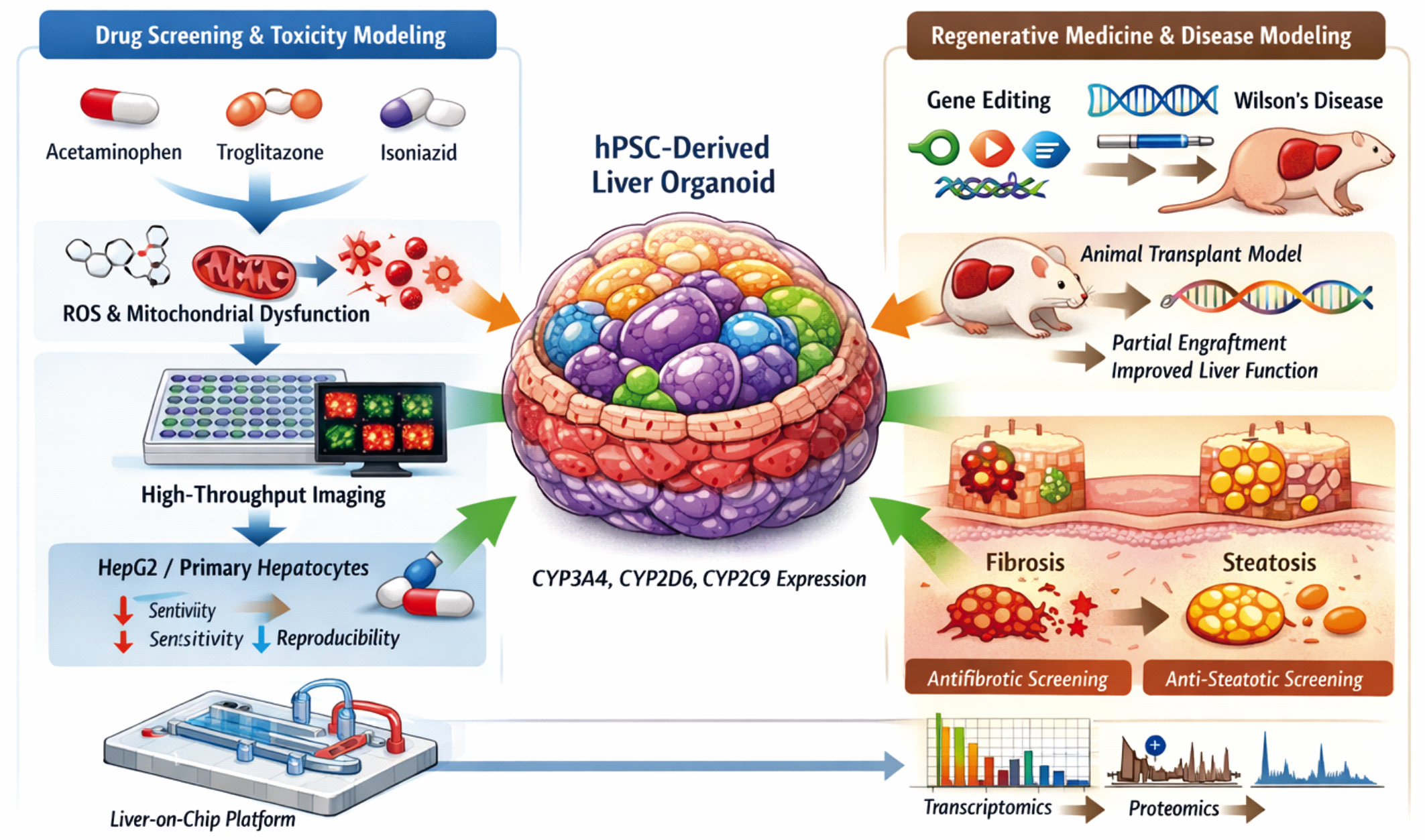
One of the most important applications of liver organoids is in assessing drug-induced liver injury (DILI), a leading cause of late-stage drug failure. Traditional hepatocyte cultures often rapidly lose their metabolic functions, while liver organoids retain the expression of cytochrome P450 enzymes and metabolic pathways crucial for pharmacokinetic studies (42, 43). When exposed to compounds such as acetaminophen, troglitazone, and isoniazid, liver organoids exhibit typical hepatotoxic effects, including mitochondrial dysfunction and oxidative stress (44). Imaging-based assays using uniformly sized liver spheroids have enabled high-throughput toxicity screening, demonstrating greater sensitivity than conventional HepG2 or primary hepatocyte models (44).
Advanced transcriptomic and proteomic analyses of organoids under toxic stress conditions have also provided mechanistic insights into drug responses. Importantly, the expression of key drug-metabolizing enzymes, including CYP3A4, CYP2D6, and CYP2C9, can be sustained during prolonged culture, increasing the predictive value of these models for pharmacological research (45, 46). The integration of liver organoids with microfluidic liver-on-chip platforms further improves physiological relevance by enabling controlled perfusion and dynamic metabolic evaluation (47, 48).
Disease Modeling
Liver organoids also serve as valuable platforms for modeling inherited metabolic disorders and chronic liver diseases. Patient-derived induced pluripotent stem cells (iPSCs) can be differentiated into organoids that retain disease-associated mutations, enabling mechanistic studies and therapeutic testing. For example, organoid models of α1-antitrypsin deficiency and Wilson’s disease have shown that CRISPR-mediated gene correction can restore normal protein expression and hepatic function in vitro (49, 50). Beyond genetic diseases, liver organoids are increasingly used to model acquired liver conditions. Exposure to transforming growth factor β (TGF-β) or free fatty acids can induce fibrotic and steatotic phenotypes, enabling mechanistic studies of liver fibrosis and metabolic liver disease and supporting antifibrotic drug screening (51, 52).
Regenerative Hepatology
In regenerative medicine, liver organoids are being explored as possible sources of transplantable tissue. Experimental transplantation studies in liver injury models have demonstrated partial engraftment of organoid-derived hepatocytes and improvements in liver function, emphasizing their potential as regenerative building blocks (51, 52). At the same time, efforts to standardize organoid manufacturing are advancing toward clinical application. Emerging guidelines highlight quality control criteria, including batch consistency, cellular composition, and functional validation, to support regulatory approval of organoid-based therapeutic products (53).
Together, these applications position liver organoids as a key experimental platform connecting pharmacology, disease modeling, and regenerative hepatology. As differentiation protocols and bioengineering tools evolve, liver organoids are expected to play a central role in precision hepatology (Figure 2).
4. Islet Organoids
Islet organoids demonstrate the dual translational potential of organoid technologies in diabetes research: enabling mechanistic studies of pancreatic endocrine biology while supporting the development of cell-based replacement therapies for glycemic control. hPSC-derived islet organoids replicate key features of pancreatic endocrine tissue, including insulin-secreting β-like cells and supporting endocrine populations organized into three-dimensional clusters that resemble native pancreatic islets (54). The translational uses of islet organoids generally fall into three main areas: endocrine disease modeling, drug discovery and metabolic pharmacology, and regenerative cell therapy.
Endocrine Disease Modeling
Early landmark studies showed that hPSC differentiation protocols could produce glucose-responsive β-like cells capable of secreting insulin in response to physiological glucose levels (55, 56). Later work has improved these strategies to create multilineage endocrine organoids with better maturation, including α-, β-, δ-, and other endocrine cell types that more closely resemble native human islets (57–60). These systems offer valuable experimental platforms for studying β-cell development, dysfunction, and metabolic regulation in diabetes.
Drug Discovery and Metabolic Pharmacology
Islet organoids have also enabled high-throughput pharmacologic screening for diabetes treatments. Organoid-based models have been utilized to identify compounds that restore β-cell function or prevent endocrine cell death. For instance, galunisertib was shown to promote β-cell survival in GLIS3 mutation-related diabetes models (61). Patient-specific iPSC-derived β-cells further facilitate the assessment of therapeutic responses in genetically defined settings, supporting precision medicine strategies. Drug responses to sulfonylureas, GLP-1 receptor agonists, and glucokinase activators have been tested in patient-derived organoid systems (62).
Emerging multi-organoid and organ-on-chip platforms further extend these capabilities by enabling investigation of systemic metabolic interactions. For example, integrated liver-islet organoid systems have been developed to study metabolic crosstalk and evaluate the effects of systemic therapies such as metformin (63, 64).
Regenerative Cell Therapy
Perhaps the most transformative application of islet organoids lies in regenerative medicine. Strategies aimed at immune evasion and improved engraftment have shown encouraging results in preclinical models. For instance, PD-L1 overexpression in engineered islet organoids enabled transplantation into immune-competent diabetic mice without immunosuppression while maintaining glucose homeostasis (65). Advances in chemical reprogramming have also accelerated progress toward clinical translation. The Deng group generated chemically induced pluripotent stem cells (CiPSCs) and demonstrated the functional efficacy of derived islet tissues in nonhuman primate diabetes models (66). Subsequent work introduced a transplantation strategy beneath the anterior rectus sheath of the abdomen, improving graft survival and enabling clinical translation through investigator-initiated trials for type 1 diabetes (67, 68). More recently, endocrine-subtype-complete islet constructs containing all major endocrine cell populations have been generated, demonstrating physiologic glucose regulation and protection from hypoglycemia through α-cell-mediated glucagon signaling (69). Together, these developments establish hPSC-derived islet organoids as promising experimental platforms that integrate metabolic disease modeling, pharmacologic testing, and regenerative cell therapy.
5. Bone Organoids
Bone organoids illustrate the growing use of hPSC-derived systems for musculoskeletal drug discovery, marrow toxicology, and regenerative skeletal engineering.
Skeletal Tissue Organization and Bone Marrow Niche Modeling
Bone organoids derived from hPSCs represent an emerging frontier in skeletal tissue engineering and drug discovery. These 3D self-organizing constructs mimic key features of bone physiology, including osteogenic lineage progression, ECM mineralization, and bone marrow niche characteristics (70, 71). Compared with conventional osteoblast monocultures, hPSC-derived bone organoids offer a more physiologically relevant model for studying human bone biology in vitro.
Drug Discovery and Marrow Toxicology
In drug screening, bone organoids provide unique advantages for testing bone-building or anti-resorption compounds. Organoid systems have been used to evaluate the effectiveness of bone morphogenetic proteins (BMPs), parathyroid hormone analogs, and Wnt signaling modulators, offering high-throughput platforms for osteoanabolic drug discovery (72, 73). Importantly, these models respond to known osteogenic triggers with increased alkaline phosphatase activity, RUNX2 expression, and matrix calcification, demonstrating their translational relevance (74, 75). Bone organoids are increasingly moving from proof-of-concept systems to scalable platforms for drug screening. Human bone marrow organoids can sustain patient-derived hematologic cancers while maintaining key stromal-hematopoietic interactions, enabling functional drug-sensitivity testing, therapy prioritization, and target discovery/validation in a physiologically relevant environment (76). Meanwhile, standardized protocols have been developed to generate multilineage bone marrow organoids from iPSCs, increasing reproducibility and throughput for efficacy and mechanistic screening.
For safety assessment, vascularized bone marrow-on-a-chip systems mimic clinically relevant features of marrow injury, including myelo-/erythro-toxicity and recovery patterns after exposure to chemotherapeutics or ionizing radiation (77). Related microfluidic bone marrow chips have also been used to predict hematopoietic risks of biologics and immunotherapeutics in preclinical development (78). Beyond marrow niche models, self-assembling human skeletal organoids, including “mini-joint” constructs, support disease modeling and tissue-level drug testing, offering a complementary approach for candidate validation against inflammation- and remodeling-related skeletal phenotypes (79). Recent research further consolidates these applications and provides technical roadmaps to advance bone organoids toward broader drug screening and translational applications (80, 81).
Regenerative Skeletal Engineering
Bone organoids are increasingly being developed as regenerative building blocks for skeletal repair, providing developmentally informed multicellular tissues that can mature and integrate in vivo. Notably, hPSC-derived cartilaginous organoids have been shown to promote scaffold-free healing of critical-size long-bone defects, supporting an endochondral-like route to bone regeneration (82). Meanwhile, advances in biofabrication are accelerating the development of practical applications. Scaffold-free, rapid creation of pre-mineralized bone organoids has been reported with improved repair in calvarial defect models, and the sequential development of vascularized and mineralized bone organoids has further enhanced cranial defect healing (83). Recent reviews summarize these advancements and highlight remaining challenges, including perfusable vascularization, mechanical strength, and scalable manufacturing for clinical use (80).
6. Vascular Organoids
Vascular organoids underscore the importance of engineering-enabled complexity, serving as platforms for vascular disease modeling, therapeutic testing, and the construction of more physiologically integrated organoid systems.
Vascular Development and Organoid Formation
Blood vessels are crucial for nearly all tissues, providing nutrients and oxygen, regulating hemostasis and inflammation, and supporting organ development and stem cell maintenance (84, 85). Therefore, recreating functional vasculature is vital for in vitro vascular biology (86). hPSC-derived vascular organoids create 3D self-organizing morphogenetic vascular structures that surpass the limitations of primary cell culture systems, offering dependable platforms for studying angiogenesis, disease modeling, and biomedical applications (87–89).
Vascular Disease Modeling
Pioneering work by Wimmer et al. established hPSC-derived vascular organoids that self-organize into basement membrane-enveloped capillary networks within 2-3 weeks, displaying morphological, functional, and molecular features of human microvasculature (90). After transplantation into immunocompromised mice, these organoids connected with the host circulation and developed into functional vascular networks. These organoids have also been utilized to model vascular disease. Exposure to hyperglycemia and inflammatory cytokines replicates diabetic vasculopathy phenotypes, revealing key signaling mechanisms and identifying potential therapeutic candidates (90). Additionally, iPSC-derived vascular organoids that retain patient-specific epigenetic information function as precision-medicine platforms for modeling vascular dysfunction and testing targeted interventions (91).
Bioengineering and Multi-Organoid Integration
Although many protocols rely on Matrigel to promote vessel sprouting, recent advances have introduced a quick five-day method for creating vascular organoids by orthogonally activating the transcription factors ETV2 and NKX3.1. This method enables controlled co-differentiation of endothelial and mural cells without ECM embedding, resulting in functional vessels capable of rescuing ischemic limbs and supporting islet grafts (92). Additionally, hPSC-derived vascular networks can serve as modular units that connect with other organoids to build tissue-vasculature integrated models (93–95). Alternatively, microfluidic platforms with vascularized organoids-on-chip enable dynamic perfusion and real-time quantitative evaluation of vascular function, supporting high-throughput drug screening in physiologically relevant settings (96, 97). As these technologies mature, vascular organoids are expected to play a central role in modeling vascular disease and enabling physiologically integrated organoid systems for translational research.
7. Brain organoids
Brain organoids offer human-relevant experimental systems for modeling neurodevelopment, neurological diseases, and drug responses in complex neural tissues. Created through directed differentiation of hPSC, these three-dimensional neural aggregates partially mimic early human brain development, including aspects of cortical organization, neural progenitor behavior, and neuronal network formation (98–100). From a translational perspective, brain organoids support three main research applications: modeling neurodevelopmental disorders, testing neuroactive compounds and neurotoxicity, and exploring regenerative strategies for neural repair.
Modeling of Neurodevelopmental and Neurological Diseases
Brain organoids have become essential tools for studying neurodevelopmental disorders that are difficult to model in animal systems. Early research showed that cerebral organoids could mimic key developmental features of the human cortex, such as progenitor zone organization and neuronal differentiation (100). These models have since been used to explore various neurological conditions, including microcephaly, autism spectrum disorders, and neurodegenerative diseases (101–103). Single-cell transcriptomic analyses have further uncovered the diversity of neuronal and glial populations produced within brain organoids, allowing comparison with fetal brain development and identifying human-specific developmental pathways (102). These abilities make brain organoids valuable systems for understanding the genetic basis of neurological diseases.
Drug Discovery and Neurotoxicity Evaluation
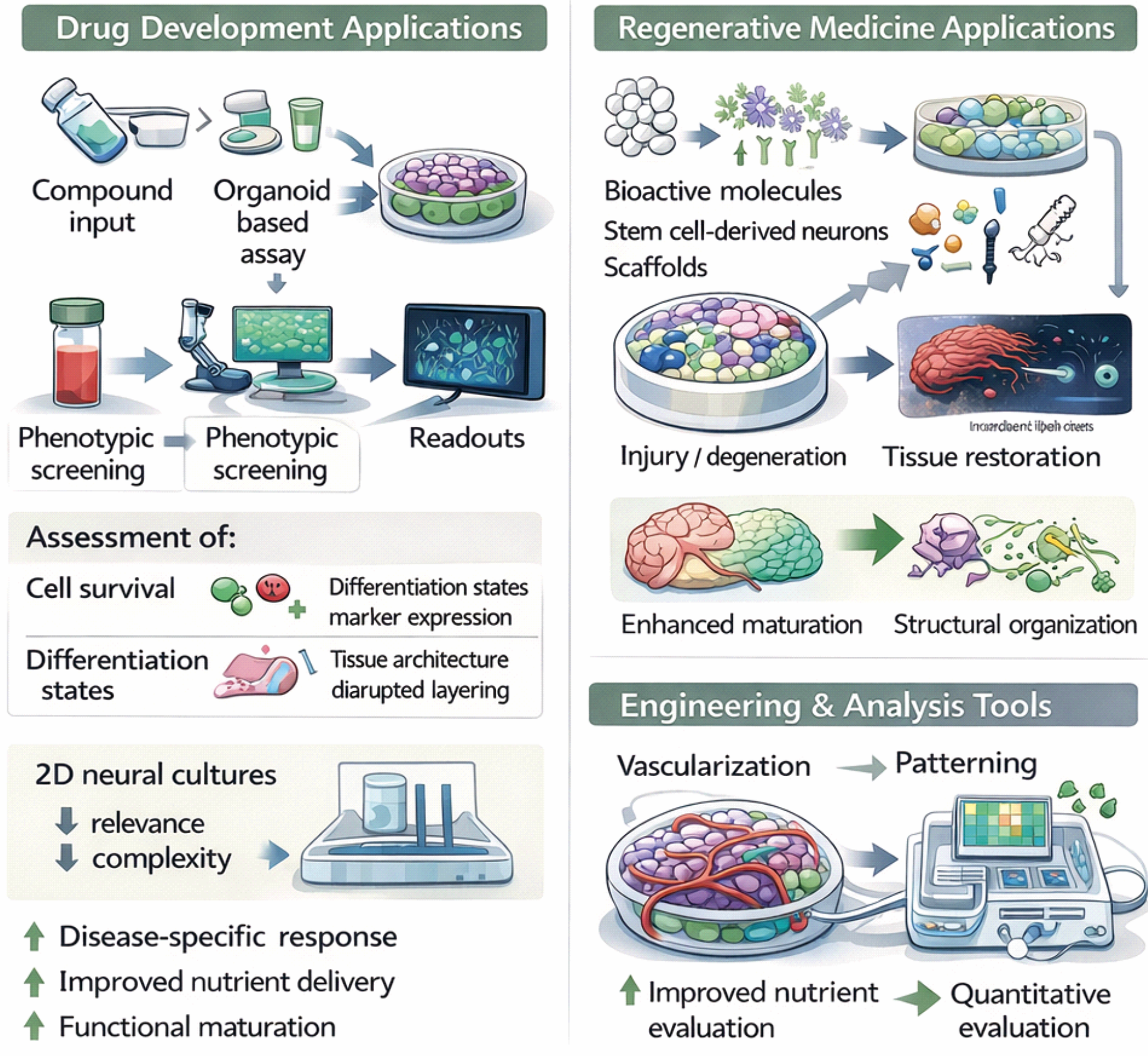
Brain organoids are increasingly used in pharmacological research. Unlike two-dimensional neural cultures, organoids enable assessment of compound effects on various cell types and tissue structures (104, 105). Disease-specific organoids derived from patient iPSCs offer platforms for testing therapeutic responses in genetically defined contexts. Crucially, organoid-based assays can detect human-specific adverse effects that are often missed in animal models. These systems have been employed to study drug-induced neuronal toxicity, neuroinflammatory responses, and vulnerabilities related to neurodegenerative disorders (106–108).
Regenerative and Engineering Applications
In addition to disease modeling and drug screening, brain organoids serve as experimental systems for studying neural regeneration and tissue repair. These models enable the assessment of stem cell-derived neural populations, biomaterial scaffolds, and bioactive molecules that support neuronal survival, differentiation, and circuit formation (109–111). Advances in engineering continuously enhance the physiological relevance of these models. Techniques such as vascularization, controlled patterning, and microfluidic integration improve nutrient delivery and reduce variability during long-term culture (112, 113). Incorporating multimodal functional readouts, including electrophysiology, calcium imaging, and high-content imaging, further allows for quantitative evaluation of neural activity and drug responses (114, 115). Despite these progresses, several limitations still exist. Brain organoids exhibit intercultural variability, limited maturation, and an incomplete representation of the in vivo microenvironment (116, 117).
Overcoming these challenges through improved differentiation protocols and bioengineering strategies will be crucial for fully unlocking the translational potential of organoid-based neural models. Continued efforts toward standardization and technological refinement are expected to further strengthen the role of brain organoids as translational platforms for drug screening and regenerative medicine (Figure 3).
8. Cardiac Organoids
Cardiac organoids are increasingly valuable as translational platforms for efficacy studies, predictive cardiotoxicity testing, and mechanistic investigation of cardiac repair strategies. hPSC-derived cardiac organoids are 3D myocardial-like tissues that reproduce key structural and functional attributes of cardiac tissue, including multicellular organization, contractile behavior, and electrophysiological activity in a tissue-relevant context. By extending beyond the constraints of 2D monolayer cultures, cardiac organoids provide an informative experimental system for cardiovascular drug development, safety pharmacology, and regenerative research (118, 119).
Drug Discovery and Pharmacology
In drug discovery, cardiac organoids enable efficacy testing under near-physiological conditions and facilitate screening of compounds that modulate cardiomyocyte function and tissue-level phenotypes with greater biological accuracy (120–122). Their organized cellular structure supports the evaluation of pharmacodynamic responses across various endpoints, including contractile performance, calcium handling, and electrophysiological parameters, and can be adapted to genetically defined or patient-specific contexts to enhance precision medicine efforts (121, 122). For safety assessment, cardiac organoids provide a manageable platform for studying both acute and chronic cardiotoxicity, including structural damage and functional irregularities. Organoid-based models have been used to mimic responses such as myocardial injury, fibrotic remodeling, and electrical conduction issues following drug exposure (123–126). Quantitative analysis of contractility, calcium transients, and arrhythmogenic potential further enhances predictive toxicology and offers mechanistic insights into liability factors (127, 128).
Regenerative Cardiology and Tissue Repair
In regenerative medicine research, cardiac organoids support the mechanistic investigation of human heart development and disease and serve as test beds for tissue repair strategies. They have been applied to model cardiac failure, cardiomyopathies, and congenital cardiac defects, enabling dissection of disease mechanisms and evaluation of therapeutic hypotheses (129–131). Integration with microfluidic platforms and multi-organ-on-chip systems further enhances their utility by enabling studies of systemic drug responses and inter-organ communication (132–134) (Figure 4).
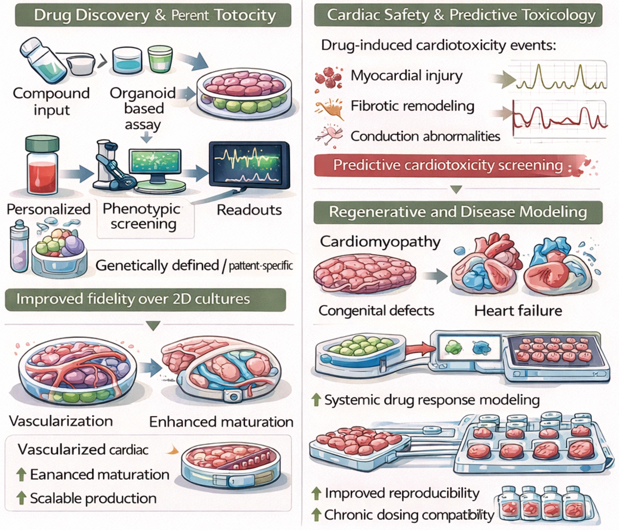
Improvements in cardiac organoid engineering, including enhanced maturation protocols, vascularization strategies, and scalable manufacturing workflows, are progressively increasing reproducibility and enabling longer-term culture, both of which are prerequisites for chronic dosing paradigms and translational toxicology studies.
9. Skin Organoids
Skin organoids demonstrate how hPSC-derived systems can integrate regenerative reconstruction with disease modeling and drug discovery in a tissue that is both experimentally accessible and clinically relevant.
Developmental Organization and Tissue Architecture
hPSC-derived skin organoids leverage the ability of ectodermal and mesenchymal progenitors to self-organize into 3D skin-like tissues. Sequential modulation of BMP, bFGF, and TGF-β signaling guides hPSCs toward surface ectoderm and neural crest-derived mesenchyme, mimicking early morphogenesis. This epithelial-mesenchymal interaction promotes epidermal polarization, stratification, and the formation of appendage progenitors. Unlike two-dimensional keratinocyte cultures, skin organoids create organized epithelial and mesenchymal layers that resemble fetal human skin (135).
Regenerative Dermatology and Wound Repair
Skin injuries from trauma, surgery, or burns impose significant physiological and psychological burdens (136). Healing occurs through hemostasis, inflammation, proliferation, and remodeling (137, 138), during which fibroblasts reorganize the ECM and facilitate collagen deposition and maturation (139). For wounds larger than 4 cm, implantation strategies are often required (140). However, artificial substitutes are limited by poor vascularization and integration, while autologous grafts are associated with donor-site morbidity and mismatch (141).
Skin organoids can model chronic wounds and serve as autologous graft sources for epithelial repair. Transplanted organoids integrate into host skin, guide hair follicle formation, and support growth (142). Human skin organoids containing follicles and glands form functional connections with the host epidermis, muscles, and nerves without rejection (143), and co-transplantation of epidermal and dermal layers further enhances wound closure (144, 145). iPSC-derived skin organoid technologies have recently been used in reconstructive strategies, including facial tissue reconstruction with improved graft integration and less scarring (146). For large defects, iPSC-derived constructs that resemble native skin demonstrate immune compatibility and improve re-epithelialization, barrier restoration, and hair regeneration.
Drug Discovery and Disease Modeling
Biomaterial innovations, such as 3D bioprinting, nanoparticles, and hydrogels, further expand therapeutic applications. iPSC-derived organoids secrete exosomes enriched in VEGF and regenerative microRNAs that enhance proliferation, differentiation, and angiogenesis (147). Because wound healing depends on precise regulation of cytokines and RNA (148), skin organoids provide valuable in vitro systems for dissecting these mechanisms and accelerating clinical translation (149). They replicate cell-cell and ECM interactions that are critical for regeneration (150).
Skin organoids also provide regenerative options for alopecia by generating complete hair follicles for transplantation. When combined with gelatin-based hydrogels, they accelerate wound healing in frostbite by reducing inflammation and promoting ECM remodeling via the integrin α5β1-FAK pathway, thereby decreasing scarring (151). iPSC-derived skin organoids function as advanced 3D systems for drug discovery, mimicking epidermal stratification and dermal-epidermal crosstalk to enhance drug efficacy and toxicity testing (152). Li et al. modeled Enterovirus A71 infection using these organoids, showing that EV-A71 causes epidermal injury via autophagy and disruption of the integrin/Hippo-YAP/TAZ pathway. The discovery of an autophagy-related target and a viral inhibitor highlights the translational potential of skin organoids for antiviral drug development (153).
10. Intestinal organoids
Intestinal organoids are among the most established translational organoid platforms, supporting studies of epithelial biology, pharmacologic response, host-pathogen interactions, and regenerative repair of intestinal injury.
Development and Epithelial Biology
hPSCs provide a renewable source for creating intestinal lineages. Advances in 3D culture techniques have facilitated the development of hPSC-derived intestinal organoids (HIOs) that replicate native intestinal structure and function (154, 155), thus bridging the gap between 2D cultures and animal models. iPSC-derived HIOs self-organize into epithelial structures that resemble crypt-villus architecture (156). HIOs are produced by guiding hPSCs through endoderm, midgut-hindgut, and intestinal developmental stages by modulating WNT, BMP, FGF, and Notch pathways (157). These organoids comprise enterocytes, goblet cells, Paneth cells, and enteroendocrine cells that perform absorptive and secretory roles (158, 159). Improvements in biomaterials, including synthetic hydrogels and engineered matrices, further enhance maturation and stability compared to Matrigel systems (160).
Disease modeling and pharmacologic testing
Traditional 2D systems poorly recapitulate intestinal complexity, whereas organoid models preserve native epithelial microenvironments and enhance translational relevance. The first hPSC differentiation protocol appeared in 2011 (161), followed by the generation of region-specific colonic organoids (162). Patient-derived FAP organoids carrying APC mutations have been used to model tumorigenesis and evaluate therapeutic strategies, including geneticin-mediated restoration of APC function (163). CFTRΔF508 HIOs similarly model cystic fibrosis, in which mutation correction restores organoid swelling responses, confirming their suitability for pharmacologic testing (164, 165).
Host-Pathogen Interactions and Antiviral Screening
During the COVID-19 pandemic, intestinal organoids proved useful for antiviral screening, as intestinal epithelial cells express ACE2 and TMPRSS2 (166). Krüger et al. showed that ramelteon inhibited viral replication, while Han et al. identified imatinib, mycophenolic acid, and quinacrine as inhibitors, establishing HIOs as biologically relevant platforms for antiviral drug testing (166, 167).
Regenerative Therapy for the Intestine
Enteroids, epithelial-only cultures derived from intestinal crypts, have shown potential to promote epithelial repair in animal models (168–170). A clinical trial (UMIN000013524) is currently evaluating autologous enteroid transplantation for the treatment of inflammatory bowel disease ulcers. Persistent ulcers caused by ischemia or radiation remain difficult to treat, highlighting the need for organoid-based regenerative approaches (171, 172). Experimental transplantation studies have demonstrated that fragmented HIOs can repair injured rodent intestines and integrate into the mucosal, muscular, and vascular layers (173). More recently, innervated HIOs containing enteric neurons have been generated, displaying peristaltic activity and stable engraftment, underscoring the importance of neuroepithelial integration for functional intestinal reconstruction (174).
11. Designing Organoid Platforms for Drug Discovery and Regenerative Medicine
Although hPSC-derived organoids are increasingly used across many fields of biomedical research, the optimal design of these systems heavily depends on their intended use. Specifically, the needs for organoids employed in drug discovery are very different from those required for regenerative medicine. Recognizing these differences is crucial for the thoughtful development of organoid platforms that are both scientifically reliable and clinically applicable.
In the context of drug discovery and safety pharmacology, organoid models must support reproducibility, scalability, and compatibility with quantitative screening workflows. For these applications, the primary goal is not necessarily to recreate the full structural complexity of native tissues but rather to capture key cellular functions and drug-response pathways in a format that can be reliably reproduced across experiments (175). Consequently, organoids used for pharmacological screening are often optimized for standardized culture conditions, uniform size, and compatibility with automated imaging or high-content analysis platforms. Such designs enable the evaluation of drug efficacy, mechanism of action, and toxicity in human-relevant biological systems while maintaining the throughput required for preclinical drug development. By contrast, regenerative medicine places greater emphasis on biological fidelity and functional maturity. Organoids intended for therapeutic applications must more closely resemble native tissues in their architecture, cellular diversity, and physiological function. In addition to structural complexity, these systems must demonstrate long-term stability, integration potential, and the capacity to survive and function following transplantation (176, 177). Achieving these properties often requires more sophisticated engineering strategies, including incorporating vascular networks, stromal cell support, immune components, or biomechanical cues to better replicate the in vivo microenvironment.
These contrasting demands emphasize an important principle in organoid engineering: the most complex model is not always the most useful. Instead, organoid systems should be designed based on their specific experimental or clinical context. For drug screening, simplified and highly standardized models may offer the most reliable and scalable platforms. In regenerative medicine, however, increased tissue complexity and functional maturation become crucial for achieving therapeutic relevance. Advances in bioengineering are beginning to close this gap between scalability and physiological fidelity. Approaches such as microfluidic organoid-on-chip systems, controlled biomaterial scaffolds, and vascularized organoid constructs are enabling better control over tissue architecture and environmental cues while maintaining experimental reproducibility (175–177). These developments suggest that the next generation of organoid platforms will increasingly combine biological self-organization with engineered control, allowing researchers to customize organoid complexity to meet specific translational goals. Taken together, these considerations highlight that hPSC-derived organoids should not be regarded as a single experimental format but rather as a versatile platform technology adaptable to various biomedical applications. Matching organoid design with the needs of drug discovery, toxicology, and regenerative medicine will be crucial for maximizing their translational potential.
Emerging Frontiers in Organoid Engineering
hPSC-derived organoids are evolving from proof-of-concept developmental models into increasingly sophisticated translational platforms. Several emerging technological and conceptual advances are likely to define the next generation of organoid systems and expand their impact in drug discovery, disease modeling, and regenerative medicine.
Immune and microenvironmental integration
Incorporating immune components such as macrophages, lymphocytes, and microglia into organoid cultures will enable modeling of inflammatory diseases, host-pathogen interactions, and immunotherapy responses in human tissue-like environments. This approach also enables investigation of dynamic immune-epithelial interactions and mechanisms of immune-mediated tissue remodeling or therapeutic resistance.
Vascularized and Multi-Organoid Platforms
Engineering perfusable vascular networks and integrating multiple organoids within microfluidic systems will allow investigation of systemic physiology, inter-organ communication, and pharmacokinetic responses across tissue interfaces. These systems further support controlled delivery of nutrients and drugs, improving physiological relevance and enabling more accurate modeling of tissue-specific exposure.
AI-Assisted Phenotyping and Quantitative Imaging
Advances in machine learning and high-content imaging are enabling automated phenotypic profiling of organoid systems. These approaches facilitate large-scale drug screening, multiparametric functional analysis, and improved reproducibility across laboratories. Importantly, AI-driven analyses can uncover subtle phenotypic patterns that are not detectable by conventional readouts, enhancing sensitivity in drug-response assessment.
Standardized and GMP-Compliant Organoid Manufacturing
Translating organoid technologies toward clinical applications will require standardized differentiation protocols, chemically defined matrices, automated production pipelines, and rigorous quality-control frameworks compatible with Good Manufacturing Practice (GMP). Establishing these standards will be essential for ensuring batch-to-batch consistency, regulatory approval, and clinical scalability.
Patient-Specific Precision Organoids
The integration of patient-derived iPSCs with genome editing and multi-omics profiling will enable personalized organoid models that capture individual genetic variation and therapeutic responses, supporting precision medicine strategies. Such models provide a platform for predicting patient-specific drug efficacy and toxicity, facilitating more tailored therapeutic decision-making.
Together, these advances are expected to transform organoid systems from experimental models into scalable platforms for predictive pharmacology, disease modeling, and regenerative therapeutics.
Discussion
Organoid technologies are revolutionizing preclinical research by creating experimental systems that mimic the complexity of human tissue while remaining compatible with scalable drug discovery processes. hPSC-derived organoids are emerging as advanced experimental platforms that integrate developmental biology, bioengineering, and translational medicine to enhance the predictive accuracy of preclinical studies. Overall, these organoids are transforming preclinical workflows by providing human-relevant assessments of efficacy, toxicity, and mechanisms across various tissues. Their value is maximized when combined with quantitative functional readouts, high-content imaging, and multi-omics profiling, enabling integrated decision-making that surpasses single-endpoint tests and increases confidence in target validation and translational efforts.
Key barriers to wider adoption still exist, including batch-to-batch variability, incomplete maturation, and limited integration of vasculature, immune components, innervation, and biomechanical cues. Overcoming these challenges requires better control of initial cell states, chemically defined and adjustable matrices, and engineering solutions such as microfluidic perfusion and precise morphogen delivery, along with standardized phenotyping panels and reference materials to compare performance across labs and platforms.
Looking ahead, scaling organoids for regulated applications will depend on comprehensive standardization, automated manufacturing processes, in-process quality checks, and fit-for-purpose acceptance criteria tailored to specific use cases (e.g., safety pharmacology versus patient-stratified efficacy testing). Integration with organ-on-chip and multi-organoid systems, combined with computational modeling and machine-learning-assisted image/omics analysis, is set to improve throughput and interpretability. Coupled with emerging consensus guidelines, these advancements should speed up regulatory approval and broaden the clinical use of organoid technologies in predictive pharmacology, toxicology, and regenerative medicine. This new approach, often called organoid-enabled translational modeling, marks a move toward human-relevant experimental platforms that connect reductionist cell-culture models to complex in vivo physiology. Future progress will probably rely on combining organoid biology with microfluidics, biomaterials engineering, and AI-driven analytics to develop more accurate human tissue models. hPSC-derived organoids are thus becoming a key experimental platform that links developmental biology, disease modeling, drug discovery, and regenerative medicine.
In this emerging framework, organoids are best seen not as miniature organs but as programmable biological systems whose structural complexity can be adjusted to address specific translational questions. As stem cell biology, bioengineering, and computational analytics come together, hPSC-derived organoids are set to become key platforms for the next generation of translational medicine.
References
1. Lancaster MA, Knoblich JA. Organogenesis in a dish: modeling development and disease using organoid technologies. Science. 2014;345(6194):1247125. https://doi.org/10.1126/science.1247125
2. Prior N, Inacio P, Huch M. Liver organoids: from basic research to therapeutic applications. Gut. 2019;68(12):2228–2237. https://doi.org/10.1136/gutjnl-2019-319256
3. Baker BM, Chen CS. Deconstructing the third dimension: how 3D culture microenvironments alter cellular cues. J Cell Sci. 2012;125(15):3015–3024. https://doi.org/10.1242/jcs.079509
4. Method of the Year 2017: Organoids. Nat Methods. 2018;15(1):1. https://doi.org/10.1038/nmeth.4575
5. Miller AJ, Dye BR, Ferrer-Torres D, Hill DR, Overeem AW, Shea LD, et al. Generation of lung organoids from human pluripotent stem cells in vitro. Nat Protoc. 2019;14(2):518–540. https://doi.org/10.1038/s41596-018-0104-8
6. Aurora M, Spence JR. hPSC-derived lung and intestinal organoids as models of human fetal tissue. Dev Biol. 2016;420(2):230–238. https://doi.org/10.1016/j.ydbio.2016.06.006
7. Han Y, Duan X, Yang L, Nilsson-Payant BE, Wang P, Duan F, et al. Identification of SARS-CoV-2 inhibitors using lung and colonic organoids. Nature. 2021;589(7841):270–275. https://doi.org/10.1038/s41586-020-2901-9
8. Guan Y, Xu D, Garfin PM, Ehmer U, Hurwitz M, Enns G, et al. Human hepatic organoids for the analysis of human genetic diseases. JCI Insight. 2017;2(17):e94954. https://doi.org/10.1172/jci.insight.94954
9. Jiang S, Xu F, Jin M, Wang Z, Xu X, Zhou Y, et al. Development of a high-throughput micropatterned agarose scaffold for consistent and reproducible hPSC-derived liver organoids. Biofabrication. 2023;15(1):015006. https://doi.org/10.1088/1758-5090/ac933c
10. Li MQ, Xu YP, Li K, Zhou C, Fan XX, Wang H, et al. Recapitulating dengue virus infection with human pluripotent stem cell-derived liver organoids for antiviral screening. Nat Commun. 2025;16(1):8069. https://doi.org/10.1038/s41467-025-63323-3
11. Pedraza-Arevalo S, Cujba AM, Alvarez-Fallas ME, Sancho R. Differentiation of beta-like cells from human induced pluripotent stem cell-derived pancreatic progenitor organoids. STAR Protoc. 2022;3(4):101656. https://doi.org/10.1016/j.xpro.2022.101656
12. Yang L, Han Y, Zhang T, Dong X, Ge J, Roy A, et al. Human vascularized macrophage-islet organoids to model immune-mediated pancreatic β cell pyroptosis upon viral infection. Cell Stem Cell. 2024;31(11):1612–1629.e8. https://doi.org/10.1016/j.stem.2024.08.007
13. Pitstick AL, Poling HM, Sundaram N, Lewis PL, Kechele DO, Sanchez JG, et al. Aggregation of cryopreserved mid-hindgut endoderm for more reliable and reproducible hPSC-derived small intestinal organoid generation. Stem Cell Reports. 2022;17(8):1889–1902. https://doi.org/10.1016/j.stemcr.2022.06.011
14. Vanslambrouck JM, Tan KS, Mah S, Little MH. Generation of proximal tubule-enhanced kidney organoids from human pluripotent stem cells. Nat Protoc. 2023;18(12):3229–3252. https://doi.org/10.1038/s41596-023-00880-1
15. Garreta E, Moya-Rull D, Marco A, Amato G, Ullate-Agote A, Tarantino C, et al. Natural hydrogels support kidney organoid generation and promote in vitro angiogenesis. Adv Mater. 2024;36(24):2400306. https://doi.org/10.1002/adma.202400306
16. Sander V, Przepiorski A, Hukriede NA, Davidson AJ. Large-scale production of kidney organoids from human pluripotent stem cells. Methods Mol Biol. 2023;2664:69–83. https://doi.org/10.1007/978-1-0716-3179-9_6
17. Frenz-Wiessner S, Fairley SD, Buser M, Goek I, Salewskij K, Jonsson G, et al. Generation of complex bone marrow organoids from human induced pluripotent stem cells. Nat Methods. 2024;21(5):868–881. https://doi.org/10.1038/s41592-024-02172-2
18. Werschler N, Penninger J. Generation of human blood vessel organoids from pluripotent stem cells. J Vis Exp. 2023;(190):64715. https://doi.org/10.3791/64715
19. Wimmer RA, Leopoldi A, Aichinger M, Kerjaschki D, Penninger JM. Generation of blood vessel organoids from human pluripotent stem cells. Nat Protoc. 2019;14(11):3082–3100. https://doi.org/10.1038/s41596-019-0213-z
20. Drakhlis L, Devadas SB, Zweigerdt R. Generation of heart-forming organoids from human pluripotent stem cells. Nat Protoc. 2021;16(12):5652–5672. https://doi.org/10.1038/s41596-021-00629-8
21. Venkateshappa R, Yildirim Z, Zhao SR, Wu MA, Vacante F, Abilez OJ, et al. Protocol to study electrophysiological properties of hPSC-derived 3D cardiac organoids using MEA and sharp electrode techniques. STAR Protoc. 2024;5(4):103406. https://doi.org/10.1016/j.xpro.2024.103406
22. Harkin J, Peña KH, Gomes C, Meyer JS. A highly reproducible and efficient method for retinal organoid differentiation from human pluripotent stem cells. Proc Natl Acad Sci U S A. 2024;121(25):e2317285121. https://doi.org/10.1073/pnas.2317285121
23. Sridhar A, Hoshino A, Finkbeiner CR, Chitsazan A, Dai L, Haugan AK, et al. Single-cell transcriptomic comparison of human fetal retina, hPSC-derived retinal organoids, and long-term retinal cultures. Cell Rep. 2020;30(5):1644–1659.e4. https://doi.org/10.1016/j.celrep.2020.01.007
24. Hergenreder E, Minotti AP, Zorina Y, Oberst P, Zhao Z, Munguba H, et al. Combined small-molecule treatment accelerates maturation of human pluripotent stem cell-derived neurons. Nat Biotechnol. 2024;42(11):1515–1525. https://doi.org/10.1038/s41587-023-02031-z
25. Walsh RM, Luongo R, Giacomelli E, Ciceri G, Rittenhouse C, Verrillo A, et al. Generation of human cerebral organoids with a structured outer subventricular zone. Cell Rep. 2024;43(4):114031. https://doi.org/10.1016/j.celrep.2024.114031
26. Xiang Y, Tanaka Y, Patterson B, Kang YJ, Govindaiah G, Roselaar N, et al. Fusion of regionally specified hPSC-derived organoids models human brain development and interneuron migration. Cell Stem Cell. 2017;21(3):383–398.e7. https://doi.org/10.1016/j.stem.2017.07.007
27. Lee J, Rabbani CC, Gao H, Steinhart MR, Woodruff BM, Pflum ZE, et al. Hair-bearing human skin generated entirely from pluripotent stem cells. Nature. 2020;582(7812):399–404. https://doi.org/10.1038/s41586-020-2352-3
28. Shafiee A, Sun J, Ahmed IA, Phua F, Rossi GR, Lin C-Y, et al. Development of physiologically relevant skin organoids from human induced pluripotent stem cells. Small. 2023;20(1):e2304879. https://doi.org/10.1002/smll.202304879
29. Vandana JJ, Manrique C, Lacko LA, Chen S. Human pluripotent stem cell-derived organoids for drug discovery and evaluation. Cell Stem Cell. 2023;30(5):571–591. https://doi.org/10.1016/j.stem.2023.04.011
30. Kim J, Koo BK, Knoblich JA. Human organoids: model systems for human biology and medicine. Nat Rev Mol Cell Biol. 2020;21(10):571–584. https://doi.org/10.1038/s41580-020-0259-3
31. Dye BR, Hill DR, Ferguson MAH, Tsai YH, Nagy MS, Dyal R, et al. In vitro generation of human pluripotent stem cell-derived lung organoids. eLife. 2015;4:e05098. https://doi.org/10.7554/elife.05098
32. Sachs N, Papaspyropoulos A, Zomer-van Ommen DD, Heo I, Böttinger L, Klay D, et al. Long-term expanding human airway organoids for disease modeling. EMBO J. 2019;38(4):e100300. https://doi.org/10.15252/embj.2018100300
33. Nikolić MZ, Caritg O, Jeng Q, Johnson JA, Sun D, Howell KJ, et al. Human embryonic lung epithelial tips are multipotent progenitors that can be expanded in vitro as long-term self-renewing organoids. eLife. 2017;6:e26575. https://doi.org/10.7554/elife.26575
34. Dekkers JF, Wiegerinck CL, de Jonge HR, Bronsveld I, Janssens HM, de Winter-de Groot KM, et al. A functional CFTR assay using primary cystic fibrosis intestinal organoids. Nat Med. 2013;19(7):939–945. https://doi.org/10.1038/nm.3201
35. Chen YW, Huang SX, Rodrigues Toste de Carvalho AL, Ho SH, Islam MN, Volpi S, et al. A three-dimensional model of human lung development and disease from pluripotent stem cells. Nat Cell Biol. 2017;19(5):542–549. https://doi.org/10.1038/ncb3510
36. Tindle C, Fuller M, Fonseca A, Taheri S, Ibeawuchi SR, Beutler N, et al. Adult stem cell-derived complete lung organoid models emulate lung disease in COVID-19. eLife. 2021;10:e66417. https://doi.org/10.7554/elife.66417
37. Youk J, Kim T, Evans KV, Jeong YI, Hur Y, Hong SP, et al. Three-dimensional human alveolar stem cell culture models reveal infection response to SARS-CoV-2. Cell Stem Cell. 2020;27(6):905–919.e10. https://doi.org/10.1016/j.stem.2020.10.004
38. Salahudeen AA, Choi SS, Rustagi A, Zhu J, van Unen V, de la O SM, et al. Progenitor identification and SARS-CoV-2 infection in human distal lung organoids. Nature. 2020;588(7839):670–675. https://doi.org/10.1038/s41586-020-3014-1
39. Wu Y, Li Y, Feng F, Chen H. Air pollutants and lung regeneration: impact on the fate of lung stem cells. Environ Int. 2025;199:109525. https://doi.org/10.1016/j.envint.2025.109525
40. Wang J, Li X, Chen H. Organoid models in lung regeneration and cancer. Cancer Lett. 2020;475:129–135. https://doi.org/10.1016/j.canlet.2020.01.030
41. Kim W, Lee Y, Kang D, Kwak T, Lee H-R, Jung S. 3D inkjet-bioprinted lung-on-a-chip. ACS Biomater Sci Eng. 2023;9(5):1295–1305. https://doi.org/10.1021/acsbiomaterials.3c00089
42. Kim H, Park HJ. Current hPSC-derived liver organoids for toxicity testing: Cytochrome P450 enzymes and drug metabolism. Toxicol Res. 2025;41(2):105–121. https://doi.org/10.1007/s43188-024-00275-8
43. Takebe T, Sekine K, Enomura M, Koike H, Kimura M, Ogaeri T, et al. Vascularized and functional human liver from an iPSC-derived organ bud transplant. Nature. 2013;499(7459):481–484. https://doi.org/10.1038/nature12271
44. Lee G, Kim H, Park JY, Kim G, Han J, Chung S, et al. Generation of uniform liver spheroids from human pluripotent stem cells for imaging-based drug toxicity analysis. Biomaterials. 2021;269:120529. https://doi.org/10.1016/j.biomaterials.2020.120529
45. Kim H, Im I, Jeon JS, Kang EH, Lee HA, Jo S, et al. Development of human pluripotent stem cell-derived hepatic organoids as an alternative model for drug safety assessment. Biomaterials. 2022;286:121575. https://doi.org/10.1016/j.biomaterials.2022.121575
46. Liu S, Wang Z, Zhu L, Wang L, Zhao T, Han P, et al. Generation and applications of an expandable and mature hiPSC-derived liver organoid. Acta Pharm Sin B. 2025;15(12):6382–6398. https://doi.org/10.1016/j.apsb.2025.09.029
47. Lucchetti M, Aina KO, Grandmougin L, Jäger C, Pérez Escriva P, Letellier E, et al. An organ-on-chip platform for simulating drug metabolism along the gut–liver axis. Adv Healthc Mater. 2024;13(20):e2303943. https://doi.org/10.1002/adhm.202303943
48. Theobald J, Ghanem A, Wallisch P, Banaeiyan AA, Andrade-Navarro MA, Taškova K, et al. Liver-kidney-on-chip to study toxicity of drug metabolites. ACS Biomater Sci Eng. 2018;4(1):188–199. https://doi.org/10.1021/acsbiomaterials.7b00417
49. Huch M, Dorrell C, Boj SF, van Es JH, Li VSW, van de Wetering M, et al. In vitro expansion of single Lgr5+ liver stem cells induced by Wnt-driven regeneration. Nature. 2013;494(7436):247–250. https://doi.org/10.1038/nature11826
50. Carolina E, Kuse Y, Okumura A, Aoshima K, Tadokoro T, Matsumoto S, et al. Generation of human iPSC-derived 3D bile duct within liver organoid by incorporating human iPSC-derived blood vessel. Nat Commun. 2024;15(1):7424. https://doi.org/10.1038/s41467-024-51487-3
51. Li Y, Yang X, Plummer R, Hayashi Y, Deng XS, Nie YZ, et al. Human pluripotent stem cell-derived hepatocyte-like cells and organoids for liver disease and therapy. Int J Mol Sci. 2021;22(19):10471. https://doi.org/10.3390/ijms221910471
52. Shin DS, Yang JY, Jeong HN, Mun SJ, Kim H, Son MJ, et al. Hepatotoxicity evaluation method through multiple-factor analysis using human pluripotent stem cell-derived hepatic organoids. Sci Rep. 2025;15(1):10804. https://doi.org/10.1038/s41598-025-95071-1
53. Moon HR, Mun SJ, Kim TH, Kim H, Kang D, Kim S, et al. Guidelines for manufacturing and application of organoids: liver. Int J Stem Cells. 2024;17(2):120–129. https://doi.org/10.15283/ijsc24044
54. Beydag-Tasoz BS, Yennek S, Grapin-Botton A. Towards a better understanding of diabetes mellitus using organoid models. Nat Rev Endocrinol. 2023;19(4):232–248. https://doi.org/10.1038/s41574-022-00797-x
55. Pagliuca FW, Millman JR, Gürtler M, Segel M, Van Dervort A, Ryu JH, et al. Generation of functional human pancreatic β cells in vitro. Cell. 2014;159(2):428–439. https://doi.org/10.1016/j.cell.2014.09.040
56. Rezania A, Bruin JE, Arora P, Rubin A, Batushansky I, Asadi A, et al. Reversal of diabetes with insulin-producing cells derived in vitro from human pluripotent stem cells. Nat Biotechnol. 2014;32(11):1121–1133. https://doi.org/10.1038/nbt.3033
57. Nair GG, Liu JS, Russ HA, Tran S, Saxton MS, Chen R, et al. Recapitulating endocrine cell clustering in culture promotes maturation of human stem-cell-derived β cells. Nat Cell Biol. 2019;21(2):263–274. https://doi.org/10.1038/s41556-018-0271-4
58. Veres A, Faust AL, Bushnell HL, Engquist EN, Kenty JH-R, Harb G, et al. Charting cellular identity during human in vitro β-cell differentiation. Nature. 2019;569(7756):368–373. https://doi.org/10.1038/s41586-019-1168-5
59. Balboa D, Barsby T, Lithovius V, Saarimäki-Vire J, Omar-Hmeadi M, Dyachok O, et al. Functional, metabolic and transcriptional maturation of human pancreatic islets derived from stem cells. Nat Biotechnol. 2022;40(7):1042–1055. https://doi.org/10.1038/s41587-022-01219-z
60. Augsornworawat P, Hogrebe NJ, Ishahak M, Schmidt MD, Marquez E, Maestas MM, et al. Single-nucleus multi-omics of human stem cell-derived islets identifies deficiencies in lineage specification. Nat Cell Biol. 2023;25(6):904–916. https://doi.org/10.1038/s41556-023-01150-8
61. Amin S, Cook B, Zhou T, Ghazizadeh Z, Lis R, Zhang T, et al. Discovery of a drug candidate for GLIS3-associated diabetes. Nat Commun. 2018;9(1):2681. https://doi.org/10.1038/s41467-018-04918-x
62. Millman JR, Xie C, Van Dervort A, Gürtler M, Pagliuca FW, Melton DA, et al. Generation of stem cell-derived β-cells from patients with type 1 diabetes. Nat Commun. 2016;7:11463. https://doi.org/10.1038/ncomms11463
63. Tsakmaki A, Fonseca Pedro P, Bewick GA. Diabetes through a 3D lens: organoid models. Diabetologia. 2020;63(6):1093–1102. https://doi.org/10.1007/s00125-020-05126-3
64. Tao T, Deng P, Wang Y, Zhang X, Guo Y, Chen W, et al. Microengineered multi-organoid system from hiPSCs to recapitulate human liver-islet axis in normal and type 2 diabetes. Adv Sci. 2022;9(5):e2103495. https://doi.org/10.1002/advs.202103495
65. Yoshihara E, O’Connor C, Gasser E, Wei Z, Oh TG, Tseng TW, et al. Immune-evasive human islet-like organoids ameliorate diabetes. Nature. 2020;586(7830):606–611. https://doi.org/10.1038/s41586-020-2631-z
66. Du Y, Liang Z, Wang S, Sun D, Wang X, Liew SY, et al. Human pluripotent stem-cell-derived islets ameliorate diabetes in non-human primates. Nat Med. 2022;28(2):272–282. https://doi.org/10.1038/s41591-021-01645-7
67. Liang Z, Sun D, Lu S, Lei Z, Wang S, Luo Z, et al. Implantation underneath the abdominal anterior rectus sheath enables effective and functional engraftment of stem-cell-derived islets. Nat Metab. 2023;5(1):29–40. https://doi.org/10.1038/s42255-022-00713-7
68. Wang S, Du Y, Zhang B, Meng G, Liu Z, Liew SY, et al. Transplantation of chemically induced pluripotent stem-cell-derived islets under abdominal anterior rectus sheath in a type 1 diabetes patient. Cell. 2024;187(22):6152–6164.e18. https://doi.org/10.1016/j.cell.2024.09.004
69. Meng G, Gu J, Liew SY, Cao J, Wang Z, Ma C, et al. Reconstruction of endocrine subtype-complete human pluripotent stem cell-derived islets with capacity for hypoglycemia protection in vivo. Cell Stem Cell. 2025;32(9):1438–1456.e7. https://doi.org/10.1016/j.stem.2025.07.006
70. Loh KM, Chen A, Koh PW, Deng TZ, Sinha R, Tsai JM, et al. Mapping the pairwise choices leading from pluripotency to human bone, heart, and other mesoderm cell types. Cell. 2016;166(2):451–467. https://doi.org/10.1016/j.cell.2016.06.011
71. Baccin C, Al-Sabah J, Velten L, Helbling PM, Grünschläger F, Hernández-Malmierca P, et al. Combined single-cell and spatial transcriptomics reveal the molecular, cellular and spatial bone marrow niche organization. Nat Cell Biol. 2020;22(1):38–48. https://doi.org/10.1038/s41556-019-0439-6
72. Olijnik A-A, Rodriguez-Romera A, Wong ZC, Shen Y, Reyat JS, Jooss NJ, et al. Generating human bone marrow organoids for disease modeling and drug discovery. Nat Protoc. 2024;19(7):2117–2146. https://doi.org/10.1038/s41596-024-00971-7
73. Panoskaltsis N, Mantalaris A. The promise of human bone marrow organoids for drug discovery and testing in myeloid and lymphoid cancers. Expert Opin Drug Discov. 2026;21(2):143–146. https://doi.org/10.1080/17460441.2025.2601109
74. Zamorano M, Aguilar-Gallardo C, Lugo A, Jimenez L, Farias JG, Mantalaris A. Engineering an integrated bioprocess to produce human dental pulp stem cell–alginate-based bone organoids. Int J Mol Sci. 2025;26(9):4348. https://doi.org/10.3390/ijms26094348
75. Negishi Y, Adili A, de Vega S, Momoeda M, Kaneko H, Cilek MZ, et al. IL-6 reduces spheroid sizes of osteophytic cells derived from osteoarthritis knee joint via induction of apoptosis. Am J Pathol. 2024;194(1):135–149. https://doi.org/10.1016/j.ajpath.2023.10.005
76. Khan AO, Rodriguez-Romera A, Reyat JS, Olijnik AA, Colombo M, Wang G, et al. Human bone marrow organoids for disease modeling, discovery, and validation of therapeutic targets in hematologic malignancies. Cancer Discov. 2023;13(2):364–385. https://doi.org/10.1158/2159-8290.cd-22-0199
77. Chou DB, Frismantas V, Milton Y, David R, Pop-Damkov P, Ferguson D, et al. On-chip recapitulation of clinical bone marrow toxicities and patient-specific pathophysiology. Nat Biomed Eng. 2020;4(4):606–611. https://doi.org/10.1038/s41551-019-0495-z
78. Koenig L, Juglair L, Tao TP, Fischer S, Clausen I, Imhof-Jung S, et al. A microfluidic bone marrow chip for the safety profiling of biologics in pre-clinical drug development. Commun Biol. 2025;8(1):754. https://doi.org/10.1038/s42003-025-08137-1
79. Abraham DM, Herman C, Witek L, Cronstein BN, Flores RL, Coelho PG. Self-assembling human skeletal organoids for disease modeling and drug testing. J Biomed Mater Res B Appl Biomater. 2021;109(12):2700–2710. https://doi.org/10.1002/jbm.b.34968
80. Hong Y, Li R, Sheng S, Zhou F, Bai L, Su J. Bone organoid construction and evolution. J Orthop Transl. 2025;53:260–273. https://doi.org/10.1016/j.jot.2025.06.011
81. Kong Y, Yang Y, Hou Y, Wang Y, Li W, Song Y. Advance in the application of organoids in bone diseases. Front Cell Dev Biol. 2024;12:1459891. https://doi.org/10.3389/fcell.2024.1459891
82. Tam WL, Mendes LF, Chen X, Lesage R, Van Hoven I, Leysen E, et al. Human pluripotent stem cell-derived cartilaginous organoids promote scaffold-free healing of critical size long bone defects. Stem Cell Res Ther. 2021;12(1):513. https://doi.org/10.1186/s13287-021-02580-7
83. Zhang L, Zhang Y, Wan Z, Yuan X, Gao Y, Song R, et al. Piezo1 mediated scaffold-free rapid generation of self-mineralized bone organoids via activating Wnt signaling. Mater Today Bio. 2026;36:102620. https://doi.org/10.1016/j.mtbio.2025.102620
84. Carmeliet P, Jain RK. Molecular mechanisms and clinical applications of angiogenesis. Nature. 2011;473(7347):298–307. https://doi.org/10.1038/nature10144
85. Bautch VL. Stem cells and the vasculature. Nat Med. 2011;17(11):1437–1443. https://doi.org/10.1038/nm.2539
86. Brunmaier LAE, Ozdemir T, Walker TW. Angiogenesis: biological mechanisms and in vitro models. Ann Biomed Eng. 2025;53(7):1543–1574. https://doi.org/10.1007/s10439-025-03721-2
87. Gao Q, Wang J, Zhang H, Wang J, Jing Y, Su J. Organoid vascularization: strategies and applications. Adv Healthc Mater. 2025;14(20):e2500301. https://doi.org/10.1002/adhm.202500301
88. Naderi-Meshkin H, Cornelius VA, Eleftheriadou M, Potel KN, Setyaningsih WA, Margariti A. Vascular organoids: unveiling advantages, applications, challenges, and disease modelling strategies. Stem Cell Res Ther. 2023;14(1):292. https://doi.org/10.1186/s13287-023-03521-2
89. Rossi G, Manfrin A, Lutolf MP. Progress and potential in organoid research. Nat Rev Genet. 2018;19(11):671–687. https://doi.org/10.1038/s41576-018-0051-9
90. Wimmer RA, Leopoldi A, Aichinger M, Wick N, Hantusch B, Novatchkova M, et al. Human blood vessel organoids as a model of diabetic vasculopathy. Nature. 2019;565(7740):505–510. https://doi.org/10.1038/s41586-018-0858-8
91. Liu C, Niu K, Xiao Q. Updated perspectives on vascular cell specification and pluripotent stem cell-derived vascular organoids for studying vasculopathies. Cardiovasc Res. 2022;118(1):97–114. https://doi.org/10.1093/cvr/cvaa313
92. Gong L, Zhang Y, Zhu Y, Lee U, Luo AC, Li X, et al. Rapid generation of functional vascular organoids via simultaneous transcription factor activation of endothelial and mural lineages. Cell Stem Cell. 2025;32(8):1200–1217.e6. https://doi.org/10.1016/j.stem.2025.05.014
93. Cakir B, Xiang Y, Tanaka Y, Kural MH, Parent M, Kang YJ, et al. Engineering of human brain organoids with a functional vascular-like system. Nat Methods. 2019;16(11):1169–1175. https://doi.org/10.1038/s41592-019-0586-5
94. Shao Y, Fu J. Engineering multiscale structural orders for high-fidelity embryoids and organoids. Cell Stem Cell. 2022;29(5):722–743. https://doi.org/10.1016/j.stem.2022.04.003
95. Jun Y, Nguyen-Ngoc KV, Sai S, Bender RHF, Gong W, Kravets V, et al. Engineered vasculature induces functional maturation of pluripotent stem cell-derived islet organoids. Dev Cell. 2025;60(18):2455–2469.e7. https://doi.org/10.1016/j.devcel.2025.04.024
96. Quintard C, Tubbs E, Jonsson G, Jiao J, Wang J, Werschler N, et al. A microfluidic platform integrating functional vascularized organoids-on-chip. Nat Commun. 2024;15(1):1452. https://doi.org/10.1038/s41467-024-45710-4
97. Salmon I, Grebenyuk S, Abdel Fattah AR, Rustandi G, Pilkington T, Verfaillie C, et al. Engineering neurovascular organoids with 3D printed microfluidic chips. Lab Chip. 2022;22(8):1615–1629. https://doi.org/10.1039/d1lc00535a
98. Jeong E, Choi S, Cho SW. Recent advances in brain organoid technology for human brain research. ACS Appl Mater Interfaces. 2022;15(1):200–219. https://doi.org/10.1021/acsami.2c17467
99. Trujillo CA, Muotri AR. Brain organoids and the study of neurodevelopment. Trends Mol Med. 2018;24(12):982–990. https://doi.org/10.1016/j.molmed.2018.09.005
100. Lancaster MA, Renner M, Martin CA, Wenzel D, Bicknell LS, Hurles ME, et al. Cerebral organoids model human brain development and microcephaly. Nature. 2013;501(7467):373–379. https://doi.org/10.1038/nature12517
101. Li C, Fleck JS, Martins-Costa C, Burkard TR, Themann J, Stuempflen M, et al. Single-cell brain organoid screening identifies developmental defects in autism. Nature. 2023;621(7978):373–380. https://doi.org/10.1038/s41586-023-06473-y
102. Kanton S, Boyle MJ, He Z, Santel M, Weigert A, Sanchís-Calleja F, et al. Organoid single-cell genomic atlas uncovers human-specific features of brain development. Nature. 2019;574(7778):418–422. https://doi.org/10.1038/s41586-019-1654-9
103. Qian X, Nguyen HN, Song MM, Hadiono C, Ogden SC, Hammack C, et al. Brain-region-specific organoids using mini-bioreactors for modeling ZIKV exposure. Cell. 2016;165(5):1238–1254. https://doi.org/10.1016/j.cell.2016.04.032
104. Lancaster MA, Knoblich JA. Generation of cerebral organoids from human pluripotent stem cells. Nat Protoc. 2014;9(10):2329–2340. https://doi.org/10.1038/nprot.2014.158
105. Yoon SJ, Elahi LS, Pașca AM, Marton RM, Gordon A, Revah O, et al. Reliability of human cortical organoid generation. Nat Methods. 2019;16(1):75–78. https://doi.org/10.1038/s41592-018-0255-0
106. Szebényi K, Wenger LMD, Sun Y, Dunn AWE, Limegrover CA, Gibbons GM, et al. Human ALS/FTD brain organoid slice cultures display distinct early astrocyte and targetable neuronal pathology. Nat Neurosci. 2021;24(11):1542–1554. https://doi.org/10.1038/s41593-021-00923-4
107. Pranty AI, Shumka S, Adjaye J. Bilirubin-induced neurological damage: current and emerging iPSC-derived brain organoid models. Cells. 2022;11(17):2647. https://doi.org/10.3390/cells11172647
108. Chen X, Sun G, Tian E, Zhang M, Davtyan H, Beach TG, et al. Modeling sporadic Alzheimer's disease in human brain organoids under serum exposure. Adv Sci. 2021;8(15):e2100371. https://doi.org/10.1002/advs.202101462
109. Mansour AA, Gonçalves JT, Bloyd CW, Li H, Fernandes S, Quang D, et al. An in vivo model of functional and vascularized human brain organoids. Nat Biotechnol. 2018;36(5):432–441. https://doi.org/10.1038/nbt.4127
110. Giandomenico SL, Sutcliffe M, Lancaster MA. Generation and long-term culture of advanced cerebral organoids for studying later stages of neural development. Nat Protoc. 2020;16(2):579–602. https://doi.org/10.1038/s41596-020-00433-w
111. Pașca AM, Sloan SA, Clarke LE, Tian Y, Makinson CD, Huber N, et al. Functional cortical neurons and astrocytes from human pluripotent stem cells in 3D culture. Nat Methods. 2015;12(7):671–678. https://doi.org/10.1038/nmeth.3415
112. Pham MT, Pollock KM, Rose MD, Cary WA, Stewart HR, Zhou P, et al. Generation of human vascularized brain organoids. Neuroreport. 2018;29(7):588–593. https://doi.org/10.1097/wnr.0000000000001014
113. Kirwan P, Turner-Bridger B, Peter M, Momoh A, Arambepola D, Robinson HPC, et al. Development and function of human cerebral cortex neural networks from pluripotent stem cells in vitro. Development. 2015;142(18):3178–3187. https://doi.org/10.1242/dev.123851
114. Trujillo CA, Gao R, Negraes PD, Gu J, Buchanan J, Preissl S, et al. Complex oscillatory waves emerging from cortical organoids model early human brain network development. Cell Stem Cell. 2019;25(4):558–569.e7. https://doi.org/10.1016/j.stem.2019.08.002
115. Quadrato G, Nguyen T, Macosko EZ, Sherwood JL, Yang SM, Berger DR, et al. Cell diversity and network dynamics in photosensitive human brain organoids. Nature. 2017;545(7652):48–53. https://doi.org/10.1038/nature22047
116. Birtele M, Lancaster M, Quadrato G. Modelling human brain development and disease with organoids. Nat Rev Mol Cell Biol. 2024;26(5):389–412. https://doi.org/10.1038/s41580-024-00804-1
117. Wu H, Feng E, Yin H, Zhang Y, Chen G, Zhu B, et al. Biomaterials for neuroengineering: applications and challenges. Regen Biomater. 2025;12:rbae137. https://doi.org/10.1093/rb/rbae137
118. Martin M, Gähwiler EKN, Generali M, Hoerstrup SP, Emmert MY. Advances in 3D organoid models for stem cell-based cardiac regeneration. Int J Mol Sci. 2023;24(6):5188. https://doi.org/10.3390/ijms24065188
119. Hyams NA, Kerr CM, Arhontoulis DC, Ruddy JM, Mei Y. Improving human cardiac organoid design using transcriptomics. Sci Rep. 2024;14(1):20147. https://doi.org/10.1038/s41598-024-61554-w
120. Mills RJ, Parker BL, Quaife-Ryan GA, Voges HK, Needham EJ, Bornot A, et al. Drug screening in human PSC-cardiac organoids identifies pro-proliferative compounds acting via the mevalonate pathway. Cell Stem Cell. 2019;24(6):895–907.e6. https://doi.org/10.1016/j.stem.2019.03.009
121. Giacomelli E, Meraviglia V, Campostrini G, Cochrane A, Cao X, van Helden RWJ, et al. Human-iPSC-derived cardiac stromal cells enhance maturation in 3D cardiac microtissues and reveal non-cardiomyocyte contributions to heart disease. Cell Stem Cell. 2020;26(6):862–879.e11. https://doi.org/10.1016/j.stem.2020.05.004
122. Filippo Buono M, von Boehmer L, Strang J, Hoerstrup SP, Emmert MY, Nugraha B. Human cardiac organoids for modeling genetic cardiomyopathy. Cells. 2020;9(7):1733. https://doi.org/10.3390/cells9071733
123. Richards DJ, Li Y, Kerr CM, Yao J, Beeson GC, Coyle RC, et al. Human cardiac organoids for the modelling of myocardial infarction and drug cardiotoxicity. Nat Biomed Eng. 2020;4(4):446–462. https://doi.org/10.1038/s41551-020-0539-4
124. Beck TC, Arhontoulis DC, Morningstar JE, Hyams N, Stoddard A, Springs K, et al. Cellular and molecular mechanisms of MEK1 inhibitor–induced cardiotoxicity. JACC CardioOncol. 2022;4(4):535–548. https://doi.org/10.1016/j.jaccao.2022.07.009
125. Hoang P, Kowalczewski A, Sun S, Winston TS, Archilla AM, Lemus SM, et al. Engineering spatial-organized cardiac organoids for developmental toxicity testing. Stem Cell Reports. 2021;16(5):1228–1244. https://doi.org/10.1016/j.stemcr.2021.03.013
126. Chen X, Lu N, Huang S, Zhang Y, Liu Z, Wang X, et al. Assessment of doxorubicin toxicity using human cardiac organoids: a novel model for evaluating drug cardiotoxicity. Chem Biol Interact. 2023;386:110777. https://doi.org/10.1016/j.cbi.2023.110777
127. Zhao D, Lei W, Hu S. Cardiac organoid - a promising perspective of preclinical model. Stem Cell Res Ther. 2021;12(1):272. https://doi.org/10.1186/s13287-021-02340-7
128. Ronaldson-Bouchard K, Ma SP, Yeager K, Chen T, Song L, Sirabella D, et al. Advanced maturation of human cardiac tissue grown from pluripotent stem cells. Nature. 2018;556(7700):239–243. https://doi.org/10.1038/s41586-018-0016-3
129. Tiburcy M, Hudson JE, Balfanz P, Schlick S, Meyer T, Liao MC, et al. Defined engineered human myocardium with advanced maturation for applications in heart failure modeling and repair. Circulation. 2017;135(19):1832–1847. https://doi.org/10.1161/circulationaha.116.024145
130. Yang KC, Breitbart A, De Lange WJ, Hofsteen P, Futakuchi-Tsuchida A, Xu J, et al. Novel adult-onset systolic cardiomyopathy due to MYH7 E848G mutation in patient-derived induced pluripotent stem cells. JACC Basic Transl Sci. 2018;3(6):728–740. https://doi.org/10.1016/j.jacbts.2018.08.008
131. Voges HK, Mills RJ, Elliott DA, Parton RG, Porrello ER, Hudson JE, et al. Development of a human cardiac organoid injury model reveals innate regenerative potential. Development. 2017;144(6):1118–1127. https://doi.org/10.1242/dev.143966
132. Oleaga C, Riu A, Rothemund S, Lavado A, McAleer CW, Long CJ, et al. Investigation of the effect of hepatic metabolism on off-target cardiotoxicity in a multi-organ human-on-a-chip system. Biomaterials. 2018;182:176–190. https://doi.org/10.1016/j.biomaterials.2018.07.062
133. Lee J, Mehrotra S, Zare-Eelanjegh E, Rodrigues RO, Akbarinejad A, Ge D, et al. A heart-breast cancer-on-a-chip platform for disease modeling and monitoring of cardiotoxicity induced by cancer chemotherapy. Small. 2021;17(15):2004258. https://doi.org/10.1002/smll.202004258
134. Yin F, Zhang X, Wang L, Wang Y, Zhu Y, Li Z, et al. HiPSC-derived multi-organoids-on-chip system for safety assessment of antidepressant drugs. Lab Chip. 2021;21(3):3361–3373. https://doi.org/10.1039/d0lc00921k
135. Lee J, van der Valk WH, Serdy SA, Deakin C, Kim J, Le AP, et al. Generation and characterization of hair-bearing skin organoids from human pluripotent stem cells. Nat Protoc. 2022;17(5):1266–1305. https://doi.org/10.1038/s41596-022-00681-y
136. Yang R, Liu F, Wang J, Chen X, Xie J, Xiong K. Epidermal stem cells in wound healing and their clinical applications. Stem Cell Res Ther. 2019;10(1):229. https://doi.org/10.1186/s13287-019-1312-z
137. Gurtner GC, Werner S, Barrandon Y, Longaker MT. Wound repair and regeneration. Nature. 2008;453(7193):314–321. https://doi.org/10.1038/nature07039
138. Sorg H, Tilkorn DJ, Hager S, Hauser J, Mirastschijski U. Skin wound healing: an update on the current knowledge and concepts. Eur Surg Res. 2017;58(1-2):81–94. https://doi.org/10.1159/000454919
139. Harding KG, Morris HL, Patel GK. Science, medicine and the future: healing chronic wounds. BMJ. 2002;324(7330):160–163. https://doi.org/10.1136/bmj.324.7330.160
140. Herndon DN, Barrow RE, Rutan RL, Rutan TC, Desai MH, Abston S, et al. A comparison of conservative versus early excision therapies in severely burned patients. Ann Surg. 1989;209(5):547–553. https://doi.org/10.1097/00000658-198905000-00006
141. Wu H, Wang G, Shang Y, Liu N, Zhen Y, Chen L, et al. Organoids and their research progress in plastic and reconstructive surgery. Aesthetic Plast Surg. 2023;47(2):880–891. https://doi.org/10.1007/s00266-022-03129-6
142. Brohem CA, da Silva Cardeal LB, Tiago M, Soengas MS, de Moraes Barros SB, Maria-Engler SS. Artificial skin in perspective: concepts and applications. Pigment Cell Melanoma Res. 2010;23(5):539–549. https://doi.org/10.1111/j.1755-148X.2010.00786.x
143. Takagi R, Ishimaru J, Sugawara A, Toyoshima KE, Ishida K, Ogawa M, et al. Bioengineering a 3D integumentary organ system from iPS cells using an in vivo transplantation model. Sci Adv. 2016;2(4):e1500887. https://doi.org/10.1126/sciadv.1500887
144. Lei M, Schumacher LJ, Lai YC, Juan WT, Yeh CY, Wu P, et al. Self-organization process in newborn skin organoid formation inspires strategy to restore hair regeneration of adult cells. Proc Natl Acad Sci U S A. 2017;114(34):E7101–E7110. https://doi.org/10.1073/pnas.1700475114
145. Lee J, Koehler KR. Skin organoids: a new human model for developmental and translational research. Exp Dermatol. 2021;30(4):613–620. https://doi.org/10.1111/exd.14292
146. Ebner-Peking P, Krisch L, Wolf M, Hochmann S, Hoog A, Vári B, et al. Self-assembly of differentiated progenitor cells facilitates spheroid human skin organoid formation and planar skin regeneration. Theranostics. 2021;11(17):8430–8447. https://doi.org/10.7150/thno.59661
147. Breitkreutz D, Koxholt I, Thiemann K, Nischt R. Skin basement membrane: the foundation of epidermal integrity—BM functions and diverse roles of bridging molecules nidogen and perlecan. Biomed Res Int. 2013;2013:179784. https://doi.org/10.1155/2013/179784
148. Tan SH, Ngo ZH, Sci DB, Leavesley D, Liang K. Recent advances in the design of three-dimensional and bioprinted scaffolds for full-thickness wound healing. Tissue Eng Part B Rev. 2022;28(1):160–181. https://doi.org/10.1089/ten.teb.2020.0339
149. Ntshingila S, Khumalo NP, Engel M, Arowolo AT. An appraisal of laboratory models of androgenetic alopecia: a systematic review. Skin Health Dis. 2021;1(2):e15. https://doi.org/10.1002/ski2.15
150. de Groot SC, Ulrich MMW, Gho CG, Huisman MA. Back to the future: from appendage development toward future human hair follicle neogenesis. Front Cell Dev Biol. 2021;9:661787. https://doi.org/10.3389/fcell.2021.661787
151. Wang W, Liu P, Zhu W, Li T, Wang Y, Wang Y, et al. Skin organoid transplantation promotes tissue repair with scarless healing in frostbite. Protein Cell. 2025;16(4):239–258. https://doi.org/10.1093/procel/pwae055
152. Xie X, Tong X, Li Z, Cheng Q, Wang X, Long Y, et al. Use of mouse primary epidermal organoids for USA300 infection modeling and drug screening. Cell Death Dis. 2023;14(1):15. https://doi.org/10.1038/s41419-022-05525-x
153. Li J, Ma J, Cao R, Zhang Q, Li M, Wang W, et al. A skin organoid-based infection platform identifies an inhibitor specific for HFMD. Nat Commun. 2025;16(1):2513. https://doi.org/10.1038/s41467-025-57610-2
154. Chen KG, Mallon BS, McKay RDG, Robey PG. Human pluripotent stem cell culture: considerations for maintenance, expansion, and therapeutics. Cell Stem Cell. 2014;14(1):13–26. https://doi.org/10.1016/j.stem.2013.12.005
155. Dedhia PH, Bertaux-Skeirik N, Zavros Y, Spence JR. Organoid models of human gastrointestinal development and disease. Gastroenterology. 2016;150(5):1098–1112. https://doi.org/10.1053/j.gastro.2015.12.042
156. Durczak PM, Fair KL, Jinks N, Cuevas Ocaña S, Sainz Zuñiga CB, Hannan NRF. Generation of hiPSC-derived intestinal organoids for developmental and disease modelling applications. J Vis Exp. 2024;(205):e61199. https://doi.org/10.3791/61199
157. McCracken KW, Catá EM, Crawford CM, Sinagoga KL, Schumacher M, Rockich BE, et al. Modelling human development and disease in pluripotent stem-cell-derived gastric organoids. Nature. 2014;516(7531):400–404. https://doi.org/10.1038/nature13863
158. Tsuruta S, Uchida H, Akutsu H. Intestinal organoids generated from human pluripotent stem cells. JMA J. 2019;2(1):3. https://doi.org/10.31662/jmaj.2019-0027
159. Xu X, Zhang Y, Huang G, Perekatt A, Wang Y, Chen L. Advances and applications of gut organoids: modeling intestinal diseases and therapeutic development. Life Med. 2025;4(2):lnaf012. https://doi.org/10.1093/lifemedi/lnaf012
160. Jeon EY, Sorrells L, Abaci HE. Biomaterials and bioengineering to guide tissue morphogenesis in epithelial organoids. Front Bioeng Biotechnol. 2022;10:1038277. https://doi.org/10.3389/fbioe.2022.1038277
161. Spence JR, Mayhew CN, Rankin SA, Kuhar MF, Vallance JE, Tolle K, et al. Directed differentiation of human pluripotent stem cells into intestinal tissue in vitro. Nature. 2011;470(7332):105–109. https://doi.org/10.1038/nature09691
162. Múnera JO, Sundaram N, Rankin SA, Hill D, Watson C, Mahe M, et al. Differentiation of human pluripotent stem cells into colonic organoids via transient activation of BMP signaling. Cell Stem Cell. 2017;21(1):51–64.e6. https://doi.org/10.1016/j.stem.2017.05.020
163. Crespo M, Vilar E, Tsai SY, Chang K, Amin S, Srinivasan T, et al. Colonic organoids derived from human induced pluripotent stem cells for modeling colorectal cancer and drug testing. Nat Med. 2017;23(7):878–884. https://doi.org/10.1038/nm.4355
164. Mithal A, Capilla A, Heinze D, Berical A, Villacorta-Martin C, Vedaie M, et al. Generation of mesenchyme-free intestinal organoids from human induced pluripotent stem cells. Nat Commun. 2020;11(1):215. https://doi.org/10.1038/s41467-019-13916-6
165. Berkers G, van Mourik P, Vonk AM, Kruisselbrink E, Dekkers JF, de Winter-de Groot KM, et al. Rectal organoids enable personalized treatment of cystic fibrosis. Clin Transl Gastroenterol. 2019;26(7):1701–1708.e3. https://doi.org/10.1016/j.celrep.2019.01.068
166. Zhou J, Li C, Liu X, Chiu MC, Zhao X, Wang D, et al. Infection of bat and human intestinal organoids by SARS-CoV-2. Nat Med. 2020;26(7):1077–1083. https://doi.org/10.1038/s41591-020-0912-6
167. Krüger J, Groß R, Conzelmann C, Müller JA, Koepke L, Sparrer KMJ, et al. Drug inhibition of SARS-CoV-2 replication in human pluripotent stem cell–derived intestinal organoids. Cell Mol Gastroenterol Hepatol. 2021;11(4):935–948. https://doi.org/10.1016/j.jcmgh.2020.11.003
168. Singh A, Poling HM, Spence JR, Wells JM, Helmrath MA. Gastrointestinal organoids: a next-generation tool for modeling human development. Am J Physiol Gastrointest Liver Physiol. 2020;319(3):G375–G381. https://doi.org/10.1152/ajpgi.00199.2020
169. Sato T, Clevers H. Growing self-organizing mini-guts from a single intestinal stem cell: mechanism and applications. Science. 2013;340(6137):1190–1194. https://doi.org/10.1126/science.1234852
170. Yui S, Nakamura T, Sato T, Nemoto Y, Mizutani T, Zheng X, et al. Functional engraftment of colon epithelium expanded in vitro from a single adult Lgr5+ stem cell. Nat Med. 2012;18(4):618–623. https://doi.org/10.1038/nm.2695
171. Kamada T, Satoh K, Itoh T, Ito M, Iwamoto J, Okimoto T, et al. Evidence-based clinical practice guidelines for peptic ulcer disease 2020. J Gastroenterol. 2021;56(4):303–322. https://doi.org/10.1007/s00535-021-01769-0
172. Elmentaite R, Ross ADB, Roberts K, James KR, Ortmann D, Gomes T, et al. Single-cell sequencing of developing human gut reveals transcriptional links to childhood Crohn’s disease. Dev Cell. 2020;55(6):771–783.e5. https://doi.org/10.1016/j.devcel.2020.11.010
173. Poling HM, Sundaram N, Fisher GW, Singh A, Shiley JR, Nattamai K, et al. Human pluripotent stem cell-derived organoids repair damaged bowel in vivo. Cell Stem Cell. 2024;31(10):1513–1523.e7. https://doi.org/10.1016/j.stem.2024.08.009
174. Kwon O, Lee H, Jung J, Son YS, Jeon S, Yoo WD, et al. Chemically-defined and scalable culture system for intestinal stem cells derived from human intestinal organoids. Nat Commun. 2024;15(1):799. https://doi.org/10.1038/s41467-024-45103-7
175. Chen KG, Mallon BS, Park K, Robey PG, McKay RDG, Gottesman MM, et al. Pluripotent stem cell platforms for drug discovery. Trends Mol Med. 2018;24(9):805–820. https://doi.org/10.1016/j.molmed.2018.06.009
176. Kumar D, Gupta S, Gupta V, Tanwar R, Chandel A. Engineering the future of regenerative medicines in gut health with stem cell-derived intestinal organoids. Stem Cell Rev Rep. 2025;21(5):1449–1470. https://doi.org/10.1007/s12015-025-10893-w
177. Takebe T, Zhang B, Radisic M. Synergistic engineering: organoids meet organs-on-a-chip. Cell Stem Cell. 2017;21(3):297–300. https://doi.org/10.1016/j.stem.2017.08.016
Declarations
Funding Statement
No funding was received for this work.
Competing Interest
The authors declare no financial or personal relationships with other individuals or organizations that could inappropriately influence or bias the content of this work. All authors have read the final version of the manuscript and confirm that there are no competing interests.
Consent for Publication
Consent for publication: All authors have approved the final version of the manuscript.
Use of Artificial Intelligence Disclosure
This article was written by human contributors. Artificial intelligence-based tools were used to improve grammar, language, and readability without affecting the article's scientific content, data interpretation, or conclusions. The authors reviewed and verified all content to ensure its accuracy and integrity.
Data Availability Statement
“No datasets were generated or analyzed in the current study.”
Ethics approval and consent to participate
Not applicable, as this study did not involve the conduct of research.
Authors’ affiliations
1. Department of Strategic Outreach, Hangzhou AimingMed Technologies Co., Ltd., Hangzhou, China
2. College of Biotechnology & Bioengineering, Zhejiang University of Technology, Hangzhou, China
3. School of Basic Medical Sciences, Shandong University, Jinan, China
4. School of Basic Medical Sciences, Chengdu University of Traditional Chinese Medicine, Chengdu, China
CRediT authorship contribution statement
Conception and design: Yawei Wang, Chongyang Shen, Mingliang You; Writing: Yuqi Wang, Zahra Nozhat, Tong Duan, Yawei Wang, Yinhua Ni; Image drawing: Yuqi Wang, Siyuan Chen. All authors contributed to the work and approved the final version of the manuscript.
ORCID ID
Yuqi Wang: 0000-0001-8431-2527
Yawei Wang: 0000-0002-2692-7029
Chongyang Shen: 0000-0002-1874-0424
Mingliang You: 0000-0002-7051-6825