Metal-Dependent Cell Death Networks Under Therapeutic Pressure: Ferroptosis-Cuproptosis Crosstalk in Drug-Resistant Cancer
1 Digestive Diseases Center, Guangdong Provincial Key Laboratory of Digestive Cancer Research, Precision Medicine Center, the Biobank, Scientific Research Center, The Seventh Affiliated Hospital, Sun Yat-sen University, Shenzhen, Guangdong, 518107, China
2 School of Medicine, Shenzhen Campus of Sun Yat-sen University, Shenzhen, Guangdong, 518107, China
Correspondence: Yuchen Liu (liuyuchen@sysush.com); Leli Zeng (zenglli6@mail.sysu.edu.cn); Yihang Pan (panyih@mail.sysu.edu.cn)
Received: January 9, 2026
Accepted: March 6, 2026
Published: May 12, 2026
© 2026 The Author(s). Published by GCINC Press, Spokane, Washington, United States. Open Access licensed under a Creative Commons Attribution 4.0 International License (CC BY 4.0), which permits unrestricted use, distribution, and reproduction in any medium, provided the original author(s) and source are credited. To view a copy of this license, visit: Creative Commons Attribution 4.0 International License (CC BY 4.0)
Abstract
Therapy resistance remains a principal cause of cancer relapse and mortality, reflecting the ability of tumor cells to remodel metabolic, redox, and stress-adaptation networks under therapeutic pressure. Ferroptosis is executed by iron-driven peroxidation of polyunsaturated phospholipids, whereas cuproptosis arises from copper-triggered mitochondrial proteotoxic stress centered on lipoylated TCA cycle proteins and Fe-S cluster disruption. Although mechanistically distinct, these programs converge through shared transition-metal redox chemistry, mitochondrial metabolism, and metabolic plasticity. Drug-selected tumor states frequently acquire a dual-shielded phenotype that couples reinforced antioxidant buffering, exemplified by NRF2 activation and SLC7A11-GSH-GPX4 maintenance, with copper-trafficking adaptations, such as upregulation of ATP7A and ATP7B and enhanced copper sequestration, thereby suppressing both lipid peroxidation and copper-induced proteotoxicity. This integrated view highlights metabolic state as a gatekeeper, with OXPHOS dependence, lipoylation capacity, and metal transport profiles shaping sensitivity windows and informing biomarker-guided stratification. We discuss actionable nodes across iron and copper handling, redox defense, and mitochondrial metabolism, and outline combination strategies that pair ferroptosis inducers with copper ionophores or copper chelators, integrate metabolic interventions, and leverage nanotechnology-enabled delivery to improve selectivity. Together, these findings support a unified model in which metal-dependent death pathways represent exploitable vulnerabilities in therapy-resistant tumors and provide a mechanistic foundation for rational resensitization strategies.
Keywords
ferroptosis; cuproptosis; therapy resistance; cancer therapeutics; metabolic reprogramming; tumor microenvironment; metabolic plasticity.
1. Introduction
Tumor drug resistance remains a major obstacle to sustained cancer control and a leading cause of treatment failure across chemotherapy, targeted therapy, endocrine therapy, and immunotherapy. Despite progress in molecular stratification and drug development, many tumors develop inherent or therapy-induced adaptations that weaken cytotoxic stress responses, leading to relapse and poor clinical outcomes (1–3). Traditional views of drug resistance have focused on increasing DNA repair (4), drug efflux (5), epithelial-mesenchymal transition (EMT) (6, 7), metabolic reprogramming (8), and changes in the tumor microenvironment (TME) (9). However, recent breakthroughs in regulated cell death (RCD) have broadened our understanding of how cancer cells survive therapeutic stress by regulating cell-fate pathways beyond apoptosis and necroptosis (10). Metal-dependent cell death involves regulated cell death programs in which transition metals, such as iron and copper, drive oxidative or proteotoxic stress, resulting in lethal cellular damage.
Among emerging RCD modalities, ferroptosis and cuproptosis have attracted significant attention because they are controlled by transition-metal chemistry and are closely linked to mitochondrial metabolism and redox regulation. Ferroptosis and cuproptosis are the two main metal-dependent cell death pathways currently associated with therapy resistance in cancer. Ferroptosis is an iron-dependent, non-apoptotic form of cell death driven by the accumulation of phospholipid hydroperoxides, and it has emerged as a key determinant of tumor susceptibility to oxidative and metabolic stress (11). Therapy-adapted cancer cells frequently raise the ferroptosis threshold through NRF2-dependent antioxidant defenses (12, 13), strengthening of solute carrier family 7 member 11 (SLC7A11) (14–16), and lipid-remodeling programs (17), thereby supporting broad drug tolerance. Conversely, pharmacological induction of ferroptosis can restore sensitivity to cytotoxic agents and enhance targeted and immune-based therapies (18, 19).
Cuproptosis, defined in 2022, represents a mechanistically distinct RCD pathway in which excess intracellular copper engages lipoylated tricarboxylic acid (TCA) cycle proteins, causing proteotoxic stress, destabilizing iron-sulfur (Fe-S) cluster proteins, and leading to mitochondrial dysfunction (20). This framework offers a molecular explanation for the long-standing observations linking copper dysregulation and malignant progression. Elevated copper levels are documented across multiple tumor types, and abnormal expression or trafficking of copper transporters, such as ATP7A, ATP7B, and CTR1, contribute to resistance to platinum-based chemotherapy (21, 22). Consistent with a causal role in copper metabolism, copper ionophores and copper chelators can enhance the effects of conventional therapies, making the circuitry associated with cuproptosis a promising therapeutic target (23).
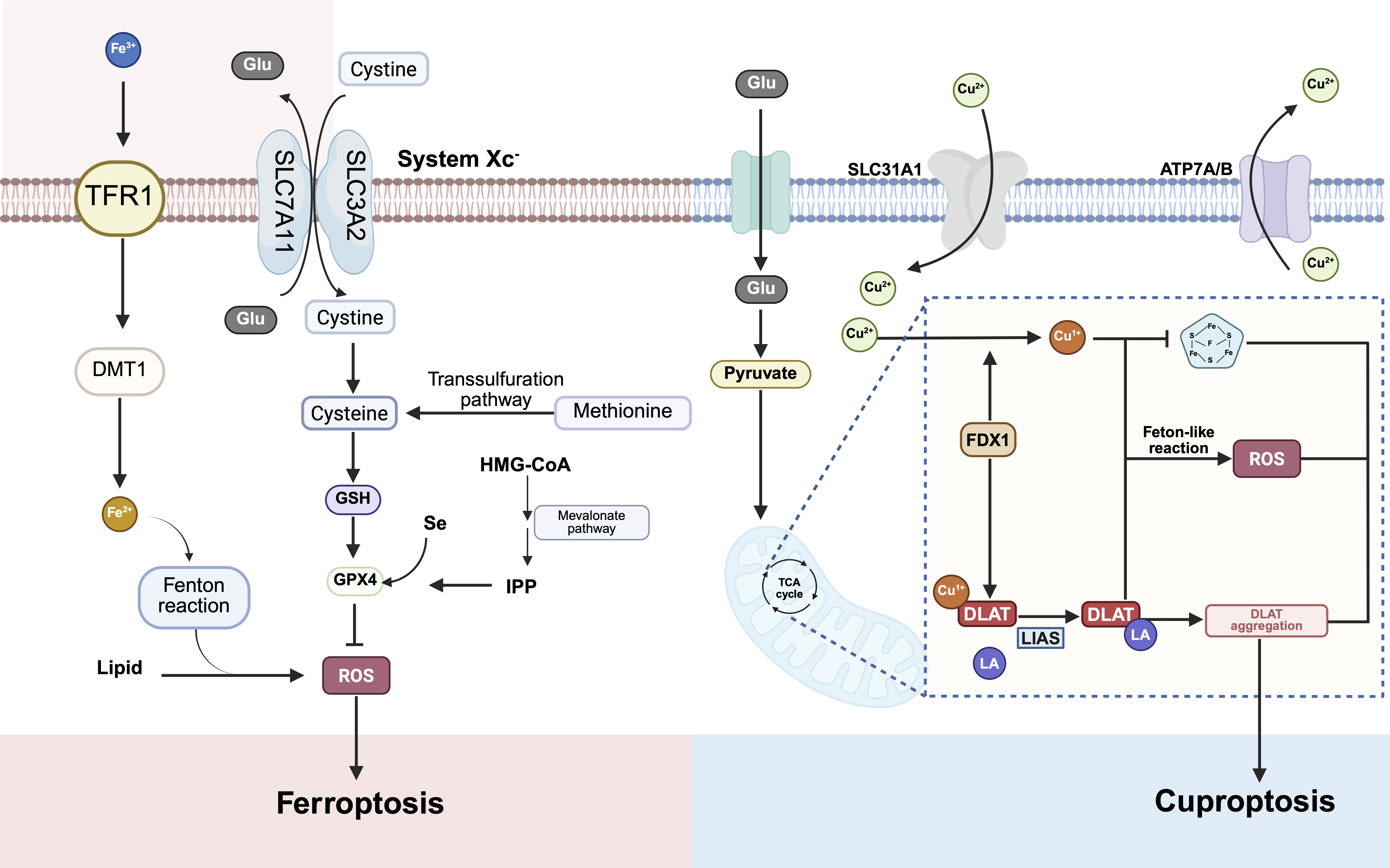
Although ferroptosis and cuproptosis are executed through distinct proximal mechanisms, lipid peroxidation versus copper-driven mitochondrial proteotoxicity, they converge on shared upstream factors, including mitochondrial activity, reactive oxygen species (ROS) flux, metal buffering capacity, and metabolic plasticity (7, 22). Transcriptional and stress-response regulators such as NRF2, p53, and HIF-1α can modulate both pathways, indicating that these metal-dependent death programs are coordinated within a broader resilience network that is often remodeled in drug-resistant tumors (24) (Figure 1). In this review, we synthesize recent advances linking ferroptosis and cuproptosis to cancer drug resistance, focusing on molecular control nodes, therapy-relevant phenotypes, and translational opportunities. We highlight mechanistic interfaces between the two pathways and discuss how combined modulation of metal handling, redox buffering, and mitochondrial metabolism may help overcome resistance in refractory disease. We propose that therapy-resistant cancers sustain survival by elevating a shared metal-redox tolerance threshold, integrating iron and copper metabolism with mitochondrial and antioxidant networks. Collectively, this integrated framework is designed to guide the development of next-generation strategies that target metal-dependent cell death vulnerabilities in oncology. We refer to this concept as the "metal-redox resilience model" of therapy resistance, where coordinated regulation of iron metabolism, copper trafficking, mitochondrial function, and antioxidant buffering raises the threshold for metal-dependent cell death.
2. Ferroptosis in Tumor Drug Resistance
Ferroptosis is an iron-dependent form of regulated cell death driven by the accumulation of lipid hydroperoxides within cellular membranes, leading to catastrophic loss of membrane integrity (25, 26). Its primary biochemical feature is the iron-catalyzed peroxidation of phospholipids containing polyunsaturated fatty acids (PUFAs), resulting in a harmful increase in lipid hydroperoxides (11, 26, 27). Central to ferroptosis control is sustained restraint of membrane peroxidation by coupled enzyme-metabolite defense systems. Glutathione peroxidase 4 (GPX4), which utilizes glutathione as an obligate cofactor, reduces phospholipid hydroperoxides into less reactive lipid alcohols, thereby preventing the escalation of lethal membrane damage (28, 29). Accordingly, inhibiting GPX4, depleting glutathione, or limiting cystine intake via system Xc⁻ (the cystine/glutamate antiporter) lowers the cell’s threshold for ferroptosis by permitting unchecked lipid peroxide accumulation (30, 31). In parallel, GPX4-independent circuits further buffer ferroptosis sensitivity by maintaining radical-trapping capabilities at membranes. These include SLC7A11-supported cystine import, ferroptosis suppressor protein 1 (FSP1)-mediated reduction of coenzyme Q (CoQ), and activity of mitochondrial dihydroorotate dehydrogenase (DHODH) (32). Overall, these layered defense mechanisms act as barriers that protect membrane integrity and prevent peroxidation-driven cell death.
Ferroptosis susceptibility is tightly coupled to intracellular iron handling. Transferrin-bound iron uptake through transferrin receptor 1 (TFRC) expands endolysosomal iron release and increases the pool of redox-active iron, which fuels radical propagation and lipid peroxidation (26). Ferritin limits this liability by sequestering excess iron, whereas nuclear receptor coactivator 4 (NCOA4)-mediated ferritinophagy mobilizes ferritin iron and enlarges the labile iron pool, thereby accelerating lipid peroxide propagation under stress (33). Under therapeutic pressure, tumor cells can therefore be pushed toward ferroptosis when iron availability coincides with heightened demand for peroxidation. Resistant states, however, frequently dampen this axis by limiting iron import, strengthening ferritin storage, or suppressing ferritinophagy to reduce bioavailable Fe2+ (34, 35). In parallel, lipid-remodeling programs determine the abundance of available oxidizable substrates. Acyl-CoA synthetase long-chain family member 4 (ACSL4) and lysophosphatidylcholine acyltransferase 3 (LPCAT3) direct PUFAs into membrane phospholipids, increasing the proportion of peroxidation-prone phospholipid species and creating lineage-specific liabilities (36). These biochemical dependencies intersect with therapy adaptation, as drug-tolerant persister cells and mesenchymal-like states can exhibit an increased requirement for GPX4 activity to restrain lipid hydroperoxides, creating a therapeutically actionable vulnerability to ferroptosis induction (26).
Evasion of ferroptosis is increasingly recognized as a core component of multidrug resistance. Many cytotoxic agents, including cisplatin, doxorubicin, sorafenib, and oxaliplatin (37–40), impose oxidative stress that would otherwise favor ferroptotic execution. Resistant tumors, however, frequently reinforce antioxidant buffering through NRF2 activation and/or SLC7A11 upregulation, enabling efficient detoxification of ROS and lipid peroxides (41, 42) and supporting cross-resistance across therapeutic classes. In targeted therapy settings, persistent PI3K-AKT-mTOR signaling can further suppress ferroptosis by coupling cyst(e)ine sufficiency to GPX4 translation and promoting lipid remodeling towards more saturated species, thereby limiting the accumulation of peroxidation-susceptible substrates under kinase-inhibitor pressure (34, 43, 44). Ferroptosis also intersects with antitumor immunity. IFN-γ released by cytotoxic T cells can suppress SLC7A11 and lower the ferroptosis threshold, whereas immune-evasive tumors often maintain heightened antioxidant capacity that blunts this vulnerability (45–48). Beyond direct tumor cell killing, ferroptosis may also reshape antitumor immunity. Lipid peroxidation products released during ferroptotic cell death can influence immune signaling and inflammatory responses within the tumor microenvironment. Moreover, recent evidence suggests that metal metabolism and oxidative stress pathways regulate immune cell activation, including macrophage polarization and T-cell function. Consequently, therapies that modulate ferroptosis may exert both tumor-intrinsic cytotoxic effects and immunomodulatory activity that enhances the efficacy of immune checkpoint blockade.
Collectively, these observations suggest that ferroptosis resistance is not simply an epiphenomenon of therapy adaptation but can serve as a mechanistic driver of treatment failure. An emerging context in which ferroptosis regulation becomes particularly relevant is the survival of drug-tolerant persister cells. These transient cell populations arise following exposure to chemotherapy or targeted therapy and survive through extensive metabolic and transcriptional reprogramming rather than stable genetic resistance. Persistent cells frequently exhibit elevated oxidative stress, altered lipid metabolism, and heightened dependence on GPX4-mediated detoxification of lipid peroxides. As a consequence, inhibition of ferroptosis defense systems can selectively eliminate persister populations and prevent the emergence of permanent resistance. These findings position ferroptosis vulnerability as a critical determinant of early therapy adaptation and minimal residual disease. Consistent with this model, pharmacological induction of ferroptosis has emerged as a promising strategy to re-sensitize refractory tumors. Small molecules like erastin, RSL3, FIN56, and ML210 can restore sensitivity to chemotherapy and targeted therapies in preclinical systems (49, 50). Combining ferroptosis inducers with cisplatin, EGFR inhibitors, or immunotherapies often enhances tumor cell killing by overwhelming antioxidant buffering and amplifying oxidative damage (51, 52). Nanotechnology-enabled ferroptosis inducers that deliver iron, GPX4 inhibitors, or ROS-generating cargos can further improve tumor selectivity while mitigating systemic toxicity. Importantly, therapeutic success is heavily influenced by the tumor’s metabolic state, with oxidative phosphorylation (OXPHOS)-high cancers, mesenchymal-like programs, and defects in lipid peroxide repair showing increased vulnerability to ferroptosis (53, 54), whereas NRF2-high tumors or states enriched for lipid saturation may remain refractory (55) (Figure 2).
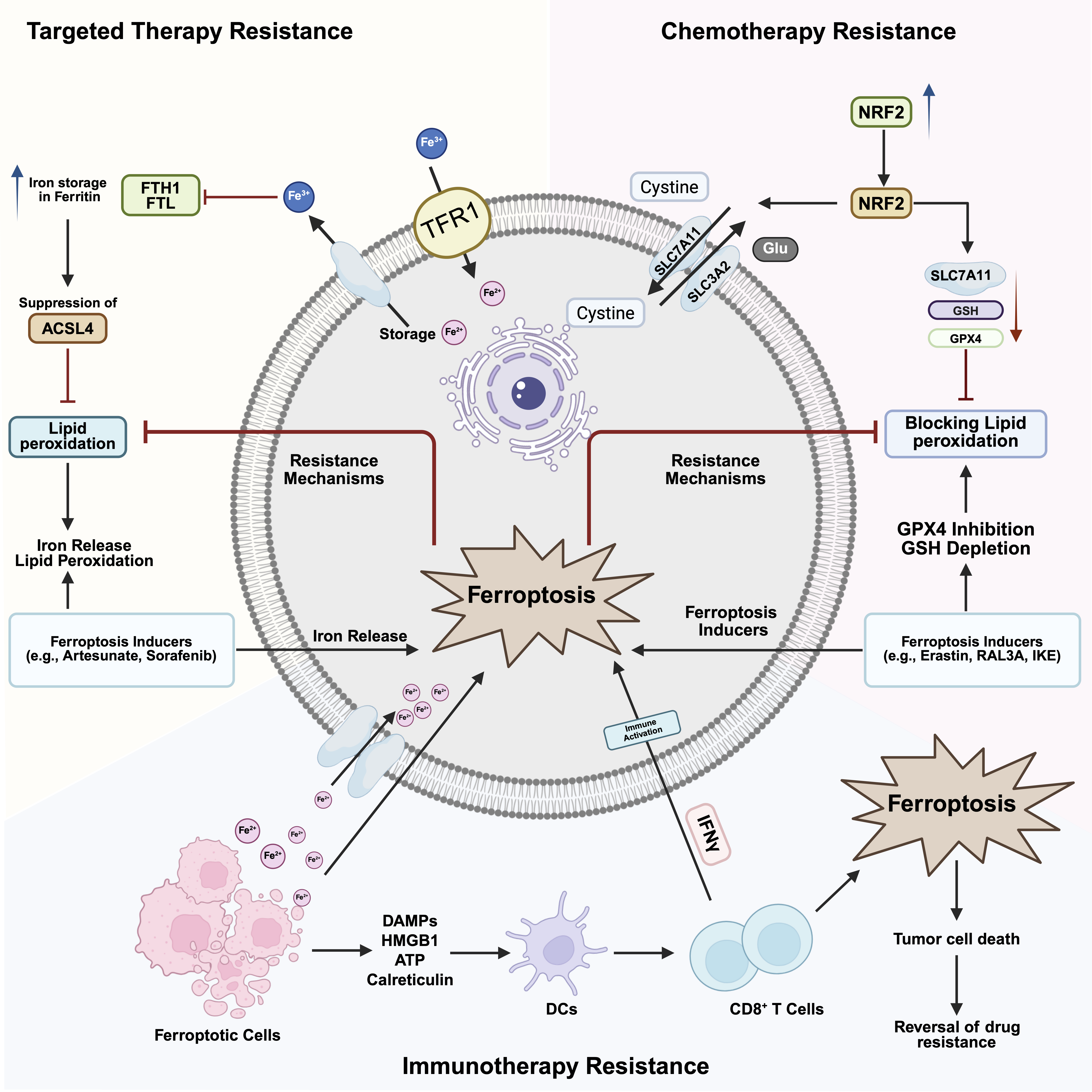
Despite these advances, the clinical translation of ferroptosis-based strategies remains difficult. Ferroptosis can elicit inflammatory sequelae through lipid peroxidation-derived mediators, raising concerns about tissue damage and systemic toxicity (56, 57). Additionally, ferroptosis sensitivity is tightly regulated by microenvironmental factors, including hypoxia, lactate accumulation, and stromal support, which can disrupt redox homeostasis and limit therapeutic efficacy. The strong context dependence of ferroptosis further complicates patient selection: tumors with similar GPX4 levels can display divergent responses due to variations in lipid composition, iron management, and antioxidant reserves (58–60). These considerations underscore the importance of developing reliable, mechanism-based biomarkers and rational combination approaches that match the tumor's metabolic profile.
3. Cuproptosis in Tumor Drug Resistance
Cuproptosis is a copper-triggered form of regulated cell death that is executed through mitochondrial metabolism and proteostasis rather than lipid peroxidation (20). When copper accumulates inside the cell, it is transported to mitochondria, where it is reduced from Cu2+ to the more reactive Cu+ by FDX1. FDX1 also supports protein lipoylation by maintaining the LIAS-GCSH lipoylation machinery (61–63). The bioavailable Cu+ subsequently engages lipoylated mitochondrial enzymes, especially the TCA cycle components DLAT and DLST, leading to abnormal aggregation and destabilization of iron-sulfur (Fe-S) cluster proteins (64). This proteotoxic stress and disruption of Fe-S-dependent mitochondrial functions weaken respiratory integrity, cause an energy crisis, and culminate in cell death (65). This mechanism creates a metabolic vulnerability in tumors with high mitochondrial respiration and OXPHOS dependence, while a shift towards glycolysis can decrease reliance on lipoylated mitochondrial enzymes and reduce susceptibility to cuproptosis (66). Copper homeostasis is maintained by a coordinated network of import, export, and intracellular distribution pathways (67). CTR1 (SLC31A1) carries out high-affinity copper uptake, while ATP7A and ATP7B regulate copper efflux and distribution to various subcellular locations (68). Copper chaperones, such as ATOX1, COX17, and CCS, ensure targeted delivery to the secretory pathway, cytochrome c oxidase, and superoxide dismutase, respectively (61, 69). In cancer, this trafficking circuitry is frequently rewired, increasing intracellular copper availability and reinforcing dependence on copper-requiring enzymes. Excess copper can, in turn, potentiate oncogenic signaling pathways such as PI3K-AKT, MAPK, and NF-κB, thereby supporting proliferation, angiogenesis, and tumor progression (70–72). These copper-driven metabolic and signaling adaptations not only improve tumor fitness but may also weaken the effectiveness of therapies that rely on mitochondrial dysfunction or ROS-mediated cell death (73). A particularly direct link between cuproptosis- related circuitry and drug resistance is seen in copper transporters that influence response to platinum drugs. While ATP7A and ATP7B were initially identified as copper efflux pumps, their structural similarity allows them to export cisplatin and carboplatin, reducing intracellular drug accumulation and promoting chemoresistance (74, 75). Clinically, elevated ATP7B expression is associated with poor cisplatin response in ovarian, gastric, and liver cancers; meanwhile, loss of ATP7B can restore sensitivity to platinum drugs (76). Conversely, downregulation of CTR 1, which limits cisplatin entry, decreases drug uptake and contributes to resistance (77). These findings highlight copper trafficking as a gatekeeper of platinum sensitivity. Metabolic flexibility further influences cuproptosis susceptibility. Tumor cells that suppress mitochondrial function or shift to glycolytic phenotypes exhibit reduced dependence on lipoylated TCA cycle enzymes, thereby reducing copper-induced proteotoxic stress (78, 79). This metabolic reprogramming, common in hypoxic or drug- resistant states, simultaneously mitigates mitochondrial stress and cuproptosis execution. In contrast, OXPHOS- high tumors, including subsets of melanoma, lung, and breast cancers, are more vulnerable to copper overload. The copper ionophore elesclomol exploits this vulnerability by transporting copper into mitochondria, causing proteotoxic collapse and selectively killing OXPHOS-dependent tumors (62, 80). Interestingly, resistance to elesclomol is frequently accompanied by a glycolytic shift, underscoring metabolic rewiring as a principal route of cuproptosis evasion (81). Resistance also involves antioxidant and copper- buffering mechanisms that limit copper toxicity. Metallothioneins (MT1/2), glutathione, and copper-binding chaperones can sequester copper within cells, reducing oxidative and mitochondrial damage. Increased metallothionein levels have been observed in drug-resistant cancers and can act as a protective sink, decreasing copper-induced oxidative stress and mitochondrial dysfunction (82). This adaptive buffering mirrors antioxidant reprogramming in ferroptosis-resistant states, suggesting that redox control constitutes a shared resilience axis across multiple metal- dependent cell death pathways (83).
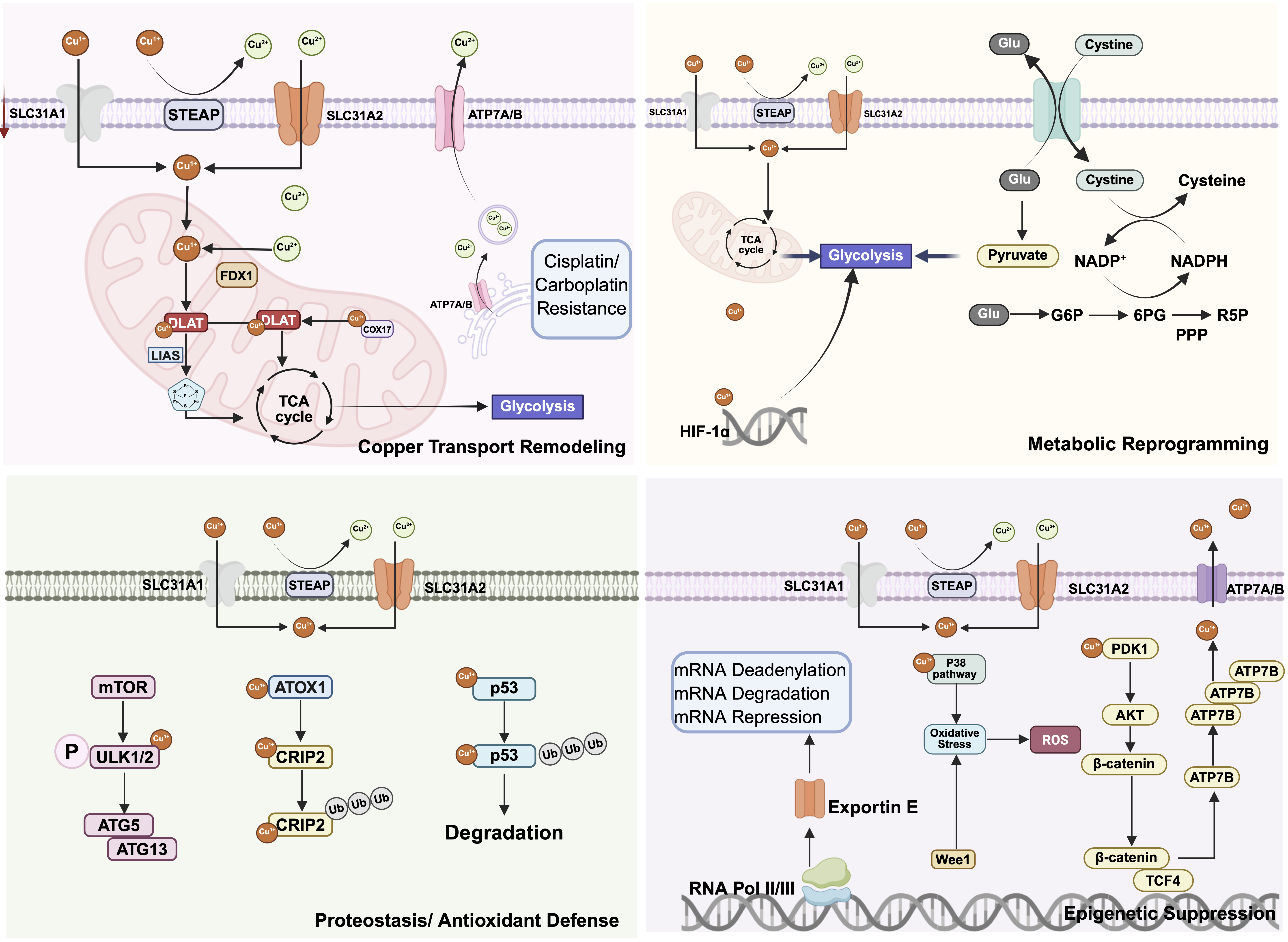
Therapeutically, the availability of copper ionophores and copper-modulating agents has catalyzed renewed interest in targeting cuproptosis. Elesclomol, disulfiram-copper complexes, and CuCl2 formulations can induce potent mitochondrial stress and cuproptosis, with particular activity in OXPHOS-reliant tumors (84). These agents can also synergize with chemotherapeutics, DNA damage response inhibitors, and immunotherapies by overwhelming mitochondrial capacity or compromising proteostasis (85–87). Conversely, copper chelators such as tetrathiomolybdate (TM) and D-penicillamine can resensitize platinum-resistant tumors by limiting copper-dependent cisplatin export and restoring intracellular drug accumulation. This context-specific duality, which induces cuproptosis in some settings while chelating copper in others, highlights both the versatility and complexity of copper-targeted strategies in oncology (Figure 3).
Despite these opportunities, significant barriers still hinder clinical translation. Cuproptosis sensitivity is highly context dependent and shaped by factors such as metabolic state, mitochondrial capacity, lipoylation levels, and copper-buffering systems. The narrow therapeutic window of copper, which is essential for normal physiological functions but toxic in excess, raises concerns about systemic toxicity (88). Additionally, the lack of validated biomarkers, including FDX1 abundance, lipoylation activity, and copper transporter profiles, limits patient stratification and trial design (89). Future research should aim to identify the molecular factors that determine cuproptosis capacity, explore how it interacts with other regulated cell death (RCD) pathways, and develop actionable metabolic signatures to enable the precise use of copper-modulating treatments (90).
4. Crosstalk Between Ferroptosis and Cuproptosis in Tumor Drug Resistance
Ferroptosis and cuproptosis interact through three major biological interfaces: metal-driven redox chemistry, mitochondrial metabolic state, and shared transcriptional stress-response networks.
Redox Convergence
Ferroptosis and cuproptosis were initially viewed as mechanistically distinct regulated cell death programs, with ferroptosis executed by iron-dependent membrane lipid peroxidation and cuproptosis driven by copper-triggered mitochondrial proteotoxic stress (11, 91, 92). However, growing evidence places them within a shared landscape shaped by transition-metal redox chemistry and stress-adaptation circuits. Iron and copper metabolism are tightly linked at the mitochondrial level. Both metals are involved in the assembly and function of iron-sulfur (Fe-S) cluster proteins and key respiratory enzymes in the electron transport chain. Perturbation of copper homeostasis can destabilize Fe-S cluster proteins, indirectly altering iron handling, mitochondrial respiration, and cellular redox balance. Conversely, alterations in iron metabolism influence mitochondrial oxidative flux and ROS production, thereby affecting copper toxicity. This biochemical interdependence suggests that iron- and copper-regulated death pathways are embedded within a shared mitochondrial metal-metabolism network rather than operating as completely independent processes. Both copper and iron can engage in Fenton or Fenton-like reactions, amplifying reactive ROS and lowering the threshold for oxidative membrane damage (93). Consistent with this convergence, copper ionophores such as elesclomol can impose mitochondrial copper stress that increases ROS and erodes antioxidant capacity, including downregulation of system Xc⁻ components and loss of GPX4, through mechanisms linked to proteostasis or autophagy (94). At a systems level, both pathways are regulated by tumor metabolic state, redox buffering, and metal trafficking, and therapy-selected persister states that reinforce antioxidant capacity can diminish ferroptosis sensitivity and potentially suppress copper-triggered stress signals (95). Integrating ferroptosis and cuproptosis, therefore, provides a unified framework for understanding how metal homeostasis and metabolic plasticity govern cell fate under therapeutic pressure and for designing combination strategies to resensitize refractory disease. Together, current evidence indicates that ferroptosis and cuproptosis function as interconnected components of a broader metal-redox network that governs cancer cell survival under therapeutic stress.
A key point of convergence is their shared reliance on redox-active transition metals that drive oxidative stress (96). Both Fe²⁺ and Cu⁺ catalyze the formation of highly reactive radicals, albeit through distinct reaction contexts. In ferroptosis, iron directly promotes lipid peroxidation through hydroxyl radical generation and propagation of PUFA-containing phospholipid oxidation (97). Copper, by contrast, can amplify intracellular redox cycling and ROS, indirectly elevating lipid peroxidation and modulating ferroptosis susceptibility (20). This reciprocity suggests that copper accumulation may potentiate ferroptotic execution, whereas iron dysregulation can perturb copper binding and trafficking, together forming an intertwined metal-redox network that shapes how resistant tumors tolerate oxidative injury.
Mitochondrial Metabolic Interface
Mitochondrial metabolism forms a second key interface (98, 99). Cuproptosis inherently depends on functional mitochondrial respiration and the presence of lipoylated TCA-cycle enzymes, whereas ferroptosis is regulated by mitochondrial ROS production, membrane lipid composition, and metabolic flux through the TCA cycle and electron transport chain (20, 22). States with high OXPHOS activity raise mitochondrial ROS levels and increase peroxidation pressure, thereby sensitizing cells to ferroptosis (100, 101). In contrast, mitochondrial suppression or a shift to glycolysis, common adaptations in hypoxic and drug-resistant tumors, can decrease both cuproptosis and ferroptosis by lowering lipoylation-dependent vulnerabilities, reducing oxidative flux, and reshaping iron-sulfur (Fe-S) cluster dynamics (102). Importantly, loss of Fe-S proteins, a hallmark of cuproptosis, can impair iron regulation and respiratory function, potentially influencing ferroptosis regulation pathways (103, 104).
Transcriptional Regulators
Shared transcriptional and signaling regulators further coordinate the two programs. NRF2 functions as a dominant antioxidant controller that can suppress ferroptosis and cuproptosis by maintaining glutathione availability, upregulating metallothioneins, and promoting the expression of iron- and copper-buffering factors (105, 106). NRF2 activation, frequently enriched in chemotherapy- and targeted therapy-resistant tumors, limits ROS accumulation and constrains both lipid and protein oxidation. p53 exerts context-dependent control over ferroptosis, for example, by repressing SLC7A11 to lower the ferroptosis threshold or, in other settings, by engaging antioxidant programs (107); through its broader influence on mitochondrial metabolism and protein quality control, p53 may also indirectly shape cuproptosis capacity (78). HIF-1α, a central mediator of hypoxic adaptation, decreases ferroptotic stress by reducing peroxidation and also lessens cuproptosis by promoting glycolytic reprogramming and reducing mitochondrial lipoylation (108–110). Collectively, these upstream regulators establish a coordinated transcriptional landscape that resistant tumors exploit to survive therapy-induced stress.
Dual-Shielded Resistance Phenotype
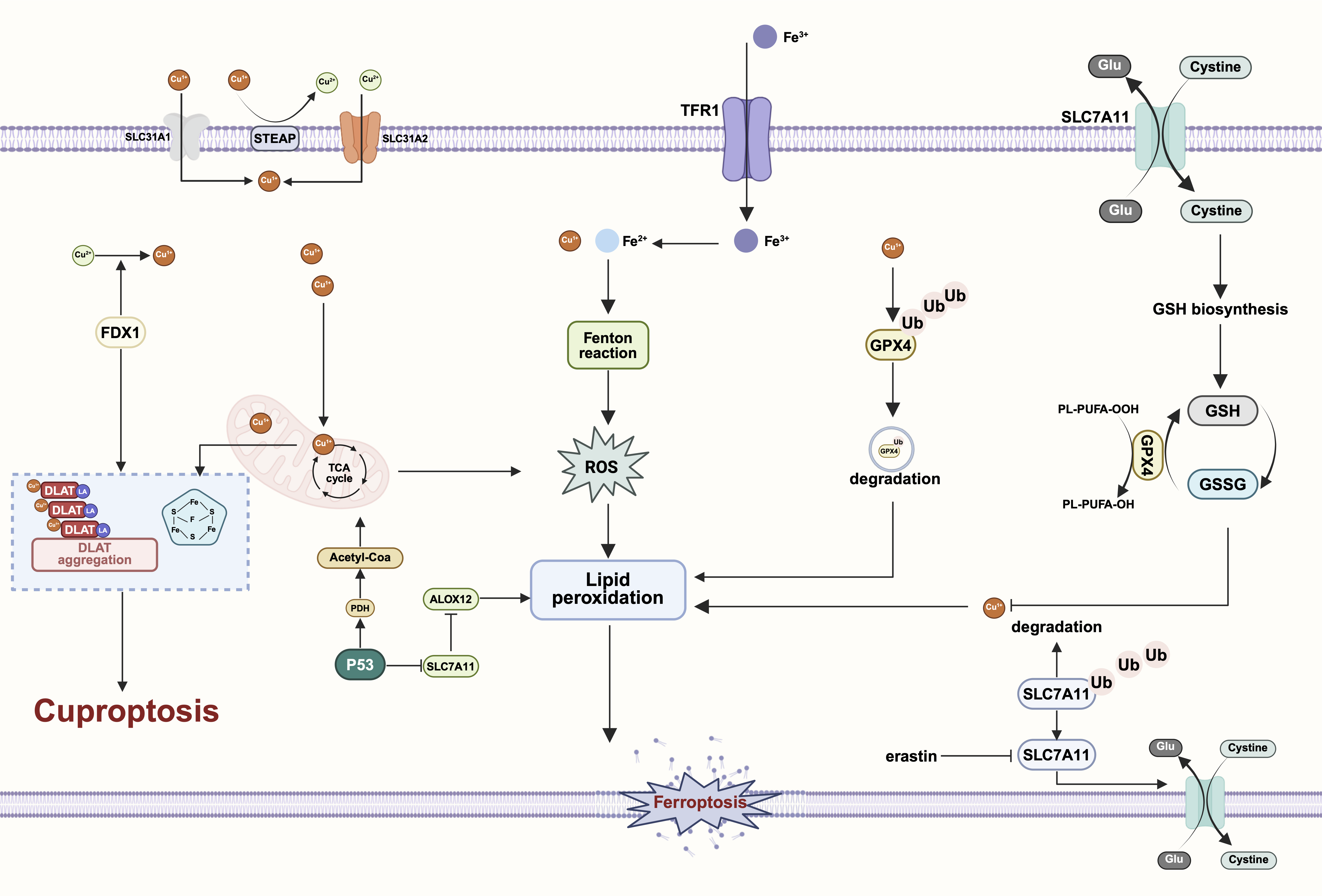
At the cellular level, therapy-resistant tumors can develop a “dual-shielded” phenotype where ferroptosis- and cuproptosis-suppressing programs are simultaneously activated. For example, co-expression of SLC7A11 and ATP7B can enhance lipid peroxide detoxification while promoting copper export, thus reducing both death pathways and supporting widespread chemoresistance (75). This coupled adaptation is most evident under sustained selective pressures such as platinum chemotherapy, tyrosine kinase inhibition, or oxidative signals within the tumor microenvironment. At the same time, mitochondrial remodeling provides a shared mechanistic scaffold for evasion of both pathways by lowering oxidative flux, reducing the abundance of lipoylated substrates, and preserving Fe-S protein integrity (111, 112). These mechanistic intersections create rational opportunities for combination targeting (113). Copper ionophores that disrupt mitochondrial proteostasis may synergize with ferroptosis inducers, including GPX4 inhibitors, by overwhelming antioxidant defenses and amplifying oxidative damage (24). Conversely, copper chelation could help restore platinum accumulation and alter metal pools in ways that make cells more susceptible to ferroptosis, although the net effect is likely to depend on the specific context. Metabolic strategies that reactivate mitochondrial respiration, such as limiting glycolysis or boosting TCA-cycle activity, might further expand the therapeutic window for co-inducing both death modalities by reinstating lipoylation-dependent vulnerabilities and oxidative stress. Collectively, these insights support dual-modality approaches that target both ferroptosis and cuproptosis to overcome metabolic flexibility and eliminate drug-resistant cancer cells (114, 115).
Although no compound has yet been formally defined as a direct and simultaneous executor of both ferroptosis and cuproptosis, emerging evidence supports the possibility of coordinated targeting. Redox-active copper ionophores can increase mitochondrial ROS and disrupt proteostasis, potentially lowering the threshold for lipid peroxidation. Meanwhile, ferroptosis inducers, such as GPX4 inhibitors, intensify membrane oxidative damage (116). Preclinical studies combining copper ionophores with GPX4 inhibition or system Xc⁻ blockade have shown a synergistic amplification of oxidative stress, indicating that dual-pathway engagement may overcome resistance mechanisms that protect against single-axis interventions (117, 118). These findings suggest that pharmacological co-induction may effectively exploit vulnerabilities within the metal-redox network in drug-resistant tumors.
Increasing evidence suggests that sensitivity to ferroptosis and cuproptosis is not determined solely by absolute iron or copper levels, but rather by the cellular threshold for metal-induced oxidative stress (20, 119). Tumor cells maintain multiple buffering systems, including ferritin-mediated iron storage, metallothionein-dependent copper sequestration, and antioxidant networks, which tightly regulate the bioavailability of redox-active metal ions. In drug-resistant tumors, these buffering mechanisms are frequently reinforced, leading to a higher tolerance to oxidative or metal stress. As a result, resistant cells may require a stronger disturbance of metal homeostasis or redox balance before ferroptotic or cuproptotic death is triggered. In this context, the effective “metal toxicity threshold” is influenced by metabolic state, mitochondrial activity, lipid composition, and antioxidant capacity rather than by a single universal concentration of iron or copper. This adaptive increase in the death threshold represents a key mechanism by which therapy-resistant tumors evade metal-dependent cytotoxicity (120, 121).
Overall, ferroptosis-cuproptosis crosstalk reflects a deeper integration of metal metabolism, mitochondrial function, and redox regulation in determining tumor survival during therapy. Their convergence suggests that resistant tumors often depend on coordinated suppression of multiple metal-dependent death pathways, and that achieving lasting resensitization may require disrupting this integrated resilience network (Figure 4). Together, these observations support a model where therapy-resistant tumors develop a dual-shielded metal-redox resistance state that both suppresses ferroptotic lipid peroxidation and prevents copper-induced mitochondrial proteotoxicity.
5. Clinical Translation and Future Directions
Therapeutic exploitation of ferroptosis and cuproptosis is emerging as a tractable strategy to counter the metabolic and oxidative resilience characteristic of drug-resistant tumors (91, 122). Both pathways are anchored in redox control, mitochondrial metabolism, and metal-ion homeostasis, making them particularly relevant for cancers that survive therapy by strengthening antioxidant buffering and rewiring bioenergetic dependencies (22). The opportunity extends beyond selective induction of either pathway alone and increasingly involves the rational co-targeting of their shared upstream liabilities to generate synergistic stress that resistant states cannot accommodate.
Alongside these mechanistic advances, early clinical research has begun to assess whether modulating metal-dependent cell death can lead to therapeutic benefits. In the copper field, the ionophore elesclomol advanced to phase II/III trials for melanoma, where clinical benefits appeared restricted to patients with preserved mitochondrial oxidative metabolism, underscoring the importance of metabolic stratification (123, 124). Disulfiram-copper combinations are being repurposed for various cancers (125), supported by their ability to trigger proteotoxic and oxidative stress. Conversely, copper chelators such as tetrathiomolybdate have been investigated for their potential to regulate copper-dependent signaling and restore platinum sensitivity in resistant tumors. On the other hand, direct ferroptosis inducers, including GPX4 inhibitors and system Xc⁻ antagonists, have not yet advanced to late-stage clinical trials, mainly due to issues with pharmacokinetics, target specificity, and systemic toxicity (126). Nanoparticle-based platforms aimed at improving tumor-specific iron delivery or lipid peroxidation are mostly still in preclinical development. These initial clinical efforts provide proof of concept and underscore the need for biomarker-guided patient stratification and rational combination strategies to maximize therapeutic benefit.
Preclinical Targeting Strategies
In the field of ferroptosis, various classes of inducers have demonstrated robust preclinical activity (104). Inhibiting GPX4 or system Xc⁻ stops lipid hydroperoxide detoxification and allows dangerous levels of membrane oxidation, making tumor cells more vulnerable to oxidative stress-producing therapies. These agents can potentiate the cytotoxic effects of chemotherapy, including cisplatin, and can amplify the effectiveness of radiotherapy and immune checkpoint blockade by increasing ROS levels and influencing inflammatory signals (127). Nanomedicine-based platforms further improve the therapeutic index by concentrating iron payloads, peroxidation catalysts, or GPX4-silencing modules directly to tumors, sustaining local oxidative stress while reducing systemic toxicity. At the same time, synthetic-lethal strategies are becoming more popular; for example, combining ferroptosis induction with inhibition of compensatory antioxidant circuits such as NRF2 or the CoQ-centered FSP1 pathway may overcome the buffered redox states that frequently accompany resistance (128).
Cuproptosis-oriented strategies provide a complementary route to resensitization, particularly in tumors with high mitochondrial respiration or copper-handling dysregulation. Copper ionophores, including elesclomol and disulfiram-Cu complexes, exploit OXPHOS dependence by delivering copper to mitochondria and causing proteotoxic collapse through aggregation of lipoylated enzymes and loss of respiratory function. These agents have shown synergy with platinum drugs and PARP inhibitors, supporting the premise that overwhelming mitochondrial proteostasis can intensify DNA damage-linked lethality in metabolically oxidative tumors (129, 130). Conversely, copper chelation offers a distinct yet clinically relevant means of reversing platinum resistance. Agents such as tetrathiomolybdate can reduce copper-dependent cisplatin efflux, restore intracellular drug levels, and resensitize tumors to DNA damage. More direct targeting of copper trafficking, such as blocking ATP7A/B or restoring CTR1-mediated uptake, may further increase intratumoral drug levels and improve therapeutic penetration. Collectively, these approaches highlight the therapeutic plasticity of copper modulation in reprogramming resistant cancer phenotypes (84).
Dual-Modality Targeting Approaches
Mechanistic convergence between ferroptosis and cuproptosis has catalyzed interest in dual-modality strategies designed to overcome the multifaceted adaptations of resistant tumors. Since both death pathways are influenced by metal-driven oxidative chemistry and mitochondrial function, simultaneous activation can impose coupled lipid and protein oxidative stress, potentially bypassing resistance mechanisms that guard against single-target approaches (131, 132). For example, combining copper ionophores with GPX4 inhibitors may simultaneously increase mitochondrial ROS flux and disable lipid peroxide repair, accelerating irreversible oxidative damage (133). Alternatively, targeting shared upstream regulators such as NRF2 or HIF-1α, or metabolic checkpoints that gate mitochondrial entry of carbon substrates, may collapse the transcriptional and metabolic programs essential for maintaining redox homeostasis, thereby sensitizing tumors to both modalities (134, 135). Another approach to improve the therapeutic selectivity of metal-dependent death is to exploit synthetic lethality between ferroptosis or cuproptosis pathways and tumor-specific genetic or metabolic vulnerabilities. Tumors often carry oncogenic mutations, metabolic rewiring, or loss of tumor suppressor functions that alter redox balance, mitochondrial function, and metal homeostasis (136, 137). These changes may create conditions in which cancer cells become especially reliant on antioxidant defenses or mitochondrial respiration to survive. For instance, tumors with high oxidative phosphorylation (138), defects in redox control, or increased polyunsaturated lipid content may be more susceptible to induction of ferroptosis or cuproptosis (139, 140). Systematic genetic screening and metabolic profiling may help identify tumor subtypes in which metal-dependent death pathways can be selectively activated. Combining these vulnerabilities with metal-targeting agents could enable synthetic lethal strategies that preferentially eliminate tumor cells while minimizing toxicity to normal tissues. These precision approaches are a promising future direction for developing metal-dependent death-based cancer therapies.
Translational Challenges and Future Perspectives
Despite these opportunities, translation faces significant challenges. The therapeutic window for metal-directed interventions is inherently narrow because iron and copper are essential micronutrients, raising concerns about systemic toxicity and damage to normal tissues. Tumor heterogeneity further complicates response prediction, as ferroptosis and cuproptosis capabilities vary with metabolic state, lipid composition, oxygen levels, and metal-buffering capacity across lesions and within subclones (141–143). Therefore, biomarker-guided stratification will be crucial. Potential markers include FDX1 abundance and lipoylation capacity for cuproptosis, as well as ACSL4, SLC7A11, and antioxidant signatures for ferroptosis. Equally important is developing clinically applicable assays that detect dynamic metal flux, mitochondrial activity, and oxidative stress in situ, combined with computational models that integrate these parameters with treatment outcomes to inform rational combination therapies. A key question emerging from recent research is how tumors coordinate iron and copper metabolism to avoid metal-dependent cell death during therapeutic stress.
Another key goal is to understand how the tumor TME influences sensitivity to metal-dependent cell death. The TME also plays a vital role in controlling iron and copper availability. Tumor-associated macrophages, stromal fibroblasts, and endothelial cells can affect metal homeostasis by releasing iron-binding proteins, modulating inflammatory signaling, and managing oxidative metabolism. For example, macrophage polarization states impact iron sequestration and release, which in turn affects ferroptosis sensitivity in nearby tumor cells. Likewise, inflammatory signaling pathways connected to copper metabolism can influence oxidative stress responses and mitochondrial activity within the tumor niche. These microenvironmental factors may therefore determine the effectiveness of therapies aimed at inducing ferroptosis or cuproptosis. The stromal and immune components, along with hypoxic niches, impose limits on redox balance and metabolic wiring, thus affecting iron and copper levels, ROS buffering, and mitochondrial function (25, 75). Effective treatment strategies will need to anticipate microenvironmental rescue signals that can hinder ferroptosis or cuproptosis and include approaches to disrupt protective interactions between cancer cells, stroma, and immune cells.
Overall, the convergence of ferroptosis and cuproptosis strengthens a broader shift in resistance biology: resistant tumors depend on integrated, metal-dependent metabolic networks. Achieving lasting eradication will likely require disrupting multiple nodes of this resilience framework. Combining ferroptosis inducers, copper modulators, metabolic interventions, and immunotherapies offers a promising path toward clinical application. As understanding of mechanisms deepens and biomarkers advance, strategies based on metal-dependent cell death may transform therapeutic approaches for drug-resistant cancer.
Discussion
Ferroptosis and cuproptosis have become two key metal-dependent forms of regulated cell death programs that change how tumor vulnerabilities are exposed under therapeutic stress. While they were originally studied as separate biochemical processes, it is now clear that iron- and copper-driven death pathways are embedded within a shared metabolic and redox landscape that is continually reshaped during cancer progression and treatment resistance. Ferroptosis, cuproptosis, mitochondrial metabolism, and redox homeostasis collectively define a metal-dependent vulnerability landscape in therapy-resistant cancer. Tumors resistant to drugs often strengthen antioxidant defenses, reprogram energy dependencies, and modify metal transport to prevent both lipid peroxidation-driven ferroptosis and copper-induced mitochondrial protein damage, showing a coordinated reliance on metal regulation and mitochondrial health to survive cytotoxic attacks (144). These findings support a metal-redox resilience model in which therapy-resistant tumors raise a common “metal-redox tolerance threshold,” allowing them to survive oxidative and metabolic stress while actively suppressing ferroptosis and cuproptosis through coordinated control of metal trafficking, mitochondrial metabolism, and antioxidant buffering. Mechanistically, ferroptosis and cuproptosis converge on upstream factors that include mitochondrial metabolism, reactive ROS flux, and stress-adaptation circuits governed by transcriptional factors such as NRF2, p53, and HIF-1α. This overlap suggests that these pathways should not be viewed separately but as interconnected components of a broader, metal-driven resilience network. In this framework, aggressive and therapy-persistent tumor states achieve cross-resistance by increasing oxidative damage thresholds in both membrane and mitochondrial areas, while disrupting metal handling can reopen death-sensitivity windows that traditional treatments often cannot target.
Therapeutically, the ability to manipulate ferroptosis and cuproptosis broadens the repertoire of resensitization strategies. Ferroptosis inducers, copper ionophores, copper chelators, metabolic interventions, and nanotechnology-based delivery platforms provide complementary approaches to disrupt the buffering systems that support disease resistance. Importantly, co-targeting ferroptosis and cuproptosis, or their common upstream regulators, may reveal synthetic-lethal vulnerabilities that are not accessible through targeting only one pathway (84). These approaches are especially promising for eliminating drug-tolerant persisters, reducing minimal residual disease, and preventing relapse.
Clinical translation, however, requires overcoming several challenges. Unmet needs include robust biomarkers that indicate ferroptosis and cuproptosis capacity, safe therapeutic windows for targeting iron and copper levels, and effective combination therapies applicable across diverse tumor microenvironments. Additionally, more detailed mechanistic understanding is needed to explain how iron and copper interact within different subcellular compartments, how metabolic heterogeneity controls pathway activation, and how microenvironmental signals influence metal-dependent cell death. More broadly, metal-dependent cell death pathways are emerging as vulnerabilities in therapy-resistant cancers and offer a theoretical basis for developing next-generation therapies that target metabolism and redox processes. Their convergence at the crossroads of metal homeostasis, mitochondrial function, and redox regulation provides a solid mechanistic foundation for rational, multi-node therapeutic design. As biomarker techniques improve and precision oncology strategies integrate metal-dependent vulnerabilities, exploiting ferroptosis and cuproptosis pathways could offer durable solutions to overcoming the adaptive resilience of refractory cancers.
References
1. Tang Y, Zang H, Wen Q, Fan S. AXL in cancer: a modulator of drug resistance and therapeutic target. J Exp Clin Cancer Res. 2023;42(1):148. https://doi.org/10.1186/s13046-023-02726-w
2. Cai M, Song XL, Li XA, Chen M, Guo J, Yang DH, et al. Current therapy and drug resistance in metastatic castration-resistant prostate cancer. Drug Resist Updat. 2023;68:100962. https://doi.org/10.1016/j.drup.2023.100962
3. Kaur P, Shankar E, Gupta S. EZH2-mediated development of therapeutic resistance in cancer. Cancer Lett. 2024;586:216706. https://doi.org/10.1016/j.canlet.2024.216706
4. Lee J, Kida K, Koh J, Liu H, Manyam GC, Gi YJ, et al. The DNA repair pathway as a therapeutic target to synergize with trastuzumab deruxtecan in HER2-targeted antibody-drug conjugate-resistant HER2-overexpressing breast cancer. J Exp Clin Cancer Res. 2024;43(1):236. https://doi.org/10.1186/s13046-024-03143-3
5. Robey RW, Pluchino KM, Hall MD, Fojo AT, Bates SE, Gottesman MM. Revisiting the role of ABC transporters in multidrug-resistant cancer. Nat Rev Cancer. 2018;18(7):452–464. https://doi.org/10.1038/s41568-018-0005-8
6. Zhou J, Wang X, Li Z, Wang F, Cao L, Chen X, et al. PIM1 kinase promotes EMT-associated osimertinib resistance via regulating GSK3β signaling pathway in EGFR-mutant non-small cell lung cancer. Cell Death Dis. 2024;15(9):644. https://doi.org/10.1038/s41419-024-07039-0
7. Jin H, He Y, Zhao P, Hu Y, Tao J, Chen J, et al. Targeting lipid metabolism to overcome EMT-associated drug resistance via integrin β3/FAK pathway and tumor-associated macrophage repolarization using legumain-activatable delivery. Theranostics. 2019;9(1):265–278. https://doi.org/10.7150/thno.27246
8. Liu S, Zhang X, Wang W, Li X, Sun X, Zhao Y, et al. Metabolic reprogramming and therapeutic resistance in primary and metastatic breast cancer. Mol Cancer. 2024;23(1):261. https://doi.org/10.1186/s12943-024-02165-x
9. Chhabra Y, Fane ME, Pramod S, Hüser L, Zabransky DJ, Wang V, et al. Sex-dependent effects in the aged melanoma tumor microenvironment influence invasion and resistance to targeted therapy. Cell. 2024;187(21):6016–6034.e25. https://doi.org/10.1016/j.cell.2024.08.013
10. Peng F, Liao M, Qin R, Zhu S, Peng C, Fu L, et al. Regulated cell death (RCD) in cancer: key pathways and targeted therapies. Signal Transduct Target Ther. 2022;7(1):286. https://doi.org/10.1038/s41392-022-01110-y
11. Dixon SJ, Lemberg KM, Lamprecht MR, Skouta R, Zaitsev EM, Gleason CE, et al. Ferroptosis: an iron-dependent form of nonapoptotic cell death. Cell. 2012;149(5):1060–1072. https://doi.org/10.1016/j.cell.2012.03.042
12. Wei R, Zhao Y, Wang J, Yang X, Li S, Wang Y, et al. Tagitinin C induces ferroptosis through PERK-Nrf2-HO-1 signaling pathway in colorectal cancer cells. Int J Biol Sci. 2021;17(11):2703–2717. https://doi.org/10.7150/ijbs.59404
13. Weiss-Sadan T, Ge M, Hayashi M, Gohar M, Yao CH, de Groot A, et al. NRF2 activation induces NADH-reductive stress, providing a metabolic vulnerability in lung cancer. Cell Metab. 2023;35(3):487–503.e7. https://doi.org/10.1016/j.cmet.2023.01.012
14. Yang J, Zhou Y, Xie S, Wang J, Li Z, Chen L, et al. Metformin induces ferroptosis by inhibiting UFMylation of SLC7A11 in breast cancer. J Exp Clin Cancer Res. 2021;40(1):206. https://doi.org/10.1186/s13046-021-02012-7
15. Shen L, Zhang J, Zheng Z, Yang F, Liu S, Wu Y, et al. PHGDH inhibits ferroptosis and promotes malignant progression by upregulating SLC7A11 in bladder cancer. Int J Biol Sci. 2022;18(14):5459–5474. https://doi.org/10.7150/ijbs.74546
16. Liu X, Nie L, Zhang Y, Yan Y, Wang C, Colic M, et al. Actin cytoskeleton vulnerability to disulfide stress mediates disulfidptosis. Nat Cell Biol. 2023;25(3):404–414. https://doi.org/10.1038/s41556-023-01091-2
17. Wang G, Qiu M, Xing X, Zhou J, Yao H, Li M, et al. Lung cancer scRNA-seq and lipidomics reveal aberrant lipid metabolism for early-stage diagnosis. Sci Transl Med. 2022;14(630):eabk2756. https://doi.org/10.1126/scitranslmed.abk2756
18. Ouyang S, Li H, Lou L, Huang Q, Zhang Z, Mo J, et al. Inhibition of STAT3-ferroptosis negative regulatory axis suppresses tumor growth and alleviates chemoresistance in gastric cancer. Redox Biol. 2022;52:102317. https://doi.org/10.1016/j.redox.2022.102317
19. Li J, Liu J, Zhou Z, Wu R, Chen X, Yu C, et al. Tumor-specific GPX4 degradation enhances ferroptosis-initiated antitumor immune response in mouse models of pancreatic cancer. Sci Transl Med. 2023;15(720):eadg3049. https://doi.org/10.1126/scitranslmed.adg3049
20. Tsvetkov P, Coy S, Petrova B, Dreishpoon M, Verma A, Abdusamad M, et al. Copper induces cell death by targeting lipoylated TCA cycle proteins. Science. 2022;375(6586):1254–1261. https://doi.org/10.1126/science.abf0529
21. Li ZH, Zheng R, Chen JT, Jia J, Qiu M. The role of copper transporter ATP7A in platinum-resistance of esophageal squamous cell cancer (ESCC). J Cancer. 2016;7(14):2085–2092. https://doi.org/10.7150/jca.16117
22. Safaei R. Role of copper transporters in the uptake and efflux of platinum containing drugs. Cancer Lett. 2006;234(1):34–39. https://doi.org/10.1016/j.canlet.2005.07.046
23. Feng L, Wu TZ, Guo XR, Wang YJ, Wang XJ, Liu SX, et al. Discovery of natural resorcylic acid lactones as novel potent copper ionophores covalently targeting PRDX1 to induce cuproptosis for triple-negative breast cancer therapy. ACS Cent Sci. 2025;11(2):357–370. https://doi.org/10.1021/acscentsci.4c02188
24. Wang W, Lu K, Jiang X, Wei Q, Zhu L, Wang X, et al. Ferroptosis inducers enhanced cuproptosis induced by copper ionophores in primary liver cancer. J Exp Clin Cancer Res. 2023;42(1):142. https://doi.org/10.1186/s13046-023-02720-2
25. Viswanathan VS, Ryan MJ, Dhruv HD, Gill S, Eichhoff OM, Seashore-Ludlow B, et al. Dependency of a therapy-resistant cancer state on the lipid peroxidase pathway. Nature. 2017;547(7664):453–457. https://doi.org/10.1038/nature23007
26. Dixon SJ, Olzmann JA. The cell biology of ferroptosis. Nat Rev Mol Cell Biol. 2024;25(6):424–442. https://doi.org/10.1038/s41580-024-00703-5
27. Yang WS, Stockwell BR. Ferroptosis: death by lipid peroxidation. Trends Cell Biol. 2016;26(3):165–176. https://doi.org/10.1016/j.tcb.2015.10.014
28. Yang WS, SriRamaratnam R, Welsch ME, Shimada K, Skouta R, Viswanathan VS, et al. Regulation of ferroptotic cancer cell death by GPX4. Cell. 2014;156(1):317–331. https://doi.org/10.1016/j.cell.2013.12.010
29. Conrad M, Friedmann Angeli JP. Glutathione peroxidase 4 (GPX4) and ferroptosis: what’s so special about it? Mol Cell Oncol. 2015;2(3):e995047. https://doi.org/10.4161/23723556.2014.995047
30. Bersuker K, Hendricks JM, Li Z, Magtanong L, Ford B, Tang PH, et al. The CoQ oxidoreductase FSP1 acts parallel to GPX4 to inhibit ferroptosis. Nature. 2019;575(7784):688–692. https://doi.org/10.1038/s41586-019-1705-2
31. Doll S, Freitas FP, Shah R, Aldrovandi M, da Silva MC, Ingold I, et al. FSP1 is a glutathione-independent ferroptosis suppressor. Nature. 2019;575(7784):693–698. https://doi.org/10.1038/s41586-019-1707-0
32. Mao C, Liu X, Zhang Y, Lei G, Yan Y, Lee H, et al. DHODH-mediated ferroptosis defence is a targetable vulnerability in cancer. Nature. 2021;593(7860):586–590. https://doi.org/10.1038/s41586-021-03539-7
33. Fang Y, Chen X, Tan Q, Zhou H, Xu J, Gu Q. Inhibiting ferroptosis through disrupting the NCOA4-FTH1 interaction: a new mechanism of action. ACS Cent Sci. 2021;7(6):980–989. https://doi.org/10.1021/acscentsci.0c01592
34. Huang G, Cai Y, Ren M, Zhang X, Fu Y, Cheng R, et al. Salidroside sensitizes triple-negative breast cancer to ferroptosis by SCD1-mediated lipogenesis and NCOA4-mediated ferritinophagy. J Adv Res. 2025;74:589–607. https://doi.org/10.1016/j.jare.2024.09.027
35. Zhao P, Yin S, Qiu Y, Sun C, Yu H. Ferroptosis and pyroptosis are connected through autophagy: a new perspective of overcoming drug resistance. Mol Cancer. 2025;24(1):23. https://doi.org/10.1186/s12943-024-02217-2
36. Doll S, Proneth B, Tyurina YY, Panzilius E, Kobayashi S, Ingold I, et al. ACSL4 dictates ferroptosis sensitivity by shaping cellular lipid composition. Nat Chem Biol. 2017;13(1):91–98. https://doi.org/10.1038/nchembio.2239
37. Hangauer MJ, Viswanathan VS, Ryan MJ, Bole D, Eaton JK, Matov A, et al. Drug-tolerant persister cancer cells are vulnerable to GPX4 inhibition. Nature. 2017;551(7679):247–250. https://doi.org/10.1038/nature24297
38. Wang B, Jin Y, Liu J, Liu Q, Shen Y, Zuo S, et al. EP1 activation inhibits doxorubicin-cardiomyocyte ferroptosis via Nrf2. Redox Biol. 2023;65:102825. https://doi.org/10.1016/j.redox.2023.102825
39. Xu J, Wang B, Liu Q, Guo S, Chen C, Wu J, et al. MVP-LCN2 axis triggers evasion of ferroptosis to drive hepatocarcinogenesis and sorafenib resistance. Drug Resist Updat. 2025;81:101246. https://doi.org/10.1016/j.drup.2025.101246
40. Zeng K, Li W, Wang Y, Zhang Z, Zhang L, Zhang W, et al. Inhibition of CDK1 overcomes oxaliplatin resistance by regulating ACSL4-mediated ferroptosis in colorectal cancer. Adv Sci (Weinh). 2023;10(25):e2301088. https://doi.org/10.1002/advs.202301088
41. Rochette L, Dogon G, Rigal E, Zeller M, Cottin Y, Vergely C. Lipid peroxidation and iron metabolism: two corner stones in the homeostasis control of ferroptosis. Int J Mol Sci. 2022;24(1):449. https://doi.org/10.3390/ijms24010449
42. Anandhan A, Dodson M, Shakya A, Chen J, Liu P, Wei Y, et al. NRF2 controls iron homeostasis and ferroptosis through HERC2 and VAMP8. Sci Adv. 2023;9(5):eade9585. https://doi.org/10.1126/sciadv.ade9585
43. Yi J, Zhu J, Wu J, Thompson CB, Jiang X. Oncogenic activation of PI3K-AKT-mTOR signaling suppresses ferroptosis via SREBP-mediated lipogenesis. Proc Natl Acad Sci U S A. 2020;117(49):31189–31197. https://doi.org/10.1073/pnas.2017152117
44. Cheng Q, Chen M, Liu M, Chen X, Zhu L, Xu J, et al. Semaphorin 5A suppresses ferroptosis through activation of PI3K-AKT-mTOR signaling in rheumatoid arthritis. Cell Death Dis. 2022;13(7):608. https://doi.org/10.1038/s41419-022-05065-4
45. Wang W, Green M, Choi JE, Gijón M, Kennedy PD, Johnson JK, et al. CD8+ T cells regulate tumour ferroptosis during cancer immunotherapy. Nature. 2019;569(7755):270–274. https://doi.org/10.1038/s41586-019-1170-y
46. Wan J, Shi JH, Shi M, Huang H, Zhang Z, Li W, et al. Lactate dehydrogenase B facilitates disulfidptosis and exhaustion of tumour-infiltrating CD8+ T cells. Nat Cell Biol. 2025;27(6):972–982. https://doi.org/10.1038/s41556-025-01673-2
47. Han C, Ge M, Xing P, Xia T, Zhang C, Ma K, et al. Cystine deprivation triggers CD36-mediated ferroptosis and dysfunction of tumor infiltrating CD8+ T cells. Cell Death Dis. 2024;15(2):145. https://doi.org/10.1038/s41419-024-06503-1
48. Lin H, Zhu S, Chen Y, Lu J, Xie C, Liao C, et al. Targeting cTRIP12 counteracts ferroptosis resistance and augments sensitivity to immunotherapy in pancreatic cancer. Drug Resist Updat. 2025;81:101240. https://doi.org/10.1016/j.drup.2025.101240
49. Yang Y, Luo M, Zhang K, Zhang J, Gao T, O'Connell D, et al. Nedd4 ubiquitylates VDAC2/3 to suppress erastin-induced ferroptosis in melanoma. Nat Commun. 2020;11(1):433. https://doi.org/10.1038/s41467-020-14324-x
50. Costa I, Barbosa DJ, Benfeito S, Silva V, Chavarria D, Borges F, et al. Molecular mechanisms of ferroptosis and their involvement in brain diseases. Pharmacol Ther. 2023;244:108373. https://doi.org/10.1016/j.pharmthera.2023.108373
51. Lv D, Zhong C, Dixit D, Yang K, Wu Q, Godugu B, et al. EGFR promotes ALKBH5 nuclear retention to attenuate N6-methyladenosine and protect against ferroptosis in glioblastoma. Mol Cell. 2023;83(23):4334–4351.e7. https://doi.org/10.1016/j.molcel.2023.10.025
52. Yang F, Xiao Y, Ding JH, Jin X, Ma D, Li DQ, et al. Ferroptosis heterogeneity in triple-negative breast cancer reveals an innovative immunotherapy combination strategy. Cell Metab. 2023;35(1):84–100.e8. https://doi.org/10.1016/j.cmet.2022.09.021
53. Friedmann Angeli JP, Schneider M, Proneth B, Tyurina YY, Tyurin VA, Hammond VJ, et al. Inactivation of the ferroptosis regulator Gpx4 triggers acute renal failure in mice. Nat Cell Biol. 2014;16(12):1180–1191. https://doi.org/10.1038/ncb3064
54. Xue C, Zhang H, Wang X, Du H, Lu L, Fei Y, et al. Bio-inspired engineered ferritin-albumin nanocomplexes for targeted ferroptosis therapy. J Control Release. 2022;351:581–596. https://doi.org/10.1016/j.jconrel.2022.09.051
55. Wang S, Zhang C, Zhou S, Liu S, Li Q, Cheng X, et al. RNF217-KEAP1-NRF2 feedback loop confers therapeutic resistance by inhibiting ferroptosis in esophageal squamous cell carcinoma. Drug Resist Updat. 2025;83:101296. https://doi.org/10.1016/j.drup.2025.101296
56. Yang L, Wang H, Yang X, Wu Q, An P, Jin X, et al. Auranofin mitigates systemic iron overload and induces ferroptosis via distinct mechanisms. Signal Transduct Target Ther. 2020;5(1):138. https://doi.org/10.1038/s41392-020-00253-0
57. Wu J, Minikes AM, Gao M, Bian H, Li Y, Stockwell BR, et al. Intercellular interaction dictates cancer cell ferroptosis via NF2-YAP signalling. Nature. 2019;572(7769):402–406. https://doi.org/10.1038/s41586-019-1426-6
58. Yao D, Bao L, Wang S, Tan M, Xu Y, Wu T, et al. Isoliquiritigenin alleviates myocardial ischemia-reperfusion injury by regulating the Nrf2/HO-1/SLC7a11/GPX4 axis in mice. Free Radic Biol Med. 2024;221:1–12. https://doi.org/10.1016/j.freeradbiomed.2024.05.012
59. Li R, Wang X, Zhang J, Hu Y, Yang Y, Zhang Y, et al. HIF-1α/HO-1-mediated ferroptosis participates in polystyrene nanoplastics-induced intergenerational cardiotoxicity. Nano Lett. 2025;25(6):2226–2235. https://doi.org/10.1021/acs.nanolett.4c05372
60. Zhang Y, Swanda RV, Nie L, Liu X, Wang C, Lee H, et al. mTORC1 couples cyst(e)ine availability with GPX4 protein synthesis and ferroptosis regulation. Nat Commun. 2021;12(1):1589. https://doi.org/10.1038/s41467-021-21841-w
61. Xue Q, Kang R, Klionsky DJ, Tang D, Liu J, Chen X. Copper metabolism in cell death and autophagy. Autophagy. 2023;19(8):2175–2195. https://doi.org/10.1080/15548627.2023.2200554
62. Tsvetkov P, Detappe A, Cai K, Keys HR, Brune Z, Ying W, et al. Mitochondrial metabolism promotes adaptation to proteotoxic stress. Nat Chem Biol. 2019;15(7):681–689. https://doi.org/10.1038/s41589-019-0291-9
63. Zulkifli M, Spelbring AN, Zhang Y, Soma S, Chen S, Li L, et al. FDX1-dependent and independent mechanisms of elesclomol-mediated intracellular copper delivery. Proc Natl Acad Sci U S A. 2023;120(10):e2216722120. https://doi.org/10.1073/pnas.2216722120
64. Chen L, Min J, Wang F. Copper homeostasis and cuproptosis in health and disease. Signal Transduct Target Ther. 2022;7(1):378. https://doi.org/10.1038/s41392-022-01229-y
65. Lei G, Sun M, Cheng J, Ye R, Lu Z, Horbath A, et al. Radiotherapy promotes cuproptosis and synergizes with cuproptosis inducers to overcome tumor radioresistance. Cancer Cell. 2025;43(6):1076–1092.e5. https://doi.org/10.1016/j.ccell.2025.03.031
66. Long S, Wang Y, Chen Y, Fang T, Yao Y, Fu K. Pan-cancer analysis of cuproptosis regulation patterns and identification of mTOR-target responder in clear cell renal cell carcinoma. Biol Direct. 2022;17(1):28. https://doi.org/10.1186/s13062-022-00340-y
67. Wu Z, Lv G, Xing F, Xiang W, Ma Y, Feng Q, et al. Copper in hepatocellular carcinoma: a double-edged sword with therapeutic potentials. Cancer Lett. 2023;571:216348. https://doi.org/10.1016/j.canlet.2023.216348
68. Li Y, Wang X. A metabolic perspective on cuproptosis. Trends Endocrinol Metab. 2026;37(3):277–287. https://doi.org/10.1016/j.tem.2025.06.007
69. Chen Y, Li C, Li M, Han B. Roles of copper transport systems members in breast cancer. Cancer Med. 2024;13(24):e70498. https://doi.org/10.1002/cam4.70498
70. Chen Y, Nan Y, Xu L, Dai A, Orteg RMM, Ma M, et al. Polystyrene nanoplastics exposure induces cognitive impairment in mice via induction of oxidative stress and ERK/MAPK-mediated neuronal cuproptosis. Part Fibre Toxicol. 2025;22(1):13. https://doi.org/10.1186/s12989-025-00633-w
71. Zhang X, Su Y, Yang W, Song Z, Sun Z, Wu X, et al. Disruption of NF-κB-mediated copper homeostasis sensitizes breast cancer to cuproptosis. Adv Sci (Weinh). 2025;12(47):e06201. https://doi.org/10.1002/advs.202506201
72. Sun Z, Xu H, Lu G, Yang C, Gao X, Zhang J, et al. AKT1 phosphorylates FDX1 to promote cuproptosis resistance in triple-negative breast cancer. Adv Sci (Weinh). 2025;12(17):e2408106. https://doi.org/10.1002/advs.202408106
73. Li Y, Qi P, Song SY, Wang Y, Wang H, Cao P, et al. Elucidating cuproptosis in metabolic dysfunction-associated steatotic liver disease. Biomed Pharmacother. 2024;174:116585. https://doi.org/10.1016/j.biopha.2024.116585
74. Jin Y, Wu Q, Pan S, Zhou Q, Liu H, Zhang Q, et al. Baicalein enhances cisplatin sensitivity in cervical cancer cells by promoting cuproptosis through the Akt pathway. Biomed Pharmacother. 2024;179:117415. https://doi.org/10.1016/j.biopha.2024.117415
75. Samimi G, Safaei R, Katano K, Holzer AK, Rochdi M, Tomioka M, et al. Increased expression of the copper efflux transporter ATP7A mediates resistance to cisplatin, carboplatin, and oxaliplatin in ovarian cancer cells. Clin Cancer Res. 2004;10(14):4661–4669. https://doi.org/10.1158/1078-0432.ccr-04-0137
76. Liu YT, Chen L, Li SJ, Wang WY, Wang YY, Yang QC, et al. Dysregulated Wnt/β-catenin signaling confers resistance to cuproptosis in cancer cells. Cell Death Differ. 2024;31(11):1452–1466. https://doi.org/10.1038/s41418-024-01341-2
77. Olaizola I, Odriozola-Gimeno M, Olaizola P, Caballero-Camino FJ, Pastor-Toyos N, Tena-Garitaonaindia M, et al. New platinum derivatives selectively cause double-strand DNA breaks and death in naïve and cisplatin-resistant cholangiocarcinomas. J Hepatol. 2025;83(5):1077–1091. https://doi.org/10.1016/j.jhep.2025.04.034
78. Liao Q, Deng J, Tong J, Gan Y, Hong W, Dong H, et al. P53 induces circFRMD4A to suppress cancer development through glycolytic reprogramming and cuproptosis. Mol Cell. 2025;85(1):132–149.e7. https://doi.org/10.1016/j.molcel.2024.11.013
79. Zhang R, Li Y, Fu H, Zhao C, Li X, Wang Y, et al. Nanomedicine strategies for cuproptosis: metabolic reprogramming and tumor immunotherapy. Acta Pharm Sin B. 2025;15(9):4582–4613. https://doi.org/10.1016/j.apsb.2025.07.007
80. Solier S, Müller S, Cañeque T, Versini A, Mansart A, Sindikubwabo F, et al. A druggable copper-signalling pathway that drives inflammation. Nature. 2023;617(7960):386–394. https://doi.org/10.1038/s41586-023-06017-4
81. Sun L, Zhang Y, Yang B, Sun S, Zhang P, Luo Z, et al. Lactylation of METTL16 promotes cuproptosis via m6A-modification on FDX1 mRNA in gastric cancer. Nat Commun. 2023;14(1):6523. https://doi.org/10.1038/s41467-023-42025-8
82. Qiu S, Zhong X, Meng X, Li S, Qian X, Lu H, et al. Mitochondria-localized cGAS suppresses ferroptosis to promote cancer progression. Cell Res. 2023;33(4):299–311. https://doi.org/10.1038/s41422-023-00788-1
83. Zhong S, Chen W, Wang B, Gao C, Liu X, Song Y, et al. Energy stress modulation of AMPK/FoxO3 signaling inhibits mitochondria-associated ferroptosis. Redox Biol. 2023;63:102760. https://doi.org/10.1016/j.redox.2023.102760
84. Gao X, Zhao H, Liu J, Wang M, Dai Z, Hao W, et al. Enzalutamide sensitizes castration-resistant prostate cancer to copper-mediated cell death. Adv Sci (Weinh). 2024;11(30):e2401396. https://doi.org/10.1002/advs.202401396
85. Wei J, Lan G, Zhang W, Ran W, Wei Y, Liu X, et al. Targeting FDX1 by trilobatin to inhibit cuproptosis in doxorubicin-induced cardiotoxicity. Br J Pharmacol. 2025;182(11):2409–2425. https://doi.org/10.1111/bph.17468
86. Huang Y, Wan X, Su Q, Zhao C, Cao J, Yue Y, et al. Ultrasound-activated piezo-hot carriers trigger tandem catalysis coordinating cuproptosis-like bacterial death against implant infections. Nat Commun. 2024;15(1):1643. https://doi.org/10.1038/s41467-024-45619-y
87. Liu WQ, Lin WR, Yan L, Xu WH, Yang J. Copper homeostasis and cuproptosis in cancer immunity and therapy. Immunol Rev. 2024;321(1):211–227. https://doi.org/10.1111/imr.13276
88. Sailer J, Nagel J, Akdogan B, Jauch AT, Engler J, Knolle PA, et al. Deadly excess copper. Redox Biol. 2024;75:103256. https://doi.org/10.1016/j.redox.2024.103256
89. Guo Z, Chen D, Yao L, Sun Y, Li D, Le J, et al. The molecular mechanism and therapeutic landscape of copper and cuproptosis in cancer. Signal Transduct Target Ther. 2025;10(1):149. https://doi.org/10.1038/s41392-025-02192-0
90. Tong X, Tang R, Xiao M, Xu J, Wang W, Zhang B, et al. Targeting cell death pathways for cancer therapy: recent developments in necroptosis, pyroptosis, ferroptosis, and cuproptosis research. J Hematol Oncol. 2022;15(1):174. https://doi.org/10.1186/s13045-022-01392-3
91. Cobine PA, Brady DC. Cuproptosis: cellular and molecular mechanisms underlying copper-induced cell death. Mol Cell. 2022;82(10):1786–1787. https://doi.org/10.1016/j.molcel.2022.05.001
92. Meng Y, Chen Q, Zhou Z, Li M. Regulated cell death in cancer: mechanisms, crosstalk, and opportunities for therapy. Cancer Lett. 2025;635:218077. https://doi.org/10.1016/j.canlet.2025.218077
93. Sies H, Jones DP. Reactive oxygen species (ROS) as pleiotropic physiological signalling agents. Nat Rev Mol Cell Biol. 2020;21(7):363–383. https://doi.org/10.1038/s41580-020-0230-3
94. Zhao C, Wang T, Lu Y, Zhou Y, Chen J, Ju R. Copper-overload promotes ferroptosis in cervical cancer cells by upregulating HMOX1 expression. Discov Oncol. 2025;16(1):1549. https://doi.org/10.1007/s12672-025-03421-2
95. Blockhuys S, Celauro E, Hildesjö C, Feizi A, Stål O, Fierro-González JC, et al. Defining the human copper proteome and analysis of its expression variation in cancers. Metallomics. 2017;9(2):112–123. https://doi.org/10.1039/c6mt00244k
96. Mao C, Wang M, Zhuang L, Gan B. Metabolic cell death in cancer: ferroptosis, cuproptosis, disulfidptosis, and beyond. Protein Cell. 2024;15(9):642–660. https://doi.org/10.1093/procel/pwae003
97. Cho S, Tam E, Nguyen K, Lei Y, Fillebeen C, Pantopoulos K, et al. ω-6 PUFA-enriched membrane phospholipid composition of cardiomyocytes increases the susceptibility to iron-induced ferroptosis and inflammation. Apoptosis. 2025;30(5-6):1614–1627. https://doi.org/10.1007/s10495-025-02121-0
98. de Beauchamp L, Himonas E, Helgason GV. Mitochondrial metabolism as a potential therapeutic target in myeloid leukaemia. Leukemia. 2022;36(1):1–12. https://doi.org/10.1038/s41375-021-01416-w
99. Sainero-Alcolado L, Liaño-Pons J, Ruiz-Pérez MV, Arsenian-Henriksson M. Targeting mitochondrial metabolism for precision medicine in cancer. Cell Death Differ. 2022;29(7):1304–1317. https://doi.org/10.1038/s41418-022-01022-y
100. Mao Y, Zhang J, Zhou Q, He X, Zheng Z, Wei Y, et al. Hypoxia induces mitochondrial protein lactylation to limit oxidative phosphorylation. Cell Res. 2024;34(1):13–30. https://doi.org/10.1038/s41422-023-00864-6
101. Jia G, Wu W, Chen L, Yu Y, Tang Q, Liu H, et al. HSF1 is a novel prognostic biomarker in high-risk prostate cancer that correlates with ferroptosis. Discov Oncol. 2023;14(1):107. https://doi.org/10.1007/s12672-023-00715-1
102. Liang FG, Zandkarimi F, Lee J, Axelrod JL, Pekson R, Yoon Y, et al. OPA1 promotes ferroptosis by augmenting mitochondrial ROS and suppressing an integrated stress response. Mol Cell. 2024;84(16):3098–3114.e6. https://doi.org/10.1016/j.molcel.2024.07.020
103. Wang Y, Lu J, Carisey AF, Chadchan SB, Lee HW, Malireddi RKS, et al. Innate immune and metabolic signals induce mitochondria-dependent membrane lysis via mitoxyperiosis. Cell. 2025;188(25):7155–7174.e25. https://doi.org/10.1016/j.cell.2025.11.002
104. Wu Y, Li H, Yue K, Jing C, Duan Y. Ferroptosis in cancer: metabolism, mechanisms and therapeutic prospects. Mol Cancer. 2025;24(1):303. https://doi.org/10.1186/s12943-025-02520-6
105. Jomova K, Alomar SY, Valko R, Nepovimova E, Kuca K, Valko M. The role of redox-active iron, copper, manganese, and redox-inactive zinc in toxicity, oxidative stress, and human diseases. EXCLI J. 2025;24:880–954. https://doi.org/10.17179/excli2025-8449
106. Wang L, Chen L, Liu Z, Wang D, Tang D. Cross-talks of GSH, mitochondria, RNA m6A modification, NRF2, and P53 between ferroptosis and cuproptosis in HCC: a review. Int J Biol Macromol. 2025;302:140523. https://doi.org/10.1016/j.ijbiomac.2025.140523
107. Jiang L, Kon N, Li T, Wang SJ, Su T, Hibshoosh H, et al. Ferroptosis as a P53-mediated activity during tumour suppression. Nature. 2015;520(7545):57–62. https://doi.org/10.1038/nature14344
108. Zhou Y, Su W, Wei X, Pan Y, Xing M, Niu L, et al. Hypoxia inducible factor‑1α drives cancer resistance to cuproptosis. Cancer Cell. 2025;43(5):937–954.e9. https://doi.org/10.1016/j.ccell.2025.02.015
109. Bhandari V, Hoey C, Liu LY, Lalonde E, Ray J, Livingstone J, et al. Molecular landmarks of tumor hypoxia across cancer types. Nat Genet. 2019;51(2):308–318. https://doi.org/10.1038/s41588-018-0318-2
110. Zhan X, Xiao H, Zhang X, Li S, Zhang W, Ma L, et al. A metallic metabolic nano-regulator reprograms the PKM2/HIF-1α/DLAT axis to amplify tumor-specific cuproptosis. Biomaterials. 2026;325:123626. https://doi.org/10.1016/j.biomaterials.2025.123626
111. Yaniv D, Mattson B, Talbot S, Gleber-Netto FO, Amit M. Targeting the peripheral neural-tumour microenvironment for cancer therapy. Nat Rev Drug Discov. 2024;23(10):780–796. https://doi.org/10.1038/s41573-024-01017-z
112. Ren S, Zhou X, Wang Z, Yuan K. Amino acids metabolism: a potential target for cancer treatment. Mol Cancer. 2025;24(1):307. https://doi.org/10.1186/s12943-025-02523-3
113. Ma L, Chen C, Zhao C, Li T, Ma L, Jiang J, et al. Targeting carnitine palmitoyl transferase 1A (CPT1A) induces ferroptosis and synergizes with immunotherapy in lung cancer. Signal Transduct Target Ther. 2024;9(1):64. https://doi.org/10.1038/s41392-024-01772-w
114. Zhou S, Liu J, Wan A, Zhang Y, Qi X. Epigenetic regulation of diverse cell death modalities in cancer: a focus on pyroptosis, ferroptosis, cuproptosis, and disulfidptosis. J Hematol Oncol. 2024;17(1):22. https://doi.org/10.1186/s13045-024-01545-6
115. Tang D, Chen X, Kroemer G. Cuproptosis: a copper-triggered modality of mitochondrial cell death. Cell Res. 2022;32(5):417–418. https://doi.org/10.1038/s41422-022-00653-7
116. Gao Y, Jin F, Zhang P, Zheng C, Zheng X, Xie J, et al. Elesclomol-copper synergizes with imidazole ketone erastin by promoting cuproptosis and ferroptosis in myelodysplastic syndromes. Biomed Pharmacother. 2024;175:116727. https://doi.org/10.1016/j.biopha.2024.116727
117. Li H, Li Y, Yu Y, Ren X, Yang C, Jin W, et al. GSH exhaustion via inhibition of xCT-GSH-GPX4 pathway synergistically enhanced DSF/Cu-induced cuproptosis in myelodysplastic syndromes. Free Radic Biol Med. 2024;222:130–148. https://doi.org/10.1016/j.freeradbiomed.2024.06.006
118. Jiang C, Li X, Wan S, Ji S, Wang Q, Hu S, et al. Copper-doped polydopamine nanoparticles-mediated GSH/GPX4-depleted ferroptosis and cuproptosis sensitizes lung tumor to checkpoint blockade immunotherapy. Small. 2025;21(23):e2503208. https://doi.org/10.1002/smll.202503208
119. Jiang X, Stockwell BR, Conrad M. Ferroptosis: mechanisms, biology and role in disease. Nat Rev Mol Cell Biol. 2021;22(4):266–282. https://doi.org/10.1038/s41580-020-00324-8
120. Ge EJ, Bush AI, Casini A, Cobine PA, Cross JR, DeNicola GM, et al. Connecting copper and cancer: from transition metal signalling to metalloplasia. Nat Rev Cancer. 2022;22(2):102–113. https://doi.org/10.1038/s41568-021-00417-2
121. Conrad M, Pratt DA. The chemical basis of ferroptosis. Nat Chem Biol. 2019;15(12):1137–1147. https://doi.org/10.1038/s41589-019-0408-1
122. Zhou Q, Meng Y, Li D, Yao L, Le J, Liu Y, et al. Ferroptosis in cancer: from molecular mechanisms to therapeutic strategies. Signal Transduct Target Ther. 2024;9(1):55. https://doi.org/10.1038/s41392-024-01769-5
123. O’Day SJ, Eggermont AMM, Chiarion-Sileni V, Kefford R, Grob JJ, Mortier L, et al. Final results of phase III SYMMETRY study: randomized, double-blind trial of elesclomol plus paclitaxel versus paclitaxel alone as treatment for chemotherapy-naive patients with advanced melanoma. J Clin Oncol. 2013;31(9):1211–1218. https://doi.org/10.1200/jco.2012.44.5585
124. O’Day S, Gonzalez R, Lawson D, Weber R, Hutchins L, Anderson C, et al. Phase II, randomized, controlled, double-blinded trial of weekly elesclomol plus paclitaxel versus paclitaxel alone for stage IV metastatic melanoma. J Clin Oncol. 2009;27(32):5452–5458. https://doi.org/10.1200/jco.2008.17.1579
125. Huang J, Chaudhary R, Cohen AL, Fink K, Goldlust S, Boockvar J, et al. A multicenter phase II study of temozolomide plus disulfiram and copper for recurrent temozolomide-resistant glioblastoma. J Neurooncol. 2019;142(3):537–544. https://doi.org/10.1007/s11060-019-03125-y
126. Chen T, Leng J, Tan J, Zhao Y, Xie S, Zhao S, et al. Discovery of novel potent covalent glutathione peroxidase 4 inhibitors as highly selective ferroptosis inducers for the treatment of triple-negative breast cancer. J Med Chem. 2023;66(14):10036–10059. https://doi.org/10.1021/acs.jmedchem.3c00967
127. Gu Y, Wang H, Xue W, Zhu L, Fu C, Zhang W, et al. Endoplasmic reticulum stress related super-enhancers suppress cuproptosis via glycolysis reprogramming in lung adenocarcinoma. Cell Death Dis. 2025;16(1):316. https://doi.org/10.1038/s41419-025-07613-0
128. Lai K, Chen Z, Lin S, Ye K, Yuan Y, Li G, et al. The IDH1-R132H mutation aggravates cisplatin-induced acute kidney injury by promoting ferroptosis through disrupting NDUFA1 and FSP1 interaction. Cell Death Differ. 2025;32(2):242–255. https://doi.org/10.1038/s41418-024-01381-8
129. Wu J, He J, Liu Z, Zhu X, Li Z, Chen A, et al. Cuproptosis: mechanism, role, and advances in urological malignancies. Med Res Rev. 2024;44(4):1662–1682. https://doi.org/10.1002/med.22025
130. Liu J, Huang H, Zhang X, Shen Y, Jiang D, Hu S, et al. Unveiling the cuproptosis in colitis and colitis-related carcinogenesis: a multifaceted player and immune moderator. Research (Wash D C). 2025;8:0698. https://doi.org/10.34133/research.0698
131. Pu Y, Li L, Peng H, Liu L, Heymann D, Robert C, et al. Drug-tolerant persister cells in cancer: the cutting edges and future directions. Nat Rev Clin Oncol. 2023;20(11):799–813. https://doi.org/10.1038/s41571-023-00815-5
132. Tolbatov I, Marrone A. Ferroptosis and cuproptosis in cancer and neurodegeneration: a comprehensive review of modulation by iron and copper chelators and related agents. Biomolecules. 2026;16(3):348. https://doi.org/10.3390/biom16030348
133. Xue Q, Yan D, Chen X, Li X, Kang R, Klionsky DJ, et al. Copper-dependent autophagic degradation of GPX4 drives ferroptosis. Autophagy. 2023;19(7):1982–1996. https://doi.org/10.1080/15548627.2023.2165323
134. Bae T, Hallis SP, Kwak MK. Hypoxia, oxidative stress, and the interplay of HIFs and NRF2 signaling in cancer. Exp Mol Med. 2024;56(3):501–514. https://doi.org/10.1038/s12276-024-01180-8
135. Reuter S, Gupta SC, Chaturvedi MM, Aggarwal BB. Oxidative stress, inflammation, and cancer: how are they linked? Free Radic Biol Med. 2010;49(11):1603–1616. https://doi.org/10.1016/j.freeradbiomed.2010.09.006
136. Tang D, Chen X, Kang R, Kroemer G. Ferroptosis: molecular mechanisms and health implications. Cell Res. 2021;31(2):107–125. https://doi.org/10.1038/s41422-020-00441-1
137. Tang D, Kroemer G, Kang R. Targeting cuproplasia and cuproptosis in cancer. Nat Rev Clin Oncol. 2024;21(5):370–388. https://doi.org/10.1038/s41571-024-00876-0
138. Wang Y, Hu M, Cao J, Wang F, Han JR, Wu TW, et al. ACSL4 and polyunsaturated lipids support metastatic extravasation and colonization. Cell. 2025;188(2):412–429.e27. https://doi.org/10.1016/j.cell.2024.10.047
139. Liu Y, Gu W. The complexity of p53-mediated metabolic regulation in tumor suppression. Semin Cancer Biol. 2022;85:4–32. https://doi.org/10.1016/j.semcancer.2021.03.010
140. Gentric G, Kieffer Y, Mieulet V, Goundiam O, Bonneau C, Nemati F, et al. PML-regulated mitochondrial metabolism enhances chemosensitivity in human ovarian cancers. Cell Metab. 2019;29(1):156–173.e10. https://doi.org/10.1016/j.cmet.2018.09.002
141. Paredes F, Williams HC, San Martin A. Metabolic adaptation in hypoxia and cancer. Cancer Lett. 2021;502:133–142. https://doi.org/10.1016/j.canlet.2020.12.020
142. Martin-Perez M, Urdiroz-Urricelqui U, Bigas C, Benitah SA. The role of lipids in cancer progression and metastasis. Cell Metab. 2022;34(11):1675–1699. https://doi.org/10.1016/j.cmet.2022.09.023
143. Nakazawa MS, Keith B, Simon MC. Oxygen availability and metabolic adaptations. Nat Rev Cancer. 2016;16(10):663–673. https://doi.org/10.1038/nrc.2016.84
144. Wang J, Li J, Liu J, Chan KY, Lee HS, Lin KN, et al. Interplay of ferroptosis and cuproptosis in cancer: dissecting metal‑driven mechanisms for therapeutic potentials. Cancers (Basel). 2024;16(3):512. https://doi.org/10.3390/cancers16030512
Declarations
Funding Statement
This work was supported by the National Natural Science Foundation of China (32270815, 82472137, 22477150, 22307151, 32270815), the Shenzhen Medical Research Fund (A2403045), the Guangdong Basic and Applied Basic Research Foundation (2025B1515020001), and the Scientific Technology Project of Shenzhen City (JCYJ20240813150506008).
Competing Interest
The authors declare no financial or personal relationships with other individuals or organizations that could inappropriately influence or bias the content of this work. All authors have read the final version of the manuscript and confirm that there are no competing interests.
Consent for Publication
Consent for publication: All authors have approved the final version of the manuscript.
Use of Artificial Intelligence Disclosure
This article was written by human contributors. Artificial intelligence-based tools were used to improve grammar, language, and readability without affecting the article's scientific content, data interpretation, or conclusions. The authors reviewed and verified all content to ensure its accuracy and integrity.
Data Availability Statement
“No datasets were generated or analyzed in the current study.”
Ethics approval and consent to participate
Not applicable, as this study did not involve the conduct of research.
Authors’ affiliations
1. Digestive Diseases Center, Guangdong Provincial Key Laboratory of Digestive Cancer Research, Precision Medicine Center, the Biobank, Scientific Research Center, The Seventh Affiliated Hospital, Sun Yat-sen University, Shenzhen, Guangdong, 518107, China
2. School of Medicine, Shenzhen Campus of Sun Yat-sen University, Shenzhen, Guangdong, 518107, China
CRediT authorship contribution statement
Huazhen Xu: Writing – original draft, Writing – review and editing. Yani Chen: Writing – original draft, Writing – review and editing. Jia Li: Writing – review and editing. Yuzhi Xu: Conceptualization, Resources, Project administration, Supervision, Writing – original draft, Writing – review and editing. Leli Zeng: Conceptualization, Resources, Project administration, Supervision, Writing – original draft, Writing – review and editing. Yihang Pan: Conceptualization, Resources, Project administration, Supervision, Writing – original draft, Writing – review and editing. All authors contributed to the work and approved the final version of the manuscript.
ORCID ID
Yuzhi Xu: https://orcid.org/0000-0001-6733-3721
Leli Zeng: https://orcid.org/0000-0002-3378-5241
Yihang Pan: https://orcid.org/0000-0002-6278-7051