Cancer Cell-Intrinsic Programs That Shape Tumor Immune Ecosystems
1 Cancer Center, Faculty of Health Sciences, University of Macau, Macau SAR, China.
2 Center for Precision Medicine Research and Training, Faculty of Health Sciences, University of Macau, Macau SAR, China.
3 Ministry of Education Frontier Science Centre for Precision Oncology, University of Macau, Macau SAR, China.
Correspondence: Kai Miao (kaimiao@um.edu.mo)
* Equal contributors to this work.
Received: January 3, 2026
Accepted: March 1, 2026
Published: May 12, 2026
© 2026 The Author(s). Published by GCINC Press, Spokane, Washington, United States. Open Access licensed under a Creative Commons Attribution 4.0 International License (CC BY 4.0), which permits unrestricted use, distribution, and reproduction in any medium, provided the original author(s) and source are credited. To view a copy of this license, visit: Creative Commons Attribution 4.0 International License (CC BY 4.0)
Abstract
The tumor immune microenvironment (TIME) is a highly dynamic ecosystem that critically influences tumor progression, metastatic dissemination, and therapeutic responsiveness. Classical models of tumor biology have long been framed by the “seed and soil” paradigm, in which malignant cells represent passive “seeds” growing within a permissive or restrictive microenvironmental “soil.” However, accumulating evidence indicates that this framework is incomplete. Tumor cells are not merely shaped by their environment but actively orchestrate the immune landscape through intrinsic molecular programs. In this review, we synthesize emerging evidence supporting a revised paradigm in which cancer cell-intrinsic drivers, including oncogenic signaling pathways, metabolic reprogramming, and epigenetic alterations, act as central regulators of the TIME. These intrinsic programs enable malignant cells to rewire immune surveillance by suppressing cytotoxic lymphocyte function, promoting the recruitment and polarization of immunosuppressive myeloid populations, and establishing metabolic and structural barriers that limit effective antitumor immunity. Recent technological advances, particularly single-cell transcriptomics, spatially resolved profiling, and multi-omic approaches, have provided unprecedented insight into how these tumor-cell programs reshape immune architecture across spatial niches within tumors. We discuss how these mechanisms collectively generate immunosuppressive microenvironments that underlie resistance to immune checkpoint blockade and other immunotherapies. Finally, we highlight emerging therapeutic strategies aimed at targeting cancer cell-intrinsic drivers to reprogram the TIME, emphasizing the potential of rational combination approaches that simultaneously modulate tumor signaling, metabolism, and immune activation. Understanding cancer cells as active architects of their immune ecosystem provides a conceptual framework for the next generation of precision immunotherapies.
Keywords
tumor microenvironment; oncogenic signaling; metabolic reprogramming; epigenetic regulation; immune evasion; cancer immunotherapy; tumor immunology.
1. Introduction
Cancer immune microenvironment research has long followed the classical "seed and soil" framework, where tumor cells are the "seeds" and the tumor microenvironment (TME) acts as the "soil" that supports and guides their growth. Traditionally, it was believed that tumor immune evasion primarily reflects dysfunction of external immune cells within the TME, such as regulatory T cell (Treg) expansion, myeloid-derived suppressor cell (MDSC) accumulation, and effector T cell exhaustion. However, this theory overlooks the proactive role of cancer cell-intrinsic drivers as "commanders." Recent breakthroughs in single-cell sequencing, spatial transcriptomics, and genetic research have revealed a revolutionary insight: cancer cells are not mere passive victims under immune surveillance but actively hijack and reprogram the immune ecosystem through their own oncogenic signals, metabolic changes, and epigenetic alterations. As suggested by the cancer immunoediting theory proposed by Schreiber and colleagues, cancer cells show a remarkable capacity to shape their environment under immune selection pressure. The tumor immune microenvironment is a constantly evolving ecosystem composed of various immune cells, including T cells, B cells, tumor-associated macrophages (TAMs), neutrophils, dendritic cells (DCs), and natural killer (NK) cells, along with non-cellular components like the extracellular matrix (ECM) and bioactive molecules. The complex interactions among these components are essential for tumor survival, with cancer cells functioning as the central regulators of this network. Understanding how cancer cell-intrinsic drivers coordinate the tumor immune microenvironment is vital to overcoming resistance to current immunotherapies. Although immune checkpoint inhibitors (ICIs) have shown remarkable success in various cancers, most patients still experience primary or acquired resistance. This resistance is largely driven by the immunosuppressive properties of the TME, often induced by cancer cells themselves. This review aims to systematically explain how cancer cell-intrinsic drivers extend beyond individual cells to orchestrate the immunosuppressive landscape of the TME, explore their therapeutic implications, and outline directions for future research. By integrating recent scientific findings, we seek to offer a comprehensive view of how cancer cells architect their ecosystem, providing new insights for precision tumor immunotherapy.
Collectively, emerging evidence supports a conceptual model in which cancer cells function as active architects of the tumor immune ecosystem. Rather than being passively shaped by immune pressure, malignant cells deploy intrinsic molecular programs that orchestrate immune suppression and spatial niche formation within tumors. These programs can be broadly categorized into three interconnected regulatory layers: oncogenic signaling pathways that regulate immune checkpoint expression and cytokine production, metabolic reprogramming that alters nutrient availability and immunometabolic competition, and epigenetic remodeling that redefines immune gene expression and antigen presentation. Through their coordinated actions, these mechanisms create immunosuppressive microenvironments marked by impaired cytotoxic lymphocyte activity, recruitment of suppressive myeloid cells, and the development of physical and metabolic barriers to immune infiltration. This framework offers a unified view of resistance to immunotherapy and emphasizes cancer cell-intrinsic drivers as key targets for reconditioning the tumor immune microenvironment (Figure 1).
Tumor cells actively shape the tumor immune microenvironment through coordinated intrinsic molecular programs rather than passively responding to immune pressure. Oncogenic signaling pathways regulate immune checkpoints and cytokine production, thereby directly linking proliferative signals to immune evasion. Simultaneously, metabolic reprogramming changes nutrient availability and creates immunometabolic competition that inhibits cytotoxic lymphocyte activity. Epigenetic remodeling further rewires antigen presentation, interferon signaling, and chemokine expression, thereby influencing immune recognition and cell recruitment. Through these processes, cancer cells reshape stromal structures, attract suppressive immune cell populations, and establish spatially diverse immune niches within tumors. This comprehensive framework underscores cancer cells as key architects of tumor immune ecosystems and provides a conceptual basis for therapies that reprogram the tumor immune microenvironment.
2. Cancer Cell-Intrinsic Drivers: From Autonomous Growth to Ecosystem Regulation
Immunoregulatory Functions of Oncogenic Signaling Pathways
Oncogenic signaling pathways were traditionally studied for their roles in controlling tumor cell growth, survival, and genetic stability. However, growing evidence indicates that these pathways also significantly affect the tumor immune microenvironment. Changes in oncogenes and tumor suppressor genes, including MYC, RAS, TP53, and others, can directly influence cytokine production, immune checkpoint expression, and the recruitment or polarization of immune cells (Figure 2).
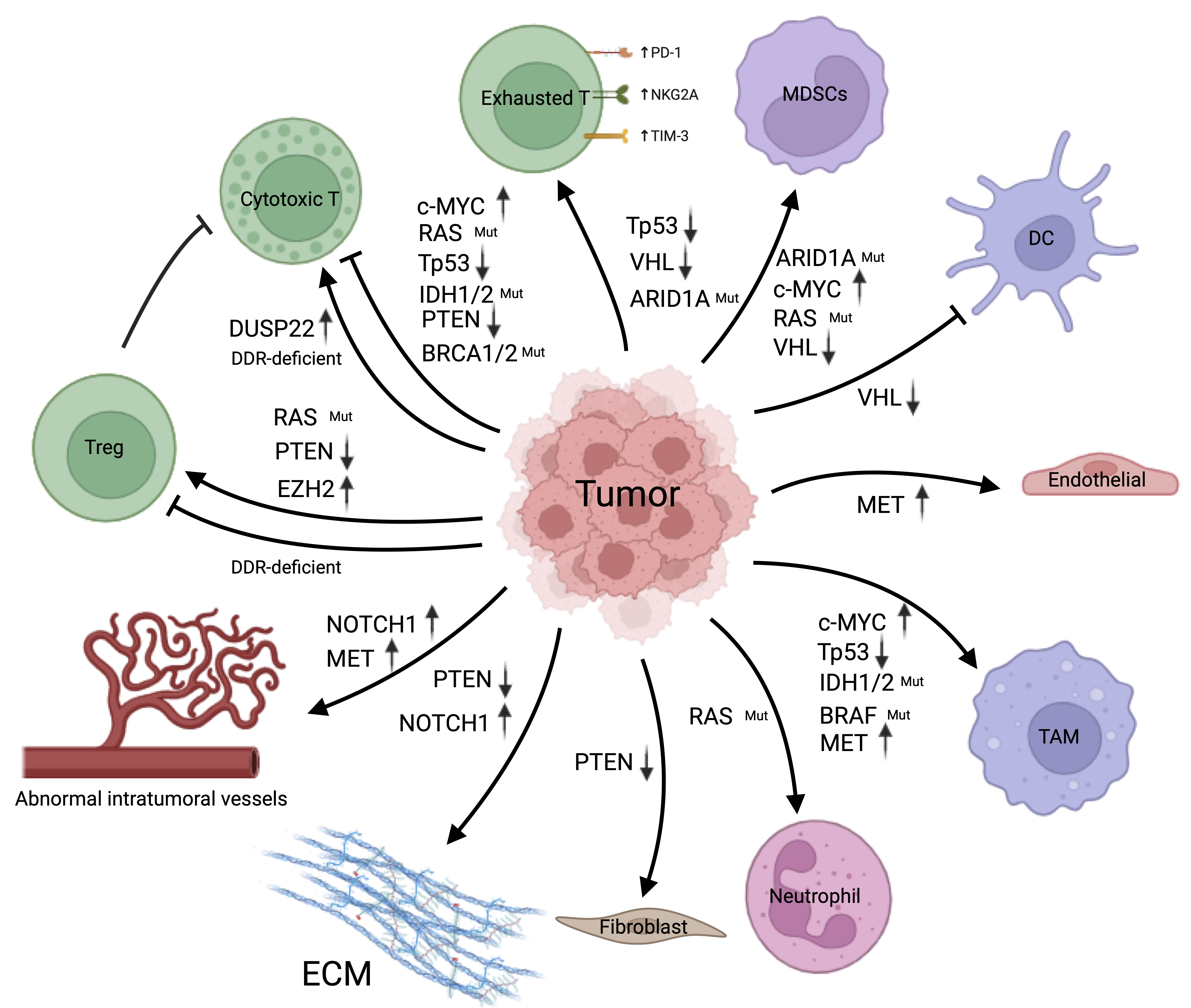
Cancer cells actively alter the tumor immune microenvironment rather than simply responding to immune attack. In the following sections, key oncogenic and tumor suppressor pathways are examined to demonstrate how cancer cell-intrinsic signaling shapes immune composition and activity within tumors.
Transcriptional oncogenes and immune checkpoint regulation
Transcriptional oncogenes play a central role in linking tumor proliferation programs with immune evasion by directly regulating immune checkpoints, cytokine networks, and stromal signaling pathways. Among these drivers, c-MYC is one of the most widely amplified oncogenes in human cancers. Beyond promoting cell-cycle progression and proliferation, MYC also coordinates tumor growth with immune suppression. MYC expression shows significant heterogeneity within tumors and contributes to intratumoral heterogeneity, which is closely linked to resistance to immunotherapy, metastasis, and recurrence (1). Mechanistically, MYC directly regulates immune checkpoint pathways and cytokine expression, thereby connecting oncogenic growth signals with immune evasion (2). MYC can directly bind to the PD-L1 promoter and increase its transcription, linking oncogene activation to immune checkpoint regulation (3). In non-small cell lung cancer, MYC expression is strongly correlated with PD-L1 levels, suggesting that tumors with both MYC and PD-L1 expression represent a separate immunological subtype (4). In addition to regulating immune checkpoints, MYC suppresses cytotoxic T-cell recruitment by modulating tumor-derived chemokine networks. In triple-negative breast cancer, MYC epigenetically represses STING expression via DNMT1-mediated mechanisms, reducing the expression of T-cell-attracting chemokines such as CCL5 and CXCL10 and thereby impairing T-cell infiltration (5). Pharmacological inhibition of MYC with JQ-1 enhances T-cell recruitment and activation, increases chemokine secretion, and synergizes with PD-1 blockade to boost antitumor immune responses (6). MYC also promotes recruitment of immunosuppressive myeloid populations by inducing cytokines such as IL-1β and IL-6, along with chemokines like CCL2, facilitating myeloid-derived suppressor cell (MDSC) accumulation within the TME (7–12). These MDSCs suppress T-cell proliferation and effector functions through multiple mechanisms, including direct cell contact, secretion of inhibitory cytokines, and metabolic disruption (13). Furthermore, MYC signaling promotes macrophage infiltration and polarization toward pro-tumorigenic M2 phenotypes by regulating factors such as CSF1 and metabolic pathways, including glycolysis-driven lactate accumulation, which induces epigenetic modifications, such as histone lactylation, in tumor-associated macrophages (14–17).
Other transcriptional oncogenes similarly reshape the immune microenvironment through interactions with stromal and endothelial cells. NOTCH1 signaling is a key regulator of tumor-stroma crosstalk, particularly in triple-negative breast cancer, where NOTCH1 activation upregulates Jagged-1 and stimulates Notch signaling in endothelial cells. This interaction promotes abnormal angiogenesis and induces stromal secretion of cytokines such as IL-1β, TGF-β, and SDF1α, thereby driving extracellular matrix remodeling and creating a desmoplastic microenvironment that excludes cytotoxic lymphocytes (18). Similarly, MET amplification contributes to immune remodeling by activating cancer-associated fibroblasts and endothelial cells through hepatocyte growth factor signaling. This process fosters extracellular matrix remodeling, macrophage recruitment via CSF-1 signaling, and polarization of tumor-associated macrophages toward immunosuppressive phenotypes, ultimately forming fibrotic tumor niches that limit T-cell infiltration and contribute to resistance to immunotherapy (19).
Canonical oncogenic signaling pathways
Classical oncogenic signaling pathways also exert substantial influence on immune composition within the tumor microenvironment. Mutations in RAS genes, including KRAS, NRAS, and HRAS, are among the most frequent oncogenic events in human cancers. KRAS mutations are especially common in pancreatic ductal adenocarcinoma, colorectal cancer, and non-small cell lung cancer (20, 21). Continuous activation of the RAS-RAF-MEK-ERK signaling pathway not only drives tumor growth but also alters immune networks by controlling cytokine and chemokine secretion (22, 23). RAS-activated tumors release factors like GM-CSF that attract immunosuppressive myeloid cells into the tumor microenvironment (24–27). In pancreatic cancer models, KRAS-driven tumor cells secrete GM-CSF, which promotes MDSC accumulation and thereby suppresses immune surveillance (28). Similar mechanisms are observed in hepatocellular carcinoma, where oncogenic NRAS signaling stimulates GM-CSF production, leading to the accumulation of inflammatory monocyte-derived cells that support tumor growth (29). Additionally, oncogenic RAS signaling can influence immune responses through stromal and inflammatory pathways; for example, RAS activation in skin tumor models leads to neutrophil recruitment, which in turn further stimulates tumor cell proliferation (30). These processes contribute to a “hot but suppressive” tumor environment characterized by immune cell infiltration along with functional immune suppression. RAS-driven signaling pathways can hamper T-cell activation by releasing immunosuppressive cytokines and fostering regulatory immune cells. For instance, KRAS-mutant pancreatic tumor cells express high levels of transglutaminase-2 and secrete cytokines such as G-CSF and GM-CSF, which inhibit T-cell activation (28, 31). Similarly, IL-33 produced by KRAS-mutant lung cancer supports regulatory T-cell growth and contributes to the exhaustion of cytotoxic CD8⁺ T cells and NK cells (32).
Other oncogenic signaling pathways also contribute to immune suppression through various mechanisms. The BRAF V600E mutation, a key driver in melanoma and colorectal cancer, promotes tumor microenvironment (TME) remodeling by sustained activation of the MAPK pathway. This signaling increases the expression of pro-angiogenic and chemotactic factors like VEGF and CCL2, which aid in recruiting monocytes and endothelial cells, while also promoting PD-L1 expression and immune evasion (33). Loss of PTEN similarly alters the tumor immune landscape by activating the PI3K-AKT-mTOR pathway. PTEN-deficient tumors display impaired antigen presentation, increased extracellular matrix remodeling, and more recruitment of immunosuppressive T-cell populations. These changes contribute to resistance to immune checkpoint blockade and encourage stromal activation via TGF-β signaling, leading to therapy-resistant tumor niches (34–36).
Tumor suppressor loss and immune remodeling
Loss of tumor suppressor genes frequently drives immune remodeling within tumors by altering cytokine signaling, antigen presentation, and immune cell recruitment. The tumor suppressor TP53 is one of the most frequently mutated genes in human cancer and functions as a central regulator of cell cycle control, apoptosis, and genomic stability (37). Mutations in TP53 often occur within the DNA-binding domain and disrupt the transcriptional activation of canonical p53 target genes (38). In addition to its traditional tumor-suppressive functions, TP53 loss significantly impacts the immune microenvironment. Mutant or deleted p53 promotes immunosuppressive macrophage polarization by inducing cytokines such as CSF-1, IL-10, and TGF-β, as well as by releasing exosomal microRNAs that reprogram macrophages toward suppressive phenotypes (39). Spatial and multi-omic analyses of lung tumors further reveal that TP53 mutations alter myeloid cell composition and promote the growth of pro-tumorigenic macrophage subpopulations associated with epithelial-mesenchymal transition and hypoxia signaling pathways (40). In addition to altering myeloid populations, TP53 loss hampers adaptive immune responses by impairing antigen presentation pathways. Loss of TP53 function reduces the expression of antigen-processing genes such as TAP1 and ERAP1, leading to lower MHC class I expression and reduced recognition by cytotoxic T cells (41). Mutant p53 can also facilitate immune evasion by increasing PD-L1 expression and suppressing interferon signaling, ultimately leading to T-cell exhaustion and regulatory T-cell expansion (42).
Other tumor suppressor alterations similarly reshape tumor immunity. In renal cell carcinoma, VHL inactivation activates HIF-1α signaling and promotes the expression of VEGF, PD-L1, and CXCR4, resulting in hypoxic and immunosuppressive tumor environments characterized by increased adenosine production and the recruitment of MDSCs and regulatory T cells (43). Mutations in ARID1A, a component of the SWI/SNF chromatin-remodeling complex, also impact immune signaling networks. ARID1A deficiency modifies chemokine expression and promotes PD-L1 upregulation via STAT3 signaling, leading to the expansion of exhausted PD-1⁺ T cells and the recruitment of regulatory T cells within the TME (44).
Genome instability and epigenetic drivers
Alterations that affect genome stability and epigenetic regulation further reshape tumor-immune interactions by modulating antigen presentation, interferon signaling, and immune checkpoint pathways. Mutations in IDH1 and IDH2 generate the oncometabolite 2-hydroxyglutarate, which blocks TET-mediated DNA demethylation and causes widespread epigenetic silencing of immune-related genes, including chemokines essential for T-cell recruitment (45). In gliomas, IDH mutations lower CXCL10 expression, reduce cytotoxic T-cell infiltration, and increase tumor-associated macrophage accumulation, leading to immunosuppressive tumor environments (45, 46).
Defects in BRCA1 and BRCA2, which impair homologous recombination DNA repair, lead to genomic instability and a higher neoantigen burden. However, the immunological effects of homologous recombination deficiency are intricate. While a higher mutation load can boost immunogenicity, the complete loss of BRCA1/2 may also suppress STING signaling and decrease immune infiltration despite increased neoantigen levels (47). Likewise, broader DNA damage repair (DDR) deficiencies activate innate immune signaling via the cGAS-STING pathway, triggering interferon responses and chemokine production that attract cytotoxic T cells. DDR-deficient tumors often show increased PD-L1 expression and may respond better to immune checkpoint blockade therapy (48).
Epigenetic regulators further contribute to immune remodeling. Overexpression of the histone methyltransferase EZH2 promotes tumor progression by silencing interferon-responsive genes and suppressing antitumor immune responses, leading to fewer tumor-infiltrating lymphocytes and increased regulatory T-cell accumulation (49). Finally, alterations in DUSP22, a dual-specificity phosphatase involved in immune signaling pathways, can influence immune checkpoint expression and T-cell infiltration. Loss of DUSP22 increases activation of EGFR and STAT3 signaling and enhances PD-L1 expression in lung cancer models, while DUSP22 overexpression can promote CD8⁺ T-cell infiltration and improve responses to immune checkpoint blockade (50–52).
Immunosuppressive Effects of Metabolic Reprogramming
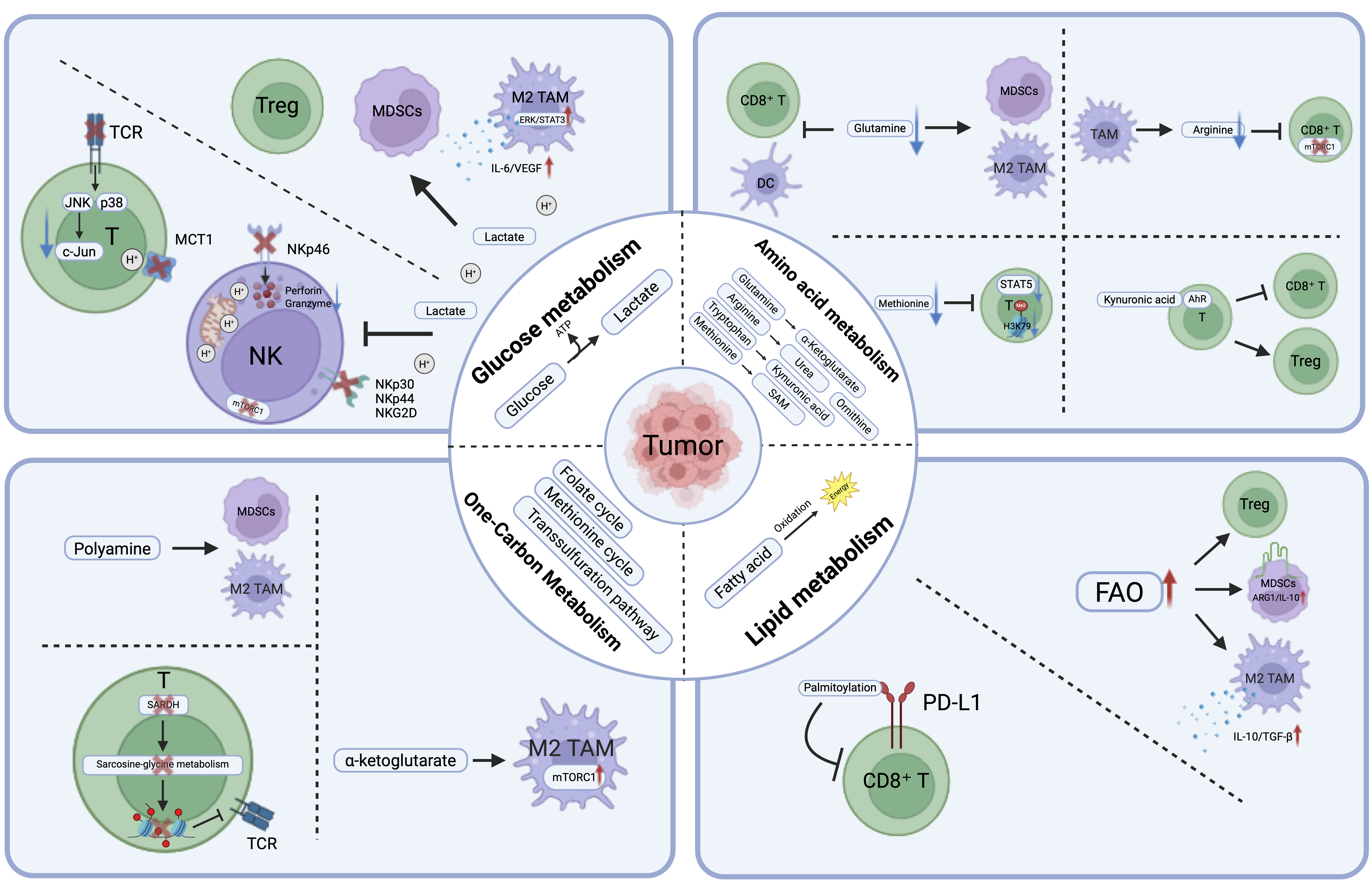
Metabolic reprogramming is a central mechanism by which cancer cells reshape the TME. In addition to supporting malignant cell growth, tumor-associated metabolic programs change nutrient levels, produce immunosuppressive metabolites, and reconfigure stromal and immune cell functions. These alterations hinder the activity of cytotoxic lymphocytes, encourage suppressive myeloid cell populations, and create metabolic barriers that restrict effective antitumor immunity. The following sections explain how major metabolic pathways, including glucose, amino acid, lipid, and one-carbon metabolism, contribute to immune evasion and tumor progression (Figure 3).
Glucose metabolism and lactate-driven immune suppression
Tumor cells undergo glucose metabolic reprogramming, characterized by aerobic glycolysis (the Warburg effect), in which they preferentially use glycolysis to produce ATP even when oxygen is sufficient. This metabolic state results in significant lactate accumulation (53, 54). Because lactate export relies on proton-coupled monocarboxylate transporters (MCTs), its release is accompanied by H+ efflux, which leads to acidification of the TME (55). This acidic, lactate-rich environment is closely associated with immune evasion, as it inhibits antitumor immune cells and promotes infiltration by immunosuppressive cell populations.
One major consequence of lactate accumulation is reduced T-cell activity. Lactate and low extracellular pH inhibit TCR-triggered signaling pathways, including p38 and JNK/c-Jun, which impairs CD8+ T-cell activation and antitumor function. Additionally, MCT1-mediated lactate transport is essential for normal T-cell metabolism, and disrupting this system decreases T-cell proliferation and cytokine production (55, 56). Competitive glucose depletion in the TME further limits nutrient supply for T cells and weakens their effector functions.
Lactate also exerts direct suppressive effects on NK cells. Accumulating lactate can decrease the expression of activating receptors such as NKp46, disrupt cytotoxic secretory pathways, and reduce perforin and granzyme production. In melanoma, high lactate dehydrogenase levels are linked to fewer NK cells and reduced cytokine secretion. TME acidification also downregulates activating receptors such as NKp30, NKp44, and NKG2D, impairs mitochondrial health, and promotes apoptosis, all of which reduce NK-cell recognition and killing ability. Additionally, lactate-driven metabolic stress inhibits mTORC1-dependent reprogramming, causing NK-cell dysfunction and exhaustion.
Meanwhile, lactate reshapes immune-cell composition by promoting immunosuppressive populations. In pancreatic cancer models, inhibiting LDHA decreases splenic MDSC frequency and enhances NK cell activity, whereas exogenous lactate promotes MDSC formation in bone marrow cultures. Lactate also drives M2 macrophage polarization through ERK/STAT3 signaling, resulting in the release of IL-6 and VEGF and further aiding immune evasion. Additionally, environments rich in lactate help Treg cells survive and function, creating a synergistic immunosuppressive network.
Amino acid metabolism and immunometabolic competition
Amino acid metabolism is another major determinant of tumor-immune interactions. In the TME, cancer cells deplete critical amino acids, compete directly with immune cells for metabolic resources, and generate immunosuppressive metabolites that impair lymphocyte and myeloid-cell function. Together, these processes create strong immunometabolic barriers that reduce antitumor immunity (Figure 2).
Among amino acid pathways, glutamine metabolism represents a central immunosuppressive hub. Tumor cells frequently overexpress glutaminase (GLS), converting glutamine into α-ketoglutarate to support biosynthesis and TCA-cycle activity, thereby promoting rapid proliferation. This glutamine consumption deprives CD8+ T cells of a critical substrate, weakening oxidative phosphorylation and reducing antitumor activity. However, inhibition of GLS can reduce MDSC infiltration, promote macrophage polarization toward a proinflammatory M1 phenotype, and restore immune surveillance (57). Tumor cells also compete with cDC1 dendritic cells for glutamine through the SLC38A2 transporter, and glutamine deprivation impairs antigen presentation and CD8+ T-cell activation (58, 59).
Arginine metabolism also plays a major role in T-cell dysfunction. Arginine is crucial for T-cell activation and proliferation, but tumor-associated macrophages frequently express high levels of arginase-1, which depletes arginine from the TME by converting it to urea and ornithine. This depletion of arginine hampers mTOR signaling, disrupts T-cell metabolic reprogramming, and prevents efficient transition from quiescence to activation, leading to decreased proliferation and cytokine production (60–62). In breast cancer, the metabolic interaction between cancer cells and macrophages makes arginine availability a key factor in disease progression (63). In this environment, tumor-derived arginine encourages pro-tumorigenic TAM polarization, while downstream polyamine metabolism further promotes this phenotype through thymine DNA glycosylase-dependent DNA demethylation, regulated by p53 signaling. Targeting this arginine-polyamine-TDG axis can significantly slow breast cancer growth (63).
Tryptophan metabolism further reinforces immunosuppression. Tryptophan is converted into kynurenine by indoleamine 2,3-dioxygenase (IDO) expressed in tumor cells or MDSCs. Kynurenine acts as an endogenous ligand for the aryl hydrocarbon receptor (AhR), activating signaling pathways that cause T-cell apoptosis, promote Treg differentiation, and inhibit effector T-cell function (64–66). Methionine metabolism also plays a role through an epigenetic mechanism. Tumor cells increase the expression of the methionine transporter SLC43A2, thereby depriving T cells of methionine and reducing H3K79me2 methylation and STAT5 signaling, leading to suppressed T-cell activation and proliferation (67). This metabolic hijacking sustains T-cell exhaustion and weakens antitumor immunity (58).
More broadly, amino acids also act as signaling molecules that control immune-cell fitness. When amino acids such as leucine and arginine are abundant, they activate mTORC1 via sensors including Sestrin2 and CASTOR1, thereby supporting T-cell growth and cytokine production (68, 69). Leucine alone can restore mTORC1 activity and induce metabolic reprogramming in T cells (70). Conversely, tumor-induced amino acid depletion triggers the GCN2-FBXO22 pathway, leading to mTOR ubiquitination and degradation, thereby reducing leucine uptake by T cells and ultimately causing T-cell dysfunction and exhaustion (71, 72). Under tryptophan-starved conditions, kynurenine-mediated activation of AhR promotes Treg differentiation and immunosuppression through coordinated upregulation of AhR and increased kynurenine intake (73–76). Additionally, new amino acid sensors such as TARS2 and HDAC6 can detect variations in threonine and valine levels, respectively, and adjust immune cell metabolism, indicating potential new therapeutic targets (77–79).
Lipid metabolism and immune dysfunction
Abnormal lipid metabolism represents a third major axis of immunometabolic remodeling in the TME. Tumor cells increase exogenous lipid uptake via receptors such as CD36 and FATP, upregulate enzymes such as ACLY and FASN to promote endogenous lipid synthesis, and redistribute lipids toward membrane biosynthesis, energy generation via fatty acid oxidation, and the synthesis of signaling molecules, such as prostaglandins (80). Over time, these changes lead to a lipid-rich, oxidative, and metabolically stressful environment that broadly influences tumor-immune interactions (Figure 2). One important consequence is dysfunction of effector T cells. Because glucose and other nutrients are limited in the TME, exhausted CD8+ T cells reduce glycolysis and exhibit impaired mitochondrial function, thereby reducing oxidative phosphorylation. Under these conditions, they become increasingly dependent on FAO to sustain residual effector activity (81–84). Similarly, enhanced FAO dependence in CD4+ T cells can promote differentiation into Tregs, thereby weakening antitumor immunity (85). Tumor cells also indirectly restrict T-cell metabolic plasticity by competing for glucose and glutamine (86, 87).
Lipid metabolism also shapes myeloid-cell behavior. Tumor-infiltrating MDSCs shift from glycolysis to FAO via CD36-mediated fatty acid uptake and increased expression of FAO enzymes, thereby maintaining high levels of immunosuppressive molecules such as ARG1 and IL-10 (88). Lipid metabolism similarly affects macrophage polarization, with M2-type TAMs utilizing FAO to produce ATP and acetyl-CoA, supporting TCA cycle activity and cholesterol synthesis, which promotes tumor progression (89). Long-chain fatty acids released from tumor-derived exosomes are taken up by macrophages through CD36 and activate ERK/STAT3 signaling, driving M2 polarization. These macrophages secrete IL-10 and TGF-β, facilitating immune evasion in liver metastasis models (90, 91). Lactate and amino acid metabolism can further reinforce this immunosuppressive M2 phenotype via ERK/STAT3 signaling (92, 93).
Another feature of lipid remodeling is the creation of a lipid peroxidation environment. The accumulation of ROS and lipids generates reactive lipid metabolites, including oxidized LDL, malondialdehyde, and 4-hydroxynonenal, which can damage immune cell membranes, interfere with signaling, and affect ferroptotic responses (94). Tumor cells adapt to environments with low glucose and high-lipid environments by increasing lipid uptake and storage in lipid droplets (95, 96). Although excessive accumulation of polyunsaturated fatty acids can trigger ferroptosis, some tumors, including non-small cell lung cancer, can resist ferroptotic death through paracrine mechanisms (97–99).
Finally, lipid-derived metabolites also regulate immune checkpoints through post-translational modification. For instance, palmitoylation of PD-L1 stabilizes the protein and maintains suppression of CD8+ T-cell cytotoxicity (99–101). Other acylation modifications driven by four-carbon metabolites, such as butyrylation and crotonylation, may also connect the metabolic state to gene regulation and signal transduction (102). Overall, these findings demonstrate how lipid metabolism contributes to immune evasion through both metabolic and epigenetic mechanisms.
One-carbon metabolism and epigenetic control of immune suppression
One-carbon metabolism is essential for nucleotide biosynthesis, methylation reactions, and cellular redox balance. By hijacking this pathway, tumor cells not only support their own proliferation but also establish immunosuppressive barriers within the TME (Figure 2).
One major consequence is the competition for the depletion of one-carbon units and related nutrients. One-carbon metabolism integrates the folate cycle, methionine cycle, and transsulfuration pathway and depends on amino acids such as serine, glycine, and methionine (103, 104). Tumor cells often overexpress enzymes such as SHMT2 and MTHFD2 to rapidly consume one-carbon units for nucleotide synthesis and methylation, thereby depriving immune cells of essential metabolic resources (105–107). Methionine deficiency in the TME greatly reduces CD8+ T cell survival and affects CD4+ T cells and Tregs. Conversely, supplementing methionine or S-adenosylmethionine can prevent T-cell apoptosis under tumor-bearing or IL-2-deficient conditions (67, 108).
One-carbon metabolism also contributes to metabolism-epigenetic crosstalk. Methionine metabolism generates S-adenosylmethionine, which is the universal methyl donor for epigenetic modifications. In immune cells, changes in one-carbon metabolism can directly influence DNA and RNA methylation states. For instance, dysregulated expression of sarcosine dehydrogenase (SARDH) shifts the sarcosine-glycine metabolic balance, reprograms cellular methylation patterns, suppresses TCR signaling, and weakens T-cell antitumor activity (109).
Furthermore, one-carbon metabolism supports the expansion and function of immunosuppressive cell populations. Tumor cells can deplete arginine levels by upregulating arginase and diverting one-carbon metabolic intermediates toward polyamine synthesis, thereby expanding MDSCs and polarizing M2 macrophages (110). The enzyme PHGDH in one-carbon metabolism also activates mTORC1 through α-ketoglutarate production, promoting M2-like TAM polarization (111). These findings position one-carbon metabolism as a crucial regulator of metabolic competition, epigenetic changes, and myeloid-mediated immune suppression. Figure 4 summarizes how tumor-associated metabolic reprogramming reshapes the tumor immune microenvironment by impairing effector immune cell function and promoting immunosuppressive cellular states.

Epigenetic Regulation
Epigenetic reprogramming is a key mechanism through which tumor cells modify the TME. By altering DNA methylation, histone modifications, and metabolism-linked chromatin remodeling, cancer cells can suppress antigen presentation, change chemokine expression, regulate immune checkpoint pathways, and promote the expansion or activity of immunosuppressive cell populations. These changes collectively enable immune evasion and contribute to therapeutic resistance.
DNA methylation and immune invisibility
During tumorigenesis, DNA methylation often exhibits a paradoxical pattern, with global hypomethylation alongside promoter-specific hypermethylation. Genome-wide CpG island hypomethylation leads to genomic instability and transposon activation, whereas hypermethylation of tumor suppressor genes and immune defense genes directly suppresses antitumor immune responses (112). Tumor cells frequently develop hypermethylated states through abnormal overexpression of DNMT1, DNMT3A, and DNMT3B, which silence tumor suppressor genes, antigen presentation genes such as B2M and HLA-DRA, and endogenous retroviruses (ERVs), all of which diminish T cell recognition of tumors. Methylation-regulated expression of immune genes can also influence immune cell infiltration; for instance, methylation of the LAG-3 promoter directly affects its expression in both tumor and immune cells. Conversely, inhibiting DNA methyltransferases can reactivate ERVs, trigger the dsRNA-interferon pathway, and restore antitumor immune signaling (113).
These methylation programs also have clinical importance. Microsatellite instability is often associated with improved immunotherapy outcomes, and in colorectal cancer, the CpG island methylator phenotype (CIMP) is an important molecular subtype characterized by widespread CpG island hypermethylation. CIMP is closely connected to microsatellite instability and BRAF or KRAS mutations, and it affects both prognosis and treatment response, making it a valuable clinical marker for patient stratification and therapy decisions.
Histone modifications and epigenetic control of tumor immunity
Histone methylation is a key epigenetic process that controls gene transcription through the coordinated actions of histone methyltransferases (HMTs) and histone demethylases (HDMs). Abnormal histone methylation can decrease tumor immunogenicity by silencing genes involved in antigen presentation, interferon signaling, and chemokine production, and by affecting the levels and differentiation of myeloid-derived suppressor cells, regulatory T cells, and tumor-associated macrophages.
Among HMTs, several enzymes inhibit tumor immunogenicity by silencing key antigen-presentation pathways. EZH2 suppresses MHC-I and MHC-II expression in diffuse large B-cell lymphoma and various cancers by depositing H3K27me3, thus directly impairing T-cell recognition of tumor antigens (114, 115). SETDB1 suppresses repetitive genomic elements and reduces neoantigen production from these sequences, thereby limiting immune recognition (116). HMTs also disrupt immune cell trafficking. For example, G9a suppresses interferon-induced chemokines such as CXCL9 and CXCL10 through H3K9me2/3, weakening CD8+ T-cell infiltration (117–120). In liver cancer, G9a also promotes MDSC chemotaxis by downregulating SLC7A2 (121). EZH2 in prostate cancer inhibits type I interferon signaling by blocking the RNA-STING-ISG pathway, further reducing immune activation and recruitment (122). Histone methyltransferases also regulate immune checkpoints and cell death pathways. KMT2A in pancreatic cancer and SETD2 in renal cell carcinoma promote PD-L1 transcription by activating H3K4me3 and H3K36me3 marks, while EZH2 and SETDB1 can suppress PD-L1 expression through H3K27me3 in a context-dependent manner (123). In colorectal cancer, G9a inhibits Fas transcription, thereby reducing Fas-FasL-mediated tumor cell killing, demonstrating how histone methylation can broadly influence susceptibility to immune attack (124). Histone demethylases also contribute to immune suppression through various mechanisms. KDM1A decreases MHC-I expression in melanoma, breast cancer, and other malignancies, and inhibits the dsRNA-type I interferon pathway by repressing ERV expression (125–127). KDM5B and KDM5C directly bind to the STING promoter and inhibit cGAS-STING signaling (128). KDM5A raises PD-L1 levels by suppressing PTEN and activating the PI3K-AKT-S6K pathway, while KDM4D amplifies STAT3-IRF1 signaling by coactivating IFNGR1 with SP-1, thus promoting PD-L1 transcription (129, 130). Additionally, KDM4C and KDM6B suppress CXCL10 or CXCL9/CXCL10 expression, limiting T-cell recruitment, and KDM4B negatively regulates immune infiltration in endometrial cancer (131–133).
Beyond their direct effects on tumor cells, HDMs also influence the function of immunosuppressive cells. KDM1A binds to the TGF-β1 promoter and promotes its expression, while synergizing with KLF5 and SMAD4 to suppress antitumor immunity. In CD8+ T cells, the LSD1/CoREST complex antagonizes TCF1 activity and modifies EOMES expression, thereby weakening T-cell function (134). KDM2A promotes JAG1 demethylation and activates Notch signaling, supporting Treg proliferation and activity (135). Along with KDM4D- and KDM5A-mediated signaling, these demethylases form part of a broader immunosuppressive network that facilitates tumor immune evasion (129, 130).
3. Cancer Cell-Derived Signals and Remodeling of the Immune Microenvironment
Cancer cells actively build microenvironments that support their survival, growth, invasion, metastasis, and immune evasion by releasing soluble factors and modifying the physical properties of the tumor microenvironment. These processes directly impact immune cell function, attract suppressive cell populations, change stromal composition, and create vascular and metabolic barriers that limit effective antitumor immunity.
Soluble factors and metabolic mediators of immune suppression
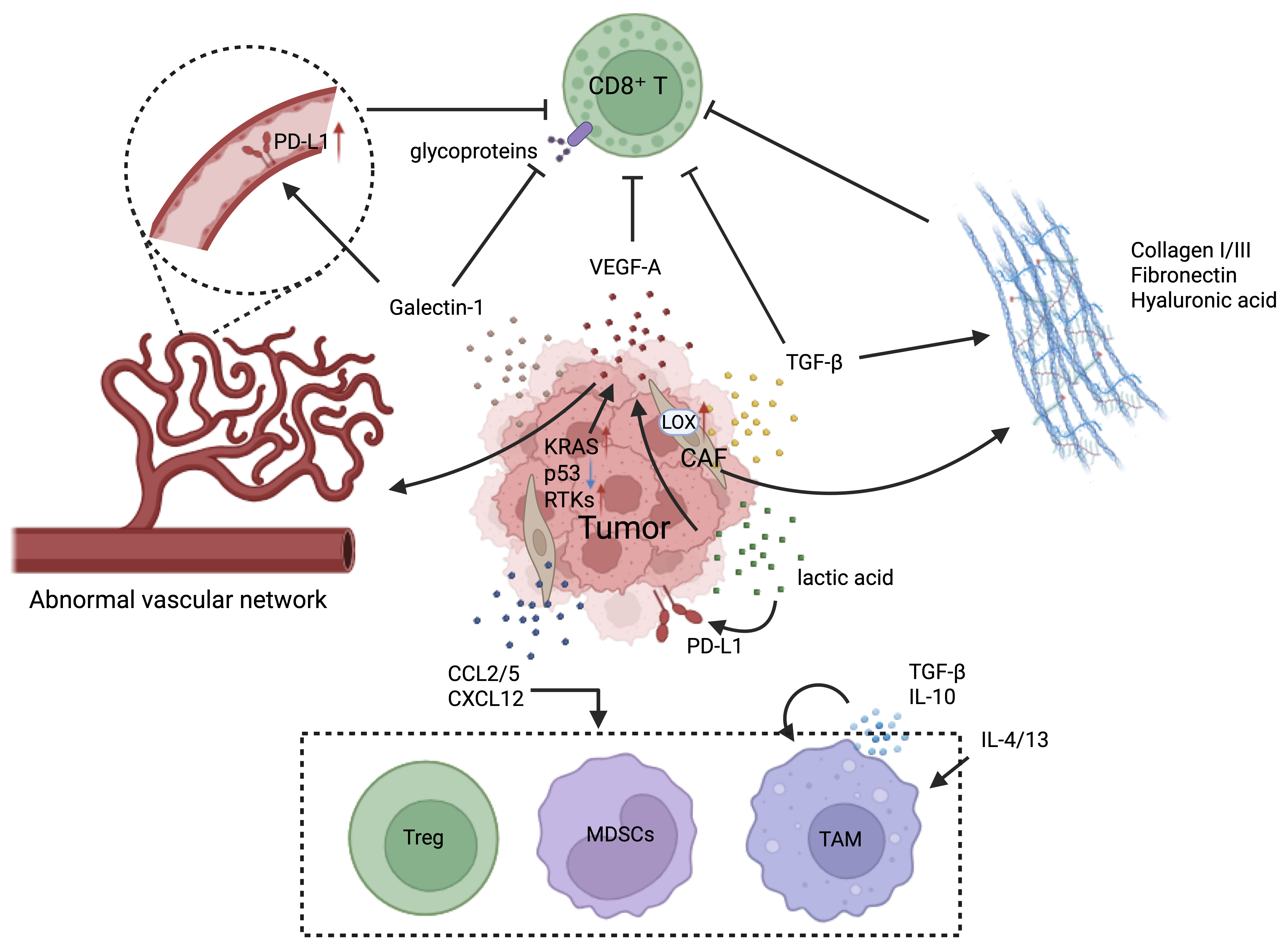
Cancer cells and their reprogrammed stromal partners, including cancer-associated fibroblasts, secrete a wide range of cytokines, chemokines, and metabolites that directly suppress effector immune cells and promote immunosuppressive cell populations. Among these factors, TGF-β is a central mediator of immune suppression. TGF-β strongly inhibits CD8+ T-cell proliferation, decreases expression of cytotoxic molecules such as perforin and granzyme B, interferes with T-cell metabolism and migration, and contributes to functional exhaustion. At the same time, TGF-β activates CAFs, which deposit extracellular matrix and form physical barriers that block T-cell infiltration, thus promoting immune exclusion. Other soluble mediators further strengthen suppression: galectin-1 directly induces apoptosis of activated T cells through interactions with cell-surface glycoproteins (136), and growing evidence indicates that the tumor-associated endothelium also functions as an active immunosuppressive barrier (137, 138). In this context, Gal1-mediated PD-L1 upregulation on vascular endothelial cells can physically restrict CD8+ T-cell infiltration (139, 140). In advanced ovarian cancer, VEGF-A directly inhibits T-cell proliferation and cytotoxic activity (141, 142), and additional studies support a broader role for tumor-induced T-cell exhaustion in this setting (143, 144). Meanwhile, cancer cells actively recruit and “tame” suppressive immune populations through chemokine signaling. Tumor-derived factors such as CCL2, CCL5, and CXCL12 recruit MDSCs, Tregs, and tumor-associated macrophages into the TME, where they are further polarized toward immunosuppressive states. For example, macrophages exposed to IL-4 and IL-13 develop pro-tumorigenic M2-like phenotypes and secrete TGF-β and IL-10, creating a self-reinforcing suppressive loop. In lung cancer, tumor-associated dendritic cells can cooperate by secreting HB-EGF and CXCL5, thereby promoting tumor growth, invasion, and migration (145).
Tumor-derived metabolites also create unfavorable physicochemical conditions for antitumor immunity. Aerobic glycolysis results in lactate accumulation and acidification of the TME, and recent research has highlighted additional signaling effects of lactate-dependent protein lactylation. Lactylation of HIF-1α increases its stability and transcriptional activity by reducing proteasomal degradation, thereby enhancing expression of hypoxia-response genes (146). In liver cancer, lactylation of PD-L1 at lysine 189 suppresses tumor growth, while delactylation promotes tumor progression through cholesterol synthesis driven by YY1-dependent upregulation of SQLE (147). Similarly, ASH2L-K312 lactylation is associated with tumor microvascular density and VEGFA expression in hepatocellular carcinoma and appears to promote tumor angiogenesis (148). More broadly, the TME causes immune dysfunction through both nutrient deprivation and metabolic waste accumulation: excessive tumor consumption of glucose and glutamine induces nutrient stress, while the accumulation of metabolites like adenosine generates suppressive signaling cues that hinder infiltrating immune cells (149–151).
Physical remodeling of the tumor microenvironment
In addition to soluble mediators, cancer cells reshape the physical architecture of the TME, creating immune-excluded and immunosuppressive niches. A key part of this process is extracellular matrix remodeling driven by cancer-associated fibroblasts. Recruited and activated CAFs secrete abundant matrix proteins, including collagen I/III, fibronectin, and hyaluronic acid, and facilitate crosslinking through enzymes such as lysyl oxidase and transglutaminases, which increases matrix stiffness (152). Under hypoxic conditions, elevated LOX expression further enhances collagen crosslinking, fostering conditions that promote recurrence and metastasis (153). Increased stiffness and altered fiber architecture impact both tumor and immune cells: YAP/TAZ activation promotes stem-like tumor phenotypes and immune evasion (154), while rigid collagen fibers form physical barriers that restrict CD8+ T-cell infiltration into the tumor core (155). Collagen has a dual role in the TME: intact fibrillar structures can impede immune-cell entry, whereas degradation products may activate immune responses. By interacting with receptors such as DDR1, DDR2, integrins, and LAIR-1, collagen can modulate immune cell exhaustion and immune evasion (156). Cancer cells also induce abnormal vasculature that acts as an immunosuppressive barrier. Through oncogenic signaling and stress responses in the microenvironment, including KRAS activation, p53 suppression, and hypoxia, tumor cells maintain VEGF production and promote the formation of aberrant vascular networks (157–161). Other receptor tyrosine kinases, such as EGFR, RET, PDGFR-α/β, and c-Kit, can also increase HIF-1α and VEGF-A levels in both hypoxic and normoxic conditions. For example, in EGFR-mutant non-small cell lung cancer, EGFR signaling can stimulate VEGF production even under normoxic conditions, contributing to a VEGF-dependent phenotype (162, 163).
Functionally, these abnormal vessels fail to effectively deliver cytotoxic T lymphocytes into the tumor core, thereby contributing to immune-desert phenotypes. Even when T-cell infiltration occurs, PD-L1 and FasL on vascular surfaces can induce exhaustion before T cells fully enter the TME (164). VEGF also directly promotes exhaustion by upregulating TOX expression in CD8+ T cells and by increasing checkpoint receptors such as TIM-3 and LAG-3 (165, 166). Therefore, stromal and vascular remodeling together strengthen the immunosuppressive barrier that shields tumors from effective immune attack.
4. Spatial Immunosuppression
Tumors are not homogeneous structures. Cancer cells in different regions of the tumor face unique metabolic conditions, stromal signals, and immune pressures. Consequently, they develop region-specific transcriptional programs and survival strategies that actively reshape the surrounding immune microenvironment. These spatially distinct tumor cell states create heterogeneous immune niches within the same tumor, affecting immune cell infiltration, immune suppression, and therapeutic responses.
Spatial heterogeneity between the tumor core and the invasive edge
Intrinsic program differences
Cancer cells in the tumor core and at the invasive edge have different intrinsic transcriptional programs that influence spatial immune heterogeneity. Mo and colleagues performed spatial transcriptomic analysis on 131 tumor sections from 78 cases across six types of cancer, including breast cancer (BRCA), colorectal cancer (CRC), pancreatic ductal adenocarcinoma (PDAC), renal cell carcinoma (RCC), uterine corpus endometrial carcinoma (UCEC), and cholangiocarcinoma (CHOL) (167). Their study found that tumor core regions show high expression of ribosome assembly genes from the RPL/RPS family and the long non-coding RNA SNHG29, indicating increased protein translation and a proliferation-focused cellular state. In contrast, tumor cells at the invasive edge are enriched for the glycolytic enzyme ENO1, the invasion-related protein TMSB10, and the immune regulatory factor ISG15, suggesting a dual program that combines migratory ability with immunomodulatory functions, thereby promoting M2 macrophage formation. Similar spatial transcriptional differences have been observed in colorectal cancer. Through integrated analysis of single-cell RNA sequencing with spatial transcriptomics, Xiao et al. showed clear differences between cancer cells in the tumor core and those at the invasive edge. Tumor-core cells mainly expressed genes associated with proliferation, matrix remodeling, and resistance to apoptosis, including TMSB4X, which has been linked to higher stemness and increased malignant potential (168). In contrast, cancer cells at the invasive edge exhibited transcriptional programs associated with cell migration and invasion, highlighting how spatial context influences functional heterogeneity within tumors.
Remodeling the immune microenvironment
These spatially distinct tumor cell programs are accompanied by equally distinct strategies of immune modulation. Cancer cells in the tumor core often face strong immune pressure and thus adopt mechanisms to actively suppress immune surveillance. A common approach is the upregulation of immune checkpoint molecules like PD-L1 and the secretion of inhibitory cytokines, including TGF-β. These strategies together inhibit the activity of cytotoxic immune cells and help form physical and biochemical barriers that prevent infiltration by NK cells and CD8⁺ T cells, creating what is often called an “immunosuppressive desert” within the tumor core (169–171). Cancer cells at the invasive edge often reshape the surrounding microenvironment by actively communicating with neighboring cells. These tumor cells release signaling molecules that alter stromal and immune cell behavior, thereby facilitating tumor growth. For instance, CXCL6⁺ tumor cells at the invasive front of liver cancer can activate the JAK-STAT3 pathway in nearby hepatocytes, prompting them to produce serum amyloid A proteins. These signals draw macrophages and promote their polarization toward an M2 pro-tumorigenic phenotype, creating a local niche that supports tumor progression and immune evasion (172).
Tertiary lymphoid structures in tumors
Another important spatial component of the tumor immune landscape is the presence of tertiary lymphoid structures (TLS). TLS are ectopic lymphoid aggregates composed of B cells, T cells, dendritic cells, and high endothelial venules that develop within tumor tissues (173). Unlike secondary lymphoid organs, TLS forms in settings of chronic inflammation and is seen in autoimmune diseases, chronic infections, and cancers. In tumors, TLS often serves as an active site for antitumor immune responses and is generally associated with increased T-cell activity and improved clinical outcomes (174). The development of TLS begins with the recruitment of lymphoid populations by signals released from lymphoid tissue inducer cells, including LTα and TNFα (173, 175). As TLS matures, they form organized structures containing B-cell follicles surrounded by T-cell zones, along with follicular dendritic cells and high endothelial venules that facilitate antigen capture and lymphocyte activation (176). Experimental models indicate that tumor cells can influence TLS formation. Joshi et al. reported spontaneous TLS development in a transgenic lung adenocarcinoma model driven by Kras LSL-G12D and Trp53 loss, showing that tumor-intrinsic oncogenic signaling can promote TLS formation (177). Other studies have found that TLS abundance correlates with tumor antigenicity. Rodriguez et al. demonstrated a positive association between antigen strength and the number of TLS (178), whereas Ng et al. reported that only highly mutated mouse lung cancer cell lines could induce TLS formation, likely due to increased tumor immunogenicity (179).
Conversely, TLS formation may be limited in specific tumor environments. In the transgenic KPC pancreatic cancer model (KrasG12D, p53R172H, Pdx-1-Cre), TLS formation is rare. However, local injection of chemokines such as CXCL13 and CCL21 in orthotopic pancreatic ductal adenocarcinoma models can trigger TLS development (180). These findings imply that TLS formation is not merely a passive immune response to tumor presence but rather a dynamic process within the microenvironment influenced by tumor antigenicity, oncogenic signaling, and chemokine-driven immune cell recruitment.
5. Discussion
This review emphasizes a conceptual shift in tumor immunology: cancer cells are not just passive targets of immune surveillance but active architects of the TIME. Through oncogenic signaling, metabolic reprogramming, and epigenetic regulation, cancer cells actively influence immune composition, inhibit antitumor responses, and create therapeutic resistance. While these insights have significantly advanced our understanding of tumor-immune interactions, turning them into long-lasting clinical benefits remains difficult. The complexity of TME changes, along with the metabolic and epigenetic flexibility of cancer cells, creates substantial barriers to therapy development. Future progress will depend on strategies that combine mechanistic insights with precise therapeutic design.
Translational challenges in targeting cancer cell-intrinsic immune regulation
Despite significant progress in understanding how cancer cell-intrinsic programs influence the TIME, translating these mechanistic insights into lasting clinical benefits remains difficult. A key challenge stems from the dynamic and diverse nature of tumor ecosystems. The TME changes both spatially and over time, creating different immune niches within the same tumor. Cancer cells in the tumor core and those at the invasive edge often exhibit distinct transcriptional programs and immunoregulatory strategies, leading to varied immune infiltration patterns and resistance mechanisms. Consequently, therapies targeting single pathways are frequently bypassed by alternative signaling networks or regional immune-escape mechanisms, thereby limiting the durability of treatment responses. Another key challenge involves tumors with sparse immune infiltration, commonly referred to as immunologically “cold” tumors. Often, immune-desert or immune-excluded phenotypes result from inherent genetic programs within cancer cells that suppress immune priming and recruitment (181). For instance, oncogenic c-MYC signaling can block chemokine secretion, while TP53 mutations disrupt antigen presentation and hinder the start of antitumor immune responses. Turning these tumors into immunologically “hot” states that respond to immunotherapy, therefore, requires treatments targeting the intrinsic drivers of immune exclusion. Simultaneously, strategies targeting metabolic or epigenetic reprogramming face additional complexity: many pathways used by cancer cells are also vital for normal immune cell function. Broadly blocking glycolysis, amino acid metabolism, or epigenetic regulators could cause systemic toxicity or inadvertently suppress antitumor immunity (182). For example, selectively blocking tumor glutamine metabolism without compromising the metabolic needs of activated T cells remains a major challenge in developing metabolic therapies.
Finally, although combination immunotherapy strategies hold some promise for overcoming resistance to immune checkpoint blockade, their effective design remains difficult. Many current combinations are mainly based on empirical reasoning or limited preclinical data. Because cancer cell-intrinsic drivers often operate through redundant and compensatory signaling pathways, blocking PD-1/PD-L1, along with a single metabolic or epigenetic target, may still be insufficient to produce lasting responses. Additionally, combination therapies can raise the risk of immune-related adverse events or promote the development of more aggressive tumor clones, which can ultimately lead to acquired resistance (183).
Future directions toward systemic remodeling of the tumor immune ecosystem
Addressing these challenges will require therapeutic strategies that systematically reshape the tumor immune ecosystem while targeting patient-specific vulnerabilities. Rather than focusing on isolated signaling pathways, future approaches should aim to intervene across multiple stages of the cancer-immunity cycle, including relieving tumor-induced immunosuppression, enhancing immune priming and recruitment, and restoring immune cell function within the TME.
Future therapeutic strategies must therefore move beyond single-target blockade to involve multi-level remodeling of the tumor immune environment. Rational combination approaches should integrate interventions that simultaneously weaken cancer cell-intrinsic immunosuppressive programs, enhance effective antigen presentation and immune cell recruitment, and restore the metabolic and functional capacity of antitumor lymphocytes.
Targeting cancer cell-intrinsic drivers at their source
Directly targeting cancer cell-intrinsic programs that establish and sustain the immunosuppressive tumor microenvironment is one of the most promising strategies for changing the tumor ecosystem at its core. Instead of merely counteracting downstream immunosuppressive effects, this approach targets the oncogenic drivers of immune evasion. One emerging strategy involves exploiting synthetic lethal interactions associated with challenging-to-target oncogenic pathways such as RAS and c-MYC. Because these oncogenes control extensive transcriptional and metabolic networks, their vulnerabilities may be found in downstream dependencies. Finding synthetic lethal partners among metabolic enzymes or epigenetic regulators could thus offer indirect strategies for targeting oncogene-driven cancers. Alternatively, treatments that interfere with oncogene-mediated immune regulation, such as blocking c-MYC-dependent transcription of immune checkpoint genes like PD-L1, might restore the immune system's ability to recognize tumor cells.
Epigenetic therapies offer another promising approach for reprogramming tumor immunogenicity. Drugs targeting epigenetic regulators, including EZH2 inhibitors and DNMT inhibitors, can reverse the silencing of immune-related genes in tumor cells and induce “viral mimicry” responses by activating endogenous retroviral elements, thereby stimulating innate immune signaling pathways (184). Emerging studies also suggest that combining epigenetic drugs with nanodelivery platforms may enhance tumor-targeted delivery while simultaneously activating the cGAS-STING pathway, promoting dendritic cell maturation, and increasing chemokine-mediated T-cell recruitment, ultimately resulting in synergistic immune activation (185). To contextualize these approaches within a broader therapeutic framework, multi-level combination strategies can be organized based on cancer cell-intrinsic drivers of immune evasion, as outlined in Table 1.
Table 1. Framework of Multi-Level Immunotherapeutic Strategies Targeting Cancer Cell-Intrinsic Drivers
| Intervention Level | Core Objective | Targeted Cancer Cell-Intrinsic Drivers | Potential Strategies and Representative Studies |
|---|---|---|---|
| Layer 1: Relieving Immunosuppression | Blocking direct suppressive signals established by cancer cells | PD-L1 expression (such as c-MYC and mutant p53-driven); metabolite-mediated suppression (lactate, kynurenine) | ICIs (anti-PD-1/PD-L1); IDO inhibitors; lactate transporter (MCT) inhibitors; targeting chemokine axes such as CCL2-CCR2 to suppress MDSC/TAM recruitment |
| Layer 2: Enhancing Immune Priming and Recruitment | Promoting antigen presentation and guiding effector cell homing to tumors | Antigen presentation defects (such as TP53 mutation); chemokine secretion suppression (such as c-MYC suppression of CXCL9/10) | STING agonists (activating cGAS-STING pathway, promoting type I interferon and chemokine production); epigenetic drugs (such as CARM1 inhibitors) activating tumor immune signals and upregulating T cell CXCR3 expression; personalized neoantigen vaccines |
| Layer 3: Improving Immune Cell Function and Survival | Reversing T cell exhaustion and providing metabolic support | Nutrient competition (glucose, amino acid depletion); immune checkpoint upregulation | Metabolic modulators (such as α-ketoglutarate supplementation, AMPK agonists); inhibitors targeting emerging immune checkpoints such as TIGIT and LAG-3; nanocarriers co-delivering siRNA and metabolic drugs |
Table 1: Multilayered therapeutic strategies targeting tumor-driven immune dysfunction. Summary of intervention strategies designed to overcome tumor-mediated immune suppression across three functional layers. Layer 1 (relieving immunosuppression) focuses on blocking direct suppressive signals established by cancer cells, including immune checkpoint activation and metabolite-mediated inhibition (e.g., lactate and kynurenine), with representative approaches such as immune checkpoint inhibitors (ICIs), IDO inhibitors, and chemokine axis targeting. Layer 2 (enhancing immune priming and recruitment) aims to restore antigen presentation and promote effector immune cell trafficking to tumors, addressing defects driven by oncogenic alterations (e.g., TP53 mutations and c-MYC-mediated chemokine suppression), using strategies such as STING agonists, epigenetic modulators, and personalized neoantigen vaccines. Layer 3 (improving immune cell function and survival) targets T-cell exhaustion and metabolic constraints within the tumor microenvironment, using approaches such as metabolic modulators, inhibitors of emerging immune checkpoints (e.g., TIGIT and LAG-3), and nanocarrier-based combinatorial delivery systems. Abbreviations: AMPK, AMP-activated protein kinase; CCL2, C-C motif chemokine ligand 2; CCR2, C-C chemokine receptor type 2; CXCL9/10, C-X-C motif chemokine ligand 9/10; IDO, indoleamine 2,3-dioxygenase; ICI, immune checkpoint inhibitor; MCT, monocarboxylate transporter; MDSC, myeloid-derived suppressor cell; siRNA, small interfering RNA; STING, stimulator of interferon genes; TAM, tumor-associated macrophage.
Precision patient stratification and dynamic monitoring
Overcoming tumor heterogeneity and enabling personalized immunotherapy will rely heavily on better patient stratification strategies. Traditional biomarkers such as PD-L1 expression and tumor mutational burden (TMB) offer only limited predictive accuracy, highlighting the need for multifaceted approaches that integrate genomic, transcriptional, and immune-ecosystem data.
One promising approach involves developing new biomarkers that reflect the dynamic state of the cancer-immunity cycle. For example, the modulators of immune infiltration (MOTIF) framework integrates multiple elements of immune regulation and has been shown to more accurately predict CD8⁺ T-cell infiltration and immunotherapy response than tumor mutational burden alone (186). Additionally, emerging evidence indicates that tumor-resident T-cell clones, those found within tumor tissues but absent from peripheral blood, are key factors in determining immunotherapy response (187).
Advances in imaging technologies also offer opportunities for non-invasive monitoring of TME evolution. Radiomics approaches combined with artificial intelligence can extract quantitative features from standard imaging modalities such as CT scans. Metrics such as the CT-TME score allow dynamic assessment of immune-inflammatory states within tumors throughout treatment, providing an alternative to repeated tissue biopsies and enabling real-time adjustment of therapeutic strategies (41).
Innovative delivery systems and spatially targeted interventions
Technological advances in drug delivery could further improve the targeting of tumor immune ecosystems. Nanotechnology-based delivery systems, including pH-responsive, enzyme-responsive, or light-activated nanocarriers, are increasingly being developed to enhance drug accumulation within tumors while reducing systemic toxicity. These platforms can deliver epigenetic drugs, metabolic modulators, and RNA-based therapeutics directly to tumor sites, enabling localized remodeling of the tumor microenvironment. For example, biomimetic nanovaccines that incorporate multiple tumor antigens and target dendritic cells have shown strong potential to boost antigen cross-presentation and induce durable antitumor immune responses (184).
Future therapeutic strategies may also benefit from spatially targeted interventions within tumors. Because different tumor regions, such as the tumor core, invasive edge, and TLS niches, exhibit distinct immune and stromal characteristics, therapies may need to be tailored to these specific areas. Methods such as intratumoral drug delivery or the development of agents that preferentially accumulate in specific tumor regions could help boost T-cell recruitment in immune-excluded zones, break down stromal barriers in tumor cores, and support TLS formation or maturation within tumors (180, 181).
In summary, recognizing cancer cells as active architects of their immune ecosystem fundamentally reshapes our understanding of tumor immunology. Instead of being passive targets of immune surveillance, tumor cells orchestrate complex networks of oncogenic signaling, metabolic reprogramming, and epigenetic regulation that together create immunosuppressive tumor microenvironments. Future immunotherapy strategies must therefore go beyond single immune checkpoint blockade and adopt integrated approaches to dismantle this ecosystem. Through thoughtfully designed combination therapies, interventions targeting cancer cell-intrinsic drivers, advanced technologies for precise patient stratification and real-time monitoring, and innovative delivery systems that enable spatially targeted therapy, it may become possible to gradually overcome the immunosuppressive barriers built by tumors and achieve longer-lasting clinical responses. Reaching this goal will require close collaboration among basic scientists, clinicians, computational biologists, and bioengineers to turn mechanistic insights into effective treatments.
References
1. Zhan H, Wang H, Pan B, Lu J, Xiao K, Lai J, et al. Reprogramming the tumor microenvironment with c-MYC-based gene circuit platform to enhance specific cancer immunotherapy. Nat Commun. 2025;16(1):7983. https://doi.org/10.1038/s41467-025-63377-3
2. Li Y, Casey SC, Felsher DW. Inactivation of MYC reverses tumorigenesis. J Intern Med. 2014;276(1):52–60. https://doi.org/10.1111/joim.12237
3. Casey SC, Tong L, Li Y, Do R, Walz S, Fitzgerald KN, et al. MYC regulates the antitumor immune response through CD47 and PD-L1. Science. 2016;352(6282):227–231. https://doi.org/10.1126/science.aac9935
4. Kim EY, Kim A, Kim SK, Chang YS. MYC expression correlates with PD-L1 expression in non-small cell lung cancer. Lung Cancer. 2017;110:63–67. https://doi.org/10.1016/j.lungcan.2017.06.006
5. Wu SY, Xiao Y, Wei JL, Xu XE, Jin X, Hu X, et al. MYC suppresses STING-dependent innate immunity by transcriptionally upregulating DNMT1 in triple-negative breast cancer. J Immunother Cancer. 2021;9(7):e002528. https://doi.org/10.1136/jitc-2021-002528
6. Jiang K, Zhang Q, Fan Y, Li J, Zhang J, Wang W, et al. MYC inhibition reprograms tumor immune microenvironment by recruiting T lymphocytes and activating the CD40/CD40L system in osteosarcoma. Cell Death Discov. 2022;8(1):117. https://doi.org/10.1038/s41420-022-00923-8
7. De Sanctis F, Adamo A, Canè S, Ugel S. Targeting tumour-reprogrammed myeloid cells: the new battleground in cancer immunotherapy. Semin Immunopathol. 2023;45(2):163–186. https://doi.org/10.1007/s00281-022-00965-1
8. Tannenbaum CS, Rayman PA, Pavicic PG, Kim JS, Wei W, Polefko A, et al. Mediators of inflammation-driven expansion, trafficking, and function of tumor-infiltrating MDSCs. Cancer Immunol Res. 2019;7(10):1687–1699. https://doi.org/10.1158/2326-6066.cir-18-0578
9. Hu H, Yang M, Huang B, Ding J, Zhu Y, Xu X, et al. Tumor cells promote immunosuppression in ovarian cancer via a positive feedback loop with MDSCs through the SAA1-IL-1β axis. J Exp Clin Cancer Res. 2025;44(1):277. https://doi.org/10.1186/s13046-025-03536-y
10. Silva-Romeiro S, Flores-Campos RDC, Sánchez-León ML, Sánchez-Margalet V, De la Cruz-Merino L. Emerging role of MDSCs as novel biomarkers and therapeutic targets for cancer immunotherapy. Immunotargets Ther. 2025;14:1267–1291. https://doi.org/10.2147/itt.S485642
11. Caetano MS, Zhang H, Cumpian AM, Gong L, Unver N, Ostrin EJ, et al. IL6 blockade reprograms the lung tumor microenvironment to limit the development and progression of K-ras-mutant lung cancer. Cancer Res. 2016;76(11):3189–3199. https://doi.org/10.1158/0008-5472.can-15-2840
12. Weber R, Riester Z, Hüser L, Sticht C, Siebenmorgen A, Groth C, et al. IL-6 regulates CCR5 expression and immunosuppressive capacity of MDSC in murine melanoma. J Immunother Cancer. 2020;8(2):e000949. https://doi.org/10.1136/jitc-2020-000949
13. Yenyuwadee S, Aliazis K, Wang Q, Christofides A, Shah R, Patsoukis N, et al. Immune cellular components and signaling pathways in the tumor microenvironment. Semin Cancer Biol. 2022;86(Pt2):187–201. https://doi.org/10.1016/j.semcancer.2022.08.004
14. Nguyen HT, Kan EL, Humayun M, Gurvich N, Offeddu GS, Wan Z, et al. Patient-specific vascularized tumor model: blocking monocyte recruitment with multispecific antibodies targeting CCR2 and CSF-1R. Biomaterials. 2025;312:122731. https://doi.org/10.1016/j.biomaterials.2024.122731
15. Zhou Y, Zeng J, Tu Y, Li L, Du S, Zhu L, et al. CSF1/CSF1R-mediated crosstalk between choroidal vascular endothelial cells and macrophages promotes choroidal neovascularization. Invest Ophthalmol Vis Sci. 2021;62(3):37. https://doi.org/10.1167/iovs.62.3.37
16. Nirala BK, Patel TD, Kurenbekova L, Shuck R, Dasgupta A, Rainusso N, et al. MYC regulates CSF1 expression via microRNA 17/20a to modulate tumor-associated macrophages in osteosarcoma. JCI Insight. 2023;8(13):e164947. https://doi.org/10.1172/jci.insight.164947
17. Dang T, You Y, Wei L, Li Q, Sun H, Sun M, et al. ICAT drives lactylation of tumor-associated macrophages via the c-Myc-ENO1 axis to promote cervical cancer progression. Free Radic Biol Med. 2025;241:316–329. https://doi.org/10.1016/j.freeradbiomed.2025.09.031
18. Colombo M, Garavelli S, Mazzola M, Platonova N, Giannandrea D, Colella R, et al. Multiple myeloma exploits Jagged1 and Jagged2 to promote intrinsic and bone marrow-dependent drug resistance. Haematologica. 2020;105(7):1925–1936. https://doi.org/10.3324/haematol.2019.221077
19. Xia Y, Huang C, Zhong M, Zhong H, Ruan R, Xiong J, et al. Targeting HGF/c-MET signaling to regulate the tumor microenvironment: implications for counteracting tumor immune evasion. Cell Commun Signal. 2025;23(1):46. https://doi.org/10.1186/s12964-025-02033-1
20. Simanshu DK, Nissley DV, McCormick F. RAS proteins and their regulators in human disease. Cell. 2017;170(1):17–33. https://doi.org/10.1016/j.cell.2017.06.009
21. Prior IA, Hood FE, Hartley JL. The frequency of Ras mutations in cancer. Cancer Res. 2020;80(14):2969–2974. https://doi.org/10.1158/0008-5472.can-19-3682
22. Bos JL. RAS oncogenes in human cancer: a review. Cancer Res. 1989;49(17):4682–4689. PMID: 2547513.
23. Downward J. RAS signalling pathways in cancer therapy. Nat Rev Cancer. 2003;3(1):11–22. https://doi.org/10.1038/nrc969
24. Sparmann A, Bar-Sagi D. Ras-induced interleukin-8 expression plays a critical role in tumor growth and angiogenesis. Cancer Cell. 2004;6(5):447–458. https://doi.org/10.1016/j.ccr.2004.09.028
25. Ancrile B, Lim KH, Counter CM. Oncogenic Ras-induced secretion of IL6 is required for tumorigenesis. Genes Dev. 2007;21(14):1714–1719. https://doi.org/10.1101/gad.1549407
26. Leslie K, Gao SP, Berishaj M, Podsypanina K, Ho H, Ivashkiv L, et al. Differential interleukin-6/Stat3 signaling as a function of cellular context mediates Ras-induced transformation. Breast Cancer Res. 2010;12(5):R80. https://doi.org/10.1186/bcr2725
27. Pylayeva-Gupta Y, Lee KE, Hajdu CH, Miller G, Bar-Sagi D. Oncogenic Kras-induced GM-CSF production promotes the development of pancreatic neoplasia. Cancer Cell. 2012;21(6):836–847. https://doi.org/10.1016/j.ccr.2012.04.024
28. Cox AD, Olive KP. Silencing the killers: paracrine immune suppression in pancreatic cancer. Cancer Cell. 2012;21(6):715–716. https://doi.org/10.1016/j.ccr.2012.05.029
29. Ramirez CFA, Taranto D, Ando-Kuri M, de Groot MHP, Tsouri E, Huang Z, et al. Cancer cell genetics shaping of the tumor microenvironment reveals myeloid cell-centric exploitable vulnerabilities in hepatocellular carcinoma. Nat Commun. 2024;15(1):2581. https://doi.org/10.1038/s41467-024-46835-2
30. Elliot AM, Ribeiro Bravo I, Zhao Y, Wang Z, Astorga Johansson J, Hutton E, et al. Oncogenic Ras activation in permissive somatic cells triggers rapid-onset phenotypic plasticity and elicits a tumor-promoting neutrophil response. Cell Rep. 2025;44(11):116478. https://doi.org/10.1016/j.celrep.2025.116478
31. Lahusen A, Minhöfer N, Lohse KA, Blechner C, Lindenmayer J, Eiseler T, et al. Pancreatic cancer cell-intrinsic transglutaminase-2 promotes T cell suppression through microtubule-dependent secretion of immunosuppressive cytokines. J Immunother Cancer. 2025;13(5):e010579. https://doi.org/10.1136/jitc-2024-010579
32. Kim BS, Clinton J, Wang Q, Chang SH. Targeting ST2 expressing activated regulatory T cells in Kras-mutant lung cancer. Oncoimmunology. 2020;9(1):1682380. https://doi.org/10.1080/2162402x.2019.1682380
33. Hua H, He W, Chen N, He Y, Wu G, Ye F, et al. Genomic and transcriptomic analysis of MSI-H colorectal cancer patients with targetable alterations identifies clinical implications for immunotherapy. Front Immunol. 2023;13:974793. https://doi.org/10.3389/fimmu.2022.974793
34. Huang K, Han Y, Chen Y, Shen H, Zeng S, Cai C. Tumor metabolic regulators: key drivers of metabolic reprogramming and the promising targets in cancer therapy. Mol Cancer. 2025;24(1):7. https://doi.org/10.1186/s12943-024-02205-6
35. Xie Z, Zhou Z, Yang S, Zhang S, Shao B. Epigenetic regulation and therapeutic targets in the tumor microenvironment. Mol Biomed. 2023;4(1):17. https://doi.org/10.1186/s43556-023-00126-2
36. Sivakumar S, Jainarayanan A, Arbe-Barnes E, Sharma PK, Leathlobhair MN, Amin S, et al. Distinct immune cell infiltration patterns in pancreatic ductal adenocarcinoma (PDAC) exhibit divergent immune cell selection and immunosuppressive mechanisms. Nat Commun. 2025;16(1):1397. https://doi.org/10.1038/s41467-024-55424-2
37. Mantovani F, Collavin L, Del Sal G. Mutant p53 as a guardian of the cancer cell. Cell Death Differ. 2019;26(2):199–212. https://doi.org/10.1038/s41418-018-0246-9
38. Chen X, Zhang T, Su W, Dou Z, Zhao D, Jin X, et al. Mutant p53 in cancer: from molecular mechanism to therapeutic modulation. Cell Death Dis. 2022;13(11):974. https://doi.org/10.1038/s41419-022-05408-1
39. Liu S, Liu T, Jiang J, Guo H, Yang R. p53 mutation and deletion contribute to tumor immune evasion. Front Genet. 2023;14:1088455. https://doi.org/10.3389/fgene.2023.1088455
40. Zhao W, Nguyen TT, Bhagwat A, Kumar A, Giotti B, Kepecs B, et al. A cellular and spatial atlas of TP53-associated tissue remodeling defines a multicellular tumor ecosystem in lung adenocarcinoma. Nat Cancer. 2025;6(11):1857–1879. https://doi.org/10.1038/s43018-025-01053-7
41. Bernatowicz K, Amat R, Prior O, Frigola J, Ligero M, Grussu F, et al. Radiomics signature for dynamic monitoring of tumor inflamed microenvironment and immunotherapy response prediction. J Immunother Cancer. 2025;13(1):e009140. https://doi.org/10.1136/jitc-2024-009140
42. Wang C, Tan JYM, Chitkara N, Bhatt S. TP53 mutation-mediated immune evasion in cancer: mechanisms and therapeutic implications. Cancers (Basel). 2024;16(17):3069. https://doi.org/10.3390/cancers16173069
43. Kim H, Shim BY, Lee SJ, Lee JY, Lee HJ, Kim IH. Loss of Von Hippel-Lindau (VHL) tumor suppressor gene function: VHL-HIF pathway and advances in treatments for metastatic renal cell carcinoma. Int J Mol Sci. 2021;22(18):9795. https://doi.org/10.3390/ijms22189795
44. Shen J, Ju Z, Zhao W. ARID1A deficiency promotes mutability and potentiates therapeutic antitumor immunity unleashed by immune checkpoint blockade. Nat Med. 2018;24(5):556–562. https://doi.org/10.1038/s41591-018-0012-z
45. Kessler J, Hohmann T, Güttler A, Petrenko M, Ostheimer C, Hohmann U, et al. Radiosensitization and a less aggressive phenotype of human malignant glioma cells expressing isocitrate dehydrogenase 1 (IDH1) mutant protein: dissecting the mechanisms. Cancers (Basel). 2019;11(6):889. https://doi.org/10.3390/cancers11060889
46. Kadiyala P, Carney SV, Gauss JC, Garcia-Fabiani MB, Haase S, Alghamri MS, et al. Inhibition of 2-hydroxyglutarate elicits metabolic reprogramming and mutant IDH1 glioma immunity in mice. J Clin Invest. 2021;131(4):e139542. https://doi.org/10.1172/jci139542
47. Kraya AA, Maxwell KN, Wubbenhorst B, Wenz BM, Pluta J, Rech AJ, et al. Genomic signatures predict the immunogenicity of BRCA-deficient breast cancer. Clin Cancer Res. 2019;25(14):4363–4374. https://doi.org/10.1158/1078-0432.ccr-18-0468
48. Shi C, Qin K, Lin A, Jiang A, Cheng Q, Liu Z, et al. The role of DNA damage repair (DDR) system in response to immune checkpoint inhibitor (ICI) therapy. J Exp Clin Cancer Res. 2022;41(1):268. https://doi.org/10.1186/s13046-022-02469-0
49. Sircar A, Singh S, Xu-Monette ZY, Coyle KM, Hilton LK, Chavdoula E, et al. Exploiting the fibroblast growth factor receptor-1 vulnerability to therapeutically restrict the MYC-EZH2-CDKN1C axis-driven proliferation in mantle cell lymphoma. Leukemia. 2023;37(10):2094–2106. https://doi.org/10.1038/s41375-023-02006-8
50. Lin HH, Chang CW, Liao YT, Yeh SD, Lin HP, Ho HM, et al. DUSP22 inhibits lung tumorigenesis by suppression of EGFR/c-Met signaling. Cell Death Discov. 2024;10(1):285. https://doi.org/10.1038/s41420-024-02038-8
51. Luchtel RA, Dasari S, Oishi N, Pedersen MB, Hu G, Rech KL, et al. Molecular profiling reveals immunogenic cues in anaplastic large cell lymphomas with DUSP22 rearrangements. Blood. 2018;132(13):1386–1398. https://doi.org/10.1182/blood-2018-03-838524
52. Wang L, Guo Y, Dai Y, Sun W, Huang X, Lei H, et al. DUSP22 dephosphorylates LGALS1 to enhance T cell-driven antitumor immunity. J Immunother Cancer. 2026;14(1):e013142. https://doi.org/10.1136/jitc-2025-013142
53. Hanahan D, Weinberg RA. Hallmarks of cancer: the next generation. Cell. 2011;144(5):646–674. https://doi.org/10.1016/j.cell.2011.02.013
54. Vander Heiden MG, Cantley LC, Thompson CB. Understanding the Warburg effect: the metabolic requirements of cell proliferation. Science. 2009;324(5930):1029–1033. https://doi.org/10.1126/science.1160809
55. Chen J, Huang Z, Chen Y, Tian H, Chai P, Shen Y, et al. Lactate and lactylation in cancer. Signal Transduct Target Ther. 2025;10(1):38. https://doi.org/10.1038/s41392-024-02082-x
56. Feng Q, Bennett Z, Grichuk A, Pantoja R, Huang T, Faubert B, et al. Severely polarized extracellular acidity around tumour cells. Nat Biomed Eng. 2024;8(7):787–799. https://doi.org/10.1038/s41551-024-01178-7
57. Liu PS, Wang H, Li X, Chao T, Teav T, Christen S, et al. α-ketoglutarate orchestrates macrophage activation through metabolic and epigenetic reprogramming. Nat Immunol. 2017;18(9):985–994. https://doi.org/10.1038/ni.3796
58. De Martino M, Rathmell JC, Galluzzi L, Vanpouille-Box C. Cancer cell metabolism and antitumour immunity. Nat Rev Immunol. 2024;24(10):654–669. https://doi.org/10.1038/s41577-024-01026-4
59. Guo C, You Z, Shi H, Sun Y, Du X, Palacios G, et al. SLC38A2 and glutamine signalling in cDC1s dictate anti-tumour immunity. Nature. 2023;620(7972):200–208. https://doi.org/10.1038/s41586-023-06299-8
60. Lin Z, Chang C, Zhao S, Fang L, Wang P. Amino acids shape the metabolic and immunologic landscape in the tumor immune microenvironment: from molecular mechanisms to therapeutic strategies. Cancer Biol Med. 2025;22(4):726–746. https://doi.org/10.20892/j.issn.2095-3941.2025.0115
61. Lv Y, Li Z, Liu S, Zhou Z, Song J, Ba Y, et al. Metabolic checkpoints in immune cell reprogramming: rewiring immunometabolism for cancer therapy. Mol Cancer. 2025;24(1):210. https://doi.org/10.1186/s12943-025-02407-6
62. Menjivar RE, Nwosu ZC, Du W, Donahue KL, Hong HS, Espinoza C, et al. Arginase 1 is a key driver of immune suppression in pancreatic cancer. eLife. 2023;12:e80721. https://doi.org/10.7554/elife.80721
63. Zhu Y, Zhou Z, Du X, Lin X, Liang ZM, Chen S, et al. Cancer cell-derived arginine fuels polyamine biosynthesis in tumor-associated macrophages to promote immune evasion. Cancer Cell. 2025;43(6):1045-1060.e7. https://doi.org/10.1016/j.ccell.2025.03.015
64. Srivastava S, Anbiaee R, Houshyari M, Laxmi, Sridhar SB, Ashique S, et al. Amino acid metabolism in glioblastoma pathogenesis, immune evasion, and treatment resistance. Cancer Cell Int. 2025;25(1):89. https://doi.org/10.1186/s12935-025-03721-1
65. Fallarino F, Grohmann U, Vacca C, Bianchi R, Orabona C, Spreca A, et al. T cell apoptosis by tryptophan catabolism. Cell Death Differ. 2002;9(10):1069–1077. https://doi.org/10.1038/sj.cdd.4401073
66. Chen W, Liang X, Peterson AJ, Munn DH, Blazar BR. The indoleamine 2,3-dioxygenase pathway is essential for human plasmacytoid dendritic cell-induced adaptive T regulatory cell generation. J Immunol. 2008;181(8):5396–5404. https://doi.org/10.4049/jimmunol.181.8.5396
67. Bian Y, Li W, Kremer DM, Sajjakulnukit P, Li S, Crespo J, et al. Cancer SLC43A2 alters T cell methionine metabolism and histone methylation. Nature. 2020;585(7824):277–282. https://doi.org/10.1038/s41586-020-2682-1
68. Wolfson RL, Chantranupong L, Saxton RA, Shen K, Scaria SM, Cantor JR, et al. Sestrin2 is a leucine sensor for the mTORC1 pathway. Science. 2016;351(6268):43–48. https://doi.org/10.1126/science.aab2674
69. Chantranupong L, Scaria SM, Saxton RA, Gygi MP, Shen K, Wyant GA, et al. The CASTOR proteins are arginine sensors for the mTORC1 pathway. Cell. 2016;165(1):153–164. https://doi.org/10.1016/j.cell.2016.02.035
70. Zhang Y, Hu H, Liu W, Yan SM, Li Y, Tan L, et al. Amino acids and RagD potentiate mTORC1 activation in CD8+ T cells to confer antitumor immunity. J Immunother Cancer. 2021;9(4):e002137. https://doi.org/10.1136/jitc-2020-002137
71. Dai X, Yan P, Wei W. Amino acid availability governs mTOR ubiquitination. Cell Res. 2024;34(4):335–336. https://doi.org/10.1038/s41422-023-00910-3
72. Ge MK, Zhang C, Zhang N, He P, Cai HY, Li S, et al. The tRNA-GCN2-FBXO22 axis-mediated mTOR ubiquitination senses amino acid insufficiency. Cell Metab. 2023;35(12):2216-2230.e8. https://doi.org/10.1016/j.cmet.2023.10.016
73. Solvay M, Holfelder P, Klaessens S, Pilotte L, Stroobant V, Lamy J, et al. Tryptophan depletion sensitizes the AHR pathway by increasing AHR expression and GCN2/LAT1-mediated kynurenine uptake, and potentiates induction of regulatory T lymphocytes. J Immunother Cancer. 2023;11(6):e006728. https://doi.org/10.1136/jitc-2023-006728
74. Mezrich JD, Fechner JH, Zhang X, Johnson BP, Burlingham WJ, Bradfield CA. An interaction between kynurenine and the aryl hydrocarbon receptor can generate regulatory T cells. J Immunol. 2010;185(6):3190–3198. https://doi.org/10.4049/jimmunol.0903670
75. Opitz CA, Litzenburger UM, Sahm F, Ott M, Tritschler I, Trump S, et al. An endogenous tumour-promoting ligand of the human aryl hydrocarbon receptor. Nature. 2011;478(7368):197–203. https://doi.org/10.1038/nature10491
76. Quintana FJ, Basso AS, Iglesias AH, Korn T, Farez MF, Bettelli E, et al. Control of Treg and TH17 cell differentiation by the aryl hydrocarbon receptor. Nature. 2008;453(7191):65–71. https://doi.org/10.1038/nature06880
77. Jin J, Meng T, Yu Y, Wu S, Jiao CC, Song S, et al. Human HDAC6 senses valine abundancy to regulate DNA damage. Nature. 2025;637(8046):215–223. https://doi.org/10.1038/s41586-024-08248-5
78. Kim SH, Choi JH, Wang P, Go CD, Hesketh GG, Gingras AC, et al. Mitochondrial threonyl-tRNA synthetase TARS2 is required for threonine-sensitive mTORC1 activation. Mol Cell. 2021;81(2):398–407.e4. https://doi.org/10.1016/j.molcel.2020.11.036
79. Sung Y, Yu YC, Han JM. Nutrient sensors and their crosstalk. Exp Mol Med. 2023;55(6):1076–1089. https://doi.org/10.1038/s12276-023-01006-z
80. Yang K, Wang X, Song C, He Z, Wang R, Xu Y, et al. The role of lipid metabolic reprogramming in tumor microenvironment. Theranostics. 2023;13(5):1774–1808. https://doi.org/10.7150/thno.82920
81. Frauwirth KA, Riley JL, Harris MH, Parry RV, Rathmell JC, Plas DR, et al. The CD28 signaling pathway regulates glucose metabolism. Immunity. 2002;16(6):769–777. https://doi.org/10.1016/S1074-7613(02)00323-0
82. Chamoto K, Chowdhury PS, Kumar A, Sonomura K, Matsuda F, Fagarasan S, et al. Mitochondrial activation chemicals synergize with surface receptor PD-1 blockade for T cell-dependent antitumor activity. Proc Natl Acad Sci U S A. 2017;114(5):E761–E770. https://doi.org/10.1073/pnas.1620433114
83. Hunt EG, Hurst KE, Riesenberg BP, Kennedy AS, Gandy EJ, Andrews AM, et al. Acetyl-CoA carboxylase obstructs CD8+ T cell lipid utilization in the tumor microenvironment. Cell Metab. 2024;36(5):969–983.e10. https://doi.org/10.1016/j.cmet.2024.02.009
84. Scharping NE, Menk AV, Moreci RS, Whetstone RD, Dadey RE, Watkins SC, et al. The tumor microenvironment represses T cell mitochondrial biogenesis to drive intratumoral T cell metabolic insufficiency and dysfunction. Immunity. 2016;45(2):701–713. https://doi.org/10.1016/j.immuni.2016.08.009
85. Siska PJ, van der Windt GJW, Kishton RJ, Cohen S, Eisner W, MacIver NJ, et al. Suppression of glut1 and glucose metabolism by decreased Akt/mTORC1 signaling drives T cell impairment in the tumor microenvironment. J Immunol. 2016 Sep 15;197(6):2532–40. https://doi.org/10.4049/jimmunol.1502464
86. Kedia-Mehta N, Finlay DK. Competition for nutrients and its role in controlling immune responses. Nat Commun. 2019;10(1):2123. https://doi.org/10.1038/s41467-019-10015-4
87. Wang Y, Liu M, Zhang L, Liu X, Ji H, Wang Y, et al. Cancer CD39 drives metabolic adaption and mal-differentiation of CD4+ T cells in patients with non-small-cell lung cancer. Cell Death Dis. 2023;14(12):804. https://doi.org/10.1038/s41419-023-06336-4
88. Wang H, Zhou F, Qin W, Yang Y, Li X, Liu R. Metabolic regulation of myeloid-derived suppressor cells in tumor immune microenvironment: targets and therapeutic strategies. Theranostics. 2025;15(5):2159–2184. https://doi.org/10.7150/thno.105276
89. Corn KC, Windham MA, Rafat M. Lipids in the tumor microenvironment: from cancer progression to treatment. Prog Lipid Res. 2020;80:101055. https://doi.org/10.1016/j.plipres.2020.101055
90. Li Y, You J, Zou Z, Sun G, Shi Y, Sun Y, et al. Decoding the tumor microenvironment: exosome-mediated macrophage polarization and therapeutic frontiers. Int J Biol Sci. 2025;21(11):4187–4214. https://doi.org/10.7150/ijbs.114222
91. Qian Y, Yin Y, Zheng X, Liu Z, Wang X. Metabolic regulation of tumor-associated macrophage heterogeneity: insights into the tumor microenvironment and immunotherapeutic opportunities. Biomark Res. 2024;12(1):1. https://doi.org/10.1186/s40364-023-00549-7
92. Xu B, Liu Y, Li N, Geng Q. Lactate and lactylation in macrophage metabolic reprogramming: current progress and outstanding issues. Front Immunol. 2024;15:1395786. https://doi.org/10.3389/fimmu.2024.1395786
93. Liang Y, He J, Chen X, Yin L, Yuan Q, Zeng Q, et al. The emerging roles of metabolism in the crosstalk between breast cancer cells and tumor-associated macrophages. Int J Biol Sci. 2023;19(15):4915–4930. https://doi.org/10.7150/ijbs.86039
94. Zhang X, Ye X, Jin H. Oxidized low-density lipoprotein as a potential target for enhancing immune checkpoint inhibitor therapy in microsatellite-stable colorectal cancer. Antioxidants (Basel). 2025;14(6):726. https://doi.org/10.3390/antiox14060726
95. Wellen KE, Thompson CB. Cellular metabolic stress: considering how cells respond to nutrient excess. Mol Cell. 2010;40(2):323–332. https://doi.org/10.1016/j.molcel.2010.10.004
96. Bensaad K, Favaro E, Lewis CA, Peck B, Lord S, Collins JM, et al. Fatty acid uptake and lipid storage induced by HIF-1α contribute to cell growth and survival after hypoxia-reoxygenation. Cell Rep. 2014;9(1):349–365. https://doi.org/10.1016/j.celrep.2014.08.056
97. Ye L, Wen X, Qin J, Zhang X, Wang Y, Wang Z, et al. Metabolism-regulated ferroptosis in cancer progression and therapy. Cell Death Dis. 2024;15(3):196. https://doi.org/10.1038/s41419-024-06584-y
98. Lancaster GI, Murphy AJ. Do physiological changes in fatty acid composition alter cellular ferroptosis susceptibility and influence cell function? J Lipid Res. 2025;66(4):100765. https://doi.org/10.1016/j.jlr.2025.100765
99. Wang P, Zhang S, Chen X, Yang XD, Huang S, Yin H, et al. Tumor cell-specific loss of GPX4 reprograms triacylglycerol metabolism to escape ferroptosis and impair antitumor immunity in non-small cell lung cancer. Protein Cell. 2025;pwaf101. https://doi.org/10.1093/procel/pwaf101
100. Li W, Li F, Zhang X, Lin HK, Xu C. Insights into the post-translational modification and its emerging role in shaping the tumor microenvironment. Signal Transduct Target Ther. 2021;6(1):422. https://doi.org/10.1038/s41392-021-00825-8
101. Yao H, Lan J, Li C, Shi H, Brosseau JP, Wang H, et al. Inhibiting PD-L1 palmitoylation enhances T-cell immune responses against tumours. Nat Biomed Eng. 2019;3(4):306–317. https://doi.org/10.1038/s41551-019-0375-6
102. Fang Y, Li X. Protein lysine four-carbon acylations in health and disease. J Cell Physiol. 2024;239(3):e30981. https://doi.org/10.1002/jcp.30981
103. Ren X, Wang X, Zheng G, Wang S, Wang Q, Yuan M, et al. Targeting one-carbon metabolism for cancer immunotherapy. Clin Transl Med. 2024;14(1):e1521. https://doi.org/10.1002/ctm2.1521
104. Sainero-Alcolado L, Liaño-Pons J, Ruiz-Pérez MV, Arsenian-Henriksson M. Targeting mitochondrial metabolism for precision medicine in cancer. Cell Death Differ. 2022;29(7):1304–1317. https://doi.org/10.1038/s41418-022-01022-y
105. Luo L, Zheng Y, Lin Z, Li X, Li X, Li M, et al. Identification of SHMT2 as a potential prognostic biomarker and correlating with immune infiltrates in lung adenocarcinoma. J Immunol Res. 2021;2021:6647122. https://doi.org/10.1155/2021/6647122
106. Cui L, Chen H, Zhao X. The prognostic significance of immune-related metabolic enzyme MTHFD2 in head and neck squamous cell carcinoma. Diagnostics (Basel). 2020;10(9):689. https://doi.org/10.3390/diagnostics10090689
107. Shang M, Yang H, Yang R, Chen T, Fu Y, Li Y, et al. The folate cycle enzyme MTHFD2 induces cancer immune evasion through PD-L1 up-regulation. Nat Commun. 2021;12(1):1940. https://doi.org/10.1038/s41467-021-22173-5
108. Hote PT, Sahoo R, Jani TS, Ghare SS, Chen T, Joshi-Barve S, et al. Ethanol inhibits methionine adenosyltransferase II activity and S-adenosylmethionine biosynthesis and enhances caspase-3-dependent cell death in T lymphocytes: relevance to alcohol-induced immunosuppression. J Nutr Biochem. 2008;19(6):384–391. https://doi.org/10.1016/j.jnutbio.2007.05.010
109. Si W, Cheng S, He H, Zhang Y, Miao Y, Yi D, et al. SARDH in the 1-C metabolism sculpts the T-cell fate and serves as a potential cancer therapeutic target. Cell Mol Immunol. 2025;22(7):1363–1378. https://doi.org/10.1038/s41423-025-01331-5
110. Miret JJ, Kirschmeier P, Koyama S, Zhu M, Li YY, Naito Y, et al. Suppression of myeloid cell arginase activity leads to therapeutic response in a NSCLC mouse model by activating anti-tumor immunity. J Immunother Cancer. 2019;7(1):32. https://doi.org/10.1186/s40425-019-0504-5
111. Cai Z, Li W, Hager S, Wilson JL, Afjehi-Sadat L, Heiss EH, et al. Targeting PHGDH reverses the immunosuppressive phenotype of tumor-associated macrophages through α-ketoglutarate and mTORC1 signaling. Cell Mol Immunol. 2024;21(3):448–465. https://doi.org/10.1038/s41423-024-01134-0
112. Zhu D, Zeng S, Su C, Li J, Xuan Y, Lin Y, et al. The interaction between DNA methylation and tumor immune microenvironment: from the laboratory to clinical applications. Clin Epigenetics. 2024;16(1):24. https://doi.org/10.1186/s13148-024-01633-x
113. Huang Z, Xu J, Wang Y, Fu Z, Zhang Y, Zhao Q, et al. The role of MHC molecules in cancer immunotherapy: insights into tumor immune evasion and therapeutic strategies. Crit Rev Oncol Hematol. 2025;218:105057. https://doi.org/10.1016/j.critrevonc.2025.105057
114. Ennishi D, Takata K, Béguelin W, Duns G, Mottok A, Farinha P, et al. Molecular and genetic characterization of MHC deficiency identifies EZH2 as therapeutic target for enhancing immune recognition. Cancer Discov. 2019;9(4):546–563. https://doi.org/10.1158/2159-8290.cd-18-1090
115. Burr ML, Sparbier CE, Chan KL, Chan YC, Kersbergen A, Lam EYN, et al. An evolutionarily conserved function of Polycomb silences the MHC class I antigen presentation pathway and enables immune evasion in cancer. Cancer Cell. 2019;36(4):385–401.e8. https://doi.org/10.1016/j.ccell.2019.08.008
116. Griffin GK, Wu J, Iracheta-Vellve A, Patti JC, Hsu J, Davis T, et al. Epigenetic silencing by SETDB1 suppresses tumour intrinsic immunogenicity. Nature. 2021;595(7866):309–314. https://doi.org/10.1038/s41586-021-03520-4
117. Kato S, Weng QY, Insco ML, Chen KY, Muralidhar S, Pozniak J, et al. Gain-of-function genetic alterations of G9a drive oncogenesis. Cancer Discov. 2020;10(7):980–997. https://doi.org/10.1158/2159-8290.cd-19-0532
118. Seier JA, Reinhardt J, Saraf K, Ng SS, Layer JP, Corvino D, et al. Druggable epigenetic suppression of interferon-induced chemokine expression linked to MYCN amplification in neuroblastoma. J Immunother Cancer. 2021;9(5):e001335. https://doi.org/10.1136/jitc-2020-001335
119. Spiliopoulou P, Spear S, Mirza H, Garner I, McGarry L, Grundland-Freile F, et al. Dual G9A/EZH2 inhibition stimulates antitumor immune response in ovarian high-grade serous carcinoma. Mol Cancer Ther. 2022;21(3):522–534. https://doi.org/10.1158/1535-7163.mct-21-0743
120. Kelly GM, Al-Ejeh F, McCuaig R, Casciello F, Ahmad Kamal N, Ferguson B, et al. G9a inhibition enhances checkpoint inhibitor blockade response in melanoma. Clin Cancer Res. 2021;27(9):2624–2635. https://doi.org/10.1158/1078-0432.ccr-20-3463
121. Xia S, Wu J, Zhou W, Zhang M, Zhao K, Liu J, et al. SLC7A2 deficiency promotes hepatocellular carcinoma progression by enhancing recruitment of myeloid-derived suppressor cells. Cell Death Dis. 2021;12(6):570. https://doi.org/10.1038/s41419-021-03853-y
122. Morel KL, Sheahan AV, Burkhart DL, Baca SC, Boufaied N, Liu Y, et al. EZH2 inhibition activates a dsRNA-STING-interferon stress axis that potentiates response to PD-1 checkpoint blockade in prostate cancer. Nat Cancer. 2021;2(4):444–456. https://doi.org/10.1038/s43018-021-00185-w
123. Lu M, Zhao B, Liu M, Wu L, Li Y, Zhai Y, et al. Pan-cancer analysis of SETD2 mutation and its association with the efficacy of immunotherapy. NPJ Precis Oncol. 2021;5(1):51. https://doi.org/10.1038/s41698-021-00193-0
124. Paschall AV, Yang D, Lu C, Choi JH, Li X, Liu F, et al. H3K9 trimethylation silences Fas expression to confer colon carcinoma immune escape and 5-fluorouracil chemoresistance. J Immunol. 2015;195(4):1868–1882. https://doi.org/10.4049/jimmunol.1402243
125. Nguyen EM, Taniguchi H, Chan JM, Zhan YA, Chen X, Qiu J, et al. Targeting lysine-specific demethylase 1 rescues major histocompatibility complex class I antigen presentation and overcomes programmed death-ligand 1 blockade resistance in SCLC. J Thorac Oncol. 2022;17(8):1014–1031. https://doi.org/10.1016/j.jtho.2022.05.014
126. Zhou Z, Van der Jeught K, Fang Y, Yu T, Li Y, Ao Z, et al. An organoid-based screen for epigenetic inhibitors that stimulate antigen presentation and potentiate T-cell-mediated cytotoxicity. Nat Biomed Eng. 2021;5(11):1320–1335. https://doi.org/10.1038/s41551-021-00805-x
127. Sheng W, LaFleur MW, Nguyen TH, Chen S, Chakravarthy A, Conway JR, et al. LSD1 ablation stimulates anti-tumor immunity and enables checkpoint blockade. Cell. 2018;174(3):549–563.e19. https://doi.org/10.1016/j.cell.2018.05.052
128. Wu L, Cao J, Cai WL, Lang SM, Horton JR, Jansen DJ, et al. KDM5 histone demethylases repress immune response via suppression of STING. PLoS Biol. 2018;16(8):e2006134. https://doi.org/10.1371/journal.pbio.2006134
129. Chen Q, Zhuang S, Hong Y, Yang L, Guo P, Mo P, et al. Demethylase JMJD2D induces PD-L1 expression to promote colorectal cancer immune escape by enhancing IFNGR1-STAT3-IRF1 signaling. Oncogene. 2022;41(10):1421–1433. https://doi.org/10.1038/s41388-021-02173-x
130. Rasmussen PB, Staller P. The KDM5 family of histone demethylases as targets in oncology drug discovery. Epigenomics. 2014;6(3):277–286. https://doi.org/10.2217/epi.14.14
131. Jie X, Chen Y, Zhao Y, Yang X, Xu Y, Wang J, et al. Targeting KDM4C enhances CD8+ T cell mediated antitumor immunity by activating chemokine CXCL10 transcription in lung cancer. J Immunother Cancer. 2022;10(2):e003716. https://doi.org/10.1136/jitc-2021-003716
132. Nagarsheth N, Peng D, Kryczek I, Wu K, Li W, Zhao E, et al. PRC2 epigenetically silences Th1-type chemokines to suppress effector T-cell trafficking in colon cancer. Cancer Res. 2016;76(2):275–282. https://doi.org/10.1158/0008-5472.can-15-1938
133. Zhang M, Liu Y, Hou S, Wang Y, Wang C, Yin Y, et al. KDM4B, a potential prognostic biomarker revealed by large-scale public databases and clinical samples in uterine corpus endometrial carcinoma. Mol Omics. 2022;18(5):506–519. https://doi.org/10.1039/d1mo00287b
134. Sheng W, Liu Y, Chakraborty D, Debo B, Shi Y. Simultaneous inhibition of LSD1 and TGFβ enables eradication of poorly immunogenic tumors with anti-PD-1 treatment. Cancer Discov. 2021;11(8):1970–1981. https://doi.org/10.1158/2159-8290.cd-20-0017
135. Zhong C, Tao B, Li X, Xiang W, Peng L, Peng T, et al. HOXA-AS2 contributes to regulatory T cell proliferation and immune tolerance in glioma through the miR-302a/KDM2A/JAG1 axis. Cell Death Dis. 2022;13(2):160. https://doi.org/10.1038/s41419-021-04471-4
136. Perillo NL, Pace KE, Seilhamer JJ, Baum LG. Apoptosis of T cells mediated by galectin-1. Nature. 1995;378(6558):736–739. https://doi.org/10.1038/378736a0
137. Motz GT, Santoro SP, Wang L-P, Garrabrant T, Lastra RR, Hagemann IS, et al. Tumor endothelium FasL establishes a selective immune barrier promoting tolerance in tumors. Nat Med. 2014;20(6):607–615. https://doi.org/10.1038/nm.3541
138. Demaria O, De Gassart A, Coso S, Gestermann N, Di Domizio J, Flatz L, et al. STING activation of tumor endothelial cells initiates spontaneous and therapeutic antitumor immunity. Proc Natl Acad Sci U S A. 2015;112(50):15408–15413. https://doi.org/10.1073/pnas.1512832112
139. Tan J, Fan W, Liu T, Zhu B, Liu Y, Wang S, et al. TREM2+ macrophages suppress CD8+ T-cell infiltration after transarterial chemoembolisation in hepatocellular carcinoma. J Hepatol. 2023;79(1):126–140. https://doi.org/10.1016/j.jhep.2023.02.032
140. Nambiar DK, Aguilera T, Cao H, Kwok S, Kong C, Bloomstein J, et al. Galectin-1-driven T cell exclusion in the tumor endothelium promotes immunotherapy resistance. J Clin Invest. 2019;129(12):5553–5567. https://doi.org/10.1172/jci129025
141. Ziogas AC, Gavalas NG, Tsiatas M, Tsitsilonis O, Politi E, Terpos E, et al. VEGF directly suppresses activation of T cells from ovarian cancer patients and healthy individuals via VEGF receptor type 2. Int J Cancer. 2012;130(4):857–864. https://doi.org/10.1002/ijc.26094
142. Gavalas NG, Tsiatas M, Tsitsilonis O, Politi E, Ioannou K, Ziogas AC, et al. VEGF directly suppresses activation of T cells from ascites secondary to ovarian cancer via VEGF receptor type 2. Br J Cancer. 2012;107(11):1869–1875. https://doi.org/10.1038/bjc.2012.468
143. Meder L, Schuldt P, Thelen M, Schmitt A, Dietlein F, Klein S, et al. Combined VEGF and PD-L1 blockade displays synergistic treatment effects in an autochthonous mouse model of small cell lung cancer. Cancer Res. 2018;78(15):4270–4281. https://doi.org/10.1158/0008-5472.can-17-2176
144. Voron T, Colussi O, Marcheteau E, Pernot S, Nizard M, Pointet AL, et al. VEGF-A modulates expression of inhibitory checkpoints on CD8+ T cells in tumors. J Exp Med. 2015;212(2):139–148. https://doi.org/10.1084/jem.20140559
145. Kuo PL, Huang MS, Hung JY, Chou SH, Chiang SY, Huang YF, et al. Synergistic effect of lung tumor-associated dendritic cell-derived HB-EGF and CXCL5 on cancer progression. Int J Cancer. 2014;135(1):96–108. https://doi.org/10.1002/ijc.28673
146. Li C, Fu C, Zhou W, Li H, Liu Z, Wu G, et al. Lactylation modification of HIF-1α enhances its stability by blocking VHL recognition. Cell Commun Signal. 2025;23(1):364. https://doi.org/10.1186/s12964-025-02366-x
147. Wang X, Li Y, Tang Y, Liu Z, Liu Y, Fu X, et al. PD-L1 delactylation-promoted nuclear translocation accelerates liver cancer growth through elevating SQLE transcription activity. Cancer Lett. 2025;630:217901. https://doi.org/10.1016/j.canlet.2025.217901
148. Han H, Wang S, Ma L, Yin H, Cheng X, Wang Y, et al. ASH2L-K312-Lac stimulates angiogenesis in tumors to expedite the malignant progression of hepatocellular carcinoma. Adv Sci (Weinh). 2025;12(40):e09477. https://doi.org/10.1002/advs.202509477
149. Heintzman DR, Fisher EL, Rathmell JC. Microenvironmental influences on T cell immunity in cancer and inflammation. Cell Mol Immunol. 2022;19(3):316–326. https://doi.org/10.1038/s41423-021-00833-2
150. Cheng J, Yan J, Liu Y, Shi J, Wang H, Zhou H, et al. Cancer-cell-derived fumarate suppresses the anti-tumor capacity of CD8+ T cells in the tumor microenvironment. Cell Metab. 2023;35(6):961–978.e10. https://doi.org/10.1016/j.cmet.2023.04.017
151. Wen Y, Hou Y, Yi X, Sun S, Guo J, He X, et al. EZH2 activates CHK1 signaling to promote ovarian cancer chemoresistance by maintaining the properties of cancer stem cells. Theranostics. 2021;11(4):1795–1813. https://doi.org/10.7150/thno.48101
152. Du W, Xia X, Hu F, Yu J. Extracellular matrix remodeling in the tumor immunity. Front Immunol. 2023;14:1340634. https://doi.org/10.3389/fimmu.2023.1340634
153. Prakash J, Shaked Y. The interplay between extracellular matrix remodeling and cancer therapeutics. Cancer Discov. 2024;14(8):1375–1388. https://doi.org/10.1158/2159-8290.cd-24-0002
154. Mai Z, Lin Y, Lin P, Zhao X, Cui L. Modulating extracellular matrix stiffness: a strategic approach to boost cancer immunotherapy. Cell Death Dis. 2024;15(5):307. https://doi.org/10.1038/s41419-024-06697-4
155. Peng DH, Rodriguez BL, Diao L, Chen L, Wang J, Byers LA, et al. Collagen promotes anti-PD-1/PD-L1 resistance in cancer through LAIR1-dependent CD8+ T cell exhaustion. Nat Commun. 2020;11(1):4520. https://doi.org/10.1038/s41467-020-18298-8
156. Sun X, Wu B, Chiang HC, Deng H, Zhang X, Xiong W, et al. Tumour DDR1 promotes collagen fibre alignment to instigate immune exclusion. Nature. 2021;599(7886):673–678. https://doi.org/10.1038/s41586-021-04057-2
157. Chen Z, Han F, Du Y, Shi H, Zhou W. Hypoxic microenvironment in cancer: molecular mechanisms and therapeutic interventions. Signal Transduct Target Ther. 2023;8(1):70. https://doi.org/10.1038/s41392-023-01332-8
158. Folkman J. Tumor angiogenesis: therapeutic implications. N Engl J Med. 1971;285(21):1182–1186. https://doi.org/10.1056/nejm197111182852108
159. Ribatti D. Judah Folkman, a pioneer in the study of angiogenesis. Angiogenesis. 2008;11(1):3–10. https://doi.org/10.1007/s10456-008-9092-6
160. Patel SA, Nilsson MB, Le X, Cascone T, Jain RK, Heymach JV. Molecular mechanisms and future implications of VEGF/VEGFR in cancer therapy. Clin Cancer Res. 2023;29(1):30–39. https://doi.org/10.1158/1078-0432.ccr-22-1366
161. Rak J, Mitsuhashi Y, Bayko L, Filmus J, Shirasawa S, Sasazuki T, et al. Mutant ras oncogenes upregulate VEGF/VPF expression: implications for induction and inhibition of tumor angiogenesis. Cancer Res. 1995;55(20):4575–4580. PMID:7553632
162. Nilsson MB, Zage PE, Zeng L, Xu L, Cascone T, Wu HK, et al. Multiple receptor tyrosine kinases regulate HIF-1alpha and HIF-2alpha in normoxia and hypoxia in neuroblastoma: implications for antiangiogenic mechanisms of multikinase inhibitors. Oncogene. 2010;29(20):2938–2949. https://doi.org/10.1038/onc.2010.60
163. Nilsson MB, Robichaux J, Herynk MH, Cascone T, Le X, Elamin Y, et al. Altered regulation of HIF-1α in naive- and drug-resistant EGFR-mutant NSCLC: implications for a vascular endothelial growth factor-dependent phenotype. J Thorac Oncol. 2021;16(3):439–451. https://doi.org/10.1016/j.jtho.2020.11.022
164. Allen E, Jabouille A, Rivera LB, Lodewijckx I, Missiaen R, Steri V, et al. Combined antiangiogenic and anti-PD-L1 therapy stimulates tumor immunity through HEV formation. Sci Transl Med. 2017;9(385):eaak9679. https://doi.org/10.1126/scitranslmed.aak9679
165. Lee WS, Yang H, Chon HJ, Kim C. Combination of anti-angiogenic therapy and immune checkpoint blockade normalizes vascular-immune crosstalk to potentiate cancer immunity. Exp Mol Med. 2020;52(9):1475–1485. https://doi.org/10.1038/s12276-020-00500-y
166. Kim CG, Jang M, Kim Y, Leem G, Kim KH, Lee H, et al. VEGF-A drives TOX-dependent T cell exhaustion in anti-PD-1-resistant microsatellite stable colorectal cancers. Sci Immunol. 2019;4(41):eaay0555. https://doi.org/10.1126/sciimmunol.aay0555
167. Mo CK, Liu J, Chen S, Storrs E, Targino da Costa ALN, Houston A, et al. Tumour evolution and microenvironment interactions in 2D and 3D space. Nature. 2024;634(8036):1178–1186. https://doi.org/10.1038/s41586-024-08087-4
168. Xiao J, Yu X, Meng F, Zhang Y, Zhou W, Ren Y, et al. Integrating spatial and single-cell transcriptomics reveals tumor heterogeneity and intercellular networks in colorectal cancer. Cell Death Dis. 2024;15(5):326. https://doi.org/10.1038/s41419-024-06598-6
169. Xie L, Kong H, Yu J, Sun M, Lu S, Zhang Y, et al. Spatial transcriptomics reveals heterogeneity of histological subtypes between lepidic and acinar lung adenocarcinoma. Clin Transl Med. 2024;14(2):e1573. https://doi.org/10.1002/ctm2.1573
170. Tavernari D, Battistello E, Dheilly E, Petruzzella AS, Mina M, Sordet-Dessimoz J, et al. Nongenetic evolution drives lung adenocarcinoma spatial heterogeneity and progression. Cancer Discov. 2021;11(6):1490–1507. https://doi.org/10.1158/2159-8290.cd-20-1274
171. Xiong X, Zheng LW, Ding Y, Chen YF, Cai YW, Wang LP, et al. Breast cancer: pathogenesis and treatments. Signal Transduct Target Ther. 2025;10(1):49. https://doi.org/10.1038/s41392-024-02108-4
172. Wu L, Yan J, Bai Y, Chen F, Zou X, Xu J, et al. An invasive zone in human liver cancer identified by Stereo-seq promotes hepatocyte-tumor cell crosstalk, local immunosuppression and tumor progression. Cell Res. 2023;33(7):585–603. https://doi.org/10.1038/s41422-023-00831-1
173. Sautès-Fridman C, Petitprez F, Calderaro J, Fridman WH. Tertiary lymphoid structures in the era of cancer immunotherapy. Nat Rev Cancer. 2019;19(6):307–325. https://doi.org/10.1038/s41568-019-0144-6
174. Schumacher TN, Thommen DS. Tertiary lymphoid structures in cancer. Science. 2022;375(6576):eabf9419. https://doi.org/10.1126/science.abf9419
175. Cheng R, Li S, Ma X, Zhuang W, Lei Y, He J, et al. Intratumoral antigen-presenting cell activation by a nanovesicle for the concurrent tertiary lymphoid structure de novo neogenesis. Sci Adv. 2025;11(8):eadr1299. https://doi.org/10.1126/sciadv.adr1299
176. Gao X, Zhao X, Li X, Zhang J, Zhao H, Ma Y. Editorial: Tertiary lymphoid structures (TLS) in the tumor immune microenvironment. Front Immunol. 2025;16:1555677. https://doi.org/10.3389/fimmu.2025.1555677
177. Lu Y, Zhao Q, Liao JY, Song E, Xia Q, Pan J, et al. Complement signals determine opposite effects of B cells in chemotherapy-induced immunity. Cell. 2020;180(6):1081–1097.e24. https://doi.org/10.1016/j.cell.2020.02.015
178. Lee-Chang C, Miska J, Hou D, Rashidi A, Zhang P, Burga RA, et al. Activation of 4-1BBL+ B cells with CD40 agonism and IFNγ elicits potent immunity against glioblastoma. J Exp Med. 2021;218(1):e20200913. https://doi.org/10.1084/jem.20200913
179. Chisca M, Choutka C, Kassiotis G. Immunogenicity of endogenous retroviruses in health and disease. Immunol Rev. 2024;317(1):168–189. https://doi.org/10.1111/imr.13378
180. Bruno TC, Ebner PJ, Moore BL, Squalls OG, Waugh KA, Eruslanov EB, et al. Antigen-presenting intratumoral B cells affect CD4+ TIL phenotypes in non-small cell lung cancer patients. Cancer Immunol Res. 2017;5(10):898–907. https://doi.org/10.1158/2326-6066.cir-17-0075
181. Ma K, Wang L, Li W, Tang T, Ma B, Zhang L, et al. Turning cold into hot: emerging strategies to fire up the tumor microenvironment. Trends Cancer. 2025;11(2):117–134. https://doi.org/10.1016/j.trecan.2024.11.011
182. Xiao C, Xiong W, Xu Y, Zou J, Zeng Y, Liu J, et al. Immunometabolism: a new dimension in immunotherapy resistance. Front Med (Lausanne). 2023;17(4):585–616. https://doi.org/10.1007/s11684-023-1012-z
183. Almawash S. Revolutionary cancer therapy for personalization and improved efficacy: strategies to overcome resistance to immune checkpoint inhibitor therapy. Cancers (Basel). 2025;17(5):880. https://doi.org/10.3390/cancers17050880
184. You Q, Wu G, Li H, Liu J, Cao F, Ding L, et al. A nanovaccine targeting cancer stem cells and bulk cancer cells for postoperative cancer immunotherapy. Nat Nanotechnol. 2025;20(8):1298–1311. https://doi.org/10.1038/s41565-025-01952-x
185. Zhao X, Qin X, Wang R, Wang Y, He Y, Wang Y, et al. An epigenetic nanoagonist facilitates T cell priming, recruitment, and reinvigoration in tumors resistant to PD-L1 therapy. Adv Mater. 2025;37(45):e02800. https://doi.org/10.1002/adma.202502800
186. Qian ZY, Pan YQ, Li XX, Chen YX, Wu HX, Liu ZX, et al. Modulator of TMB-associated immune infiltration (MOTIF) predicts immunotherapy response and guides combination therapy. Sci Bull (Beijing). 2024;69(6):803–822. https://doi.org/10.1016/j.scib.2024.01.025
187. Shorer O, Pinhasi A, Yizhak K. Single-cell meta-analysis of T cells reveals clonal dynamics of response to checkpoint immunotherapy. Cell Genom. 2025;5(5):100842. https://doi.org/10.1016/j.xgen.2025.100842
Declarations
Funding Statement
This work was supported by funding from The Science and Technology Development Fund, Macau S.A.R (FDCT) under grants 0087/2024/RIB2 and 0109/2025/RIA2.
Competing Interest
The authors declare no financial or personal relationships with other individuals or organizations that could inappropriately influence or bias the content of this work. All authors have read the final version of the manuscript and confirm that there are no competing interests.
Consent for Publication
Consent for publication: All authors have approved the final version of the manuscript.
Use of Artificial Intelligence Disclosure
This article was written by human contributors. Artificial intelligence-based tools were used to improve grammar, language, and readability without affecting the article's scientific content, data interpretation, or conclusions. The authors reviewed and verified all content to ensure its accuracy and integrity.
Data Availability Statement
“No datasets were generated or analyzed in the current study.”
Ethics approval and consent to participate
Not applicable, as this study did not involve the conduct of research.
Authors’ affiliations
1. Cancer Center, Faculty of Health Sciences, University of Macau, Macau SAR, China.
2. Center for Precision Medicine Research and Training, Faculty of Health Sciences, University of Macau, Macau SAR, China.
3. Ministry of Education Frontier Science Centre for Precision Oncology, University of Macau, Macau SAR, China.
CRediT authorship contribution statement
Lijian Wang: Conceptualization, Writing- Original draft preparation, Writing- Reviewing and Editing. Yutong Guo: Visualization, Writing- Reviewing and Editing. Lingchuan Ma: Writing- Original draft preparation. Kai Miao: Supervision, Writing- Reviewing and Editing, Project administration, Funding acquisition. All authors contributed to the work and approved the final version of the manuscript.
ORCID ID
Lijian Wang: https://orcid.org/0000-0002-2726-9654
Yutong Guo: https://orcid.org/0000-0003-4423-9008
Lingchuan Ma: https://orcid.org/0009-0005-2760-225X
Kai Miao: https://orcid.org/0000-0003-4832-3354