Antibody-Drug Conjugates in Oncology: Mechanisms, Clinical Progress, and Future Directions
1 Wenzhou Medical University, Wenzhou, 325000, P. R. China; Department of Radiation Oncology, Zhejiang Cancer Hospital, P. R. China; Hangzhou Institute of Medicine (HIM), Chinese Academy of Sciences, Zhejiang, 310000, P. R. China; Zhejiang Key Laboratory of Particle Radiotherapy Equipment, Hangzhou, Zhejiang, China.
2 Zhejiang Cancer Hospital, Department of Radiation Oncology, P. R. China; Hangzhou Institute of Medicine (HIM), Chinese Academy of Sciences, Hangzhou, Zhejiang, 310000, P. R. China; Zhejiang Key Laboratory of Particle Radiotherapy Equipment, Hangzhou, Zhejiang, China.
3 Key Laboratory of Carcinogenesis and Cancer Invasion, Ministry of Education, Zhongshan Hospital, Liver Cancer Institute, Fudan University, Shanghai, China
4 Cosmos Wisdom Biotech Co., Ltd., Hangzhou, 311215, China
5 Zhejiang Engineering Research Center for Intelligent Manufacturing of Clinical Diagnostic Equipment, Hangzhou, 311215, China
Correspondence: Nan Wang (wangnan@cwmda.com); Minghua Bai (baimh2004@126.com); Ji Zhu (zhuji@zjcc.org.cn)
* Equal contributors to this work.
Received: December 22, 2025
Accepted: February 24, 2026
Published: May 12, 2026
© 2026 The Author(s). Published by GCINC Press, Spokane, Washington, United States. Open Access licensed under a Creative Commons Attribution 4.0 International License (CC BY 4.0), which permits unrestricted use, distribution, and reproduction in any medium, provided the original author(s) and source are credited. To view a copy of this license, visit: Creative Commons Attribution 4.0 International License (CC BY 4.0)
Abstract
Antibody-drug conjugates (ADCs) are a rapidly evolving therapeutic modality that integrates the target specificity of monoclonal antibodies with the potent cytotoxicity of small-molecule drugs, thereby redefining precision oncology. By enabling selective delivery of highly toxic payloads to malignant cells, ADCs aim to maximize antitumor efficacy while limiting systemic toxicity. This review provides a comprehensive overview of ADC development, focusing on the rational design and functional interplay of the four core components: tumor-associated antigens, antibody scaffolds, linker chemistries, and cytotoxic payloads. Key considerations in target selection, antibody isotype choice, linker stability and cleavage mechanisms, and payload potency are discussed in detail. In addition, we summarize the principal in vivo mechanisms underlying ADC activity, including canonical intracellular payload release, bystander killing, antibody-mediated immune effector functions, and immunogenic cell death. These mechanisms collectively contribute to therapeutic efficacy, particularly in heterogeneous and treatment-resistant tumors. The review also provides an updated overview of 19 antibody-drug conjugates that have received regulatory approval worldwide, highlighting their clinical indications, linker-payload combinations, and representative efficacy outcomes. Safety profiles and treatment-related toxicities are examined, with emphasis on class-specific adverse events and tolerability considerations observed in clinical practice. Finally, we discuss emerging strategies and unresolved challenges in ADC development, including resistance mechanisms, linker-payload optimization, novel target discovery, and integration with immunotherapy and combination regimens. By synthesizing current clinical progress with mechanistic and technological insights, this review serves as a comprehensive reference to support future preclinical research, clinical trial design, and the rational advancement of next-generation ADC-based cancer therapies.
Keywords
antibody-drug conjugates; ADC; linker design; immunogenic cell death; ICD; tumor microenvironment; targeted therapy; translational oncology.
1. Introduction
Cancer is a major disease that threatens human health and is the second leading cause of death globally. By 2025, an estimated 2,041,910 new cancer cases will be diagnosed in the United States, and 618,120 people will die from the disease (1). Chemotherapy, historically a cornerstone of antineoplastic therapy, was the first systemic approach to cancer treatment. However, its mechanism of action, which involves non-specific targeting of both neoplastic and benign cells, inherently limits its therapeutic index and often causes toxicity, thereby raising a series of adverse effects (2). In the post-1980s era, as the molecular intricacies of cancer biology have been progressively unraveled, targeted therapeutic strategies have become a focus of translational oncology. These strategies aim to target genetic driver aberrations and dysregulated signaling pathways. A key milestone in this field was the approval of rituximab (an anti-CD20 antibody) in 1997, representing the first targeted therapy for lymphoid malignancies. While these targeted agents have demonstrated enhanced clinical efficacy and reduced toxicity relative to conventional chemotherapy, challenges remain. These primarily include the emergence of acquired drug resistance mechanisms and the intrinsic biological heterogeneity of tumors (3, 4). To overcome these obstacles, a novel therapeutic paradigm has been conceptualized that focuses on the on-target delivery of potent cytotoxic agents to neoplastic cells via carrier systems. This innovative approach has led to the development of antibody-drug conjugates (ADCs), a next-generation class of agents in personalized oncology (5). Antibody-drug conjugates are targeted therapeutic agents that couple monoclonal antibodies with highly potent cytotoxic payloads, enabling selective delivery of chemotherapy directly to tumor cells while minimizing systemic toxicity. Although ADCs show promise in cancer treatment, their development faces several challenges. For example, ADC manufacturing involves complex steps, including antibody production, small-molecule toxin synthesis, and intricate conjugation, all of which require strict quality control. Biologically, tumor cells can develop resistance by downregulating drug targets or overexpressing drug efflux pumps, which reduces the cytotoxic effect of ADCs (6). Additionally, ADC therapy often causes adverse reactions due to factors such as off-target toxicity (7).
This review covers the four main ADC components (targets, antibodies, linkers, payloads) and discusses their in vivo mechanisms, including the general mechanism of action, bystander effects, immunogenic cell death, and antibody-mediated immune functions. It also highlights the clinical use of currently approved ADCs (as monotherapies and in combination), safety issues, challenges, and future directions. Together, these advances position ADCs as a central platform technology in modern precision oncology, integrating targeted therapy, cytotoxic chemotherapy, and immunomodulation into a single therapeutic modality.
2. ADC design and structure components
Target Selection
Target antigens on tumor cell surfaces act as baits, allowing the antibody to bind with high specificity. This precisely guides ADCs within the tumor microenvironment (TME), promotes drug uptake through endocytosis, and facilitates the release of cytotoxic agents into lysosomes, enabling their action via standard chemotherapy (8). Primarily, selecting the right target antigen is crucial for ADCs to achieve high specificity, usability, and internalization efficiency (9). Although ADCs target different antigens, the core principles of effective antigen selection remain consistent. The therapeutic performance of antibody-drug conjugates is determined by the coordinated interactions among four structural modules: target antigen, antibody scaffold, linker chemistry, and cytotoxic payload, which together define the conjugate's pharmacological behavior and clinical efficacy.
Four key rules apply: First, strong specificity is vital. To minimize off-target toxicity, target antigens should be highly expressed on tumor cells but absent or minimally present in normal tissues (10). This enhances tumor-specific targeting, reduces extratumoral toxicity, and improves the therapeutic index. Second, the target antigen or epitope should ideally be expressed on the cell surface or in the extracellular space, allowing circulating ADCs to recognize and bind it (11). Third, target antigens should not be secreted. Secreted antigens entering the circulation can cause ADCs to bind off-site, reducing tumor specificity and increasing side effects (12). Fourth, efficient internalization is necessary.
When ADC-antigen complexes form, rapid internalization via endocytosis ensures their entry into cancer cells. Inside, they are trafficked to lysosomes where enzymes degrade the complexes, releasing cytotoxic payloads into the cytoplasm and triggering multiple chemotherapeutic mechanisms (13).
Since the approval of first-generation ADCs in the early 21st century, research and development in this field have expanded rapidly. To date, 19 ADCs have received regulatory approval (Tables 1A and 1B), with over 100 clinical development programs ongoing (14).
| ADC Name | Indication | Approved Date | Developer | Brand Name | Target |
|---|---|---|---|---|---|
| Gemtuzumab ozogamicin | AML | May 2000 (FDA) | Pfizer/Wyeth | Mylotarg | CD33 |
| Brentuximab vedotin | ALCL, CTCL, HL, PTCL | August 2011 (FDA) | Seagen/Takeda | Adcetris | CD30 |
| Trastuzumab emtansine | BC, HER2+ BC, Metastatic BC | August 2011 (FDA) | Genentech/Roche | Kadcyla | HER2 |
| Inotuzumab ozogamicin | ALL | August 2017 (FDA) | Pfizer/Wyeth | Besponsa | CD22 |
| Moxetumomab pasudotox | HCL | September 2018 (FDA) Withdrawn 2021 | AstraZeneca | Lumoxiti | CD22 |
| Polatuzumab vedotin | DLBCL | June 2019 (FDA) | Genentech/Roche | Polivy | CD79b |
| Enfortumab vedotin | UC | December 2019 (FDA) | Astellas/Seagen | Padcev | Nectin-4 |
| Trastuzumab deruxtecan | GOJ, HER2+ BC, HER2+ GC, HER2low BC, HER2mu NSCLC | December 2019 (FDA) | Daiichi Sankyo/AstraZeneca | Enhertu | HER2 |
| Sacituzumab govitecan | HR+/HER2- BC, TNBC, UC | April 2020 (FDA) | Immunomedics | Trodelvy | TROP-2 |
| Belantamab mafodotin | MM | August 2020 (FDA) withdrawn 2022 | GlaxoSmithKline | Blenrep | BCMA |
| Cetuximab sarotalocan | HNSCC | September 2020 (PMDA) | Rakuten | Akalux | EGFR |
| Loncastuximab tesirine | BCL, DLBCL | April 2021 (FDA) | ADC Therapeutics SA | Zynlonta | CD19 |
| Disitamab vedotin | HER2+ GC, HER2+ UC | June 2021 (NMPA) | RemeGen | Aidixi | HER2 |
| Tisotumab vedotin | Cervical cancer | September 2021 (FDA) | Genmab/Seagen | Tivdak | TF |
| Mirvetuximab soravtansine | OC | November 2022 (FDA) | ImmunoGen/ Zhongmei Huadong | Elahere | FRα |
| Sacituzumab tirumotecan | TNBC, NSCLC | November 2024 (NMPA) | Kelun-Biotech/MSD | Ji Tailai | TROP-2 |
| Datopotamab deruxtecan | HR+/HER2- BC, NSCLC | December 2024 (PMDA) | Daiichi Sankyo/ AstraZeneca | Datroway | TROP-2 |
| Telisotuzumab vedotin | NSCLC | May 2025 (FDA) | AbbVie | Emrelis | c-MET |
| SHR-A1811 | NSCLC | May 2025 (NMPA) | Hengrui | Aivda | HER2 |
Table 1A: This table summarizes the approved indications, initial regulatory approval dates, developers, brand names, and molecular targets of ADCs authorized by major regulatory agencies, including the U.S. Food and Drug Administration (FDA), the National Medical Products Administration of China (NMPA), and the Pharmaceuticals and Medical Devices Agency of Japan (PMDA). Approval dates refer to the date of the first regulatory approval. ADCs that have been withdrawn from the market are indicated accordingly. Data are current as of September 1, 2025. Abbreviations: AML, acute myeloid leukemia; ALCL, anaplastic large-cell lymphoma; CTCL, cutaneous T-cell lymphoma; HL, Hodgkin lymphoma; PTCL, peripheral T-cell lymphoma; BC, breast cancer; ALL, acute lymphoblastic leukemia; HCL, hairy cell leukemia; DLBCL, diffuse large B-cell lymphoma; GOJ, gastro-oesophageal junction cancer; UC, urothelial carcinoma; GC, gastric cancer; NSCLC, non-small-cell lung cancer; TNBC, triple-negative breast cancer; MM, multiple myeloma; HNSCC, head and neck squamous cell carcinoma; BCL, B-cell lymphoma; OC, ovarian cancer. SHR-A1811 is a development code name; an International Nonproprietary Name has not yet been assigned.
| ADC Name | Conjugate Type | Payload | Linker | DAR |
|---|---|---|---|---|
| Gemtuzumab ozogamicin | Lysine | Calicheamicin | AcButDMH | 2-3 |
| Brentuximab vedotin | Cysteine | MMAE | Mc-Val-Cit-PABC | 4 |
| Trastuzumab emtansine | Lysine | DM1 | SMCC | 3.5 |
| Inotuzumab ozogamicin | Lysine | Calicheamicin | AcButDMH | 6 |
| Moxetumomab pasudotox | Amide bonds | PE38 | Mc-Val-Cit-PABC | NA |
| Polatuzumab vedotin | Cysteine | MMAE | Mc-Val-Cit-PABC | 3.5 |
| Enfortumab vedotin | Cysteine | MMAE | Mc-Val-Cit-PABC | 3.8 |
| Trastuzumab deruxtecan | Cysteine | DXd | Maleimide-GGFG peptide | 7.7 |
| Sacituzumab govitecan | Cysteine | SN38 | CL2A | 7.6 |
| Belantamab mafodotin | Cysteine | MMAF | Mc-MMAF | 4 |
| Cetuximab sarotalocan | NA (photosensitizer) | IRDye700DX | Linear alkyl | 1.3-3.8 |
| Loncastuximab tesirine | Cysteine | SG3199 | Mal-PEG8-Val-Ala-PABC | 2.3 |
| Disitamab vedotin | Cysteine | MMAE | Mc-Val-Cit-PABC | 4 |
| Tisotumab vedotin | Cysteine | MMAE | Mc-Val-Cit-PABC | 4 |
| Mirvetuximab soravtansine | Lysine | DM4 | Sulfo-SPDB | 3.4 |
| Sacituzumab tirumotecan | Sulfonylpyrimidine | KL610023 | CL2A | 7.4 |
| Datopotamab deruxtecan | Cysteine | DXd | Mc-Gly-Gly-Phe-Gly | 4 |
| Telisotuzumab vedotin | Cysteine | MMAE | Mc-Val-Cit-PABC | 3.1 |
| SHR-A1811 | Cysteine | SHR9265 | Mc-Gly-Gly-Phe-Gly | 5.7 |
Table 1B: This table summarizes the conjugation chemistry, cytotoxic payloads, linker types, and drug-to-antibody ratios (DARs) of ADCs that have received regulatory approval from major agencies. Conjugation type refers to the predominant amino acid residue or chemical bond used to attach the payload. DAR values indicate reported averages or ranges. ADCs are listed with development code names. SHR-A1811 is a development code name; an International Nonproprietary Name has not yet been assigned. Data are current as of September 1, 2025. Abbreviations: DAR, drug-to-antibody ratio; MMAE, monomethyl auristatin E; MMAF, monomethyl auristatin F; NA: not applicable; DM1, mertansine; DM4, soravtansine; DXd, deruxtecan; SN-38, 7-ethyl-10-hydroxycamptothecin.
While most ADC-related research focuses on clinically validated targets, especially those confirmed by marketed products or other therapies, recent studies show that molecules in neovascular systems, subendothelial extracellular matrices, and tumor stroma could serve as new target antigens for ADC development (17). For instance, emerging targets like folate receptor alpha (FRα) and tissue factor (TF) are broadening traditional clinical therapeutic approaches (18, 19).
Therefore, exploring new target antigens is likely to expand ADC indications and unlock greater market potential. Notably, 18 candidate drugs, including both approved and investigational ADCs, have entered Phase III/IV clinical trials. These target antigens include CD33, CD30, CD22, CD79b, Nectin-4, HER2, and Trop-2 (13–16) (Figure 1) (Table 2).
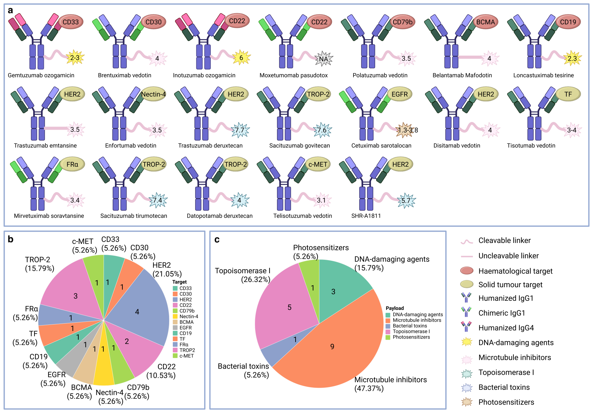
Table 2. Antibody-drug conjugates (ADCs) in phase III and phase IV clinical trials
| ADC Name | Clinical Phase | NCT Number | Representative Indication | Sponsor | Outcomes |
|---|---|---|---|---|---|
| FDA018 | III | NCT06519370 | TNBC | FDZJ | Ongoing |
| Disitamab vedotin | III | NCT05302284 | UC | RemeGen | Ongoing |
| Disitamab vedotin | III | NCT04714190 | GC | RemeGen | Ongoing |
| Disitamab vedotin | III | NCT04400695 | BC | RemeGen | Ongoing |
| Disitamab vedotin | III | NCT05904964 | HR+/HER2Low BC | Sun Yat-Sen Memorial Hospital | Ongoing |
| Disitamab vedotin | III | NCT05911295 | UC | Seagen | Ongoing |
| TQB2102 | III | NCT07008976 | metastatic BC | Nanjing Shunxin | Ongoing |
| TQB2102 | III | NCT07043725 | HER2+ BC | Nanjing Shunxin | Ongoing |
| Enfortumab vedotin | IV | NCT06764095 | BLCA | Mayo Clinic | Ongoing |
| GSK5764227 | III | NCT07099898 | SCLC | GlaxoSmithKline | Ongoing |
| Trastuzumab deruxtecan | III | NCT05950945 | BC | Daiichi Sankyo | Ongoing |
| Trastuzumab deruxtecan | III | NCT03734029 | HER2low BC | Daiichi Sankyo | PFS, 8.8 vs. 4.2 months, OS, 23.4 vs. 16.8 months, ORR, 51.7% vs. 16.8% |
| Trastuzumab deruxtecan | III | NCT04494425 | HER2low/HR+ BC | AstraZeneca | PFS, 12.8 vs. 7.0 months, OS, 28.9 vs. 27.4 months, ORR, 65.8% vs. 31.6% |
| Trastuzumab emtansine | III | NCT03529110 | HER2+/unresectable/ metastatic BC | Daiichi Sankyo | PFS, 25.1 vs. 7.2 months, ORR, 77.0% vs. 36.9% |
| MRG002 | III | NCT05754853 | advanced or metastatic UC | Shanghai Miracogen | Ongoing |
| SHR-A2102 | III | NCT06738251 | advanced UC | Shanghai Hengrui | Ongoing |
| SHR-A1811 | III | NCT06123494 | HER2+ advanced GC, GOJ | Jiangsu HengRui | Ongoing |
| Mirvetuximab soravtansine | III | NCT05445778 | OC, PC, FTC | AbbVie | Ongoing |
| Mirvetuximab soravtansine | III | NCT02631876 | OC, FTC | ImmunoGen | PFS, 4.14 vs. 4.44 months, OS, 15.57 vs. 13.93 months, ORR, 22% vs. 12% |
| Sacituzumab tirumotecan | III | NCT06074588 | NSCLC | MSD | Ongoing |
| Sacituzumab tirumotecan | III | NCT06841354 | TNBC | MSD | Ongoing |
| BNT323/DB-1303 | III | NCT06340568 | EC | BioNTech SE | Ongoing |
| Sacituzumab govitecan | III | NCT05609968 | NSCLC | MSD | Ongoing |
| Sacituzumab govitecan | III | NCT06431633 | NSCLC | Fundación GECP | Ongoing |
| Sacituzumab govitecan | IV | NCT04319198 | metastatic solid tumors | Gilead Sciences | SAE, 66.7% vs. 100% vs. 91.7% |
| Loncastuximab tesirine | III | NCT05991388 | B-cell NHL | University of Birmingham | Ongoing |
| Datopotamab deruxtecan | III | NCT06103864 | BC | AstraZeneca | Ongoing |
| Brentuximab vedotin | III | NCT05675410 | HL | NCI | Ongoing |
| Brentuximab vedotin | III | NCT02166463 | HL | NCI | EFS, 82.5% vs. 92.1% |
| Brentuximab vedotin | III | NCT03907488 | HL | NCI | EFS, 8.4% vs. 16.8%, OS, 1.4% vs. 2.9% |
| Inotuzumab ozogamicin | IV | NCT03677596 | ALL | Pfizer | PFS, 6.3 vs. 6.3 months, OS, 9.6 vs. 8.1 months |
Table 2: This table describes Phase III and IV clinical trials evaluating antibody-drug conjugates (ADCs) across solid tumors and hematologic malignancies. The listed information includes the ADC name, clinical phase, ClinicalTrials.gov identifier (NCT number), representative indication, trial sponsor, and reported clinical outcomes, where available (http://clinicaltrials.gov). Efficacy endpoints include progression-free survival (PFS), overall survival (OS), objective response rate (ORR), and event-free survival (EFS); safety outcomes include serious adverse events (SAE). In comparative trials, outcomes are reported as experimental versus control. For phase IV studies with multiple safety cohorts, serious adverse event (SAE) rates are reported separately for each cohort and do not represent head-to-head comparisons. Ongoing studies are indicated where final efficacy data have not yet been reported. Abbreviations: BLCA, bladder urothelial carcinoma; BC, breast cancer; EC, endometrial cancer; FTC, fallopian tube cancer; GC, gastric cancer; GOJ, gastro-oesophageal junction cancer; HL, Hodgkin lymphoma; HR, hormone receptor; NSCLC, non-small cell lung cancer; OC, ovarian cancer; PC, peritoneal cancer; SCLC, small cell lung cancer; UC, urothelial carcinoma; ALL, acute lymphoblastic leukemia; PFS, progression-free survival; OS, overall survival; ORR, objective response rate; EFS, event-free survival; SAE, serious adverse event; NCI, National Cancer Institute; FDZJ, Shanghai Fudan-Zhangjiang Bio-Pharmaceutical; MSD, Merck Sharp & Dohme.
Antibody Selection
Antibodies, also known as immunoglobulins (Ig), are large proteins made by plasma cells. Their primary function is to detect and neutralize foreign invaders, such as bacteria and viruses, as well as other substances called neoantigens. Structurally, antibodies usually take a “Y” shape, made of four polypeptide chains, two identical heavy chains and two identical light chains. These chains form two distinct functional regions: the Fab and Fc fragments (20). The Fab region has variable domains with different amino acid sequences that create antigen-binding sites, giving antibodies their specificity. The more conserved Fc region interacts with other parts of the immune system. In cancer treatment, therapeutic monoclonal antibodies like trastuzumab act as targeted agents. They exhibit direct cytotoxic activity against tumor cells, often by triggering apoptosis, a common mechanism used in clinical settings (21). Antibody isotypes mainly include IgG1, IgG2, IgG3, and IgG4 (22). Among them, IgG1 is the most common subclass, with a serum half-life of about 21 days. It triggers antitumor effects through Fc receptor-mediated mechanisms, such as antibody-dependent cellular cytotoxicity (ADCC), antibody-dependent cellular phagocytosis (ADCP), and complement-dependent cytotoxicity (CDC) (23). Currently, most ADCs use humanized IgG1 antibodies, such as sacituzumab govitecan, whereas some earlier ADCs, such as gemtuzumab ozogamicin (GO), use a humanized IgG4 backbone (24, 25). Although some chimeric antibodies, such as brentuximab vedotin (BV), are still in use, their murine variable regions paired with human constant domains can lead to immunogenicity (26). Additionally, bispecific antibodies, which simultaneously target two distinct antigens, are a promising area of research, offering improved tumor targeting and reduced off-target effects (27). In ADC design, four essential criteria guide antibody selection. First, reducing immunogenicity lowers the risk of anti-drug antibody (ADA) formation, thereby decreasing neutralization and clearance and enhancing safety. Second, antibodies must demonstrate strong binding avidity and high selectivity for target antigens to enable precise delivery of cytotoxic payloads. Third, effective induction of endocytosis is necessary after antigen engagement to promote cellular uptake of ADC complexes. Additionally, prolonged circulatory persistence ensures ADC stability and sufficient tumor eradication (8, 22). Standard IgG antibodies, with a molecular weight of approximately 150 kDa, have limited penetration into solid tumors due to their large size. Engineered smaller formats, including bispecific antibodies or single-chain variable fragments generated by Fc domain removal, provide improved tissue penetration and reduce adverse immune activation (8, 28).
Design of linker
The linker forms covalent bonds between monoclonal antibodies and cytotoxic payloads, enabling precise payload delivery and serving as a key part of ADC function, acting as a “biological missile” (9). To reduce off-target toxicity, the linker must be highly stable in systemic circulation, while rapid payload release upon tumor-cell internalization is crucial for maximizing cytotoxic effectiveness (29). The linker's physicochemical properties, including hydrophilicity and hydrophobicity, greatly affect the solubility, tendency to aggregate, and in vivo clearance of ADCs (30). Increased hydrophilicity can improve pharmacokinetic profiles, greatly decreasing nonspecific uptake and off-target side effects (31).
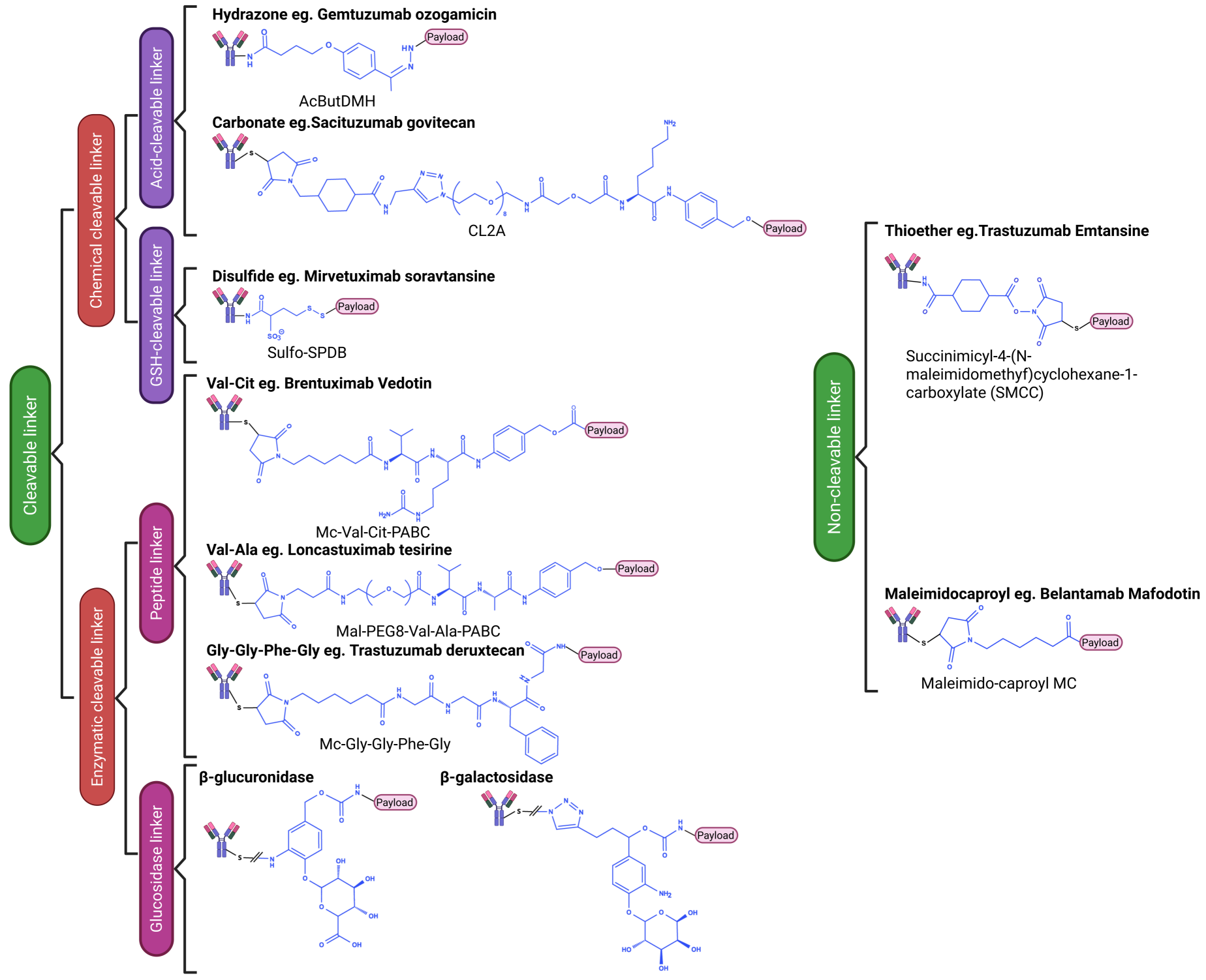
Additionally, the linker's chemical structure should be designed to avoid introducing immunogenic regions, thereby reducing the risk of immune recognition and ADA development. This approach maintains consistent pharmacokinetics and therapeutic potency while enabling safe metabolic clearance and preventing the buildup of toxic byproducts. Linkers are generally classified into two main types based on their cleavage: cleavable and non-cleavable (Figure 2). Cleavable linkers exploit microenvironmental differences between tumor cells and the systemic circulation to achieve selective payload release, facilitating the bystander effect (discussed in detail later). This feature is especially beneficial for treating tumors with diverse antigen expression. Chemically cleavable and enzymatically cleavable linkers are the two main types of cleavable linkers (32).
Chemically cleavable linkers
Chemically cleavable linkers that rely on tumor-specific chemical triggers for disruption include acid-labile and reduction-labile types (33). Acid-sensitive linkers exploit the acidic microenvironment (pH 4.5–5.0) of tumor cell endosomes and lysosomes for hydrolytic cleavage, often via hydrazone bonds. For example, GO uses a 4-(4-acetylphenoxy) butanoic acid (AcBut) linker to conjugate a calicheamicin derivative (CalichDMH) to a humanized IgG4 anti-CD33 antibody (34). Sacituzumab govitecan (SG) uses a carbonate linker, which is also acid-cleavable (31). However, their clinical use is limited by poor stability; a ketone-derived hydrazone linker showed a hydrolysis half-life of only 2 days in human and murine plasma (35). Reduction-sensitive linkers utilize the significant difference in glutathione (GSH) concentration between intracellular (1–10 mM) and extracellular (~5 μM) environments to promote disulfide bond reduction (9,36). They remain stable in circulation but undergo reductive cleavage upon internalization into tumor cells. Mirvetuximab soravtansine (MIRV), which uses a sulfo-SPDB linker, illustrates this mechanism and received accelerated FDA approval in 2022 for the treatment of platinum-resistant ovarian cancer (37).
Enzymatically cleavable linkers
Enzymatically cleavable linkers in ADCs exploit hydrolases that are overexpressed in tumors for degradation and are predominantly peptide- or glycosidase-based. Peptide linkers, recognized by lysosomal proteases such as cathepsin B, include sequences like Val-Cit, Val-Ala, and Gly-Gly-Phe-Gly. These linkers are a cornerstone of ADC technology, ensuring high selectivity and stability (38,39). For example, BV, an FDA-approved ADC, combines a CD30-specific antibody, a cleavable dipeptide linker (Mc-Val-Cit-PABC), and MMAE to treat Hodgkin lymphoma and systemic anaplastic large cell lymphoma (40). Val-Ala is more hydrophilic than Val-Cit, providing clear benefits for use with lipophilic payloads like pyrrolobenzodiazepine (PBD), as seen with the approved ADC loncastuximab tesirine (Lonca) (39). The MC-GGFG tetrapeptide linker used in trastuzumab deruxtecan (T-DXd) allows proteolytic degradation in lysosomes to release the payload (41). Glycosidase linkers, cleaved by enzymes such as β-glucuronidase or β-galactosidase, improve ADC solubility and stability, enabling higher drug-to-antibody ratios (DARs). Burke et al. showed that anti-CD30 conjugates with glucuronic acid-AE linkers demonstrated efficacy and immunospecificity (42). Kolodych et al. reported that galactosidase-based ADCs outperformed trastuzumab emtansine (T-DM1) in vitro (43).
Non-cleavable linkers
Non-cleavable linkers, including thioether and maleimidocaproyl variants, form strong covalent bonds between antibodies and cytotoxic payloads. These linkers preserve structural integrity during systemic circulation and within cellular compartments. Payload release occurs only after lysosomal proteolysis of the antibody into amino acids, creating “amino acid-linker-payload” conjugates. Unlike cleavable-linker ADCs, the metabolites of non-cleavable-linker ADCs have limited membrane permeability, which prevents the bystander effect—an advantage for tumors that uniformly express the antigen (44, 45). For example, T-DM1 uses the SMCC thioether linker to attach maytansinoid DM1 to trastuzumab (46), while belantamab mafodotin employs an MC linker to couple MMAF to its antibody via an amide bond (47). However, these ADCs face challenges, including suboptimal payload release and potential therapeutic limitations. Future research should focus on optimizing linkers, exploring combination immunotherapies, and developing new conjugation methods to mitigate resistance and improve efficacy.
Design of the Payload
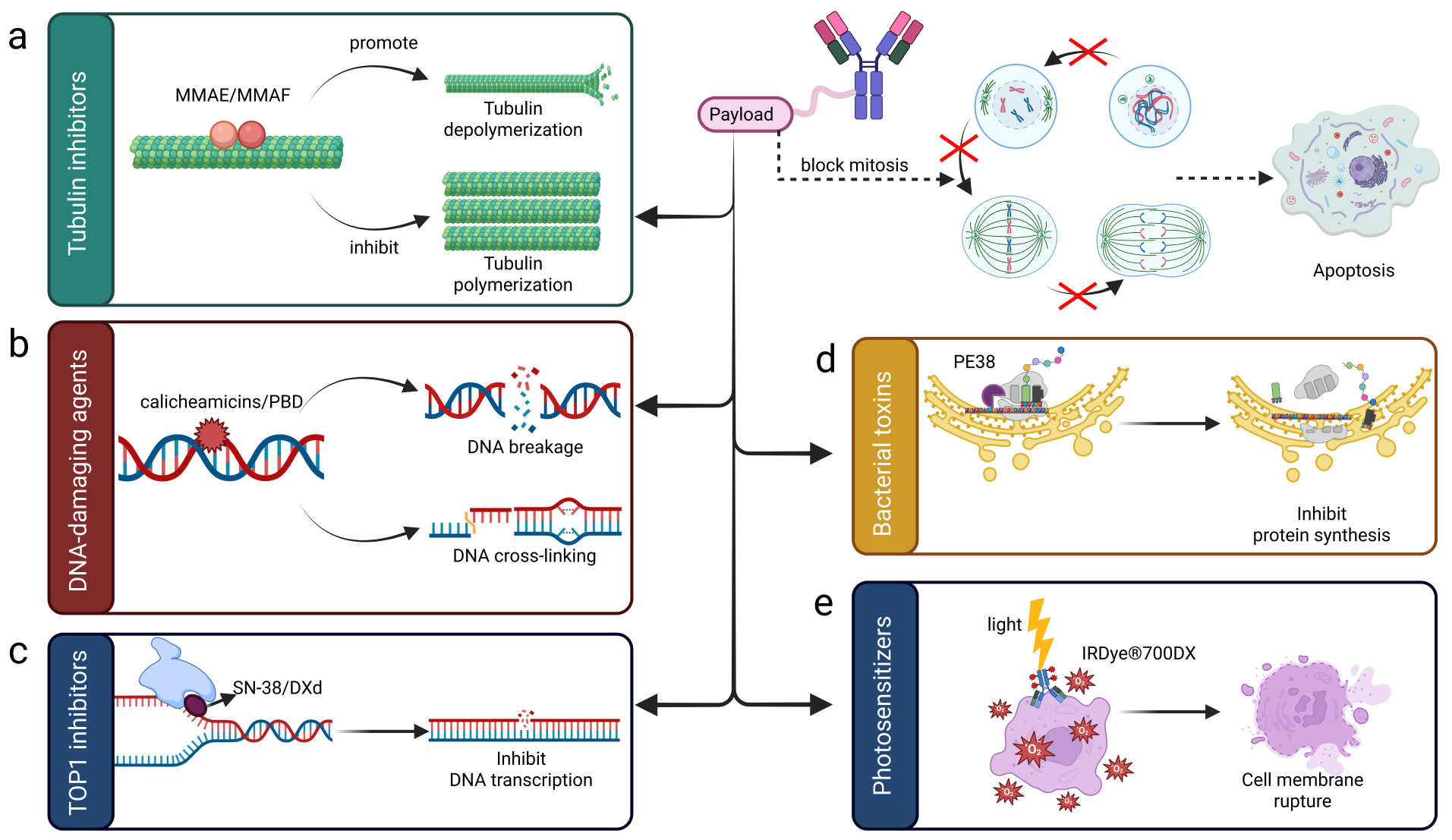
The anti-tumor activity of ADCs mainly relies on their cytotoxic payloads, which inhibit tumor growth through various mechanisms. These include interfering with DNA replication, causing structural alterations in the genome, and preventing microtubule assembly, as previously documented (48). An ideal payload must have seven key attributes: high cytotoxic effectiveness, low immunogenicity, strong stability during manufacturing and in systemic circulation, and adjustable functional groups for conjugation (49). The current range of approved ADC payloads includes five main categories: Topoisomerase I (Topo I) inhibitors, microtubule-targeting agents, DNA-damaging compounds, photosensitizers, and bacterial toxins (Figure 3).
In preclinical and clinical development, new payload classes are expanding this list, introducing Topoisomerase II (Topo II) inhibitors, RNA-targeting agents, Bcl-xL antagonists, and immunomodulatory molecules (9, 52).
Microtubule inhibitors
Microtubule inhibitors are essential in the mechanism of ADCs. By binding to tubulin subunits, these inhibitors either block microtubule polymerization or cause significant disruption. This prevents proper cell division, leading to cell cycle arrest at the G2/M checkpoint and subsequent programmed cell death (53) (Figure 3a). Among approved ADC payloads, natural products dominate, with microtubule inhibitors comprising over half of this category. Auristatins and maytansinoids are two main subclasses. Auristatins, such as monomethyl auristatin E (MMAE) and monomethyl auristatin F (MMAF), are preferred due to their high cytotoxicity, good aqueous solubility, stability under physiological conditions, and compatibility with various linker chemistries, making them popular for ADC toxin development (48). Examples of related ADCs include commercially available drugs like BV and polatuzumab vedotin (Pola), as well as Phase III candidates such as MK-2140 and ABBV-399 (25). Maytansinoids, represented by DM1 and DM4, also show excellent stability and solubility. T-DM1, approved for HER2-positive breast cancer (BC), uses DM1 with a very low IC₅₀ (10⁻¹¹–10⁻⁹ M) (54), while MIRV employs DM4 to target FRα in ovarian cancer (55).
DNA-damaging agents
DNA-damaging agents cause direct structural damage to DNA via alkylation, cross-linking, or the induction of double-strand breaks, thereby triggering cell death. These highly potent payloads are active against both proliferating and quiescent cells and are accompanied by a pronounced bystander effect (56). They are primarily classified into calicheamicins, PBD dimers, and duocarmycins. Calicheamicins, as enediyne antibiotics, generate sequence-specific DNA double-strand breaks, and derivatives are used in FDA-approved ADCs such as GO and inotuzumab ozogamicin (InO) (57–59). PBD dimers bind to the minor groove of DNA, forming irreversible cross-links that impede replication and transcription (Fig. 3b). Lonca, the first PBD-containing ADC, received approval in 2021 (60). Duocarmycins induce DNA alkylation via adenine adducts, as exemplified by trastuzumab duocarmazine (Phase III), which employs seco-DUBA to treat advanced HER2+ BC (61).
Topo I inhibitors
Topo I inhibitors increase their affinity for the Topo I-DNA complex, thereby blocking DNA re-ligation. This mechanism causes replication forks to stall and eventually leads to double-strand DNA breaks. SN-38 and DXd are examples of this class of inhibitors (62) (Figure 3c). Regulatory approvals for these agents began with T-DXd, followed by SG (63, 64). T-DXd features a humanized anti-HER2 IgG1 monoclonal antibody (mAb) conjugated to DXd through a cleavable mc-Gly-Gly-Phe-Gly linker, promoting cancer cell apoptosis and bystander killing. SG, using the anti-TROP2 IgG1 mAb hRS7 and a hydrolyzable CL2A carbonate linker for SN-38 delivery, shows efficacy in metastatic triple-negative breast cancer (TNBC) and other malignancies (62).
Bacterial toxins
Bacterial toxins, classified as exotoxins and endotoxins, are bioactive compounds produced by bacteria that can harm human and other biological systems. Exotoxins, mainly protein-based, are actively secreted by bacteria into their surroundings, while endotoxins are essential parts of the bacterial cell wall and are usually released when bacteria break apart (65). Pseudomonas aeruginosa exotoxin A (PEA) is a particularly important virulence factor of P. aeruginosa (66). As shown by the FDA-approved moxetumomab pasudotox, which combines the anti-CD22 mAb's variable fragment (Fv) with PE38 (a truncated PEA variant), PE38 causes ADP-ribosylation of the diphthamide residue in elongation factor 2 (EF-2). This post-translational modification prevents EF-2 from functioning, thereby blocking polypeptide chain elongation, hindering protein synthesis, and ultimately causing cell death (67) (Figure 3d).
Photosensitizers
Photosensitizers, an important class of compounds, can absorb electromagnetic radiation within specific spectral ranges, spanning 250–420 nm (ultraviolet) or 400–800 nm (visible light). Upon absorption, they undergo electronic excitation, producing reactive intermediates such as free radicals and cations that trigger photochemical processes. In photodynamic therapy (PDT), the synergistic interaction of photosensitizers, molecular oxygen, and appropriate light wavelengths induces therapeutic photochemical reactions (68, 69) (Figure 3e). Akalux (ASP-1929), an FDA-approved photoimmunotherapy agent, demonstrates this mechanism. Consisting of cetuximab and IRDye® 700 DX, a water-soluble silicon phthalocyanine-based photosensitizer, it targets EGFR-positive tumor cells. Upon near-infrared irradiation, the photosensitizer produces singlet oxygen, leading to tumor cell death and microvascular disruption, providing a treatment option for advanced head and neck cancer (50, 70, 71).
New potential payload
Topo II inhibitors
Topo II, a crucial nuclear endonuclease, mediates topological transformations in double-stranded DNA by temporarily cutting and rejoining the helix. This enzyme activity is vital for resolving topological problems during key genomic processes, including DNA replication, transcription, recombination, and mitotic chromosome segregation (72). Exploiting the higher Topo II expression and activity in cancer cells compared to normal tissues, this enzyme has become a validated target for anticancer drug development. Topo II inhibitors are a class of oncolytic agents that block Topo II's catalytic activity, thereby hindering DNA replication and transcription, and producing antitumor effects (62). These chemotherapeutics targeting Topo II are mainly divided into two categories: Topo II poisons (e.g., etoposide, doxorubicin) and catalytic inhibitors (73). Analysis of patent literature from 2016 to 2025 shows significant progress in the design of Topo II inhibitors, with new chemotypes such as quinolines (WO18107112) and lapidipines (CN108947916 B) highlighting the therapeutic promise of this drug class in cancer treatment (72).
RNA inhibitors
RNA inhibitors that target RNA synthesis or function have emerged as a new class of anticancer agents. Their ability to target both actively dividing and non-cycling cancer cells provides a significant therapeutic benefit, potentially preventing tumor recurrence linked to dormant cancer cells and certain drug resistance pathways (74). Key examples of RNA inhibitors used in ADCs include RNA polymerase II inhibitors, such as amatoxins, and RNA splicing inhibitors, which interfere with gene transcription and RNA maturation, ultimately stopping protein synthesis and inducing apoptosis. An example is HDP-101, a BCMA-targeted ADC that uses an amatoxin derivative as its cytotoxic payload. This ADC is currently being studied in a Phase II clinical trial for the treatment of multiple myeloma (75).
BCL-XL inhibitors
BCL-XL, a pivotal anti-apoptotic protein, plays a central role in tumor progression, metastasis, and drug resistance. BCL-XL inhibitors, as payloads in ADCs, offer a promising strategy for inducing tumor cell apoptosis. However, a critical challenge in ADC development is mitigating platelet toxicity. Because platelet survival depends on BCL-XL expression, targeting this protein expands the therapeutic index (76). ABBV-155 (mirzotamab clezutoclax) is a significant achievement, as it is the first ADC targeting the cell-surface protein B7-H3 (CD276) with a highly selective and potent BCL-XL inhibitor as its payload. Ongoing clinical investigations are exploring its efficacy in treating glioma, advanced non-small cell lung cancer (NSCLC), and BC (62, 77).
Immune stimulant
Immune stimulant payloads, unlike cytotoxic agents, are designed to activate innate immune responses within the TME. Examples include STING activators and Toll-like receptor (TLR7/8/9) agonists. These compounds trigger signaling pathways in tumors, including the STING and TLR pathways, leading to increased production of type I interferons and other cytokines. This cytokine surge activates macrophages, dendritic cells, natural killer cells, and T lymphocytes, thereby boosting anti-tumor immune surveillance (78–80). A notable example is XMT-2056, an ADC carrying a STING agonist payload. Currently in Phase I clinical trials, this HER2-targeting ADC, which recognizes a novel epitope, shows enhanced anti-tumor effects when combined with trastuzumab or pertuzumab (81).
3. Mechanism of action of ADC in vivo
ADCs are a new class of targeted therapeutic agents that combine the specificity of monoclonal antibodies with the potent cytotoxicity of small-molecule drugs. The multifaceted mode of action of ADCs comprises three main parts: the mAb, which specifically binds to tumor-associated antigens; the linker, which securely attaches the payload to the antibody while maintaining its stability in circulation; and the cytotoxic payload, which exerts anti-tumor effects upon internalization by target cells. This modular design enables ADCs to achieve both accurate tumor targeting and strong therapeutic effects while reducing systemic toxicity compared with traditional chemotherapy. Through this modular design, ADCs bridge targeted therapy, cytotoxic chemotherapy, and tumor immunology, enabling both direct tumor cell killing and modulation of the tumor microenvironment.
The canonical model of ADC action
The therapeutic action of ADCs begins with intravenous injection, resulting in systemic circulation in three forms: intact conjugates, unbound antibodies, and free payloads. The immunoglobulin domain (e.g., IgG1) remains structurally stable in circulation while exhibiting high specificity and affinity for tumor-associated antigens. These antigens are either overexpressed or uniquely found on tumor cell membranes or within the TME (82, 83). When the antigen is recognized, the ADC-antigen complex is internalized into the cell via endocytosis, primarily via clathrin- or caveolin-mediated pathways, forming early endosomes. These vesicles mature along the cytoskeleton, changing from early to late endosomes before reaching lysosomes (84) (Figure 4).
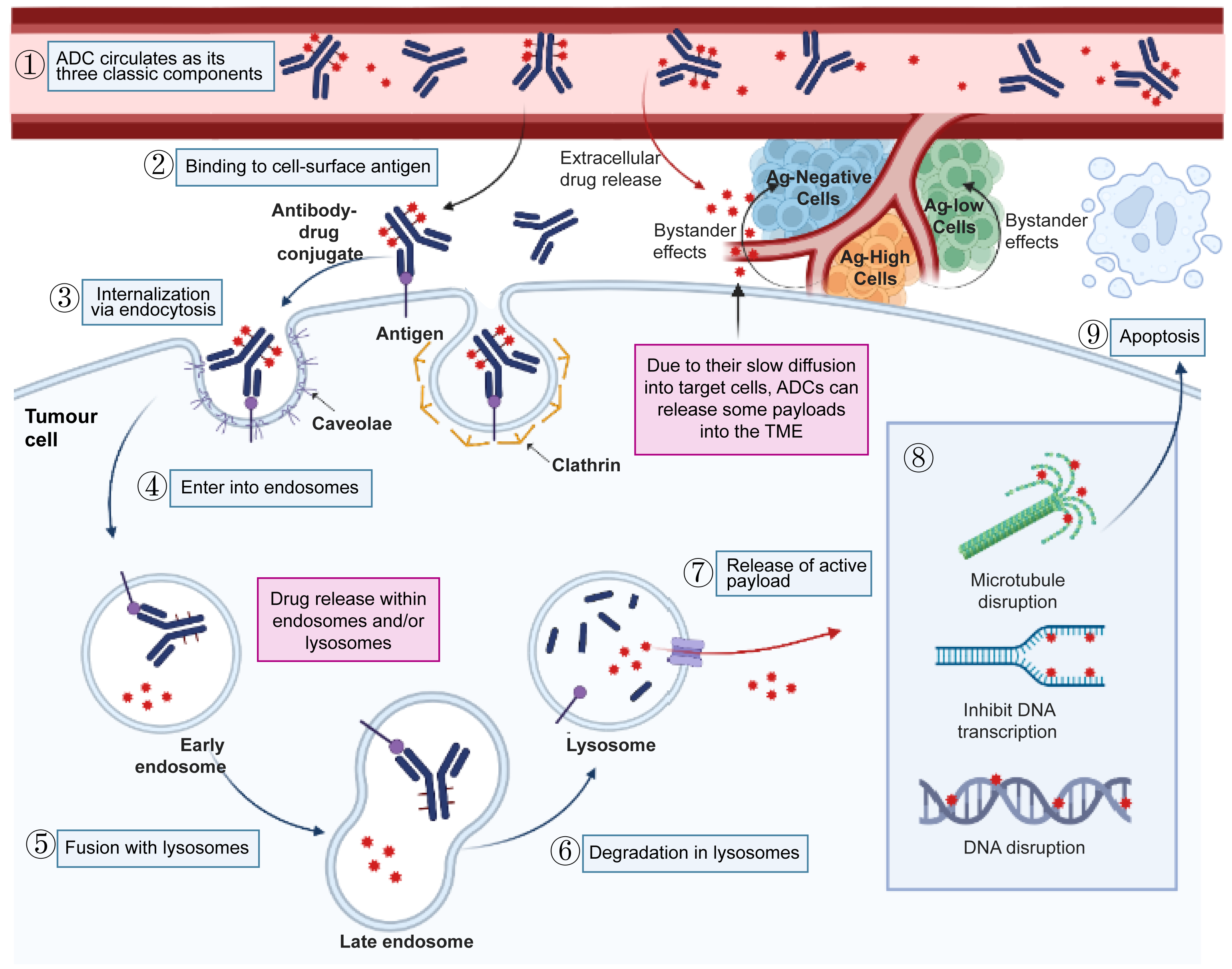
A portion of the early endosome associates with Fc receptors and is transported back outside the cell via transcytosis, while the remainder matures into late endosomes that eventually fuse with lysosomes. Not all ADCs require internalization, but this process is necessary for those using non-cleavable linkers or membrane-impermeable payloads (25). Lysosomes contain abundant proteases (e.g., Cathepsin B), acid hydrolases, and a low-pH environment (~pH 4.5–5.0), where ADC structures break down. Cleavable linkers enable specific disassembly; for example, peptide linkers are cleaved by proteases, and hydrazone bonds hydrolyze under acidic conditions, releasing cytotoxic payloads. In contrast, ADCs with non-cleavable linkers undergo complete degradation of their antibody component into amino acids by lysosomal enzymes, thereby releasing cytotoxins attached to residual amino acid residues (29). The cytotoxic payload is released into the cytoplasm in its active form. These highly potent toxins (e.g., MMAE, DM1, DXd, SN-38) subsequently act on their respective intracellular targets. This series of reactions finally induces apoptotic or necrotic cell death, exerting cytotoxic effects on tumor cells.
Bystander effects of ADCs
The bystander effect, characterized by the diffusion of lipophilic, membrane-permeable payloads from antigen-positive tumor cells to adjacent antigen-negative tumor cells, extends ADC payload delivery beyond strictly antigen-positive cells (85). This effect effectively overcomes resistance mechanisms related to target antigens, such as antigen heterogeneity, downregulation, or loss.
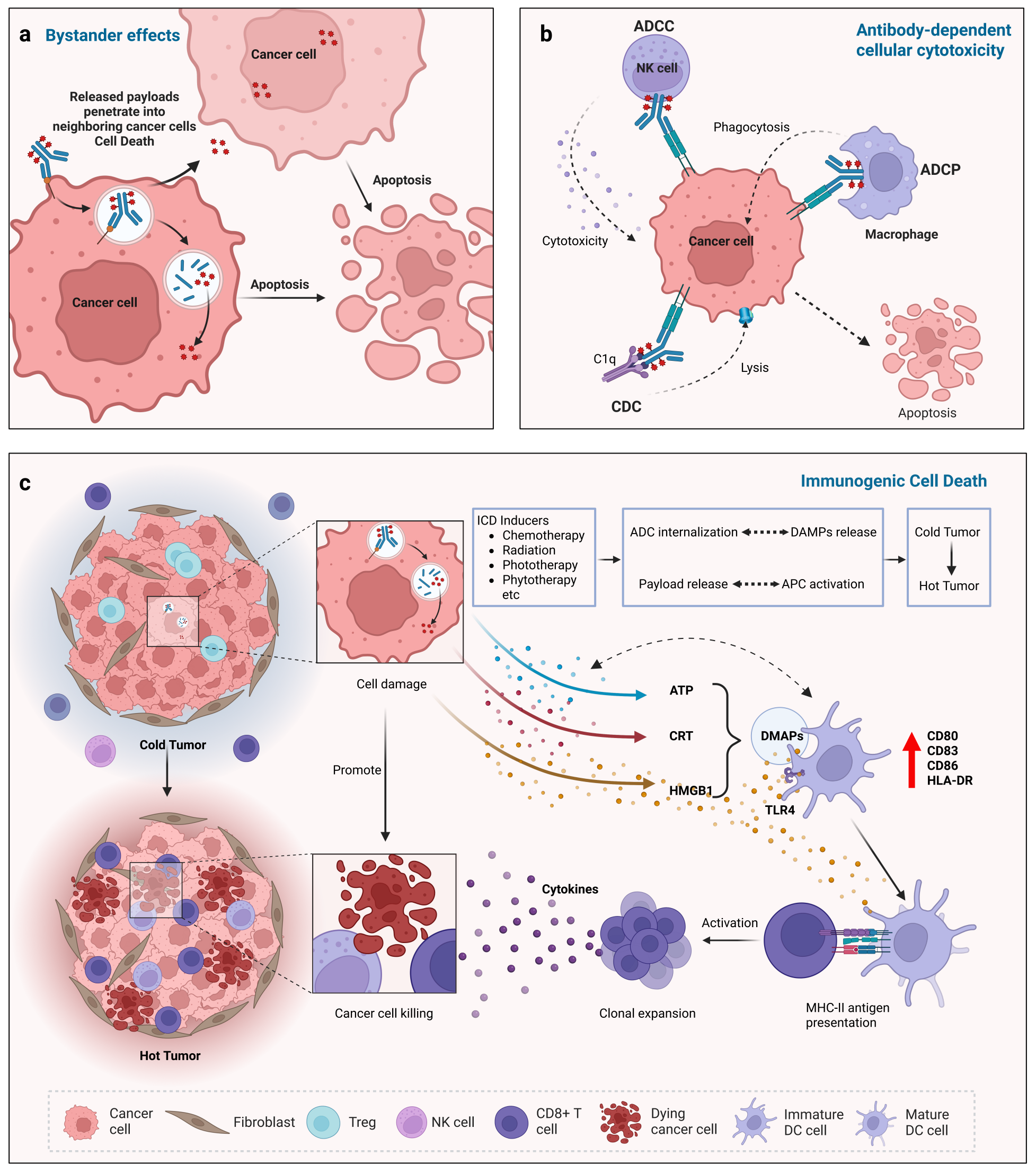
Tumor cells can decrease surface antigen expression (e.g., HER2, CD33) through transcriptional repression, proteasomal degradation, or alternative splicing. Traditionally, ADCs were thought to require binding to the target antigen and internalization to release the payload (86). However, ADCs capable of the bystander effect can operate via an alternative pathway. In the TME, acidic pH, reducing agents, and proteases can induce premature linker cleavage, releasing the payload before ADC internalization (87). Lipophilic, membrane-permeable payloads like MMAE and SN-38 diffuse from targeted cells into the surrounding microenvironment, thereby killing neighboring antigen-negative tumor cells (85, 88). This process is shown in Figure 5a, illustrating how the released payload (red dots) penetrates neighboring antigen-negative cells, emphasizing the spatial advantage of the bystander effect in addressing tumor heterogeneity.
The bystander effect is strongly influenced by factors such as DAR and the TME. Clinically, higher DAR values are typically associated with greater effectiveness in heterogeneous tumors (89). Additionally, the complex TME, including factors such as pH and protease activity, regulates the efficiency of payload release and diffusion. For instance, T-DXd (DAR = 7) showed a higher objective response rate (ORR) in HER2-mutated NSCLC than ADCs with stable, non-cleavable linkers such as T-DM1 (DAR = 5) (90, 91). Cleavable linkers and permeable payloads are vital for this, whereas ADCs with non-cleavable linkers typically lack significant bystander activity because they rely on lysosomal degradation to release the payload (92). The effectiveness of ADCs largely depends on the synergy between their linkers and payloads, as the structural and functional features of these components directly influence payload delivery, release timing, and antitumor activity. Table 3 outlines the efficacy profiles of key linker/payload combinations in approved ADCs, focusing on the main clinical endpoints across major uses. Payload diffusion can also affect normal cells within the TME, increasing the risk of off-target toxicity (e.g., corneal damage or hepatocyte injury) (89). Therefore, optimizing ADC design is essential to balance effectiveness and safety.
Table 3. Comparative efficacy outcomes of representative approved antibody-drug conjugates (ADCs) stratified by linker chemistry and cytotoxic payload class.
| ADC Name | Linker Type | Payload Class | Target | Indication | Efficacy Endpoints |
|---|---|---|---|---|---|
| Trastuzumab deruxtecan (T-DXd) | Cleavable (mc-Gly-Gly-Phe-Gly) | Topo I inhibitor (DXd) | HER2 | HER2+ BC | PFS, median: 28.8 months (vs. 7.2 months for T-DM1), ORR: 79.7% |
| Sacituzumab govitecan (SG) | Cleavable (CL2A carbonate) | Topo I inhibitor (SN-38) | TROP2 | TNBC | PFS, median: 5.6 months (vs. 1.7 months for chemotherapy), ORR: 31.7% |
| Brentuximab vedotin (BV) | Cleavable (Mc-Val-Cit-PABC) | Microtubule inhibitor (MMAE) | CD30 | HL/ALCL | ORR: 86% (sALCL), DOR, median: 12.6 months |
| Trastuzumab emtansine (T-DM1) | Non-cleavable (SMCC) | Microtubule inhibitor (DM1) | HER2 | HER2+ BC | PFS, median: 9.6 months, OS, median: 30.9 months (second-line setting) |
| Loncastuximab tesirine (Lonca) | Cleavable (Mal-PEG8-Val-Ala-PABC) | DNA-damaging agent (SG3199) | CD19 | DLBCL | ORR: 48.3%, OS, median: 10.3 months (relapsed/refractory) |
| Mirvetuximab soravtansine (MIRV) | Cleavable (sulfo-SPDB) | Microtubule inhibitor (DM4) | FRα | Platinum-resistant OC | PFS, median: 5.6 months (vs. 3.9 months for chemotherapy), ORR: 32.4% |
Table 3. Comparative efficacy outcomes of selected approved antibody-drug conjugates (ADCs), organized by linker chemistry and cytotoxic payload class. This table summarizes key clinical efficacy endpoints reported for FDA-approved ADCs, including median progression-free survival (PFS), objective response rate (ORR), overall survival (OS), and duration of response (DOR), as measured in pivotal phase II or phase III trials. Indications cover HER2-positive breast cancer (HER2+ BC), triple-negative breast cancer (TNBC), Hodgkin lymphoma (HL), anaplastic large-cell lymphoma (ALCL), diffuse large B-cell lymphoma (DLBCL), and platinum-resistant ovarian cancer (OC). Payload types include topoisomerase I inhibitors, microtubule inhibitors, and DNA-damaging agents. Data are sourced from original trial publications and associated ClinicalTrials.gov records. Because of variations in study design, patient populations, and prior treatments, cross-trial comparisons should be made cautiously. Abbreviations: ADC, antibody-drug conjugate; PFS, progression-free survival; ORR, objective response rate; OS, overall survival; DOR, duration of response; HER2, human epidermal growth factor receptor 2; TROP2, trophoblast cell-surface antigen 2; FRα, folate receptor alpha; BC, breast cancer; TNBC, triple-negative breast cancer; HL, Hodgkin lymphoma; ALCL, anaplastic large-cell lymphoma; DLBCL, diffuse large B-cell lymphoma; OC, ovarian cancer.
Antibody-Mediated Immune Effector Functions
Beyond payload-induced killing, ADC-mediated anticancer activity also involves antibody-mediated immune effector functions: ADCC, ADCP, and CDC (Figure 5b). ADCC occurs when the ADC antibody’s Fc region binds to Fcγ receptors on immune effector cells (e.g., NK cells), activating these cells to release cytotoxic substances (perforin, granzymes) that kill antibody-targeted tumor cells (93). It depends on the ADC antibody’s integrity and Fc functionality— for example, a new fucosylated anti-BCMA IgG1 enhances ADCC by increasing Fc binding to FcγRIIIa (94). ADCP occurs when myeloid cells (mainly macrophages) with Fcγ receptors are activated by binding to the Fc region of the ADC antibody, leading to phagocytosis of antibody-coated tumor cells (95). Tumor-bound antibodies promote macrophage phagocytosis via FcγR, a key mechanism of action for many therapeutic antibodies. For example, the CD47-SIRPα interaction blocks maximum antibody-dependent phagocytosis. CD47 blockade therapy removes the inhibitory signal mediated by SIRPα, boosting macrophage activation and increasing ADCP (96). CDC occurs when ADC antibody-antigen binding changes the Fc conformation to bind complement C1q, triggering the complement cascade to form MAC, which lyses target cells through osmotic pressure (97). HexaBody-CD38 (GEN3014) is a next-generation CD38-targeting IgG1 antibody with an E430G mutation in its Fc domain that promotes hexamer formation upon cell-surface binding, greatly increasing complement-dependent cytotoxicity (CDC) (98).
Immunogenic cell death (ICD)
ADCs not only deliver cytotoxic payloads but also employ diverse strategies to modulate the immune system, thereby enhancing antitumor efficacy. Immunogenic cell death (ICD), a precisely regulated form of cell death, stimulates host adaptive immunity during the cell death process. Certain payloads, especially DNA-damaging agents, can trigger ICD in tumor cells (99). This process involves apoptotic cells releasing damage-associated molecular patterns (DAMPs), such as extracellular adenosine triphosphate (ATP), high-mobility group box 1 protein (HMGB1), and surface-exposed calreticulin (CRT) (100). These DAMPs help attract and activate antigen-presenting cells (APCs), including dendritic cells (DCs), potentially initiating a tumor-specific immune response. Combining ADCs with immune checkpoint inhibitors (ICIs) results in synergistic therapeutic effects (100, 101). Extracellular ATP acts as a chemoattractant, guiding DCs and other APCs to tumor sites (102). Released HMGB1 binds to Toll-like receptor 4 (TLR4) on DCs, increasing the expression of major histocompatibility complex class II (MHC-II) and co-stimulatory molecules (CD80, CD83, CD86, HLA-DR), thereby boosting DC activation and maturation (103–105). CRT serves as an “eat-me” signal to enhance the phagocytosis of apoptotic cells by DCs (106). Activated DCs then present processed tumor antigens to naive CD8+ T cells, promoting T cell infiltration and activation, and ultimately triggering systemic antitumor immunity (107) (Figure 5c).
ICD results from cells’ inadequate adaptation to specific stressors and plays a crucial role in the development of new anticancer strategies. Current treatments like chemotherapy, radiation, and targeted oncotherapeutics mimic viral signatures to stress malignant cells, induce apoptosis, and activate tumor-specific immune responses. ICD inducers improve tumor response to immune checkpoint inhibitors (ICIs). Ongoing research explores new ICD compounds, some FDA-approved (108, 109) and others (e.g., LTX-315/VC-315) in clinical trials (110) for combined immunotherapy. The ICD pathway serves as a molecular link between the direct cytolytic activity of ADCs and systemic antitumor immune responses. It not only destroys target cells but also modulates the immunosuppressive tumor microenvironment, transforming “cold” tumors into “hot” ones. By shifting tumors from immune-excluded to immune-inflamed phenotypes, ICD facilitates effective combination therapies with ICIs. This synergistic approach offers a promising strategy to advance cancer immunotherapy. These mechanisms illustrate that ADCs operate through a multilayered therapeutic model combining targeted cytotoxicity, bystander killing, and immune activation.
4. Clinical applications of marketed ADCs
Single-Agent Clinical Application
ADCs in hematological malignancies
To date, seven ADCs have been approved by regulatory agencies for the treatment of hematologic malignancies. Moxetumomab pasudotox, the first FDA-approved therapy for hairy cell leukemia in over 20 years (2018) (111), was withdrawn from the U.S. market in 2023 due to toxicity, low usage, and concerns about efficacy (10). The remaining six ADCs target acute myeloid leukemia (AML), lymphoma, multiple myeloma (MM), and other malignancies. A central question in the clinical development of ADCs is how to optimize target selection, payload potency, and linker stability to maximize tumor specificity while minimizing systemic toxicity.
Gemtuzumab ozogamicin
Gemtuzumab ozogamicin (GO), a monoclonal antibody conjugate, targets CD33 and plays a key role in AML treatment (112). Approved for newly diagnosed CD33-positive AML in adults and relapsed or refractory cases in both adults and children aged ≥ 2 years, GO's therapeutic history has been eventful. Initially, it was given as a single 9 mg/m² dose but was halted due to severe complications, especially hepatic veno-occlusive disease (VOD) (113). Later research showed that fractionated dosing (3 mg/m² on days 1, 4, and 7) maintained effectiveness while lowering peak plasma levels, reducing VOD and other serious side effects (114). This dosing approach has made GO a practical option, particularly for elderly patients and those who cannot undergo intensive chemotherapy, such as individuals over 75 years old or those aged 61-75 with a WHO performance status score > 2 who refuse aggressive treatment (115). The optimized dosing schedule was crucial to GO's subsequent regulatory reapproval.
Brentuximab vedotin
Brentuximab vedotin (BV), a CD30-targeting ADC, has transformed treatment options for many CD30-positive lymphoma subtypes, including classical Hodgkin lymphoma (cHL), anaplastic large cell lymphoma (ALCL), primary cutaneous ALCL (pcALCL), and mycosis fungoides (MF) (116, 117). In 2025, its use was expanded to include relapsed or refractory diffuse large B-cell lymphoma (DLBCL) (118). Multiple randomized clinical trials have demonstrated BV's safety and effectiveness, with a recommended dose of 1.8 mg/kg every three weeks. For example, a key monotherapy trial involving 102 patients with relapsed or refractory Hodgkin lymphoma (HL) after autologous stem cell transplantation (auto-SCT) showed an ORR of 75% and a median response duration of 5.6 months among responders, with a good safety profile (119). Similarly, in a group of 58 patients with relapsed or refractory systemic ALCL (sALCL), single-agent BV achieved an ORR of 86% and a median response duration of 12.6 months, significantly outperforming traditional chemotherapy (120). These results highlight BV's clinical utility as a primary treatment for patients with CD30-positive lymphomas, especially those with relapsed or refractory sALCL and HL.
Inotuzumab ozogamicin
Inotuzumab ozogamicin (InO), the pioneering ADC approved for clinical use, has transformed the treatment approach for relapsed or refractory B-cell precursor acute lymphoblastic leukemia (B-ALL). This innovative therapy is approved for adult and pediatric patients aged 1 year or older and specifically targets CD22-positive B-ALL that is resistant to initial treatment regimens. For Philadelphia chromosome-positive (Ph+) patients, InO treatment is recommended only after failure of tyrosine kinase inhibitor therapy (121, 122). Results from a pivotal phase III randomized controlled trial (NCT01564784) involving 326 patients showed significant effectiveness of InO. With a dosing regimen of 1.8 mg/m² given in three divided intravenous infusions over a 15-day cycle, InO treatment resulted in notable improvements in complete remission (80.7% vs 29.4%) and minimal residual disease negativity (78.4% vs 28.1%) rates, as well as a median overall survival (OS) benefit (7.7 vs 6.7 months) compared to standard care (123).
Polatuzumab vedotin
Pola, the world's first ADC targeting CD79b, has demonstrated significant clinical utility. Mainly used in combination regimens, with monotherapy as an option, this agent has transformed the treatment approach for DLBCL. Clinical evidence shows that Pola-based therapy greatly increases complete remission rates and overall survival, especially in patients with relapsed or refractory DLBCL (124). These findings highlight the drug's key role in improving treatment outcomes and its significance in modern hematologic oncology practice.
Loncastuximab tesirine
Lonca, a new CD19-targeted ADC, marks a major breakthrough in the treatment of relapsed or refractory DLBCL. This innovative therapy is especially useful for patients who have tried multiple treatments without success (125). Through its unique mechanism, Lonca delivers potent cytotoxic agents directly to CD19-positive tumor cells, enhancing treatment efficacy while reducing systemic side effects. As a result, it fills a critical gap in the treatment options for this difficult patient group, providing a promising alternative when standard therapies fail.
Belantamab mafodotin
Belantamab mafodotin, an ADC targeting the B-cell maturation antigen (BCMA), has become a significant therapeutic breakthrough for patients with relapsed or refractory MM (126). Initially approved for monotherapy, the drug was later withdrawn from the market. Current clinical studies mainly assess its effectiveness when combined with other treatments. It is crucial that Belantamab mafodotin is administered strictly according to the approved indications, prescribed dosing regimens, and supporting clinical evidence to ensure its safety and efficacy profiles (127, 128).
ADCs in solid tumors
As of the latest data, 12 ADCs have been approved for the treatment of solid tumors. Clinical evidence shows that HER2-positive cancers, including breast cancer and other solid tumor types, make up a significant portion of patients who benefit from ADC therapies.
Trastuzumab emtansine
Trastuzumab emtansine (T-DM1), the first HER2-targeted ADC to achieve commercial success, has proven clinical utility in early breast cancer (EBC) and metastatic breast cancer (MBC) (129). In EBC, it is given to HER2-positive patients with residual invasive disease after neoadjuvant treatment (e.g., trastuzumab plus a taxane). Data from Trial NCT01772472 showed that T-DM1 increased 3-year invasive disease-free survival from 77.0% to 88.3% compared to trastuzumab, reducing the risk of recurrence or death by 50% (130). For MBC, a key Phase III trial (NCT00829166) found that, in HER2-positive advanced breast cancer patients previously treated with trastuzumab/taxane, T-DM1 achieved a median PFS of 9.6 months (versus 6.4 months) and a median OS of 30.9 months (versus 25.1 months), making it the standard second-line therapy (15).
Trastuzumab deruxtecan
Trastuzumab deruxtecan (T-DXd), a second-generation HER2-targeted ADC, provides distinct advantages. With a high DAR (DAR = 7.7) and strong bystander effects from its cytotoxic payload, it effectively tackles treatment challenges in HER2-positive and HER2-low-expressing cancers, showing efficacy across breast, gastric, and lung cancers. A key, open-label, randomized Phase III trial (NCT03529110) demonstrated that T-DXd significantly extended progression-free survival (PFS) compared to T-DM1 in HER2-positive recurrent/metastatic breast cancer (BC) (131). In HER2-low unresectable or metastatic BC, it outperformed physician-selected chemotherapy (NCT03734029) (132). Furthermore, it has shown activity in heavily pretreated HER2-low gastric or gastroesophageal junction adenocarcinoma (133). The randomized, double-blind Phase II DESTINY-Lung02 trial (NCT04644237) involving 152 platinum-pretreated HER2-mutated metastatic non-small cell lung cancer (mNSCLC) patients reported an ORR of 49.0%, a median PFS of 9.9 months, and a median OS of 19.5 months with T-DXd at 5.4 mg/kg (134), establishing T-DXd as the first targeted ADC for this mNSCLC subtype.
Disitamab vedotin
Disitamab vedotin (DV), the first domestically developed anti-HER2 antibody-drug conjugate (ADC) in China, provides a notable advantage. It shows strong targeted cell-killing effects against tumors with low-to-moderate HER2 levels while maintaining an acceptable safety profile. Currently, its main clinical use is in HER2-positive gastric cancer (GC), with ongoing trials investigating its potential in breast cancer (BC), urothelial carcinoma (UC), and other cancers. In a Phase II study (RC48-C008) (135), DV achieved an ORR of 24.8%, a median PFS of 4.1 months, and a median OS of 7.9 months in HER2-overexpressing GC patients who had at least two prior chemotherapy treatments. Mild-to-moderate side effects, such as leukopenia, alopecia, and neutropenia, were well managed. Two additional Phase II trials (RC48-C005, NCT03507166; RC48-C009, NCT03809013) (136) confirmed its effectiveness and safety in platinum-pretreated, HER2-overexpressing (IHC 2+/3+) UC, leading to its approval for this use in China in December 2021.
Enfortumab vedotin
Enfortumab vedotin (EV) marks a significant breakthrough in targeted cancer therapy as the first ADC designed to directly target nectin-4, a transmembrane cell adhesion molecule that is overexpressed in various solid tumors, including UC and BC (137). This innovative treatment provides a precise mechanism of action that selectively destroys nectin-4-positive cancer cells and shows considerable efficacy even in patients with chemotherapy-resistant disease. Its main clinical use is the treatment of locally advanced or metastatic UC. The U.S. FDA granted accelerated approval in December 2019 based on the promising results of the Phase II EV-201 trial (NCT03219333). Later, in April 2022, the European Union approved EV for patients with locally advanced or metastatic UC who experienced disease progression after platinum-based chemotherapy and PD-1/PD-L1 inhibitor therapy (138).
Sacituzumab Govitecan
Sacituzumab govitecan (SG), a Trop-2-directed ADC, consists of a humanized anti-TROP2 monoclonal antibody linked to SN-38, an active Topo I inhibitor and irinotecan metabolite, via a cleavable CL2A linker with a DAR of 7.6. This design allows targeted delivery of SN-38 to cancer cells. In a pivotal global, open-label, randomized Phase III trial (NCT02574455), 468 patients with unresectable locally advanced or metastatic TNBC who had received at least two prior systemic treatments were enrolled. The study showed that SG significantly prolonged PFS compared with chemotherapy, with median PFS of 5.6 and 1.7 months, respectively (139). As a result, the U.S. FDA approved SG for patients with metastatic or advanced TNBC who had prior treatment failure in April 2020 (140). Emerging evidence also indicates its potential in UC (141), HR+/HER2− BC (142), and recurrent or metastatic cervical cancer (143).
Cetuximab Saratolacan
Cetuximab saratolacan (RM-1929; Akalux) is a targeted photoimmunotherapy agent directed against EGFR-positive tumors. It consists of cetuximab, an anti-EGFR monoclonal antibody, conjugated to a light-activatable photoabsorber. Following binding to tumor cells, exposure to near-infrared light induces rapid, localized damage to the cell membrane and tumor cell death. Regulatory approval in Japan for unresectable locally advanced or recurrent head and neck cancer was supported by an international Phase I/II study and a Japanese Phase I trial (144). In the international Phase IIa trial (NCT02422979), RM-1929 achieved an ORR of 43.3% and median OS of 9.3 months in heavily pretreated patients. The Japanese Phase I study reported an ORR of 66.7% in a smaller cohort (145).
Tisotumab vedotin
Tisotumab vedotin (TV), the pioneering ADC targeting TF, consists of a humanized anti-TF mAb covalently linked to the microtubule-disrupting agent MMAE via a cleavable valine-citrulline (Val-Cit) linker, with a DAR of 4. Approved for adult patients with recurrent or metastatic cervical cancer progressing after prior chemotherapy, TV represents a significant advance in targeted cancer therapy. In the Phase II InnovaTV 204 trial (NCT03438396), TV demonstrated efficacy in patients with recurrent or metastatic cervical cancer who had received prior treatment, achieving an ORR of 24% (7% complete response), a median duration of response (DOR) of 8.3 months, and a median OS of 12.1 months (19). These findings supported the FDA’s accelerated approval, which was later confirmed by a Phase III trial (NCT04697628) (146). Preclinical studies have shown TF overexpression across multiple solid tumor types, including ovarian cancer (147), lung cancer, pancreatic cancer, colorectal cancer, and head and neck cancers (148). Based on these findings, clinical trials of TV for these cancers have been initiated.
Mirvetuximab soravtansine
MIRV, a groundbreaking ADC, marks a major step forward in targeted cancer treatment. Designed to target FRα, MIRV is made of a humanized anti-FRα mAb linked to DM4, a powerful microtubule inhibitor, through a cleavable disulfide linker with a DAR of about 3.4. MIRV is approved for treating adult patients with FRα-positive, platinum-resistant epithelial ovarian, fallopian tube, or primary peritoneal cancer who have received 1 to 3 previous systemic therapies. The multicenter, open-label, randomized MIRASOL trial (NCT04209855) enrolled 453 patients and showed that MIRV monotherapy significantly outperformed investigator-selected chemotherapy (paclitaxel, pegylated liposomal doxorubicin, topotecan) in PFS, OS, and ORR, thereby slowing disease progression, extending survival, and improving tumor reduction (37). Additionally, the SORAYA trial (NCT04296890) confirmed its effectiveness in patients with high FRα expression, reporting an ORR of 32.4% and a median DOR of 6.9 months, supporting MIRV's regulatory approval (149).
Sacituzumab tirumotecan
Sacituzumab tirumotecan (sac-TMT) is an ADC targeting TROP2. It consists of a humanized anti-TROP2 monoclonal antibody conjugated via a cleavable CL2A linker to tiromotecan, a Topo I inhibitor and derivative of SN-38 (the active metabolite of irinotecan), with a DAR of 7.4. In clinical trials, sac-TMT has been extensively studied, primarily in TNBC and NSCLC, and has shown significant therapeutic potential across multiple solid tumor types. The pivotal Phase III OptiTROP-Breast01 trial provided strong evidence of its superiority in prolonging survival compared with conventional chemotherapy. In China, sac-TMT is approved for adult patients with unresectable, locally advanced, or metastatic TNBC who have received at least two prior systemic therapies, including one for advanced or metastatic disease (150). A clinical study led by Fang and Zhang from Sun Yat-sen University demonstrated that sac-TMT monotherapy showed promising antitumor activity with acceptable safety in previously treated patients with advanced NSCLC, regardless of EGFR mutation status (151). Regulatory approval for EGFR-mutated NSCLC was sought in China in October 2024 (152). The U.S. FDA designated sac-TMT as a Breakthrough Therapy for pre-treated EGFR-mutated non-squamous NSCLC (153).
Datopotamab deruxtecan
Datopotamab deruxtecan (Dato-DXd), a TROP2-targeting ADC, has shown significant clinical usefulness. Its indications include unresectable or metastatic HR+/HER2- breast cancer patients who have been pretreated with endocrine therapy and at least one line of chemotherapy, as well as locally advanced or metastatic NSCLC patients with previous systemic treatment. Clinical data have demonstrated the effectiveness of datopotamab deruxtecan (Dato-DXd) as a monotherapy in breast cancer and NSCLC. In HR+/HER2− advanced breast cancer, Dato-DXd significantly lowered the hazard ratio for progression or death compared to investigator-selected chemotherapy (154). In NSCLC, Dato-DXd reduced the rate of ≥ Grade 3 treatment-emergent adverse events by 16.5% compared with docetaxel (155).
Telisotuzumab vedotin
Telisotuzumab vedotin (Teliso-V) is the world’s first ADC targeting c-MET (mesenchymal-epithelial transition factor), which is often overexpressed or amplified in solid tumors like NSCLC and GC. Teliso-V is composed of a humanized anti-c-MET mAb linked to MMAE via a cleavable Val-Cit linker, with a DAR of 3.1. Data from an open-label, single-arm trial (NCT03539536) show that in 84 c-MET-high patients, Teliso-V achieved a 35% ORR, including 100% partial responses, with a median DOR of 7.2 months (156). In May 2025, the U.S. FDA granted accelerated approval to Teliso-V for adult patients with locally advanced or metastatic non-squamous NSCLC. Eligible candidates had prior systemic treatment and high c-MET protein expression levels (157).
Trastuzumab rezetecan
Trastuzumab Rzetecan (SHR-A1811), an ADC targeting HER2, consists of a humanized anti-HER2 monoclonal antibody, a cleavable linker, and a potent Topo I inhibitor as the cytotoxic payload. According to available clinical data, SHR-A1811 has demonstrated significant efficacy in HER2-positive breast cancer (post-multiline therapy) and HER2-mutant NSCLC, achieving ORRs of 50-70%. Ongoing studies are exploring broader populations, including those with low HER2 expression. In neoadjuvant treatment for HER2-positive breast cancer, the pathological complete response (pCR) rate reached 63.2% with SHR-A1811 monotherapy (50% in HR+ and 74.5% in HR− subgroups) (158). For patients with advanced HER2-mutant NSCLC, the independent review committee-determined ORR was 73.4%, the disease control rate (DCR) was 98.9%, and the median PFS was 11.5 months (159). In May 2025, SHR-A1811 received regulatory approval in China for the treatment of adult patients with unresectable locally advanced or metastatic NSCLC harboring HER2-activating mutations who had received at least 1 prior systemic therapy.
Combination Clinical Application
In recent years, an increasing number of preclinical and clinical studies have investigated combination therapies involving ADCs, primarily by combining conventional chemotherapy, targeted therapy, immunotherapy, and other modalities. These methods aim to improve treatment effectiveness across different tumor types through potential synergistic effects.
In combination with chemotherapy
Combining GO with standard chemotherapy regimens (e.g., daunorubicin and cytarabine) in newly diagnosed AML significantly increases the complete remission rate and extends event-free survival (EFS), making it a key component of current standard therapy. In the ALFA-0701 trial, patients aged 50-70 with newly diagnosed, de novo AML who received the combination of daunorubicin, cytarabine, and GO had a median EFS of 17.3 months, significantly longer than the 9.5 months observed in those treated with chemotherapy alone (160). Clinical evidence shows that the BV combination therapeutic strategy outperforms monotherapy with chemotherapeutic agents in improving survival among patients with HL. Replacing bleomycin with BV in the traditional ABVD protocol results in the BV-AVD regimen. Multiple phase III trials (NCT01712490, NCT02292979) (161, 162) have demonstrated its superior efficacy and safety profile across disease stages, as well as a reduced risk of bleomycin-induced pulmonary complications. These findings highlight the role of BV-AVD as a preferred first-line treatment option, especially for high-risk Hodgkin lymphoma patients. In relapsed or refractory DLBCL, the pivotal ECHELON-3 clinical trial (NCT04404283) showed that the triple combination of BV, lenalidomide, and rituximab outperformed the control regimen. The combination significantly extended median OS (13.8 vs. 8.5 months) and PFS (4.2 vs. 2.6 months), achieving an ORR of 64.3%. In February 2025, the U.S. FDA approved this therapeutic approach for adult patients with relapsed/refractory DLBCL who had received at least 2 prior systemic treatments and were not candidates for autologous hematopoietic stem cell transplantation or chimeric antigen receptor T-cell therapy (118).
In combination with immunotherapy
ADCs not only enable targeted elimination of neoplastic cells but also induce ICD, which releases tumor-associated antigens that activate the host immune system. When combined with immune checkpoint blockade, this approach reduces T-cell suppression, boosts antitumor immune responses, and shifts the TME from immunosuppressive to immunostimulatory. The ADC plus immunotherapy (IO + ADC) strategy has the potential to create synergistic antitumor effects, improve immunotherapy outcomes, and avoid drug resistance, as previously reported (163). Results from a Phase 1b, open-label study (NCT04042701) were presented at the 2024 ESMO Immuno-Oncology Congress. The study evaluated the safety profile and therapeutic efficacy of T-DXd combined with the PD-1 inhibitor pembrolizumab (PEM) in immunotherapy-naive patients with HER2-positive or HER2-mutated NSCLC. Significant anti-tumor activity was seen in both groups, with an ORR over 50% and a median DOR over 15 months, regardless of HER2 status. The treatment combination was well tolerated (164).
EV, combined with immunotherapy, has transformed the treatment of advanced UC. Based on results from the Phase Ib/II EV-103 trial (NCT03288545), the U.S. FDA granted Breakthrough Therapy designation in February 2020 for this regimen (with PEM) as a first-line treatment for cisplatin-ineligible patients with locally advanced or metastatic UC (165). Guo et al. reported the final results of the Phase Ib/II RC48-C014 trial, an investigator-initiated study assessing the combination of DV and toripalimab in patients with advanced UC. As of March 1, 2024, the confirmed ORR was 73.2%, with a median PFS of 9.3 months and OS of 33.1 months. The DV and toripalimab combination showed significant efficacy and manageable safety, especially in HER2-expressing patients. These results support further research into this regimen for advanced UC (166). OptiTROP-Lung01 (NCT05351788) is a multicenter, multicohort, open-label Phase II trial evaluating the combination of sac-TMT and tagitanlimab (an anti-PD-L1 antibody) as first-line treatment for patients with advanced or metastatic NSCLC lacking actionable genomic alterations. In this trial, Cohort 1A patients received sac-TMT at 5 mg/kg every 3 weeks, along with tagitanlimab at 1200 mg every 3 weeks. Conversely, Cohort 1B received sac-TMT (5 mg/kg biweekly) and tagitanlimab (900 mg biweekly). The confirmed ORR was 40.0% in Cohort 1A and 66.7% in Cohort 1B, with median PFS of 15.4 months and not yet reached, respectively. Both cohorts showed low discontinuation rates due to adverse events, indicating good tolerability, and no treatment-related deaths occurred. These findings suggest the combination's effectiveness and manageable safety in NSCLC patients, warranting further study (24).
In combination with targeted drugs
The combination of ADCs and targeted agents is a very active area in oncology, focusing on improving effectiveness by blocking multiple cancer-driving pathways simultaneously or leveraging synergistic mechanisms. For instance, T-DXd is a HER2-targeting ADC, while pertuzumab is a monoclonal antibody that binds to a different HER2 epitope; their combined use allows for more thorough suppression of HER2 signaling. The important Phase III DESTINY-Breast09 trial showed that the “T-DXd + pertuzumab” combo performed better than the traditional “taxane + trastuzumab + pertuzumab (THP)” treatment in patients with unresectable or metastatic HER2+ breast cancer. It significantly increased median PFS from 26.9 to 40.7 months, lowering the risk of disease progression or death by 44% (167). With the FDA's approval of its supplemental New Drug Application (sBLA) for priority review, this combo could soon become a new first-line standard.
In a single-arm, open-label Phase IIa clinical trial (NCT00875979), Miller KD et al. studied the combination of T-DM1 and pertuzumab in patients with HER2+ locally advanced or metastatic breast cancer. The overall ORR was 41%, with first-line therapy showing higher effectiveness (57% vs. 33% for second-line and beyond). The median PFS was 6.6 months, indicating sustained antitumor activity (168). Unlike conventional monospecific ADCs, bispecific ADCs demonstrate improved target selectivity by recognizing either two different antigens or different epitopes of the same antigen. This mechanism enhances therapeutic efficacy while reducing systemic toxicity. Izalontamab brengitecan (BL-B01D1), a new bispecific ADC targeting EGFR and HER3, marks a significant progress in the field (28). Osimertinib, a well-established third-generation EGFR tyrosine kinase inhibitor, works synergistically with the bispecific ADC BL-B01D1. This combination targets EGFR-mutant signaling and kills EGFR/HER3-positive tumor cells, potentially overcoming resistance to osimertinib. Phase II data from WCLC 2025 showed a 100% ORR as first-line therapy for NSCLC, leading to an ongoing Phase III trial (169).
5. Exploration of ADC Safety
Systemic Pharmacokinetics
ADCs have pharmacokinetic properties that differ from those of traditional chemotherapy and monoclonal antibodies due to their modular structure (antibody, linker, payload) and their processing in the body. These principles control exposure to intact ADCs, free payloads, and metabolites, which influence toxicity. Understanding how ADC pharmacokinetics affect exposure and toxicity is essential for optimizing dosing, reducing side effects, and ensuring patient safety (83). After an IV injection, ADCs first circulate in the blood, exit blood vessels, enter tumor tissues, diffuse through the tumor microenvironment (TME), bind to surface antigens on tumor cells, and are taken up by endocytosis. Inside lysosomes, the linker breaks, releasing the toxin, which then enters the cytoplasm or nucleus to kill the cell. Any failure here can lower treatment success (170). A key overlooked fact is that most ADCs administered never reach the tumor; instead, they are taken up and broken down by healthy tissues. The safety of ADCs mainly depends on their unique pharmacokinetics, since an ADC is basically a "three-part system": the antibody, the linker, and the payload. The combined behavior of these parts determines how much of the drug is in the body, where it goes, how it is released, and the potential for toxicity (171).
The stability of the linker is a key factor influencing pharmacokinetic properties, as the rate and extent of payload release depend on both the linker type and the tissue microenvironment. Cleavable linkers, such as those made from peptides or hydrazones, release their payload within acidic lysosomal compartments (pH 4.5–5.0) or through tumor-specific proteases, while non-cleavable linkers require complete antibody degradation to release the payload-linker-amino acid conjugate (29). Premature cleavage of cleavable linkers in circulation increases exposure to free payload, thereby amplifying off-target toxicity (172). Conversely, non-cleavable linkers rely on lysosomal degradation after internalization; although this improves plasma stability, it may reduce payload-release efficiency (44). An important factor in assessing ADC efficiency is the DAR, which measures the number of drug molecules attached to each mAb. DAR values can affect drug effectiveness, as low drug loading may decrease potency, while high drug loading can influence toxicity and pharmacokinetics (173). Therefore, many clinical ADCs use a compromise with a DAR of approximately 4, followed by adjustments to pharmacokinetics and stability via hydrophilic linkers and site-specific conjugation (174). From a molecular weight and spatial volume perspective, the main component of an ADC is the antibody, resulting in pharmacokinetic profiles that are largely similar to those of the unmodified antibody. The tissue distribution of ADCs depends on both antibody-mediated targeted delivery and nonspecific uptake mechanisms, including Fcγ receptor-mediated endocytosis, macropinocytosis, and charge-based interactions (175, 176). These processes can result in both on-target and off-target toxicity.
The target antigens for ADCs are often proteins highly expressed on tumor cells, although they may also be expressed at varying levels in normal tissues. Therefore, while ADCs are designed to selectively bind to and eliminate tumor cells, they can also bind to and destroy normal cells that express the target antigen. This accidental binding can cause damage to healthy tissues and organs, a phenomenon known as on-target toxicity (177). As a result, ADCs may harm normal cells expressing the same antigen during cancer treatment, creating a significant challenge for target selection (178). For example, Trop2, which is overexpressed in many tumors, is also found at low levels in normal ocular surface and oral mucosal cells. Trop2-targeted ADCs, such as SG, sac-TMT, and Dato-DXd, can cause ocular toxicity and oral mucositis by binding to Trop2 on these cells. Reported incidences include 5% ocular toxicity (179) and 10% oral mucositis (139) for SG, 49.2% oral mucositis for sac-TMT (150), and 36.4% ocular toxicity with 72.7% oral mucositis for Dato-DXd (180). Interestingly, when researchers analyzed data from over 100 clinical ADC trials (181), they discovered that ADCs within the same payload class showed very similar toxicity profiles, regardless of the specific target. This indicates that toxicity is often "payload-driven" rather than solely determined by the antibody’s localization. Off-target toxicity can result from premature payload release into the bloodstream due to linker instability (172), nonspecific uptake of the ADC by normal cells, and nonspecific binding of the ADC's antibody to normal cells (182). Additionally, ADCs undergo recycling via the neonatal Fc receptor (FcRn), which extends the half-life of the antibody component and may increase exposure to the payload (183).
Payload Toxicity
As the “ammunition” of ADCs, payloads determine their toxicity profile. Each payload exhibits distinct cytotoxicity, leading to organ-specific damage. Microtubule inhibitors such as MMAE, DM1, and MMAF disrupt microtubule-dependent processes in neurons and platelets, causing neurotoxicity and thrombocytopenia (184). Clinical trials report high rates of peripheral neuropathy with MMAE-based ADCs (185), while T-DM1-induced thrombocytopenia limits its dosage (186). DNA-damaging payloads in ADCs, such as calicheamicin and PBD dimers, exhibit off-target toxicity. Because of their affinity for rapidly dividing cells, these agents mainly affect hematopoietic stem cells in the bone marrow and mucosal epithelial cells, resulting in myelosuppression and gastrointestinal toxicity (187). PBD dimers, with strong DNA crosslinking ability, can cause DNA damage in both cancerous and normal tissues. Calicheamicin payloads, which induce DNA double-strand breaks, may lead to hepatic sinusoidal obstruction syndrome if released prematurely from the ADC conjugate (188, 189). The pulmonary system, especially alveolar epithelial cells, is also at risk from these payloads, potentially causing interstitial lung disease (ILD) characterized by respiratory symptoms (190). ADCs that incorporate Topo I inhibitors (e.g., SN-38, DXd) as payloads mainly cause gastrointestinal and blood-related toxicities (191). By inducing DNA double-strand breaks, these payloads trigger apoptosis in rapidly dividing cells (192). Significant hematological side effects, such as leukopenia, anemia, and neutropenia, are observed in many Phase III trial ADCs, including YL201, HS-20093, MHB088C, and BL-B01D1 (193–196).
Strategies for Reducing Toxicity
Leveraging the unique pharmacokinetic principles of ADCs and the relationship between exposure and toxicity, multiple strategies can be used to reduce ADC-related toxicity.
The first strategy is dose optimization, which can be approached from five aspects. First, identify the optimal therapeutic dose that ensures efficacy while minimizing ADC-related toxicity. For T-DXd in HER2-positive breast cancer (BC) and HER2-mutant non-small cell lung cancer (NSCLC), randomized studies comparing 5.4 mg/kg and 6.4 mg/kg showed similar ORRs (49% vs. 56%) and overlapping PFS curves, indicating no significant difference in efficacy. However, there was a notable difference in ILD incidence: the higher-dose group reported an ILD incidence of 28%, whereas the low-dose group reported only 13% (134). Second, establish a maximum dose limit to prevent excessive drug exposure in patients with larger body sizes. For example, EV initially lacked a dose cap, leading to three treatment-related fatalities in patients with a baseline weight ≥100 kg. Implementing a 125 mg dose cap eliminated excessive free payload (MMAE) exposure, reducing fatal toxicities while maintaining ORR. Additionally, treatment duration can be limited. For ADCs with cumulative toxicities, such as neurotoxicity, restricting the number of treatment cycles can reduce toxicity risk (197). Furthermore, dose adjustments can be made based on treatment response. For example, InO uses a two-step dose adjustment: an initial dose of 1.8 mg/m² to induce remission, followed by a maintenance dose of 1.6 mg/m² (198). Finally, fractionated dosing can be applied. GO was initially withdrawn from the market due to excessive toxicity but was later re-approved after improving its safety profile through low-dose fractionated administration, changing from a single dose of 9 mg/m² to divided doses of 3 mg/m² on days 1, 4, and 7. This adjustment significantly reduced hepatic and hematological toxicities while maintaining therapeutic efficacy (114).
The second strategy is patient stratification. A meta-analysis of 999 subjects with UGT1A1 genotypes found that individuals carrying the UGT1A1*28/*28 genotype have an increased risk of severe SG-related toxicity. Pre-treatment UGT1A1 genotyping is recommended to identify patients who may benefit from personalized dosing, closer monitoring, or alternative therapies. Dosing adjustments or the selection of alternative treatment regimens based on genotype can help reduce toxicity risks (199).
The third strategy involves developing biomarkers to predict severe toxicity. Currently, there are no diagnostic tools to manage ADC-related toxicities, although multiple development efforts are underway. One early-stage project focuses on detecting and monitoring ADC-associated ILD by measuring lung-derived cell-free DNA (cfDNA). This method relies on observations that patients treated with T-Dxd show significant methylation spikes when ILD develops, which then decrease after corticosteroid treatment. By identifying specific methylation signatures in lung-derived cfDNA, this approach can provide objective criteria for grading ILD severity and guiding treatment decisions, including drug rechallenge protocols (200).
Discussion
The potential and challenges of ADCs
ADCs have become a revolutionary method in oncology, using antibody specificity to precisely target and deliver toxic payloads to tumor cells. This targeted approach has shown great therapeutic promise, but challenges in drug design, effectiveness, and safety remain. Future research should aim to optimize ADC features to enhance clinical results. The development of ADC technology has occurred in three main stages. Although the idea of ADCs was introduced in the early 20th century, technological limitations delayed their clinical use until 2000, when GO, the first approved ADC, gained regulatory approval (113). First-generation ADCs, which used murine or chimeric antibodies, random conjugation methods, unstable linkers, high immunogenicity, poor stability, and narrow therapeutic windows, have been replaced by later versions. Second-generation ADCs, such as BV and T-DM1, use humanized antibodies and random conjugation techniques, greatly reducing immunogenicity. However, attaching drugs randomly to native amino acids (cysteine/lysine) caused product heterogeneity, affected pharmacokinetics, and increased toxicity (201). Third-generation ADCs, including Pola and EV, use site-specific conjugation methods to produce uniform products, thereby broadening therapeutic windows and marking a new chapter in precision ADC development (202).
Mechanisms of Resistance and Cross-Resistance in ADC Therapy
As formats diversify, more ADCs target the same antigens or carry similar payloads. This highlights a key issue of cross-resistance, in which resistance to one ADC can reduce the effectiveness of subsequent ADCs sharing the same molecular vulnerabilities. Understanding the mechanisms behind cross-resistance is crucial for optimizing ADC sequencing, positioning, and combination strategies to improve clinical outcomes. Since ADCs are targeted cytotoxic agents, resistance usually comes from either the antibody or the payload component. These can generally be divided into three types.
Target-centered cross-resistance: ADCs targeting the same antigen share vulnerabilities to resistance caused by target modifications, including loss or downregulation of the target antigen, expression heterogeneity, mutations, or masking (203). Under therapeutic pressure, tumor cells may reduce or even cease to express the target antigen, rendering the ADC's "guidance head" unable to find its target. For example, the DAISY trial provided additional clinical evidence that approximately 65% of HER2-positive breast cancer patients experienced a decrease in HER2 expression during treatment with T-DXd (204). Tumors are not uniform; within a single tumor, only some cells may express the antigen at high levels, while others express it at low levels or not at all. ADCs can kill only cells with high antigen levels, allowing low-expressing tumor cells to survive through clonal expansion and become a source of resistance. Studies have shown that patients with highly heterogeneous HER2 expression respond minimally to T-DM1 therapy (205).
Payload-centered cross-resistance: ADCs using the same payload class are vulnerable to resistance mechanisms that reduce payload activity (206). For microtubule inhibitors (such as MMAE and DM1), overexpression of drug efflux pumps (e.g., P-glycoprotein) can reduce intracellular accumulation or payload-binding affinity (207, 208). For Topo I inhibitors (such as DXd and SN-38), upregulation of DNA repair pathways (204) or mutations in Topo I (209) can counteract their cytotoxic effects. Linker/transport-mediated cross-resistance: Shared defects in internalization (e.g., increased caveolae-mediated pathways (210) or linker cleavage issues (e.g., reduced lysosomal proteolytic activity (211)) may confer cross-resistance to ADCs with similar linker designs, regardless of payload type. As previously mentioned, despite notable advancements in ADC development, challenges such as safety and drug resistance persist in clinical practice. Addressing these complex issues requires a comprehensive approach, including optimizing ADC design, personalizing treatment strategies, and developing innovative combination therapies. Currently, the field of ADC design and engineering is moving toward diversification and innovation.
Emerging ADC Platforms and Future Directions
Despite the clinical success of multiple antibody-drug conjugates, several unresolved challenges still limit their therapeutic index and broader applicability. Tumor heterogeneity, antigen downregulation, and variable internalization kinetics remain major barriers to achieving lasting responses, especially in spatially diverse solid tumors. Off-target toxicity arising from premature linker cleavage, payload diffusion, and Fc-mediated uptake by healthy tissues underscores the need for next-generation linker chemistries and conditionally activated payloads. At the same time, emerging resistance mechanisms such as antigen loss, drug efflux, lysosomal dysfunction, and adaptive changes in cell death pathways underscore the importance of rational combination strategies that involve immune checkpoint inhibitors, DNA damage response agents, or tumor microenvironment-targeting therapies. Advances in single-cell profiling, spatial transcriptomics, and functional imaging are expected to improve patient selection and enable biomarker-guided use of ADCs, moving the field from empirical target-payload pairing toward precisely engineered conjugates optimized for specific tumor environments.
New constructs have appeared, including bispecific ADCs, probody-drug conjugates (PDCs), immune-stimulating ADCs (ISACs), protein-degrader ADCs (DACs), and dual-payload ADCs, marking an era of "universal conjugation" (212). Currently, about 10 bispecific ADCs are in clinical development worldwide. These agents target either two different epitopes of the same tumor antigen or two separate antigens. For example, BL-B01D1 by Biokin Pharmaceutical, which targets EGFR×HER3, has entered Phase III trials (213). Biparatopic ADCs mainly target established key molecules such as HER2, exemplified by JSKN003, TQB-2102, and KM-501 (214). In targeted therapy, PDCs use advanced strategies to achieve tumor specificity. The N-terminus of PDCs is either linked to a self-blocking component through a labile linker or engineered to show pH-responsive changes in the antigen-recognition domain. When reaching the TME, breaking of the masking element or local reconfiguration of the antigen-binding site restores receptor affinity, allowing payload release (215, 216). Clinical candidates CX-2051 (EpCAM-directed) and CX-2009 (ALCAM-targeted) exemplify this approach (217, 218). ISACs harness the immune system's anti-tumor potential by targeting Toll-like receptors (TLR7, TLR8, and TLR9). Activation of these receptors on APCs triggers the presentation of tumor-derived DAMPs, initiating both innate and adaptive immune responses (219). BDC-1001, a HER2-targeted ISAC with FDA orphan drug designation, is currently under clinical evaluation across multiple solid tumor types (220).
DACs employ proteolysis-targeting chimeras (PROTACs) to harness the endogenous intracellular protein-degradation machinery and selectively target proteins of interest. By simultaneously binding to a target protein ligand and an E3 ubiquitin ligase, DACs initiate targeted ubiquitination and subsequent proteasomal degradation, thereby conferring high tumor specificity (221). This approach circumvents the need for direct antagonism of the target, thereby expanding the druggable proteome. Dual-payload ADCs deliver two distinct cytotoxic payloads with synergistic mechanisms, addressing tumor resistance and heterogeneity (222). In addition to developing smarter ADC armaments through strategies such as selecting superior targets, optimizing antibody structures for enhanced targeting precision, and employing dual- or multi-payload ADCs, future advancements should also focus on integrating ADC platforms with other interventional approaches. This includes exploring novel combinations with immunotherapy and targeted agents to achieve multi-pathway tumor suppression, thereby offering more effective and personalized treatment options for cancer patients. Concurrently, leveraging precision medicine tools, including biomarker-guided therapeutic selection and AI-driven efficacy prediction models, will facilitate patient stratification for tailored treatments, optimize dosage-adjustment protocols to manage toxicity, and ultimately advance the goal of efficient and safe cancer therapy while improving therapeutic windows and clinical outcomes.
In conclusion, ADC drugs have significantly changed cancer treatment and will likely remain a main focus of research in the next decade. Their potential resides in precise targeting and powerful killing mechanisms, while challenges include inherent toxicity, tumor resistance, and complex technical barriers. Future success relies on continuous technological innovation and clinical creativity to find the best balance between effectiveness and toxicity, ultimately offering new hope to more patients. More broadly, antibody–drug conjugates represent one of the most promising platforms for next-generation targeted cancer therapy, offering a versatile framework for integrating precision targeting, potent cytotoxic agents, and immune-modulating strategies.
References
1. Siegel RL, Kratzer TB, Giaquinto AN, Sung H, Jemal A. Cancer statistics, 2025. CA Cancer J Clin. 2025;75(1):10–45. https://doi.org/10.3322/caac.21871
2. Yang Y, Wang S, Ma P, Jiang Y, Cheng K, Yu Y, et al. Drug conjugate-based anticancer therapy - current status and perspectives. Cancer Lett. 2023;552:215969. https://doi.org/10.1016/j.canlet.2022.215969
3. Kelley B. The history and potential future of monoclonal antibody therapeutics development and manufacturing in four eras. MAbs. 2024;16(1):2373330. https://doi.org/10.1080/19420862.2024.2373330
4. Salles G, Barrett M, Foà R, Maurer J, O’Brien S, Valente N, et al. Rituximab in B-cell hematologic malignancies: a review of 20 years of clinical experience. Adv Ther. 2017;34(10):2232–2273. https://doi.org/10.1007/s12325-017-0612-x
5. Beck A, Goetsch L, Dumontet C, Corvaïa N. Strategies and challenges for the next generation of antibody-drug conjugates. Nat Rev Drug Discov. 2017;16(5):315–337. https://doi.org/10.1038/nrd.2016.268
6. Lalli G, Sabatucci I, Paderno M, Martinelli F, Signorelli M, Maruccio M, et al. Navigating the landscape of resistance mechanisms in antibody-drug conjugates for cancer treatment. Target Oncol. 2025;20(3):419–430. https://doi.org/10.1007/s11523-025-01140-w
7. Liu K, Li M, Li Y, Li Y, Chen Z, Tang Y, et al. A review of the clinical efficacy of FDA-approved antibody-drug conjugates in human cancers. Mol Cancer. 2024;23(1):62. https://doi.org/10.1186/s12943-024-01963-7
8. Fu Z, Li S, Han S, Shi C, Zhang Y. Antibody drug conjugate: the “biological missile” for targeted cancer therapy. Signal Transduct Target Ther. 2022;7(1):93. https://doi.org/10.1038/s41392-022-00947-7
9. Wang R, Hu B, Pan Z, Mo C, Zhao X, Liu G, et al. Antibody-Drug Conjugates (ADCs): current and future biopharmaceuticals. J Hematol Oncol. 2025;18(1):51. https://doi.org/10.1186/s13045-025-01704-3
10. Jiang M, Li Q, Xu B. Spotlight on ideal target antigens and resistance in antibody-drug conjugates: Strategies for competitive advancement. Drug Resist Updat. 2024;75:101086. https://doi.org/10.1016/j.drup.2024.101086
11. Ritchie M, Tchistiakova L, Scott N. Implications of receptor-mediated endocytosis and intracellular trafficking dynamics in the development of antibody drug conjugates. MAbs. 2013;5(1):13–21. https://doi.org/10.4161/mabs.22854
12. Chau CH, Steeg PS, Figg WD. Antibody-drug conjugates for cancer. Lancet. 2019;394(10200):793–804. https://doi.org/10.1016/S0140-6736(19)31774-x
13. Criscitiello C, Morganti S, Curigliano G. Antibody-drug conjugates in solid tumors: a look into novel targets. J Hematol Oncol. 2021;14(1):20. https://doi.org/10.1186/s13045-021-01035-z
14. Peters S, Loi S, André F, Chandarlapaty S, Felip E, Finn SP, et al. Antibody–drug conjugates in lung and breast cancer: current evidence and future directions—a position statement from the ETOP IBCSG Partners Foundation. Ann Oncol. 2024;35(7):607–629. https://doi.org/10.1016/j.annonc.2024.04.002
15. Verma S, Miles D, Gianni L, Krop IE, Welslau M, Baselga J, et al. Trastuzumab emtansine for HER2-positive advanced breast cancer. N Engl J Med. 2012;367(19):1783–1791. https://doi.org/10.1056/nejmoa1209124
16. Nelson BE, Meric-Bernstam F. Leveraging TROP2 Antibody-Drug Conjugates in Solid Tumors. Annu Rev Med. 2024;75:31–48. https://doi.org/10.1146/annurev-med-071322-065903
17. Xiao Y, Yu D. Tumor microenvironment as a therapeutic target in cancer. Pharmacol Ther. 2021;221:107753. https://doi.org/10.1016/j.pharmthera.2020.107753
18. Furuuchi K, Rybinski K, Fulmer J, Moriyama T, Drozdowski B, Soto A, et al. Antibody–drug conjugate MORAb-202 exhibits long-lasting antitumor efficacy in TNBC PDX models. Cancer Sci. 2021;112(6):2467–2480. https://doi.org/10.1111/cas.14898
19. Coleman RL, Lorusso D, Gennigens C, González-Martín A, Randall L, Cibula D, et al. Efficacy and safety of tisotumab vedotin in previously treated recurrent or metastatic cervical cancer (innovaTV 204/GOG-3023/ENGOT-cx6): a multicentre, open-label, single-arm, phase 2 study. Lancet Oncol. 2021;22(5):609–619. https://doi.org/10.1016/S1470-2045(21)00056-5
20. Sela-Culang I, Kunik V, Ofran Y. The structural basis of antibody-antigen recognition. Front Immunol. 2013;4:302. https://doi.org/10.3389/fimmu.2013.00302
21. Lambert JM, Chari RVJ. Ado-trastuzumab Emtansine (T-DM1): an antibody-drug conjugate (ADC) for HER2-positive breast cancer. J Med Chem. 2014;57(16):6949–6964. https://doi.org/10.1021/jm500766w
22. Peters C, Brown S. Antibody-drug conjugates as novel anti-cancer chemotherapeutics. Biosci Rep. 2015;35(4):e00225. https://doi.org/10.1042/bsr20150089
23. Qian L, Lin X, Gao X, Khan RU, Liao J-Y, Du S, et al. The dawn of a new era: targeting the “undruggables” with antibody-based therapeutics. Chem Rev. 2023;123(12):7782–7853. https://doi.org/10.1021/acs.chemrev.2c00915
24. Hong S, Wang Q, Cheng Y, Luo Y, Qu X, Zhu H, et al. First-line sacituzumab tirumotecan with tagitanlimab in advanced non-small-cell lung cancer: a phase 2 trial. Nat Med. 2025;31(11):3654–3661. https://doi.org/10.1038/s41591-025-03883-5
25. Dumontet C, Reichert JM, Senter PD, Lambert JM, Beck A. Antibody-drug conjugates come of age in oncology. Nat Rev Drug Discov. 2023;22(8):641–661. https://doi.org/10.1038/s41573-023-00709-2
26. Katz J, Janik JE, Younes A. Brentuximab Vedotin (SGN-35). Clin Cancer Res. 2011;17(20):6428–6436. https://doi.org/10.1158/1078-0432.ccr-11-0488
27. Labrijn AF, Janmaat ML, Reichert JM, Parren PWHI. Bispecific antibodies: a mechanistic review of the pipeline. Nat Rev Drug Discov. 2019;18(8):585–608. https://doi.org/10.1038/s41573-019-0028-1
28. Klein C, Brinkmann U, Reichert JM, Kontermann RE. The present and future of bispecific antibodies for cancer therapy. Nat Rev Drug Discov. 2024;23(4):301–319. https://doi.org/10.1038/s41573-024-00896-6
29. Tsuchikama K, An Z. Antibody-drug conjugates: recent advances in conjugation and linker chemistries. Protein Cell. 2018;9(1):33–46. https://doi.org/10.1007/s13238-016-0323-0
30. King HD, Dubowchik GM, Mastalerz H, Willner D, Hofstead SJ, Firestone RA, et al. Monoclonal antibody conjugates of doxorubicin prepared with branched peptide linkers: inhibition of aggregation by methoxytriethyleneglycol chains. J Med Chem. 2002;45(19):4336–4343. https://doi.org/10.1021/jm020149g
31. Su Z, Xiao D, Xie F, Liu L, Wang Y, Fan S, et al. Antibody-drug conjugates: recent advances in linker chemistry. Acta Pharm Sin B. 2021;11(12):3889–3907. https://doi.org/10.1016/j.apsb.2021.03.042
32. Anand U, Dey A, Chandel AKS, Sanyal R, Mishra A, Pandey DK, et al. Cancer chemotherapy and beyond: current status, drug candidates, associated risks and progress in targeted therapeutics. Genes Dis. 2023;10(4):1367–1401. https://doi.org/10.1016/j.gendis.2022.02.007
33. Chuprakov S, Ogunkoya AO, Barfield RM, Bauzon M, Hickle C, Kim YC, et al. Tandem-cleavage linkers improve the in vivo stability and tolerability of antibody-drug conjugates. Bioconjug Chem. 2021;32(4):746–754. https://doi.org/10.1021/acs.bioconjchem.1c00029
34. DiJoseph JF, Armellino DC, Boghaert ER, Khandke K, Dougher MM, Sridharan L, et al. Antibody-targeted chemotherapy with CMC-544: a CD22-targeted immunoconjugate of calicheamicin for the treatment of B-lymphoid malignancies. Blood. 2004;103(5):1807–1814. https://doi.org/10.1182/blood-2003-07-2466
35. Govindan SV, Cardillo TM, Sharkey RM, Tat F, Gold DV, Goldenberg DM. Milatuzumab-SN-38 conjugates for the treatment of CD74-expressing cancers. Mol Cancer Ther. 2013;12(6):968–978. https://doi.org/10.1158/1535-7163.mct-12-1170
36. Ma W, Wang X, Zhang D, Mu X. Research progress of disulfide bond based tumor microenvironment targeted drug delivery system. Int J Nanomedicine. 2024;19:7547–7566. https://doi.org/10.2147/ijn.s471734
37. Moore KN, Angelergues A, Konecny GE, García Y, Banerjee S, Lorusso D, et al. Mirvetuximab soravtansine in FRα-positive, platinum-resistant ovarian cancer. N Engl J Med. 2023;388(24):2285–2296. https://doi.org/10.1056/nejmoa2309169
38. Balamkundu S, Liu C-F. Lysosomal-cleavable peptide linkers in antibody-drug conjugates. Biomedicines. 2023;11(11):3080. https://doi.org/10.3390/biomedicines11113080
39. Li M, Zhao X, Yu C, Wang L. Antibody-drug conjugate overview: a state-of-the-art manufacturing process and control strategy. Pharm Res. 2024;41(3):419–440. https://doi.org/10.1007/s11095-023-03649-z
40. Castellino SM, Pei Q, Parsons SK, Hodgson D, McCarten K, Horton T, et al. Brentuximab vedotin with chemotherapy in pediatric high-risk Hodgkin’s lymphoma. N Engl J Med. 2022;387(18):1649–1660. https://doi.org/10.1056/nejmoa2206660
41. Yver A, Agatsuma T, Soria J-C. The art of innovation: clinical development of trastuzumab deruxtecan and redefining how antibody-drug conjugates target HER2-positive cancers. Ann Oncol. 2020;31(3):430–434. https://doi.org/10.1016/j.annonc.2019.11.019
42. Burke PJ, Hamilton JZ, Pires TA, Setter JR, Hunter JH, Cochran JH, et al. Development of novel quaternary ammonium linkers for antibody-drug conjugates. Mol Cancer Ther. 2016;15(5):938–945. https://doi.org/10.1158/1535-7163.mct-16-0038
43. Kolodych S, Michel C, Delacroix S, Krezel W, Erbacher P, Jullien L, et al. Development and evaluation of β-galactosidase-sensitive antibody-drug conjugates. Eur J Med Chem. 2017;142:376–382. https://doi.org/10.1016/j.ejmech.2017.08.008
44. Su D, Zhang D. Linker design impacts antibody-drug conjugate pharmacokinetics and efficacy via modulating the stability and payload release efficiency. Front Pharmacol. 2021;12:687926. https://doi.org/10.3389/fphar.2021.687926
45. Hamblett KJ, Jacob AP, Gurgel JL, Tometsko ME, Rock BM, Patel SK, et al. SLC46A3 is required to transport catabolites of noncleavable antibody maytansine conjugates from the lysosome to the cytoplasm. Cancer Res. 2015;75(24):5329–5340. https://doi.org/10.1158/0008-5472.can-15-1610
46. García-Alonso S, Ocaña A, Pandiella A. Trastuzumab emtansine: mechanisms of action and resistance, clinical progress, and beyond. Trends Cancer. 2020;6(2):130–146. https://doi.org/10.1016/j.trecan.2019.12.010
47. Markham A. Belantamab mafodotin: first approval. Drugs. 2020;80(15):1607–1613. https://doi.org/10.1007/s40265-020-01404-x
48. Sasso JM, Tenchov R, Bird R, Iyer KA, Ralhan K, Rodriguez Y, et al. The evolving landscape of antibody–drug conjugates: in‑depth analysis of recent research progress. Bioconjug Chem. 2023;34(11):1951–2000. https://doi.org/10.1021/acs.bioconjchem.3c00374
49. Wang Z, Li H, Gou L, Li W, Wang Y. Antibody-drug conjugates: recent advances in payloads. Acta Pharm Sin B. 2023;13(10):4025–4059. https://doi.org/10.1016/j.apsb.2023.06.015
50. Kobayashi H, Furusawa A, Rosenberg A, Choyke PL. Near-infrared photoimmunotherapy of cancer: a new approach that kills cancer cells and enhances anti-cancer host immunity. Int Immunol. 2021;33(1):7–15. https://doi.org/10.1093/intimm/dxaa037
51. Dhillon S. Moxetumomab pasudotox: first global approval. Drugs. 2018;78(16):1763–1767. https://doi.org/10.1007/s40265-018-1000-9
52. Diamantis N, Banerji U. Antibody-drug conjugates—an emerging class of cancer treatment. Br J Cancer. 2016;114(4):362–367. https://doi.org/10.1038/bjc.2015.435
53. Dumontet C, Jordan MA. Microtubule-binding agents: a dynamic field of cancer therapeutics. Nat Rev Drug Discov. 2010;9(10):790–803. https://doi.org/10.1038/nrd3253
54. Hurvitz SA, Martin M, Symmans WF, Jung KH, Huang CS, Thompson AM, et al. Neoadjuvant trastuzumab, pertuzumab, and chemotherapy versus trastuzumab emtansine plus pertuzumab in patients with HER2-positive breast cancer (KRISTINE): a randomised, open-label, multicentre, phase 3 trial. Lancet Oncol. 2018;19(1):115–126. https://doi.org/10.1016/S1470-2045(17)30716-7
55. Moore KN, Oza AM, Colombo N, Oaknin A, Scambia G, Lorusso D, et al. Phase III, randomized trial of mirvetuximab soravtansine versus chemotherapy in patients with platinum-resistant ovarian cancer: primary analysis of FORWARD I. Ann Oncol. 2021;32(6):757–765. https://doi.org/10.1016/j.annonc.2021.02.017
56. Fu Y, Ho M. DNA damaging agent-based antibody-drug conjugates for cancer therapy. Antib Ther. 2018;1(2):43–53. https://doi.org/10.1093/abt/tby007
57. Wiedemeyer WR, Gavrilyuk J, Schammel A, Zhao X, Sarvaiya H, Pysz M, et al. ABBV-011, a novel, calicheamicin-based antibody-drug conjugate, targets SEZ6 to eradicate small cell lung cancer tumors. Mol Cancer Ther. 2022;21(6):986–998. https://doi.org/10.1158/1535-7163.mct-21-0851
58. Larson RA, Sievers EL, Stadtmauer EA, Löwenberg B, Estey EH, Dombret H, et al. Final report of the efficacy and safety of gemtuzumab ozogamicin (Mylotarg) in patients with CD33-positive acute myeloid leukemia in first recurrence. Cancer. 2005;104(7):1442–1452. https://doi.org/10.1002/cncr.21326
59. Yurkiewicz IR, Muffly L, Liedtke M. Inotuzumab ozogamicin: a CD22 mAb-drug conjugate for adult relapsed or refractory B-cell precursor acute lymphoblastic leukemia. Drug Des Devel Ther. 2018;12:2293–2300. https://doi.org/10.2147/dddt.s150317
60. Lee A. Loncastuximab tesirine: first approval. Drugs. 2021;81(10):1229–1233. https://doi.org/10.1007/s40265-021-01550-w
61. van der Lee MM, Groothuis PG, Ubink R, van der Vleuten MAJ, van Achterberg TA, Loosveld EM, et al. The preclinical profile of the duocarmycin-based HER2-targeting ADC SYD985 predicts for clinical benefit in low HER2-expressing breast cancers. Mol Cancer Ther. 2015;14(3):692–703. https://doi.org/10.1158/1535-7163.mct-14-0881-t
62. Conilh L, Sadilkova L, Viricel W, Dumontet C. Payload diversification: a key step in the development of antibody-drug conjugates. J Hematol Oncol. 2023;16(1):3. https://doi.org/10.1186/s13045-022-01397-y
63. Keam SJ. Trastuzumab deruxtecan: first approval. Drugs. 2020;80(5):501–508. https://doi.org/10.1007/s40265-020-01281-4
64. Syed YY. Sacituzumab govitecan: first approval. Drugs. 2020;80(10):1019–1025. https://doi.org/10.1007/s40265-020-01337-5
65. Misra R, Gupta R, Nayyar N, Baweja R, Sharma V, Singh Y, et al. Bacterial protein toxins as anticancer agents: clinical potential of Pseudomonas exotoxin A and anthrax toxin. Toxins (Basel). 2025;17(9):459. https://doi.org/10.3390/toxins17090459
66. Tu Z, Xiao R, Xiong J, Tembo KM, Deng X, Xiong M, et al. CCR9 in cancer: oncogenic role and therapeutic targeting. J Hematol Oncol. 2016;9:10. https://doi.org/10.1186/s13045-016-0236-7
67. Perez HL, Cardarelli PM, Deshpande S, Gangwar S, Schroeder GM, Vite GD, et al. Antibody-drug conjugates: current status and future directions. Drug Discov Today. 2014;19(7):869–881. https://doi.org/10.1016/j.drudis.2013.11.004
68. Larue L, Myrzakhmetov B, Ben-Mihoub A, Moussaron A, Thomas N, Arnoux P, et al. Fighting hypoxia to improve PDT. Pharmaceuticals (Basel). 2019;12(4):163. https://doi.org/10.3390/ph12040163
69. Aebisher D, Serafin I, Batóg-Szczęch K, Dynarowicz K, Chodurek E, Kawczyk-Krupka A, et al. Photodynamic therapy in the treatment of cancer—the selection of synthetic photosensitizers. Pharmaceuticals (Basel). 2024;17(7):932. https://doi.org/10.3390/ph17070932
70. Mitsunaga M, Ogawa M, Kosaka N, Rosenblum LT, Choyke PL, Kobayashi H. Cancer cell-selective in vivo near infrared photoimmunotherapy targeting specific membrane molecules. Nat Med. 2011;17(12):1685–1691. https://doi.org/10.1038/nm.2554
71. Sato K, Ando K, Okuyama S, Moriguchi S, Ogura T, Totoki S, et al. Photoinduced ligand release from a silicon phthalocyanine dye conjugated with monoclonal antibodies: a mechanism of cancer cell cytotoxicity after near-infrared photoimmunotherapy. ACS Cent Sci. 2018;4(11):1559–1569. https://doi.org/10.1021/acscentsci.8b00565
72. Turrini E, Maffei F, Milelli A, Fimognari C. Topoisomerase II inhibitors in oncology: an updated patent review (2016-present). Expert Opin Ther Pat. 2025;35(11):1141–1154. https://doi.org/10.1080/13543776.2025.2558899
73. Okoro CO, Fatoki TH. A mini review of novel topoisomerase II inhibitors as future anticancer agents. Int J Mol Sci. 2023;24(3):2532. https://doi.org/10.3390/ijms24032532
74. Li C, Shi K, Zhao S, Liu J, Zhai Q, Hou X, et al. Natural-source payloads used in the conjugated drugs architecture for cancer therapy: recent advances and future directions. Pharmacol Res. 2024;207:107341. https://doi.org/10.1016/j.phrs.2024.107341
75. Figueroa-Vazquez V, Ko J, Breunig C, Baumann A, Giesen N, Pálfi A, et al. HDP-101, an anti-BCMA antibody-drug conjugate, safely delivers amanitin to induce cell death in proliferating and resting multiple myeloma cells. Mol Cancer Ther. 2021;20(2):367–378. https://doi.org/10.1158/1535-7163.mct-20-0287
76. Kale J, Osterlund EJ, Andrews DW. BCL-2 family proteins: changing partners in the dance towards death. Cell Death Differ. 2018;25(1):65–80. https://doi.org/10.1038/cdd.2017.186
77. Fernandez EG, Mai WX, Song K, Bayley NA, Kim J, Zhu H, et al. Integrated molecular and functional characterization of the intrinsic apoptotic machinery identifies therapeutic vulnerabilities in glioma. Nat Commun. 2024;15(1):10089. https://doi.org/10.1038/s41467-024-54138-9
78. Amouzegar A, Chelvanambi M, Filderman JN, Storkus WJ, Luke JJ. STING agonists as cancer therapeutics. Cancers (Basel). 2021;13(11):2695. https://doi.org/10.3390/cancers13112695
79. Wang Y, Zhang S, Li H, Wang H, Zhang T, Hutchinson MR, et al. Small-molecule modulators of Toll-like receptors. Acc Chem Res. 2020;53(5):1046–1055. https://doi.org/10.1021/acs.accounts.9b00631
80. Patel AM, Willingham A, Cheng AC, Tomazela D, Bowman E, Kofman E, et al. Design and optimization of selectivity-tunable Toll-like receptor 7/8 agonists as novel antibody-drug conjugate payloads. J Med Chem. 2024;67(17):15756–15779. https://doi.org/10.1021/acs.jmedchem.4c01384
81. Bukhalid RA, Duvall JR, Lancaster K, Catcott KC, Malli Cetinbas N, Monnell T, et al. XMT-2056, a HER2-directed STING agonist antibody-drug conjugate, induces innate antitumor immune responses by acting on cancer cells and tumor-resident immune cells. Clin Cancer Res. 2025;31(9):1766–1782. https://doi.org/10.1158/1078-0432.ccr-24-2449
82. Lin CI, Merley A, Sciuto TE, Li D, Dvorak AM, Melero-Martin JM, et al. TM4SF1: a new vascular therapeutic target in cancer. Angiogenesis. 2014;17(4):897–907. https://doi.org/10.1007/s10456-014-9437-2
83. Birrer MJ, Moore KN, Betella I, Bates RC. Antibody-drug conjugate-based therapeutics: state of the science. J Natl Cancer Inst. 2019;111(6):538–549. https://doi.org/10.1093/jnci/djz035
84. Huotari J, Helenius A. Endosome maturation. EMBO J. 2011;30(17):3481–3500. https://doi.org/10.1038/emboj.2011.286
85. Li F, Emmerton KK, Jonas M, Zhang X, Miyamoto JB, Setter JR, et al. Intracellular released payload influences potency and bystander-killing effects of antibody-drug conjugates in preclinical models. Cancer Res. 2016;76(9):2710–2719. https://doi.org/10.1158/0008-5472.can-15-1795
86. Kovtun YV, Goldmacher VS. Cell killing by antibody-drug conjugates. Cancer Lett. 2007;255(2):232–240. https://doi.org/10.1016/j.canlet.2007.04.010
87. Staudacher AH, Brown MP. Antibody drug conjugates and bystander killing: is antigen-dependent internalisation required? Br J Cancer. 2017;117(12):1736–1742. https://doi.org/10.1038/bjc.2017.367
88. Bardia A, Mayer IA, Vahdat LT, Tolaney SM, Isakoff SJ, Diamond JR, et al. Sacituzumab govitecan-hziy in refractory metastatic triple-negative breast cancer. N Engl J Med. 2019;380(8):741–751. https://doi.org/10.1056/nejmoa1814213
89. Giugliano F, Corti C, Tarantino P, Michelini F, Curigliano G. Bystander effect of antibody-drug conjugates: fact or fiction? Curr Oncol Rep. 2022;24(7):809–817. https://doi.org/10.1007/s11912-022-01266-4
90. Suzuki M, Yagishita S, Sugihara K, Ogitani Y, Nishikawa T, Ohuchi M, et al. Visualization of intratumor pharmacokinetics of [fam-] trastuzumab deruxtecan (DS-8201a) in HER2 heterogeneous model using phosphor-integrated dots imaging analysis. Clin Cancer Res. 2021;27(14):3970–3979. https://doi.org/10.1158/1078-0432.ccr-21-0397
91. Girish S, Gupta M, Wang B, Lu D, Krop IE, Vogel CL, et al. Clinical pharmacology of trastuzumab emtansine (T-DM1): an antibody-drug conjugate in development for the treatment of HER2-positive cancer. Cancer Chemother Pharmacol. 2012;69(5):1229–1240. https://doi.org/10.1007/s00280-011-1817-3
92. Bargh JD, Isidro-Llobet A, Parker JS, Spring DR. Cleavable linkers in antibody-drug conjugates. Chem Soc Rev. 2019;48(16):4361–4374. https://doi.org/10.1039/c8cs00676h
93. Vincken R, Armendáriz-Martínez U, Ruiz-Sáenz A. ADCC: the rock band led by therapeutic antibodies, tumor and immune cells. Front Immunol. 2025;16:1548292. https://doi.org/10.3389/fimmu.2025.1548292
94. Tai Y-T, Mayes PA, Acharya C, Zhong MY, Cea M, Cagnetta A, et al. Novel anti-B-cell maturation antigen antibody-drug conjugate (GSK2857916) selectively induces killing of multiple myeloma. Blood. 2014;123(20):3128–3138. https://doi.org/10.1182/blood-2013-10-535088
95. Su S, Zhao J, Xing Y, Zhang X, Liu J, Ouyang Q, et al. Immune checkpoint inhibition overcomes ADCP-induced immunosuppression by macrophages. Cell. 2018;175(2):442–457.e23. https://doi.org/10.1016/j.cell.2018.09.007
96. Weiskopf K, Weissman IL. Macrophages are critical effectors of antibody therapies for cancer. MAbs. 2015;7(2):303–310. https://doi.org/10.1080/19420862.2015.1011450
97. Wang S-Y, Weiner G. Complement and cellular cytotoxicity in antibody therapy of cancer. Expert Opin Biol Ther. 2008;8(6):759–768. https://doi.org/10.1517/14712598.8.6.759
98. Hiemstra IH, Santegoets KCM, Janmaat ML, De Goeij BECG, Ten Hagen W, van Dooremalen S, et al. Preclinical anti-tumour activity of HexaBody-CD38, a next-generation CD38 antibody with superior complement-dependent cytotoxic activity. EBioMedicine. 2023;93:104663. https://doi.org/10.1016/j.ebiom.2023.104663
99. Galluzzi L, Guilbaud E, Schmidt D, Kroemer G, Marincola FM. Targeting immunogenic cell stress and death for cancer therapy. Nat Rev Drug Discov. 2024;23(6):445–460. https://doi.org/10.1038/s41573-024-00920-9
100. Krysko DV, Garg AD, Kaczmarek A, Krysko O, Agostinis P, Vandenabeele P. Immunogenic cell death and DAMPs in cancer therapy. Nat Rev Cancer. 2012;12(12):860–875. https://doi.org/10.1038/nrc3380
101. Kroemer G, Galassi C, Zitvogel L, Galluzzi L. Immunogenic cell stress and death. Nat Immunol. 2022;23(4):487–500. https://doi.org/10.1038/s41590-022-01132-2
102. Elliott MR, Chekeni FB, Trampont PC, Lazarowski ER, Kadl A, Walk SF, et al. Nucleotides released by apoptotic cells act as a find-me signal to promote phagocytic clearance. Nature. 2009;461(7261):282–286. https://doi.org/10.1038/nature08296
103. Apetoh L, Ghiringhelli F, Tesniere A, Obeid M, Ortiz C, Criollo A, et al. Toll-like receptor 4-dependent contribution of the immune system to anticancer chemotherapy and radiotherapy. Nat Med. 2007;13(9):1050–1059. https://doi.org/10.1038/nm1622
104. Galluzzi L, Buqué A, Kepp O, Zitvogel L, Kroemer G. Immunogenic cell death in cancer and infectious disease. Nat Rev Immunol. 2017;17(2):97–111. https://doi.org/10.1038/nri.2016.107
105. Fucikova J, Moserova I, Truxova I, Hermanova I, Vancurova I, Partlova S, et al. High hydrostatic pressure induces immunogenic cell death in human tumor cells. Int J Cancer. 2014;135(5):1165–1177. https://doi.org/10.1002/ijc.28766
106. Obeid M, Tesniere A, Ghiringhelli F, Fimia GM, Apetoh L, Perfettini J-L, et al. Calreticulin exposure dictates the immunogenicity of cancer cell death. Nat Med. 2007;13(1):54–61. https://doi.org/10.1038/nm1523
107. Lim JYH, Gerber SA, Murphy SP, Lord EM. Type I interferons induced by radiation therapy mediate recruitment and effector function of CD8+ T cells. Cancer Immunol Immunother. 2014;63(3):259–271. https://doi.org/10.1007/s00262-013-1506-7
108. Li L, Zou C, Dong S, Wu Z-X, Ashby CR, Chen Z-S, et al. Lurbinectedin for the treatment of small cell lung cancer. Drugs Today (Barc). 2021;57(6):377–385. https://doi.org/10.1358/dot.2021.57.6.3294559
109. Baines AC, Ershler R, Kanapuru B, Xu Q, Shen G, Li L, et al. FDA Approval Summary: Belantamab Mafodotin for Patients with Relapsed or Refractory Multiple Myeloma. Clin Cancer Res. 2022;28(21):4629–4633. https://doi.org/10.1158/1078-0432.ccr-22-0618
110. Li X-Q, Yamazaki T, He T, Alam MM, Liu J, Trivett AL, et al. LTX-315 triggers anticancer immunity by inducing MyD88-dependent maturation of dendritic cells. Front Immunol. 2024;15:1332922. https://doi.org/10.3389/fimmu.2024.1332922
111. Kreitman RJ, Dearden C, Zinzani PL, Delgado J, Karlin L, Robak T, et al. Moxetumomab pasudotox in relapsed/refractory hairy cell leukemia. Leukemia. 2018;32(8):1768–1777. https://doi.org/10.1038/s41375-018-0210-1
112. Jen EY, Ko C-W, Lee JE, Del Valle PL, Aydanian A, Jewell C, et al. FDA Approval: Gemtuzumab Ozogamicin for the Treatment of Adults with Newly Diagnosed CD33-Positive Acute Myeloid Leukemia. Clin Cancer Res. 2018;24(14):3242–3246. https://doi.org/10.1158/1078-0432.ccr-17-3179
113. Nabhan C, Rundhaugen L, Jatoi M, Riley MB, Boehlke L, Peterson LC, et al. Gemtuzumab ozogamicin (Mylotarg) is infrequently associated with sinusoidal obstructive syndrome/veno-occlusive disease. Ann Oncol. 2004;15(8):1231–1236. https://doi.org/10.1093/annonc/mdh324
114. Norsworthy KJ, Ko C, Lee JE, Liu J, John CS, Przepiorka D, et al. FDA Approval Summary: Mylotarg for Treatment of Patients with Relapsed or Refractory CD33-Positive Acute Myeloid Leukemia. Oncologist. 2018;23(9):1103–1108. https://doi.org/10.1634/theoncologist.2017-0604
115. Amadori S, Suciu S, Selleslag D, Aversa F, Gaidano G, Musso M, et al. Gemtuzumab ozogamicin versus best supportive care in older patients with newly diagnosed acute myeloid leukemia unsuitable for intensive chemotherapy: results of the randomized phase III EORTC-GIMEMA AML-19 trial. J Clin Oncol. 2016;34(9):972–979. https://doi.org/10.1200/jco.2015.64.0060
116. Ansell SM. Brentuximab vedotin. Blood. 2014;124(22):3197–3200. https://doi.org/10.1182/blood-2014-06-537514
117. Welborn M, Duvic M. Antibody-Based Therapies for Cutaneous T-Cell Lymphoma. Am J Clin Dermatol. 2019;20(1):115–122. https://doi.org/10.1007/s40257-018-0402-5
118. Bartlett NL, Hahn U, Kim W-S, Fleury I, Laribi K, Bergua J-M, et al. Brentuximab vedotin combination for relapsed diffuse large B-cell lymphoma. J Clin Oncol. 2025;43(9):1061–1072. https://doi.org/10.1200/jco-24-02242
119. Younes A, Gopal AK, Smith SE, Ansell SM, Rosenblatt JD, Savage KJ, et al. Results of a pivotal phase II study of brentuximab vedotin for patients with relapsed or refractory Hodgkin’s lymphoma. J Clin Oncol. 2012;30(18):2183–2189. https://doi.org/10.1200/jco.2011.38.0410
120. Pro B, Advani R, Brice P, Bartlett NL, Rosenblatt JD, Illidge T, et al. Brentuximab vedotin (SGN-35) in patients with relapsed or refractory systemic anaplastic large-cell lymphoma: results of a phase II study. J Clin Oncol. 2012;30(18):2190–2196. https://doi.org/10.1200/jco.2011.38.0402
121. Lamb YN. Inotuzumab ozogamicin: first global approval. Drugs. 2017;77(14):1603–1610. https://doi.org/10.1007/s40265-017-0802-5
122. O’Brien MM, Ji L, Shah NN, Rheingold SR, Bhojwani D, Yuan CM, et al. Phase II trial of inotuzumab ozogamicin in children and adolescents with relapsed or refractory B-cell acute lymphoblastic leukemia: Children’s Oncology Group protocol AALL1621. J Clin Oncol. 2022;40(9):956–967. https://doi.org/10.1200/jco.21.01693
123. Kantarjian HM, DeAngelo DJ, Stelljes M, Martinelli G, Liedtke M, Stock W, et al. Inotuzumab Ozogamicin versus Standard Therapy for Acute Lymphoblastic Leukemia. N Engl J Med. 2016;375(8):740–753. https://doi.org/10.1056/nejmoa1509277
124. Deeks ED. Polatuzumab vedotin: first global approval. Drugs. 2019;79(13):1467–1475. https://doi.org/10.1007/s40265-019-01175-0
125. Caimi PF, Ai W, Alderuccio JP, Ardeshna KM, Hamadani M, Hess B, et al. Loncastuximab tesirine in relapsed or refractory diffuse large B-cell lymphoma (LOTIS-2): a multicentre, open-label, single-arm, phase 2 trial. Lancet Oncol. 2021;22(6):790–800. https://doi.org/10.1016/s1470-2045(21)00139-x
126. Lonial S, Lee HC, Badros A, Trudel S, Nooka AK, Chari A, et al. Belantamab mafodotin for relapsed or refractory multiple myeloma (DREAMM-2): a two-arm, randomised, open-label, phase 2 study. Lancet Oncol. 2020;21(2):207–221. https://doi.org/10.1016/S1470-2045(19)30788-0
127. Kaplon H, Crescioli S, Chenoweth A, Visweswaraiah J, Reichert JM. Antibodies to watch in 2023. MAbs. 2023;15(1):2153410. https://doi.org/10.1080/19420862.2022.2153410
128. Dimopoulos MA, Beksac M, Pour L, Delimpasi S, Vorobyev V, Quach H, et al. Belantamab mafodotin, pomalidomide, and dexamethasone in multiple myeloma. N Engl J Med. 2024;391(5):408–421. https://doi.org/10.1056/nejmoa2403407
129. Barok M, Joensuu H, Isola J. Trastuzumab emtansine: mechanisms of action and drug resistance. Breast Cancer Res. 2014;16(2):209. https://doi.org/10.1186/bcr3621
130. von Minckwitz G, Huang C-S, Mano MS, Loibl S, Mamounas EP, Untch M, et al. Trastuzumab emtansine for residual invasive HER2-positive breast cancer. N Engl J Med. 2019;380(7):617–628. https://doi.org/10.1056/nejmoa1814017
131. Hurvitz SA, Hegg R, Chung W-P, Im S-A, Jacot W, Ganju V, et al. Trastuzumab deruxtecan versus trastuzumab emtansine in patients with HER2-positive metastatic breast cancer: updated results from DESTINY-Breast03, a randomised, open-label, phase 3 trial. Lancet. 2023;401(10371):105–117. https://doi.org/10.1016/s0140-6736(22)02420-5
132. Modi S, Jacot W, Yamashita T, Sohn J, Vidal M, Tokunaga E, et al. Trastuzumab deruxtecan in previously treated HER2-low advanced breast cancer. N Engl J Med. 2022;387(1):9–20. https://doi.org/10.1056/nejmoa2203690
133. Yamaguchi K, Bang Y-J, Iwasa S, Sugimoto N, Ryu M-H, Sakai D, et al. Trastuzumab deruxtecan in anti-human epidermal growth factor receptor 2 treatment-naive patients with human epidermal growth factor receptor 2-low gastric or gastroesophageal junction adenocarcinoma: exploratory cohort results in a phase II trial. J Clin Oncol. 2023;41(4):816–825. https://doi.org/10.1200/jco.22.00575
134. Goto K, Goto Y, Kubo T, Ninomiya K, Kim SW, Planchard D, et al. Trastuzumab deruxtecan in patients with HER2-mutant metastatic non-small-cell lung cancer: primary results from the randomized, phase II DESTINY-Lung02 trial. J Clin Oncol. 2023;41(31):4852–4863. https://doi.org/10.1200/jco.23.01361
135. Peng Z, Liu T, Wei J, Wang A, He Y, Yang L, et al. Efficacy and safety of a novel anti-HER2 therapeutic antibody RC48 in patients with HER2-overexpressing, locally advanced or metastatic gastric or gastroesophageal junction cancer: a single-arm phase II study. Cancer Commun (Lond). 2021;41(11):1173–1182. https://doi.org/10.1002/cac2.12214
136. Sheng X, Wang L, He Z, Shi Y, Luo H, Han W, et al. Efficacy and safety of disitamab vedotin in patients with human epidermal growth factor receptor 2-positive locally advanced or metastatic urothelial carcinoma: a combined analysis of two phase II clinical trials. J Clin Oncol. 2024;42(12):1391–1402. https://doi.org/10.1200/jco.22.02912
137. Heath EI, Rosenberg JE. The biology and rationale of targeting nectin-4 in urothelial carcinoma. Nat Rev Urol. 2021;18(2):93–103. https://doi.org/10.1038/s41585-020-00394-5
138. Yu EY, Petrylak DP, O’Donnell PH, Lee JL, van der Heijden MS, Loriot Y, et al. Enfortumab vedotin after PD-1 or PD-L1 inhibitors in cisplatin-ineligible patients with advanced urothelial carcinoma (EV-201): a multicentre, single-arm, phase 2 trial. Lancet Oncol. 2021;22(6):872–882. https://doi.org/10.1016/s1470-2045(21)00094-2
139. Bardia A, Hurvitz SA, Tolaney SM, Loirat D, Punie K, Oliveira M, et al. Sacituzumab govitecan in metastatic triple-negative breast cancer. N Engl J Med. 2021;384(16):1529–1541. https://doi.org/10.1056/nejmoa2028485
140. Nardin S, Del Mastro L. Sacituzumab govitecan in HR-positive HER2-negative metastatic breast cancer. Ann Transl Med. 2023;11(5):228. https://doi.org/10.21037/atm-22-6266
141. Loriot Y, Petrylak DP, Rezazadeh Kalebasty A, Fléchon A, Jain RK, Gupta S, et al. TROPHY-U-01, a phase II open-label study of sacituzumab govitecan in patients with metastatic urothelial carcinoma progressing after platinum-based chemotherapy and checkpoint inhibitors: updated safety and efficacy outcomes. Ann Oncol. 2024;35(4):392–401. https://doi.org/10.1016/j.annonc.2024.01.002
142. Rugo HS, Bardia A, Marmé F, Cortés J, Schmid P, Loirat D, et al. Overall survival with sacituzumab govitecan in hormone receptor-positive and human epidermal growth factor receptor 2-negative metastatic breast cancer (TROPiCS-02): a randomised, open-label, multicentre, phase 3 trial. Lancet. 2023;402(10411):1423–1433. https://doi.org/10.1016/s0140-6736(23)01245-x
143. An J, Li G, Zhang Y, Feng M, Kong W, Jiang H, et al. Sacituzumab govitecan in Chinese patients with recurrent/metastatic cervical cancer: results from the phase 2 EVER-132-003 basket study (NCT05119907). Gynecol Oncol. 2025;202:33–40. https://doi.org/10.1016/j.ygyno.2025.09.001
144. Gomes-da-Silva LC, Kepp O, Kroemer G. Regulatory approval of photoimmunotherapy: photodynamic therapy that induces immunogenic cell death. Oncoimmunology. 2020;9(1):1841393. https://doi.org/10.1080/2162402x.2020.1841393
145. Cognetti DM, Johnson JM, Curry JM, Kochuparambil ST, McDonald D, Mott F, et al. Phase 1/2a, open-label, multicenter study of RM-1929 photoimmunotherapy in patients with locoregional, recurrent head and neck squamous cell carcinoma. Head Neck. 2021;43(12):3875–3887. https://doi.org/10.1002/hed.26885
146. Vergote I, González-Martín A, Fujiwara K, Kalbacher E, Bagaméri A, Ghamande S, et al. Tisotumab vedotin as second- or third-line therapy for recurrent cervical cancer. N Engl J Med. 2024;391(1):44–55. https://doi.org/10.1056/nejmoa2313811
147. Bogani G, Coleman RL, Vergote I, Raspagliesi F, Lorusso D, Monk BJ. Tisotumab vedotin in recurrent or metastatic cervical cancer. Curr Probl Cancer. 2023;47(3):100952. https://doi.org/10.1016/j.currproblcancer.2023.100952
148. Hong DS, Arkenau H-T, de Bono J, Lassen UN, Drew Y, Slomovitz BM, et al. Tisotumab vedotin (TV) in patients with previously treated recurrent or metastatic cervical cancer: updated safety and efficacy results from the full cervical cohort of the phase II innovaTV 201 study (NCT02001623). Gynecol Oncol. 2019;154(3):503–510. https://doi.org/10.1016/j.ygyno.2019.04.034
149. Dilawari A, Shah M, Ison G, Gittleman H, Fiero MH, Shah A, et al. FDA approval summary: mirvetuximab soravtansine-gynx for FRα-positive, platinum-resistant ovarian cancer. Clin Cancer Res. 2023;29(19):3835–3840. https://doi.org/10.1158/1078-0432.ccr-23-0991
150. Yin Y, Fan Y, Ouyang Q, Song L, Wang X, Li W, et al. Sacituzumab tirumotecan in previously treated metastatic triple-negative breast cancer: a randomized phase 3 trial. Nat Med. 2025;31(6):1969–1975. https://doi.org/10.1038/s41591-025-03630-w
151. Zhao S, Cheng Y, Wang Q, Li X, Liao J, Rodon J, et al. Sacituzumab tirumotecan in advanced non-small-cell lung cancer with or without EGFR mutations: phase 1/2 and phase 2 trials. Nat Med. 2025;31(6):1976–1986. https://doi.org/10.1038/s41591-025-03638-2
152. Fong JY, Phuna Z, Chong DY, Heryanto CM, Low YS, Oh KC, et al. Advancements in antibody-drug conjugates as cancer therapeutics. J Natl Cancer Cent. 2025;5(4):362–378. https://doi.org/10.1016/j.jncc.2025.01.007
153. Fang W, Li X, Wang Q, Meng X, Zheng W, Sun L, et al. Sacituzumab tirumotecan versus docetaxel for previously treated EGFR-mutated advanced non-small cell lung cancer: multicentre, open label, randomised controlled trial. BMJ. 2025;389:e085680. https://doi.org/10.1136/bmj-2025-085680
154. Bardia A, Jhaveri K, Im SA, Pernas S, De Laurentiis M, Wang S, et al. Datopotamab deruxtecan versus chemotherapy in previously treated inoperable/metastatic hormone receptor-positive human epidermal growth factor receptor 2-negative breast cancer: primary results from TROPION-Breast01. J Clin Oncol. 2025;43(3):285–296. https://doi.org/10.1200/jco.24.00920
155. Ahn MJ, Tanaka K, Paz-Ares L, Cornelissen R, Girard N, Pons-Tostivint E, et al. Datopotamab deruxtecan versus docetaxel for previously treated advanced or metastatic non-small cell lung cancer: the randomized, open-label phase III TROPION-Lung01 study. J Clin Oncol. 2025;43(3):260–272. https://doi.org/10.1200/jco-24-01544
156. Camidge DR, Bar J, Horinouchi H, Goldman J, Moiseenko F, Filippova E, et al. Telisotuzumab vedotin monotherapy in patients with previously treated c-Met protein-overexpressing advanced nonsquamous EGFR-wildtype non-small cell lung cancer in the phase II LUMINOSITY trial. J Clin Oncol. 2024;42(25):3000–3011. https://doi.org/10.1200/jco.24.00720
157. Blair HA. Telisotuzumab vedotin: first approval. Drugs. 2025;85(9):1171–1176. https://doi.org/10.1007/s40265-025-02210-z
158. Li JJ, Wang ZH, Chen L, Zhang WJ, Ma LXX, Wu J, et al. Efficacy and safety of neoadjuvant SHR-A1811 with or without pyrotinib in women with locally advanced or early HER2-positive breast cancer: a randomized, open-label, phase II trial. Ann Oncol. 2025;36(6):651–659. https://doi.org/10.1016/j.annonc.2025.02.011
159. Li Z, Wang Y, Sun Y, Wang L, Li X, Sun L, et al. Trastuzumab rezetecan, a HER2-directed antibody-drug conjugate, in patients with advanced HER2-mutant non-small-cell lung cancer (HORIZON-Lung): phase 2 results from a multicentre, single-arm study. Lancet Oncol. 2025;26(4):437–446. https://doi.org/10.1016/s1470-2045(25)00012-9
160. Castaigne S, Pautas C, Terré C, Raffoux E, Bordessoule D, Bastie JN, et al. Effect of gemtuzumab ozogamicin on survival of adult patients with de-novo acute myeloid leukaemia (ALFA-0701): a randomised, open-label, phase 3 study. Lancet. 2012;379(9825):1508–1516. https://doi.org/10.1016/s0140-6736(12)60485-1
161. Connors JM, Jurczak W, Straus DJ, Ansell SM, Kim WS, Gallamini A, et al. Brentuximab vedotin with chemotherapy for stage III or IV Hodgkin’s lymphoma. N Engl J Med. 2018;378(4):331–344. https://doi.org/10.1056/nejmoa1708984
162. Fornecker LM, Lazarovici J, Aurer I, Casasnovas RO, Gac AC, Bonnet C, et al. Brentuximab vedotin plus AVD for first-line treatment of early-stage unfavorable Hodgkin lymphoma (BREACH): a multicenter, open-label, randomized, phase II trial. J Clin Oncol. 2023;41(2):327–335. https://doi.org/10.1200/jco.21.01281
163. Nicolò E, Giugliano F, Ascione L, Tarantino P, Corti C, Tolaney SM, et al. Combining antibody-drug conjugates with immunotherapy in solid tumors: current landscape and future perspectives. Cancer Treat Rev. 2022;106:102395. https://doi.org/10.1016/j.ctrv.2022.102395
164. Italiano A, Besse B, Borghaei H, Popat S, Palacios GA, Goncalves A, et al. Trastuzumab deruxtecan (T-DXd) and pembrolizumab in immuno-oncology (IO)-naive HER2-expressing or HER2-mutant non-small cell lung cancer (NSCLC): Interim analysis of a phase Ib study. Immuno Oncol Technol. 2024;24:100747. https://doi.org/10.1016/j.iotech.2024.100747
165. Zhu Y, Liu K, Zhu H, Li S, Yuan D. Enfortumab vedotin plus pembrolizumab for previously untreated locally advanced or metastatic urothelial carcinoma: a cost-effectiveness analysis. Ther Adv Med Oncol. 2025;17:17588359241295544. https://doi.org/10.1177/17588359241295544
166. Zhou L, Yang KW, Zhang S, Yan XQ, Li SM, Xu HY, et al. Disitamab vedotin plus toripalimab in patients with locally advanced or metastatic urothelial carcinoma (RC48-C014): a phase Ib/II dose-escalation and dose-expansion study. Ann Oncol. 2025;36(3):331–339. https://doi.org/10.1016/j.annonc.2024.12.002
167. Li J, Hao C, Wang H, Pan Y, Jiang Z. DESTINY-Breast09, new breakthroughs in first-line therapy for HER2-positive advanced breast cancer. Transl Breast Cancer Res. 2025;6:28. https://doi.org/10.21037/tbcr-25-35
168. Miller KD, Diéras V, Harbeck N, Andre F, Mahtani RL, Gianni L, et al. Phase IIa trial of trastuzumab emtansine with pertuzumab for patients with human epidermal growth factor receptor 2-positive, locally advanced, or metastatic breast cancer. J Clin Oncol. 2014;32(14):1437–1444. https://doi.org/10.1200/jco.2013.52.6590
169. Zhou H, Zhao H, Hou X, Wang Y, He Z, Li Y, et al. Izalontamab brengitecan in locally advanced or metastatic non-small cell lung cancer with actionable genomic alterations outside of classical EGFR mutations: a phase Ib study. J Clin Oncol. 2026;44(10):893-902. https://doi.org/10.1200/jco-25-01929
170. Kalim M, Chen J, Wang S, Lin C, Ullah S, Liang K, et al. Intracellular trafficking of new anticancer therapeutics: antibody-drug conjugates. Drug Des Devel Ther. 2017;11:2265–2276. https://doi.org/10.2147/dddt.s135571
171. Zippelius A, Tolaney SM, Tarantino P, Balthasar JP, Thurber GM. Unveiling the molecular and immunological drivers of antibody-drug conjugates in cancer treatment. Nat Rev Cancer. 2025;25(12):925–944. https://doi.org/10.1038/s41568-025-00869-w
172. Lu J, Jiang F, Lu A, Zhang G. Linkers having a crucial role in antibody-drug conjugates. Int J Mol Sci. 2016;17(4):561. https://doi.org/10.3390/ijms17040561
173. Pettinato MC. Introduction to antibody-drug conjugates. Antibodies (Basel). 2021;10(4):42. https://doi.org/10.3390/antib10040042
174. Hamblett KJ, Senter PD, Chace DF, Sun MMC, Lenox J, Cerveny CG, et al. Effects of drug loading on the antitumor activity of a monoclonal antibody drug conjugate. Clin Cancer Res. 2004;10(20):7063–7070. https://doi.org/10.1158/1078-0432.ccr-04-0789
175. Conner SD, Schmid SL. Regulated portals of entry into the cell. Nature. 2003;422(6927):37–44. https://doi.org/10.1038/nature01451
176. Boswell CA, Tesar DB, Mukhyala K, Theil FP, Fielder PJ, Khawli LA. Effects of charge on antibody tissue distribution and pharmacokinetics. Bioconjug Chem. 2010;21(12):2153–2163. https://doi.org/10.1021/bc100261d
177. Drago JZ, Modi S, Chandarlapaty S. Unlocking the potential of antibody-drug conjugates for cancer therapy. Nat Rev Clin Oncol. 2021;18(6):327–344. https://doi.org/10.1038/s41571-021-00470-8
178. Hinrichs MJM, Dixit R. Antibody drug conjugates: nonclinical safety considerations. AAPS J. 2015;17(5):1055–1064. https://doi.org/10.1208/s12248-015-9790-0
179. Rugo HS, Tolaney SM, Loirat D, Punie K, Bardia A, Hurvitz SA, et al. Safety analyses from the phase 3 ASCENT trial of sacituzumab govitecan in metastatic triple-negative breast cancer. NPJ Breast Cancer. 2022;8(1):98. https://doi.org/10.1038/s41523-022-00467-1
180. Bardia A, Krop IE, Kogawa T, Juric D, Tolcher AW, Hamilton EP, et al. Datopotamab deruxtecan in advanced or metastatic HR+/HER2- and triple-negative breast cancer: results from the phase I TROPION-PanTumor01 study. J Clin Oncol. 2024;42(19):2281–2294. https://doi.org/10.1200/jco.23.01909
181. Masters JC, Nickens DJ, Xuan D, Shazer RL, Amantea M. Clinical toxicity of antibody drug conjugates: a meta-analysis of payloads. Invest New Drugs. 2018;36(1):121–135. https://doi.org/10.1007/s10637-017-0520-6
182. Mahalingaiah PK, Ciurlionis R, Durbin KR, Yeager RL, Philip BK, Bawa B, et al. Potential mechanisms of target-independent uptake and toxicity of antibody-drug conjugates. Pharmacol Ther. 2019;200:110–125. https://doi.org/10.1016/j.pharmthera.2019.04.008
183. Mellman I, Plutner H, Ukkonen P. Internalization and rapid recycling of macrophage Fc receptors tagged with monovalent antireceptor antibody: possible role of a prelysosomal compartment. J Cell Biol. 1984;98(4):1163–1169. https://doi.org/10.1083/jcb.98.4.1163
184. Cheng Y, Lu J, Zhang C, Yan W, Zhu P, Qin Q, et al. Overview of antibody-drug conjugates nonclinical and clinical toxicities and related contributing factors. Antib Ther. 2025;8(2):124–144. https://doi.org/10.1093/abt/tbaf004
185. Fu Z, Gao C, Wu T, Wang L, Li S, Zhang Y, et al. Peripheral neuropathy associated with monomethyl auristatin E-based antibody-drug conjugates. iScience. 2023;26(10):107778. https://doi.org/10.1016/j.isci.2023.107778
186. Uppal H, Doudement E, Mahapatra K, Darbonne WC, Bumbaca D, Shen BQ, et al. Potential mechanisms for thrombocytopenia development with trastuzumab emtansine (T-DM1). Clin Cancer Res. 2015;21(1):123–133. https://doi.org/10.1158/1078-0432.ccr-14-2093
187. Hartley JA, Flynn MJ, Bingham JP, Corbett S, Reinert H, Tiberghien A, et al. Pre-clinical pharmacology and mechanism of action of SG3199, the pyrrolobenzodiazepine (PBD) dimer warhead component of antibody-drug conjugate (ADC) payload tesirine. Sci Rep. 2018;8:10479. https://doi.org/10.1038/s41598-018-28533-4
188. Ikemoto N, Kumar RA, Ling TT, Ellestad GA, Danishefsky SJ, Patel DJ. Calicheamicin-DNA complexes: warhead alignment and saccharide recognition of the minor groove. Proc Natl Acad Sci U S A. 1995;92(23):10506–10510. https://doi.org/10.1073/pnas.92.23.10506
189. Wadleigh M, Richardson PG, Zahrieh D, Lee SJ, Cutler C, Ho V, et al. Prior gemtuzumab ozogamicin exposure significantly increases the risk of veno-occlusive disease in patients who undergo myeloablative allogeneic stem cell transplantation. Blood. 2003;102(5):1578–1582. https://doi.org/10.1182/blood-2003-01-0255
190. Joubert N, Beck A, Dumontet C, Denevault-Sabourin C. Antibody-drug conjugates: the last decade. Pharmaceuticals (Basel). 2020;13(9):245. https://doi.org/10.3390/ph13090245
191. Udofa E, Sankholkar D, Mitragotri S, Zhao Z. Antibody drug conjugates in the clinic. Bioeng Transl Med. 2024;9(6):e10677. https://doi.org/10.1002/btm2.10677
192. Pommier Y. Topoisomerase I inhibitors: camptothecins and beyond. Nat Rev Cancer. 2006;6(10):789–802. https://doi.org/10.1038/nrc1977
193. Ma Y, Yang Y, Huang Y, Fang W, Xue J, Meng X, et al. A B7H3-targeting antibody-drug conjugate in advanced solid tumors: a phase 1/1b trial. Nat Med. 2025;31(6):1949–1957. https://doi.org/10.1038/s41591-025-03600-2
194. Feustel K, Martin J, Falchook GS. B7-H3 inhibitors in oncology clinical trials: a review. J Immunother Precis Oncol. 2024;7(1):53–66. https://doi.org/10.36401/jipo-23-18
195. Ji X, Yang Y, Ma C, Huang W, Guo S, Wang L, Zheng H, Wu X. Recent advances in bispecific antibody-drug conjugates for breast cancer therapy. Cancer Chemother Pharmacol. 2026;96(1):15. https://doi.org/10.1007/s00280-025-04863-9
196. Ma Y, Huang Y, Zhao Y, Zhao S, Xue J, Yang Y, et al. BL-B01D1, a first-in-class EGFR-HER3 bispecific antibody-drug conjugate, in patients with locally advanced or metastatic solid tumours: a first-in-human, open-label, multicentre, phase 1 study. Lancet Oncol. 2024;25(7):901–911. https://doi.org/10.1016/S1470-2045(24)00159-1
197. Hanna KS. Clinical overview of enfortumab vedotin in the management of locally advanced or metastatic urothelial carcinoma. Drugs. 2020;80(1):1–7. https://doi.org/10.1007/s40265-019-01241-7
198. Tarantino P, Ricciuti B, Pradhan SM, Tolaney SM. Optimizing the safety of antibody-drug conjugates for patients with solid tumours. Nat Rev Clin Oncol. 2023;20(8):558–576. https://doi.org/10.1038/s41571-023-00783-w
199. Dello Russo C, Asiimwe IG, Pushpakom S, Palmieri C, Pirmohamed M. UGT1A1 and sacituzumab govitecan toxicity: a systematic review and meta-analysis. Clin Pharmacol Ther. 2026;119(1):63–73. https://doi.org/10.1002/cpt.70060
200. Grinshpun A, Zick A, Perri T, Naim A, Tarantino P, Tolaney SM, et al. Detection of antibody-drug conjugate-induced interstitial lung disease using circulating cell-free DNA. ESMO Open. 2024;9(10):103715. https://doi.org/10.1016/j.esmoop.2024.103715
201. Abdollahpour-Alitappeh M, Lotfinia M, Gharibi T, Mardaneh J, Farhadihosseinabadi B, Larki P, et al. Antibody-drug conjugates (ADCs) for cancer therapy: strategies, challenges, and successes. J Cell Physiol. 2019;234(5):5628–5642. https://doi.org/10.1002/jcp.27419
202. Farràs M, Miret J, Camps M, Román R, Martínez Ó, Pujol X, et al. Homogeneous antibody-drug conjugates: DAR 2 anti-HER2 obtained by conjugation on isolated light chain followed by mAb assembly. MAbs. 2020;12(1):1702262. https://doi.org/10.1080/19420862.2019.1702262
203. Khoury R, Saleh K, Khalife N, Saleh M, Chahine C, Ibrahim R, et al. Mechanisms of resistance to antibody-drug conjugates. Int J Mol Sci. 2023;24(11):9674. https://doi.org/10.3390/ijms24119674
204. Mosele F, Deluche E, Lusque A, Le Bescond L, Filleron T, Pradat Y, et al. Trastuzumab deruxtecan in metastatic breast cancer with variable HER2 expression: the phase 2 DAISY trial. Nat Med. 2023;29(8):2110–2120. https://doi.org/10.1038/s41591-023-02478-2
205. Filho OM, Viale G, Stein S, Trippa L, Yardley DA, Mayer IA, et al. Impact of HER2 heterogeneity on treatment response of early-stage HER2-positive breast cancer: phase II neoadjuvant clinical trial of T-DM1 combined with pertuzumab. Cancer Discov. 2021;11(10):2474–2487. https://doi.org/10.1158/2159-8290.cd-20-1557
206. Du D, Wang-Kan X, Neuberger A, van Veen HW, Pos KM, Piddock LJV, et al. Multidrug efflux pumps: structure, function and regulation. Nat Rev Microbiol. 2018;16(9):523–539. https://doi.org/10.1038/s41579-018-0048-6
207. Chang K, Delavan H, Porten S, Feng F, Chou J. Mechanisms and strategies to overcome resistance to enfortumab vedotin in bladder cancer. Urol Oncol. 2024;42:S28–S29. https://doi.org/10.1016/j.urolonc.2024.01.104
208. Loganzo F, Tan X, Sung M, Jin G, Myers JS, Melamud E, et al. Tumor cells chronically treated with a trastuzumab-maytansinoid antibody-drug conjugate develop varied resistance mechanisms but respond to alternate treatments. Mol Cancer Ther. 2015;14(4):952–963. https://doi.org/10.1158/1535-7163.mct-14-0862
209. Abelman RO, Wu B, Barnes H, Medford A, Norden B, Putur A, et al. TOP1 mutations and cross-resistance to antibody-drug conjugates in patients with metastatic breast cancer. Clin Cancer Res. 2025;31(10):1966–1974. https://doi.org/10.1158/1078-0432.ccr-24-2771
210. Sung M, Tan X, Lu B, Golas J, Hosselet C, Wang F, et al. Caveolae-mediated endocytosis as a novel mechanism of resistance to trastuzumab emtansine (T-DM1). Mol Cancer Ther. 2018;17(1):243–253. https://doi.org/10.1158/1535-7163.mct-17-0403
211. Chen Y-F, Xu Y-Y, Shao Z-M, Yu K-D. Resistance to antibody-drug conjugates in breast cancer: mechanisms and solutions. Cancer Commun (Lond). 2023;43(3):297–337. https://doi.org/10.1002/cac2.12387
212. Tsuchikama K, Anami Y, Ha SYY, Yamazaki CM. Exploring the next generation of antibody-drug conjugates. Nat Rev Clin Oncol. 2024;21(3):203–223. https://doi.org/10.1038/s41571-023-00850-2
213. Li X, Liang L, Zhu Z, Hua H, Qiu Y. DB-1310, a HER3-targeting antibody-drug conjugate, has synergistic anti-tumor activity with trastuzumab in HER2- and HER3-expressing breast cancer. Cancer Biol Med. 2025;22(3):231. https://doi.org/10.20892/j.issn.2095-3941.2024.0586
214. Ruan DY, Wu HX, Meng Q, Xu RH. Development of antibody-drug conjugates in cancer: overview and prospects. Cancer Commun (Lond). 2024;44(1):1–22. https://doi.org/10.1002/cac2.12517
215. Sulea T, Rohani N, Baardsnes J, Corbeil CR, Deprez C, Cepero-Donates Y, et al. Structure-based engineering of pH-dependent antibody binding for selective targeting of solid-tumor microenvironment. MAbs. 2020;12(1):1682866. https://doi.org/10.1080/19420862.2019.1682866
216. Geiger M, Stubenrauch K-G, Sam J, Richter WF, Jordan G, Eckmann J, et al. Protease-activation using anti-idiotypic masks enables tumor specificity of a folate receptor 1-T cell bispecific antibody. Nat Commun. 2020;11(1):3196. https://doi.org/10.1038/s41467-020-16838-w
217. Desnoyers LR, Vasiljeva O, Richardson JH, Yang A, Menendez EE, Liang TW, et al. Tumor-specific activation of an EGFR-targeting probody enhances therapeutic index. Sci Transl Med. 2013;5(207):207ra144. https://doi.org/10.1126/scitranslmed.3006682
218. Johnson M, El-Khoueiry A, Hafez N, Lakhani N, Mamdani H, Rodon J, et al. Phase I, First-in-human study of the probody therapeutic CX-2029 in adults with advanced solid tumor malignancies. Clin Cancer Res. 2021;27(16):4521–4530. https://doi.org/10.1158/1078-0432.ccr-21-0194
219. Urban-Wojciuk Z, Khan MM, Oyler BL, Fåhraeus R, Marek-Trzonkowska N, Nita-Lazar A, et al. The role of TLRs in anti-cancer immunity and tumor rejection. Front Immunol. 2019;10:2388. https://doi.org/10.3389/fimmu.2019.02388
220. Shi R, Jia L, Lv Z, Cui J. Another power of antibody-drug conjugates: immunomodulatory effect and clinical applications. Front Immunol. 2025;16:1632705. https://doi.org/10.3389/fimmu.2025.1632705
221. Hong KB, An H. Degrader-Antibody Conjugates: Emerging new modality. J Med Chem. 2023;66(1):140–148. https://doi.org/10.1021/acs.jmedchem.2c01791
222. Levengood MR, Zhang X, Hunter JH, Emmerton KK, Miyamoto JB, Lewis TS, et al. Orthogonal cysteine protection enables homogeneous multi-drug antibody-drug conjugates. Angew Chem Int Ed Engl. 2017;56(3):733–737. https://doi.org/10.1002/anie.201608292
Declarations
Funding Statement
The study was funded by Beijing Xislike Clinical Oncology Research Foundation (Y-Gilead2024-PT-0092), China Postdoctoral Science Foundation (2024M763331), Zhejiang Provincial Natural Science Foundation of China (LQN25H160009), Jiaxing Key Research and Development Program (Number 2024BZ20004), the China National Postdoctoral Program for Innovative Talents (BX20250225), National Natural Science Foundation of China (NSFC) (82574019) and Young Scientists Fund of the National Natural Science Foundation of China (82503661).
Competing Interest
The authors declare no financial or personal relationships with other individuals or organizations that could inappropriately influence or bias the content of this work. All authors have read the final version of the manuscript and confirm that there are no competing interests.
Consent for Publication
Consent for publication: All authors have approved the final version of the manuscript.
Use of Artificial Intelligence Disclosure
This article was written by human contributors. Artificial intelligence-based tools were used to improve grammar, language, and readability without affecting the article's scientific content, data interpretation, or conclusions. The authors reviewed and verified all content to ensure its accuracy and integrity.
Data Availability Statement
“No datasets were generated or analyzed in the current study.”
Ethics approval and consent to participate
Not applicable, as this study did not involve the conduct of research.
Authors’ affiliations
1. Wenzhou Medical University, Wenzhou, 325000, P. R. China; Department of Radiation Oncology, Zhejiang Cancer Hospital, P. R. China; Hangzhou Institute of Medicine (HIM), Chinese Academy of Sciences, Zhejiang, 310000, P. R. China; Zhejiang Key Laboratory of Particle Radiotherapy Equipment, Hangzhou, Zhejiang, China.
2. Zhejiang Cancer Hospital, Department of Radiation Oncology, P. R. China; Hangzhou Institute of Medicine (HIM), Chinese Academy of Sciences, Hangzhou, Zhejiang, 310000, P. R. China; Zhejiang Key Laboratory of Particle Radiotherapy Equipment, Hangzhou, Zhejiang, China.
3. Key Laboratory of Carcinogenesis and Cancer Invasion, Ministry of Education, Zhongshan Hospital, Liver Cancer Institute, Fudan University, Shanghai, China
4. Cosmos Wisdom Biotech Co., Ltd., Hangzhou, 311215, China
5. Zhejiang Engineering Research Center for Intelligent Manufacturing of Clinical Diagnostic Equipment, Hangzhou, 311215, China
CRediT authorship contribution statement
Zichun Zhu: Writing - original draft; Visualization (Figures). Qingping Zhong: Visualization (Tables). Weixin Wang: Visualization (Tables). Ji Zhu: Writing - review & editing. Minghua Bai: Writing - review & editing. Siwei Wang: Supervision; Writing - review & editing. Weifeng Hong: Supervision; Writing - review & editing. Nan Wang: Supervision; Writing - review & editing. All authors contributed to the work and approved the final version of the manuscript.
ORCID ID
Minghua Bai: https://orcid.org/0009-0003-2664-4533
Ji Zhu: https://orcid.org/0000-0001-7134-9419
Nan Wang: https://orcid.org/0000-0001-5801-7399