Monocyte-Macrophage Lineages in Cancer: Biology, Plasticity, and Therapeutic Targeting
1 Department of Cell and Molecular Biology, University of Mississippi Medical Center, Jackson, MS 39216, USA
2 Cancer Center and Research Institute, University of Mississippi Medical Center, Jackson, MS 39216, USA
3 Center for Immunology and Microbial Research, University of Mississippi Medical Center, Jackson, MS 39216, USA
Correspondence: Ramona Moles (rmoles@umc.edu)
Received: October 8, 2025
Accepted: February 3, 2026
Published: May 12, 2026
© 2026 The Author(s). Published by GCINC Press, Spokane, Washington, United States. Open Access licensed under a Creative Commons Attribution 4.0 International License (CC BY 4.0), which permits unrestricted use, distribution, and reproduction in any medium, provided the original author(s) and source are credited. To view a copy of this license, visit: Creative Commons Attribution 4.0 International License (CC BY 4.0)
Abstract
Monocytes are highly plastic innate immune cells that serve as critical sentinels of host defense, tissue homeostasis, and inflammatory regulation. Beyond their canonical functions in phagocytosis, cytokine and chemokine production, and antigen presentation, monocytes play increasingly recognized roles in shaping the tumor ecosystem. In cancer, malignant cells and their associated stromal networks profoundly reprogram monocyte development, trafficking, and functional states, converting these cells from immune defenders into key facilitators of tumor initiation, progression, and therapeutic resistance. Upon infiltration into the tumor microenvironment (TME), circulating monocytes differentiate into heterogeneous populations of tumor-associated macrophages (TAMs) that support cancer cell survival, angiogenesis, immune suppression, and metastatic dissemination. Recent advances in single-cell transcriptomics, spatial profiling, lineage tracing, and high-dimensional cytometry have transformed our understanding of monocyte and macrophage biology, revealing previously unappreciated diversity in their ontogeny, phenotypic states, and context-dependent functions across tumor types and disease stages. These technologies have uncovered dynamic crosstalk between monocytes, TAMs, cancer cells, and other immune and stromal components, highlighting monocyte-derived populations as central regulators of the immunosuppressive TME. In this Review, we synthesize current insights into the functional heterogeneity of monocyte and macrophage subsets in cancer, emphasizing their roles in tumorigenesis, immune evasion, and therapeutic responses. We further discuss emerging therapeutic strategies aimed at targeting monocyte recruitment, differentiation, and effector functions, including macrophage reprogramming, depletion, and combinatorial approaches with immunotherapy. Collectively, these advances position monocytes and their progeny as both biomarkers and actionable targets, offering new opportunities to reshape the tumor immune landscape for durable cancer control.
Keywords
myeloid cells; macrophages; tumor-associated macrophages; TAM; myeloid-derived suppressor cells; MDSC; myeloid reprogramming; tumor microenvironment; cancer immunotherapy; nanomedicine.
1. Introduction
The immune system exerts a paradoxical influence on cancer, functioning both as a barrier to malignant transformation and as an active architect of tumor progression. Central to this duality is the remarkable plasticity of myeloid cells, particularly monocytes and macrophages, whose phenotypes and functions are shaped by tissue context, metabolic cues, and inflammatory signals. Traditional binary classifications of macrophages into pro-inflammatory “M1” and anti-inflammatory “M2” states have provided a useful conceptual framework but increasingly fail to capture the complexity observed in human tumors. Monocyte-macrophage lineages refer to the continuum of circulating monocytes, tissue-resident macrophages, and tumor-associated macrophages that collectively regulate immune surveillance, tissue remodeling, and tumor progression within the tumor microenvironment. Emerging evidence shows that tumor-associated myeloid cells occupy a continuum of activation states, often co-expressing inflammatory, immunosuppressive, and tissue-remodeling programs. This functional heterogeneity is further amplified by spatial constraints within the tumor microenvironment, temporal evolution during disease progression, and therapeutic pressure. As a result, macrophage behavior cannot be reliably inferred from canonical markers or static polarization models. Recognizing the inadequacy of simplified paradigms is therefore essential for understanding how monocytes and macrophages orchestrate immune evasion, therapeutic resistance, and metastatic dissemination in cancer.
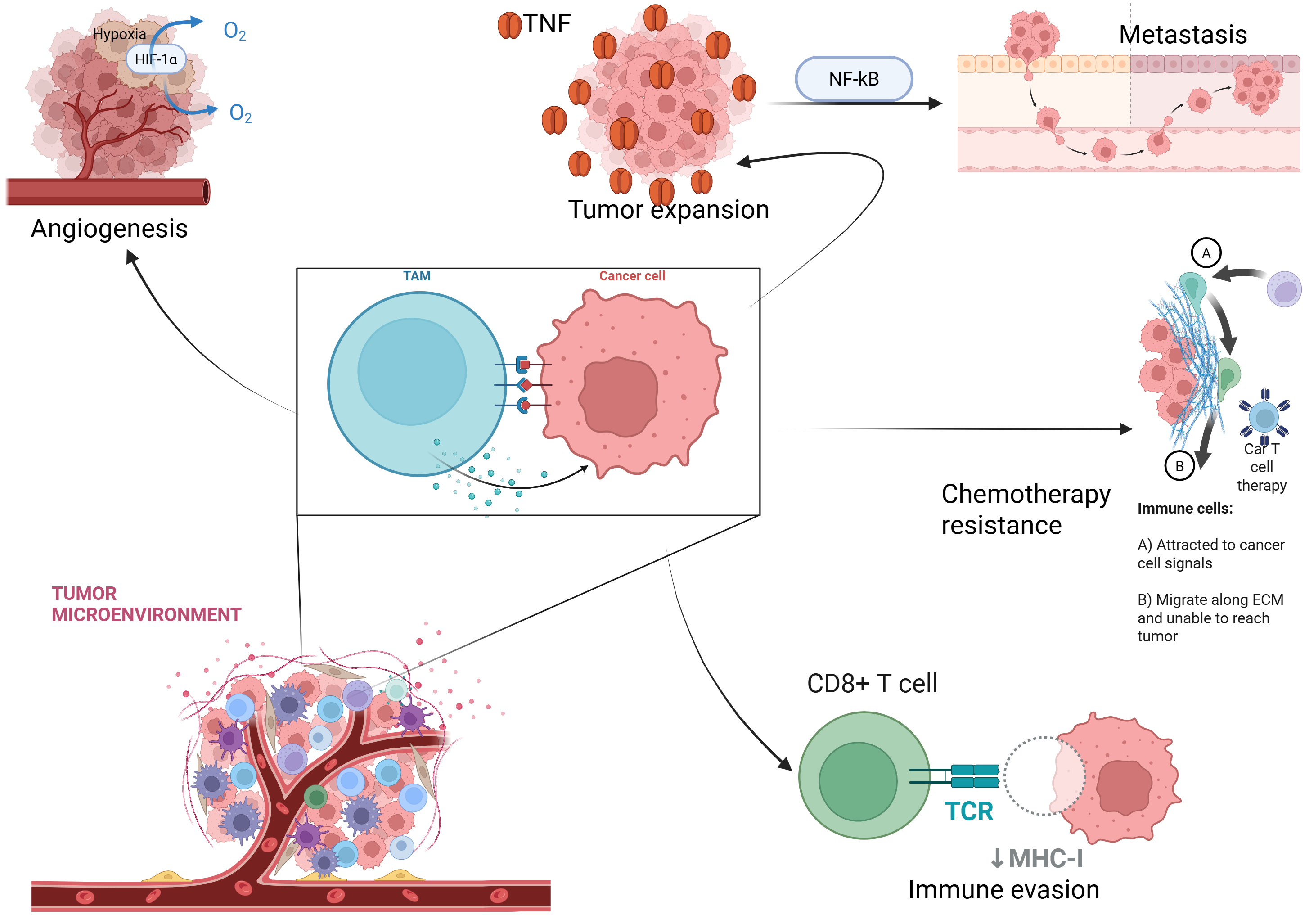
The interaction between tumor cells and the immune system is a central determinant of cancer development and progression. Through a complex network of cytokines, chemokines, and metabolic cues, tumors actively shape immune responses that can either restrain or promote malignant growth. Monocytes are key mediators of this crosstalk, as they are efficiently recruited to tumor sites in response to tumor- and stroma-derived signals, thereby accumulating within primary tumors and metastatic niches (1–3). These short-lived, circulating mononuclear phagocytes infiltrate tissues and differentiate into diverse myeloid populations, including myeloid-derived suppressor cells (MDSCs) and tumor-associated macrophages (TAMs), which constitute major cellular components of the tumor microenvironment (TME). Accumulating evidence indicates that these monocyte-derived populations contribute to angiogenesis, immune suppression, metastatic dissemination, and resistance to chemotherapy (4–6). While macrophages retain the capacity to eliminate tumor cells through phagocytosis, exposure to the TME frequently reprograms them toward tumor-supportive states (7, 8), underscoring their functional duality and context dependence during tumor progression (9) (Fig. 1). Together, these observations position monocyte-derived populations as central architects of the tumor microenvironment and key regulators of tumor immunity, angiogenesis, and therapeutic resistance.
Recent technological advances have fundamentally reshaped our understanding of monocyte and macrophage diversity in cancer. Single-cell transcriptomic and epigenomic profiling have revealed extensive heterogeneity within circulating monocytes and tumor-associated macrophages, uncovering discrete developmental trajectories, activation states, and functional programs that were previously obscured by bulk analyses. Spatially resolved transcriptomics and multiplex imaging further demonstrate that macrophage phenotypes are tightly coupled to their anatomical localization within the tumor microenvironment, with distinct niches supporting immunosuppressive, angiogenic, or inflammatory programs. Complementing these approaches, lineage-tracing and fate-mapping studies have clarified the ontogeny of tumor-associated macrophage populations, distinguishing monocyte-derived cells from tissue-resident macrophages and revealing context-dependent contributions to tumor growth and immune regulation. Together, these high-resolution technologies highlight that monocyte and macrophage states are dynamic, reversible, and shaped by local cues, challenging static models of myeloid identity and underscoring the need for integrated, multidimensional frameworks to interpret myeloid function in cancer.
Despite these advances, major conceptual and translational gaps remain. It remains unclear which monocyte-derived and macrophage subsets are causally linked to tumor progression, therapeutic resistance, or durable clinical responses, and how these populations evolve under treatment pressure. Furthermore, the field lacks unified criteria to functionally define myeloid subsets across tumor types and disease stages, complicating biomarker development and therapeutic targeting. Emerging strategies that modulate monocyte recruitment, reprogram macrophage states, or selectively deplete tumor-promoting subsets have shown promise, yet their clinical success has been variable, reflecting incomplete mechanistic insight and context dependence. In this Review, we synthesize current knowledge on monocyte and macrophage heterogeneity in cancer, highlight unresolved challenges in classification and functional attribution, and discuss therapeutic approaches that exploit myeloid plasticity. By integrating insights from cutting-edge technologies with clinical observations, we aim to provide a conceptual framework to guide the rational design of next-generation myeloid-targeted therapies. These advances motivate a closer examination of how innate myeloid cells are organized and reprogrammed within the tumor microenvironment. In this Review, we propose that tumor progression is strongly shaped by a dynamic myeloid plasticity network in which monocytes, macrophages, and related myeloid populations continuously adapt to tumor-derived inflammatory, metabolic, and spatial cues.
2. Innate Myeloid Landscapes in the Tumor Microenvironment
Innate myeloid cells constitute a dominant and functionally diverse component of the tumor microenvironment (TME), integrating tumor-intrinsic signals with local stromal and immune cues. In this section, we outline the organizational principles of innate myeloid landscapes within the TME, focusing on monocyte recruitment, macrophage reprogramming, and the microenvironmental signals that define tumor-associated myeloid states. Understanding how monocytes and macrophages are recruited, reprogrammed, and spatially organized within the tumor microenvironment provides a conceptual framework for interpreting their roles in tumor progression and therapeutic response.
Cancer progression is strongly associated with genetic and epigenetic changes, but recent studies show that TME is also a key determinant (10). TME is a niche within the body that contains both cellular and non-cellular components (11–15). Cellular components include cancer, stromal, and immune cells (such as T and B lymphocytes, neutrophils, natural killer (NK) cells, dendritic cells (DCs), MDSCs, and macrophages). Non-cellular components encompass the extracellular matrix (ECM) (collagens, fibronectin, and laminins), soluble mediators (cytokines, chemokines, and growth factors), extracellular vesicles, and physicochemical features such as hypoxia, acidity, and metabolites (10). Immune cells in the TME are not malignant but contribute to tumor suppression or progression (11, 12) through cell-surface receptor expression and production of soluble factors. Recent spatial transcriptomics analyses, often integrated with single-cell RNA sequencing, have revealed that immune and myeloid cells are organized into spatially distinct niches within the TME, shaping macrophage localization, functional states, and tumor progression (14, 15). Within the TME, monocytes are polarized into distinct phenotypes and functions (16–19). For example, Th2 signals drive macrophage differentiation toward an M2 anti-inflammatory phenotype that promotes tumor progression (20). Notably, macrophages present in the tumor site, referred to as TAMs, are transcriptionally distinct from both monocytes and their tissue-resident counterparts. This distinct identity arises from active reprogramming within the TME, where hypoxia, metabolic stress, cell-cell interactions, cytokines, and growth factors drive context-dependent differentiation programs (21). In cancer patients, altered chemokine levels, including changes in C-X3-C motif chemokine ligand 1 (CX3CL1) and reduced C-C motif chemokine ligand 2 (CCL2) in the circulation, are associated with changes in monocyte mobilization and recruitment to tumors. As a result, TAMs acquire tumor-associated transcriptional states enriched for genes involved in immune regulation, extracellular matrix remodeling, angiogenesis, and tissue repair, and are accompanied by reduced expression of genes involved in inflammatory and antigen presentation pathways (22–24).
Tumor-derived signals such as colony-stimulating factor 1 (CSF1), interleukin-10 (IL-10), transforming growth factor beta (TGF-β), hypoxia-inducible factors (HIFs), and metabolic mediators including lactate, lipids, and oxidative stress activate multiple transcriptional regulators in TAMs, including signal transducer and activator of transcription 3 (STAT3), hypoxia-inducible factor-1 alpha (HIF-1α), peroxisome proliferator-activated receptor gamma (PPARγ), and nuclear factor kappa B (NF-κB).
These pathways collectively shape TAM transcriptional programs, leading to sustained expression of genes commonly associated with tumor macrophages, such as complement component 1q (C1Q), apolipoprotein E (APOE), triggering receptor expressed on myeloid cells 2 (TREM2), galectin-3 (LGALS3), cluster of differentiation 163 (CD163), and V-set and immunoglobulin domain-containing protein 4 (VSIG4) (21). In several human cancers, TAMs also show elevated expression of sialic acid-binding immunoglobulin-like lectin 1 (SIGLEC1), which encodes cluster of differentiation 169 (CD169), and C-C motif chemokine ligand 8 (CCL8), a macrophage-derived chemokine linked to tumor progression and metastasis (25–27). In contrast, circulating monocytes maintain transcriptional programs associated with migration, pathogen sensing, and inflammatory cytokine production, whereas tissue-resident macrophages preserve lineage-specific programs related to tissue development and homeostasis (28, 29).
In this context, the TME is a complex, dynamic niche that integrates immune, stromal, and tumor-derived elements. The cellular component is primarily composed of immune cells, including T cells, TAMs, dendritic cells, NK cells, MDSCs, and neutrophils, which together define the immune compartment of the TME and are discussed in detail in later sections of this review. There are also stromal cells, such as fibroblasts and adipocytes, that can acquire altered phenotypes to promote cancer progression. Malignant tumor cells can also actively influence surrounding stromal and immune cells. The non-cellular component comprises the ECM, a network of structural proteins and bioactive molecules that forms a scaffold promoting tumor cell adhesion, migration, and invasion. In addition, bioactive molecules contribute to the chemical and physical properties of the TME. The presence of cytokines and chemokines, combined with a hypoxic environment, contributes to TME complexity and heterogeneity, thereby increasing resistance to chemotherapy (30–33). As a consequence of this organization, the TME also contributes to therapy resistance, tumor development, and metastasis, largely through immune cells and mediators such as CCL2, CXCR4, and TGF-β (34–37). This supportive niche is maintained by non-tumor cells, ECM remodeling, and soluble mediators. The ECM is frequently remodeled and stiffened, thereby serving as a physical and biochemical barrier that limits T-cell infiltration and the penetration of chemotherapeutic drugs. Nutrient-deprived conditions within the TME impose metabolic stress that favors the selection of aggressive tumor phenotypes. Cell-ECM interactions induce the release of soluble factors that promote immune evasion and ECM remodeling, thereby contributing to therapy resistance. In addition, the hypoxic nature of the niche, the presence of tumor-derived exosomes, and deregulated metabolic pathways play key roles in supporting the TME and promoting cancer progression (38–40).
Because cancer cell survival and chemotherapy resistance depend on a supportive niche, the TME has become a major focus of cancer research. Studying the microenvironment beyond tumor cells alone has revealed how interactions among cancer cells, immune cells, stromal components, and the ECM support tumor growth and therapy resistance, thereby guiding the development of therapeutic strategies that disrupt these tumor-supportive interactions and improve treatment responses in cancer patients.
Tumor-Driven Reprogramming of Monocyte Phenotypes
Tumor cells actively reprogram infiltrating monocytes, reshaping their phenotype, metabolic state, and differentiation potential to support immune suppression and tumor progression. This reprogramming is driven by tumor-derived cytokines, metabolic stress, and aberrant activation of intracellular signaling pathways, altering monocyte abundance, function, and clinical impact. In this section, we discuss the mechanisms by which tumors remodel monocyte phenotypes and the prognostic and therapeutic implications of these changes.
Myeloid cells are involved in homeostasis, protection against pathogens, tissue remodeling, and activation of adaptive immunity (41–43). Myeloid cell populations within the TME broadly regulate tumor elimination or survival (44). The presence of infiltrating innate myeloid immune cells, such as monocytes, DCs, and macrophages, initiates a cycle of responses at the tumor site. These cells can act as antitumor effectors or promote cancer growth (45). Tumor-associated myeloid cells include monocytes, MDSCs, tumor-associated DCs, and TAMs (46).
Monocytes, a crucial component of the innate immune system, are divided into three subpopulations in humans: classical, non-classical, and intermediate (47, 48). Originating in the bone marrow, monocytes migrate to sites of inflammation, where they differentiate into macrophages or DCs. Monocytes and their derivatives display three main functions: antigen presentation, phagocytosis, and cytokine/chemokine production (49). However, in the context of cancer, tumor cells can exploit the TME to reprogram myeloid cells to favor cancer progression. The release of tumor-associated cytokines, such as G-CSF and GM-CSF, alters the metabolic profile of myeloid cells, including upregulation of lipid transport and uptake pathways. This increased lipid uptake drives metabolic and functional reprogramming, promoting immunosuppressive mechanisms and enhancing the survival and expansion of myeloid cells within the tumor environment (50, 51). As a result, tumor-derived signals alter monocyte abundance, phenotype, differentiation trajectories, and overall immune function. Within this altered environment, monocytes are preferentially driven to differentiate into TAMs rather than into immunostimulatory dendritic cells. This skewed differentiation is mediated by tumor-derived cytokines and aberrant activation of intracellular signaling pathways. For example, ovarian cancer cells release leukemia inhibitory factor (LIF) and IL-6 into the TME, which promote TAM generation by redirecting monocyte differentiation toward TAM-like phenotypes. In parallel, tumor-induced activation of the MAPK and PI3K/AKT pathways in myeloid cells further reinforces M2-like polarization, suppressing differentiation toward antigen-presenting dendritic cells (25, 52).
Cancer development typically involves inflammation, which can be both a driver and a consequence of tumorigenesis. This inflammation recruits inflammatory cells, including monocytes, from the bloodstream to the tumor site, and tumors can also promote monocytosis by releasing cytokines and chemokines that enhance myelopoiesis and monocyte mobilization. This may contribute to increases in the total count and relative number of monocytes, which are commonly observed in cancer patients. The connection between cancer and inflammation creates a conducive environment for tumor initiation, with about 20% of cancer cases arising from infection and chronic inflammation. Chronic inflammation primarily suppresses the antitumor response and drives tissue remodeling by releasing cytokines and chemokines, such as VEGF, TGF-β, and TNF-α, and by activating pathways such as NF-κB and STAT3, thereby promoting cancer progression (53–56). Studies have shown that an elevated absolute monocyte count (AMC) at diagnosis is associated with worse overall survival. Higher circulating monocyte counts are associated with poor clinical outcomes and may reflect increased TAM activity, which is linked to angiogenesis, immunosuppression, and metastasis (Fig. 1) (53, 57–59). Ratios reflecting monocyte proportion, such as the low lymphocyte-to-monocyte ratio (LMR), predict poor outcomes in many solid tumors (53, 60–62). Pretreatment values of AMC or LMR can help identify patients at higher or lower risk. For example, elevated AMC has been linked to poor survival in prostate and pancreatic cancers (63, 64). A low LMR is associated with adverse outcomes in oral and head and neck cancers (HNCs) (65, 66). Monitoring these measures during therapy may also provide insight into treatment response; however, their clinical utility requires further validation in large, prospective studies to determine their predictive value across different tumor types and treatment settings.
Additional studies highlight the potential role of monocytes as prognostic markers. Signal regulatory protein α (SIRPα) is a receptor that suppresses monocyte/macrophage phagocytic function and inflammatory signaling. The interaction between SIRPα and cluster of differentiation 47 (CD47, the “self” signal expressed by normal cells) prevents phagocytic clearance. Consistent with this, tumor cells often overexpress CD47 to evade phagocytosis by monocytes and macrophages (67). Clinical studies further show that increased numbers of CD14⁺ SIRPαhigh monocytes are associated with reduced survival in follicular lymphoma, whereas an increased CD14⁻ SIRPαlow monocyte subset correlates with better survival (68). Monocyte numbers are not only associated with cancer survival but can also predict responsiveness. Patysheva et al. reported that the percentages of CD163-expressing CD14low CD16⁺ and CD14⁺ CD16⁺ monocytes are increased in breast cancer compared with healthy women; moreover, CD163⁺ monocytes increase in tumors after neoadjuvant chemotherapy (NAC), and the proportion of CD14low CD16⁺ monocytes positively correlates with response to NAC (69). The TME shapes monocyte phenotypes to favor cancer progression. These alterations can serve as indicators of tumor grade and prognosis (70). In pancreatic cancer, circulating monocytes exhibit constitutive phosphorylation of signal transducer and activator of transcription (STAT) family proteins and a diminished response to stimulation, indicating abnormal activation and impaired immunity (71). Serum levels of monocyte-associated cytokines, including interleukin-6 (IL-6), granulocyte colony-stimulating factor (G-CSF), and granulocyte-macrophage colony-stimulating factor (GM-CSF), are elevated in lung cancer. Notably, increased G-CSF levels correlate with adverse clinical outcomes and are associated with advanced disease stage, increased tumor burden, and greater disease severity (72).
The direct effects of tumor cells on monocyte phenotype have been validated ex vivo. Co-culturing monocytes from cancer patients with tumor cells resulted in downregulation of the pro-inflammatory macrophage marker CD86, suggesting compromised antitumor function (71). An additional study showed increased expression of the cancer stemness-promoting factor CD51 in monocytes after co-culture with pancreatic tumor cells (73). Taken together, the TME profoundly shapes monocyte populations and phenotypes, driving their differentiation toward immunosuppressive states.
Monocyte-Derived DCs and MDSCs in Cancer
In addition to giving rise to tumor-associated macrophages, monocytes represent a critical source of dendritic cells and myeloid-derived suppressor cells (MDSCs) in cancer, with profound consequences for antitumor immunity and disease progression. Tumor-derived inflammatory and metabolic cues skew monocyte differentiation toward immunosuppressive MDSC and macrophage lineages rather than immunostimulatory dendritic cell lineages. In this section, we examine the mechanisms governing monocyte fate decisions in cancer and discuss how monocyte-derived DCs and MDSCs contribute to immune evasion, therapy resistance, and metastatic dissemination.
Monocytes can differentiate into DCs or macrophages in response to cytokine stimulation. DCs generally support antitumor immunity by presenting tumor-associated antigens and priming cytotoxic CD8⁺ T cells. Tumor-derived factors shift this balance. For example, retinoic acid in the TME has been reported to favor differentiation of intratumoral monocytes toward TAMs, which support tumor growth, in part by suppressing the DC-associated transcription factor IRF4 (74). Although the mechanisms remain incompletely understood, soluble factors in the TME appear to drive infiltrating monocytes toward immunosuppressive TAMs rather than toward immunostimulatory DCs. Tumor-derived cytokines and growth factors, including IL-10, IL-6, TGF-β, and vascular endothelial growth factor (VEGF), suppress DC differentiation while promoting immunosuppressive macrophage phenotype (75, 76). For example, in human melanoma, melanoma cell supernatants rich in IL-10 have been shown to induce a shift in monocyte polarization toward a CD163+, programmed death-ligand 1 (PD-L1)+ M2-like phenotype and to inhibit monocyte differentiation into DCs, which are associated with tumor progression (77). In cancer, circulating monocytes are a major source of MDSCs. Tumor-driven inflammation can reprogram these cells toward immunosuppression (41). MDSCs include granulocytic/polymorphonuclear (G-MDSCs or PMN-MDSCs) and monocytic (M-MDSCs). M-MDSCs are defined as CD11b⁺Ly6G⁻Ly6Chigh in mice and CD11b⁺CD14⁺HLA-DR−/loCD15⁻ in humans, and low MHC-II further helps distinguish them from monocytes. During tumor development, persistent signals such as GM-CSF, IL-6, and IL-1β pathologically activate monocytes and drive the expansion of M-MDSCs (41, 78).
The development and suppressive function of MDSCs are regulated by multiple transcriptional and signaling pathways. Key regulators of MDSC development include cellular Rel proto-oncogene (c-Rel), STAT3, S100 calcium-binding protein A8 and S100 calcium-binding protein A9 (S100A8/9), TNF-α-induced protein 8-like 2 (TIPE2), and Interferon Regulatory Factor 8 (IRF8) (79–82). c-Rel is a central driver of M-MDSC growth and suppressive function (83). c-Rel belongs to the Nuclear Factor κ-light-chain-enhancer of activated B cells (NF-κB) family and turns on immunosuppressive enzymes and other M-MDSC genes by forming a c-Rel/ CCAAT/enhancer-binding protein β (C-EBPβ)/pSTAT3/p65 enhanceosome (79). A c-Rel-programmed subset of M-MDSCs in mouse and human melanoma, termed c-Rel-dependent monocytes (rMos), has been described. These c-Rel⁺, IL-1βhigh, Arginase-1 (Arg-1)⁻ rMos promote tumor growth by blocking T-cell function and maintaining a suppressive microenvironment through IL-1β/CCL2 crosstalk (84). MDSCs play an essential role in tumorigenesis and cancer progression by inhibiting T-cell proliferation and activation through multiple mechanisms, including cytokine modulation, metabolic depletion, and direct interactions (85, 86). For example, in acute myeloid leukemia (AML), increased M-MDSC counts are associated with poor prognosis (87). Notably, elevated M-MDSC levels predict reduced immunotherapy efficacy, including CAR-T, and are associated with chemoresistance (88, 89). Within the TME, multiple tumor-derived signals further reinforce M-MDSC suppressive phenotypes. In glioblastoma, the cell adhesion molecule integrin β1 and the dipeptidyl peptidase-4 (DPP-4), which are upregulated in M-MDSCs, act as central regulators that promote their tumor-promoting functions (90). Leukocyte immunoglobulin-like receptor B4 (LILRB4) drives the polarization of M-MDSCs and downregulates miR-1 family microRNAs, thereby promoting tumor cell migration and invasion (91, 92). Soluble Heat Shock Protein 90 α (HSP90α) can program human monocytes into immunosuppressive M-MDSCs via Toll-like Receptor 4 (TLR4) signaling, which, in turn, upregulates PD-L1 on these cells (93). Post-transcriptional mechanisms also contribute to M-MDSC function. Functional suppression of the type I interferon (IFN-I) axis is necessary for MDSCs to develop immunosuppressive activity in cancer, whereas stabilizing Interferon α/β Receptor Subunit 1 (IFNAR1) and combining it with interferon-based therapy produces a strong antitumor response (94). Finally, Ubiquitin-Specific Peptidase 12 (USP12)-mediated deubiquitination and stabilization of p65 diminish both the infiltration and suppressive capacity of M-MDSCs, enhancing CD8⁺ T-cell activity and slowing tumor growth (95).
M-MDSCs exhibit lower surface MHC-II expression than monocytes. Increased autophagy in these cells accelerates lysosomal degradation of MHC-II, thereby reducing surface expression and impairing antitumor immunity. When autophagy is reduced, lysosomal processing is diminished, MHC-II surface levels rise, and tumor-specific CD4⁺ T cells are more effectively activated. Inhibiting the E3 ligase Membrane-Associated RING-CH-type Finger 1 (MARCH 1) stabilizes MHC-II at the plasma membrane, indirectly weakening the suppressive program of M-MDSCs and leading to potent antitumor responses and marked tumor reduction (96, 97). Together, these findings indicate that the TME reprograms monocyte differentiation into M-MDSCs through coordinated transcriptional and post-transcriptional mechanisms. In fibrotic liver, M-MDSC recruitment correlates with diminished tumor-infiltrating lymphocytes and increased tumorigenicity, as observed in both mouse models and human disease (98). In patients with hepatocellular carcinoma (HCC), the liver is notably enriched for M-MDSCs, and CD33 expression associates with more aggressive tumor features and poorer survival (99). Mechanistically, activated hepatic stellate cells (HSCs) engage monocyte-intrinsic p38 Mitogen-Activated Protein Kinase (MAPK) signaling to drive M-MDSC development and immunosuppression. Consistently, pharmacologic p38 MAPK inhibition disrupts this HSC-M-MDSC circuit and suppresses HCC growth (98).
Metastatic dissemination further highlights the context-dependent roles of monocyte subsets. Metastasis refers to the dissemination of tumor cells to distant organs and their subsequent colonization. “Classical” inflammatory monocytes support metastatic relapse when systemic or local inflammation rises during therapeutic intervention for the primary tumor. For example, in breast cancer, these monocytes are recruited to the tumor site, differentiate into macrophages, and drive metastatic progression by releasing cytokines such as CCL2, which recruit CCR2⁺ inflammatory monocytes to the tumor, where they differentiate into TAMs under the influence of CSF-1 (M-CSF)/CSF-1R signaling and other tumor-associated cytokines. These TAMs promote metastatic progression by creating a pro-tumor environment that supports tumor cell survival, invasion, and dissemination (100). The Chitinase-like protein 3-positive, Lymphocyte antigen 6 complex locus C-high monocyte (Ym1⁺Ly6Chigh) subset, which favors tissue repair, plays a role in this process (101). Consistently, depleting Ly6Chigh immunoregulatory monocytes suppresses inflammation-driven metastasis, whereas transferring these cells into naïve mice enhances lung metastasis. Additionally, these monocytes express high levels of Matrix Metalloproteinase 9 (MMP-9) and C-X-C chemokine receptor type 4 (CXCR4), and inhibiting either pathway reduces the metastasis-promoting effect (102).
Monocytes and macrophages can promote tumor invasiveness and metastatic potential by engaging adhesion- and stemness-associated pathways, including Cluster of Differentiation 44 (CD44) signaling and interactions with tumor-cell Vascular Cell Adhesion Molecule 1 (VCAM-1), which support cancer cell survival in metastatic niches. Additionally, tumor-intrinsic mechanisms involving ezrin and Phosphoinositide 3-kinase (PI3K) signaling have been implicated in aggressive behavior across multiple cancer types, including breast, prostate, and head-and-neck cancers (103–107). Notably, interrupting monocyte-cancer interactions reverses this invasive phenotype (37). Inflammation-induced monocyte-derived macrophages (CD11b⁺CD11c⁺) increase the efficiency of early metastatic colonization in mouse models. These macrophages secrete hepatocyte growth factor (HGF), which supports tumor cell survival under stress in vitro. Blocking HGF signaling prevents the formation of early micrometastatic lesions that arise during inflammation in vivo. These findings show that HGF-producing macrophages create a pre-metastatic niche and support early survival of tumor cells (108). Moreover, M-MDSCs promote tumor metastasis, coordinating complex interactions between breast cancer cells and stromal cells (109–112).
Conversely, specific monocyte populations can directly inhibit metastasis. Co-culturing undifferentiated monocytes with pancreatic ductal adenocarcinoma (PDAC) cells markedly suppresses invadopodia formation via tissue inhibitor of metalloproteinase-2 (TIMP2) secreted by monocytes. These findings suggest that increasing TIMP2⁺ monocytes at the primary tumor site may block invasion and metastasis (113). In metastatic breast cancer, IFN-γ- producing monocytes are recruited to the lung and activate the TMEM173/STING pathway in neutrophils. This boosts neutrophil killing and prevents the outgrowth of disseminated tumor cells (114). Monocytes induced by IFN-γ (IFN-IMos) also limit lung metastasis by expanding NK cells through an IL-27-dependent mechanism. Forkhead box O1 (FOXO1) and nuclear receptor subfamily 4 group A member 1 (NR4A1) regulate IFN-γ-driven differentiation and the antimetastatic function of IFN-IMos. These findings identify two potential therapeutic targets to improve IFN-γ-based treatment for cancer metastasis (115). Non-classical “patrolling” monocytes, unlike classical monocytes, gather in the lung microvasculature and limit lung metastasis in several mouse models (116).
Together, these findings underscore the functional heterogeneity of monocyte-derived populations in cancer. Although many tumor-associated myeloid cells promote immune suppression and tumor progression, distinct monocyte states can, in specific contexts, exert potent antimetastatic functions, highlighting their therapeutic potential (113, 114, 117).
Together, these findings indicate that monocyte fate decisions in cancer are governed by integrated inflammatory, metabolic, and transcriptional programs rather than linear differentiation pathways.
3. Tumor-Driven Reprogramming of Macrophages
Macrophages are professional phagocytes with remarkable plasticity and arise from multiple developmental origins. In cancer, macrophages can derive from bone marrow-produced monocytes that circulate in the bloodstream and are recruited into tissues, where they differentiate into tumor-associated macrophages or undergo programmed cell death (118, 119). In addition, tissue-resident macrophages originate from embryonic progenitors in the yolk sac or fetal liver and persist as long-lived populations with distinct transcriptional and functional identities (120–123). These macrophage populations are widely distributed across tissues and contribute to host defense, immune surveillance, and tissue repair; however, within tumors, they are frequently co-opted to support malignant growth, immune suppression, and remodeling of the tumor microenvironment (124, 125). A central question in tumor immunology is how distinct macrophage populations are reprogrammed by tumor-derived signals to shift from antitumor immune surveillance toward tumor-promoting and immunosuppressive states.
Ontogeny, Polarization, and Functional Plasticity of Macrophages
Macrophage states in tumors reflect both ontogeny (tissue-resident versus monocyte-derived) and dynamic polarization programs induced by local signals. Although the M1/M2 framework has been widely used, tumor-associated macrophages exhibit a continuum of phenotypes, coupled with metabolic remodeling and niche-specific cues. Here, we summarize how the origin and polarization of macrophages intersect to shape macrophage function within the TME and influence whether tumors are controlled or promoted.
Macrophages can be broadly divided into two groups: monocyte-derived macrophages (MDMs) and TRMs. MDMs, derived from the bone marrow, are recruited from the circulation to tissue sites, typically following inflammation. By contrast, TRMs are found in various tissues and arise from embryonic progenitors that seed organs before birth and subsequently self-renew locally. Inflammation and aging can alter tissue macrophage composition by favoring recruitment of bone marrow-derived monocytes, which differentiate and partially replace TRMs in some organs (126). Tissue-resident and monocyte-derived macrophages have distinct origins and consequently display distinct transcriptional programs. As a result, changes in their relative abundance can modify local immune regulation and TME interactions. In tumors, the accumulation of monocyte-derived macrophages often promotes immunosuppressive, pro-angiogenic, and tissue-remodeling functions. This, in part, is mediated by pathways such as CSF-1R signaling, which drive tumor growth, immune evasion, and metastasis. However, the consequences of replacing resident cells with monocyte-derived macrophages are tissue- and context-dependent, and both populations can adopt pro- or antitumor phenotypes depending on the microenvironmental conditions (127–129). Beyond developmental origin, macrophage behavior in tumors is further shaped by functional polarization programs driven by local signals within the TME. Macrophages have classically been categorized into two phenotypes: M1 (“classically activated”) and M2 (“alternatively activated”). M1 macrophages are involved in proinflammatory responses, pathogen clearance, and antitumoral activity. They express CD80 and CD86 cell-surface receptors and secrete IL-1β, tumor necrosis factor α (TNF-α), IL-12, and IL-23. Additionally, M1 macrophages secrete reactive oxygen species (ROS) and have greater antigen-presenting capacity. While these properties support antitumor immunity, they can contribute to chronic inflammation and tissue injury (130).
M2 macrophages are involved in anti-inflammatory responses, tissue repair, angiogenesis, and tumor progression. They express CD206 and CD163 cell-surface receptors, display increased phagocytic activity, express higher levels of Arg-1, and secrete transforming growth factor β (TGF-β), IL-10, and vascular endothelial growth factor (VEGF) (131–134). The secretion of IL-10 and TGF-β by macrophages inhibits T-cell activity, thereby promoting tumor progression rather than clearance. Importantly, macrophage activation is highly plastic, and both pro-inflammatory and anti-inflammatory programs can influence cancer in context-dependent ways. While pro-inflammatory M1 macrophages can support antitumor immunity by enhancing antigen presentation and producing cytotoxic mediators, chronic inflammation can also promote tumor initiation and progression. Persistent inflammation increases mutagenic stress by releasing ROS from myeloid cells, thereby promoting DNA damage and genetic instability. Moreover, inflammatory cytokines, such as TNF-α, activate survival and proliferation pathways, including NF-κB, in tumor and stromal cells. Over time, these processes can favor more aggressive and therapy-resistant tumor cells, helping explain how chronic inflammation promotes tumor progression rather than clearance (135–138).
Macrophage polarization is also tightly linked to metabolic reprogramming. Macrophage activation leads to profound changes in cellular metabolism. M1 activation is characterized by glycolysis and fatty acid synthesis, whereas M2 activation is associated with the tricarboxylic acid cycle, fatty acid oxidation, and glutaminolysis (139). Although widely used, the M1-M2 designation is now recognized as an oversimplification, since it captures only extreme phenotypes along a spectrum of macrophage states. Macrophages and monocytes exhibit considerable diversity and plasticity, enabling them to adapt to various functional states in response to diverse environmental conditions. They exhibit a broader spectrum of activation states rather than the two extreme representatives, M1 and M2. Macrophage complexity is shaped by activation markers, environmental signals, external factors such as substrate stiffness, and epigenetic controls. The continuum of macrophage polarization enables them to adapt to various physiological and pathological conditions. This flexibility complicates classification, as various markers can be expressed across multiple activation states and tissues are exposed to complex stimuli, resulting in mixed macrophage populations. The imbalance in macrophage populations is associated with several immune-related diseases. The continuum is crucial for developing therapeutic strategies that target macrophage polarization to balance inflammation and repair (140, 141).
Within the TME, skewing of this polarization continuum toward M2-like states has profound functional consequences. M2 macrophage polarization promotes tumor-cell proliferation and immune evasion by increasing PD-L1 expression on tumor cells, enhancing secretion of immunosuppressive cytokines such as IL-10 and TGF-β, and driving metabolic changes, including depletion of metabolites such as L-arginine, that inhibit cytotoxic T-cell function (142, 143). Tumor-associated macrophages (TAMs) represent the most abundant immune population within the TME. Despite its antitumor properties, the TME can polarize macrophage phenotypic differentiation and induce tumor-promoting activities, including proliferation, progression, and metastasis (144). Macrophages play numerous roles in tumor progression, and TAMs are crucial at different stages of cancer progression. TAMs induce the expression of cell-surface receptors, cytokine production, and the secretion of growth factors that promote tumor invasion and metastasis (145–148). During the early stages of tumor formation, macrophages control cancer growth through cytotoxicity, phagocytosis, and activation of other innate and adaptive immune responses. As the tumor develops and becomes well established, these macrophages respond to external stimuli, differentiating into phenotypes that suppress immune-cell cytotoxicity and promote tumor survival, proliferation, and progression (149–152). M2 TAMs shape the TME by secreting chemokines that drive immune cell infiltration. M2 TAMs favor immunosuppressive cell subsets, such as regulatory T cells (Treg cells), and inhibit antigen-presenting cells, such as DCs, thereby creating an immunosuppressive TME. M2 TAMs also upregulate receptors involved in the “don’t eat me” signaling pathway, thereby blocking phagocytic uptake and clearance of cancer cells (153). On the other hand, M1 TAMs can participate in a robust adaptive anticancer immune response by enhancing antigen presentation and engaging adaptive immunity.
However, microenvironmental conditions within tumors limit the persistence of these antitumor programs. Hypoxia within the TME reshapes macrophage functional states, suppressing classical M1-associated immune-stimulatory programs while promoting immunosuppressive and tissue-remodeling activities (130). Current TAM-targeted strategies aim to reprogram macrophages toward an M1 pro-inflammatory state or to block “don’t eat me” signaling to restore anticancer activity. It remains unclear which macrophage subsets most strongly drive suppression versus control, in part because clear phenotypic markers are lacking (154, 155).
Tumor-Associated Macrophages as Drivers of Tumor Progression and Therapy Resistance
Tumor-associated macrophages (TAMs) constitute a heterogeneous macrophage compartment that is often skewed toward immunosuppressive, pro-angiogenic, and tissue-remodeling programs. Beyond suppressing antitumor immunity, TAMs directly promote cancer stemness, epithelial-mesenchymal transition, metastatic competence, and therapeutic resistance. In this subsection, we discuss key tumor-promoting functions of TAMs and evidence linking TAM abundance or gene signatures to clinical outcomes.
The presence of TAMs at the tumor site supports cancer progression by promoting M2-like programs that secrete cytokines and growth factors that enhance tumor growth and survival, and TAMs can also contribute to chemotherapy resistance. Mechanistically, TAMs promote drug-resistant cancer stem/initiating cell phenotypes; for example, TAM-derived MFG-E8 activates STAT3 signaling and can cooperate with IL-6 to amplify chemoresistance (156). In addition, TAMs can promote epithelial-to-mesenchymal transition (EMT) programs that confer chemoresistance and metastatic traits, as shown in peritoneal dissemination of pancreatic cancer (157). Clinically, elevated TAM infiltration and/or TAM-associated gene signatures have been associated with poorer therapeutic response and worse outcomes in patient cohorts (158).
Collectively, these tumor-promoting activities reflect a functional shift in macrophages, in which intrinsic antitumor programs are suppressed and replaced by immunosuppressive and pro-tumorigenic states. Macrophages’ primary antitumor function is phagocytosis and clearance of cancer cells; however, the TME reprograms macrophages, impairing their function. TAMs encompass a broad spectrum of M1-M2 macrophages, but their activation and polarization are skewed toward the alternative M2 rather than the classical M1, which is mostly associated with anti-inflammatory responses (159). Macrophage plasticity and heterogeneity are highly diverse and complex; even slight changes in the microenvironment can alter their types, numbers, and functions, thereby affecting their ability to control tumor development and response to therapy (160). The TME's ability to influence monocyte/macrophage function and phenotype depends on its composition, which is directly influenced by tumor stage, site, and type (161). Notably, M2-polarized TAMs are mostly localized in the hypoxic areas of the TME (162). Overall, tumor-associated immune cells, especially TAMs, have been associated with poor prognosis and reduced patient survival (163–165). TAMs release immunosuppressive factors that inhibit T-cell proliferation, thereby blocking activation of the adaptive immune response, which is crucial for controlling tumor growth and clearance (166, 167). Collectively, these studies position TAM abundance and functional skewing as key determinants of therapeutic resistance across tumor types.
Macrophage Receptors and Immune Checkpoints Shaping the Tumor Microenvironment
Macrophage behavior in tumors is shaped not only by soluble cues but also by receptor-ligand interactions that regulate recruitment, activation, antigen presentation, and immune suppression. Tumor-associated macrophages frequently upregulate inhibitory receptors and checkpoint ligands while retaining activating pathways that can be therapeutically leveraged to reprogram. Here, we highlight key macrophage receptors in the TME and discuss how targeting these pathways can restore phagocytosis and antitumor immunity.
During tumor formation, innate immune cells are recruited to the tumor site via chemokines and receptor-ligand-mediated signaling, where they recognize tumor cells and initiate antitumor responses. However, these same immune cells can be reprogrammed to drive tumor-related inflammation, promoting progression rather than control and clearance (45, 168). Monocyte development and differentiation in the bone marrow are governed by CSF1R signaling through its ligands CSF-1 and IL-34 (169). Once in circulation, chemokine receptors such as CX3CR1 guide monocyte trafficking into tumors, where they phagocytose cancer cells and associated material (116). Within the TME, particularly in hypoxic niches, monocytes/macrophages often polarize toward an M2-like phenotype, and CD209 expression is observed on M2-skewed TAMs in hypoxic regions (170). In addition, M2 TAMs express CD204, which is correlated with high CCL2 levels and a tumor-promoting microenvironment (171, 172).
Most TAMs induce immunosuppressive signaling by expressing inhibitory receptors, such as the non-classical MHC-I molecules HLA-E and HLA-G, and the immune checkpoint ligands PD-L1, PD-L2, CD80, and CD86 (144). M2 TAMs express PD-L1 to support cancer cells’ immune evasion (142). M2-skewed TAMs commonly express high levels of PD-L1 (Table 1). Functionally, PD-L1 expressed on TAMs engages PD-1 on T cells, suppressing activation and cytotoxicity, while CD80/CD86 binding to cytotoxic T-lymphocyte-associated protein 4 (CTLA-4) further inhibits T-cell responses (Table 1) (173). In addition, TAMs that release IL-1β induce upregulation of PD-L1 expression on tumor cells, contributing to immune escape (174). TAMs help maintain an immunosuppressive environment by secreting IL-10 and TGF-β, which inhibit both CD4 and CD8 T cells and promote the differentiation of regulatory T (Treg) cells. TAMs recruit Treg cells within the TME through the production of numerous chemokines and by inhibiting T-cell cytotoxicity, either by depleting L-arginine via Arg-1 release or by depleting tryptophan via indoleamine 2,3-dioxygenase (IDO) expression (175). Tregs are immunosuppressive lymphocytes that promote tumor progression by limiting antitumor immune responses within the tumor microenvironment. They are actively recruited to tumor sites by chemokines such as CCL1, CCL22, and CCL28, or can be induced from conventional CD4⁺ T helper cells in the presence of TGF-β. Once established in the TME, Tregs suppress effector T-cell activity, promote immune tolerance, and contribute to tumor immune evasion (176–180).
PD-L1 expression is often increased in M2-like tumor-associated macrophages (TAMs), particularly CD163⁺ macrophages. Nasopharyngeal carcinoma, prostate cancer, and cervical carcinoma show a positive correlation between CD163⁺ M2-type macrophage infiltration and PD-L1 expression. In cervical cancer, M2-type TAMs upregulate PD-L1 expression by activating the PI3K/AKT pathway. The interaction between PD-L1 on TAMs and PD-1 on T cells inhibits T cells, thereby promoting immune evasion. This is a key immunosuppressive mechanism by which tumors progress. PD-1/PD-L1 checkpoint inhibitors restore antitumor T-cell activity by interrupting inhibitory signals driven by PD-L1 on tumor cells and immune cells, including tumor-associated macrophages. However, patient responses vary widely, and PD-L1-expressing myeloid cells can contribute to an immunosuppressive TME, potentially limiting therapeutic efficacy in some patients. This has led to the development of combination approaches that target TAM recruitment or polarization to improve responses to checkpoint blockade. For example, inhibition of CSF1R has been shown to reprogram TAMs toward a less immunosuppressive phenotype and enhance the efficacy of anti-PD-1 therapy in colorectal cancer models. Together, these findings support combining myeloid-targeted therapies with PD-1/PD-L1 blockade to achieve more durable and effective clinical responses (181–185). CD40, a member of the TNF receptor superfamily, is expressed on DCs, B cells, and macrophages (including TAMs) (Table 1). When CD40 binds its ligand CD40L, it triggers tumor-specific T-cell immune responses. Consistent with this, agonistic anti-CD40 antibodies, designed to mimic CD40L binding to CD40, can inhibit tumor growth by activating DCs and repolarizing TAMs toward an M1 phenotype (186, 187). The CD40/CD40L axis leads to DC activation and upregulates co-stimulatory molecules (CD80, CD86) and MHC molecules (Table 1). Interestingly, CD40 agonists have been shown to elicit tumor-specific immune responses and, in some malignancies, to directly induce tumor-cell death (188–192). Therapeutically, CD40 can be targeted to induce pro-inflammatory cytokines and activate cytotoxic T cells (187, 193).
Recognition of tumor-derived danger signals or dsRNA by TLR3 skews macrophages toward an M1 phenotype while suppressing M2 features (Table 1). Activation of TLRs on macrophages promotes M1 polarization, the release of pro-inflammatory cytokines such as IL-1β, IL-6, IL-12, and TNF-α, and the activation of the type I interferon response. Through IL-12-driven activation of NK and T cells, this pathway can also promote IFN-γ-associated Th1 immunity. This response involves the production of pro-inflammatory cytokines, which enhance antitumor immunity through mechanisms such as antigen presentation, nitric oxide production, and the activation of Th1-polarized and cytotoxic immune cells. However, when TLR signaling persists or becomes excessive, inflammatory cytokines such as IL-6 and IL-1β are produced at elevated levels. These cytokines activate signaling pathways, most notably the JAK/STAT3 pathway, in both tumor cells and surrounding stromal cells. Activation of this pathway supports tumor cell survival and proliferation and is associated with chemotherapy resistance. Additionally, endothelial cell activation induces VEGF expression, promoting angiogenesis and altering vascular structure, thereby facilitating tumor invasion and metastatic spread. Chronic inflammation also affects the extracellular matrix by inducing matrix-degrading proteases, gradually removing physical barriers that would otherwise limit tumor cell invasion. Sustained inflammatory activity further contributes to hypoxia in the TME. Under hypoxic conditions, hypoxia-inducible factors such as HIF-1α and HIF-2α drive transcriptional programs associated with angiogenesis, immune evasion, and metabolic adaptation. These hypoxia-driven changes favor macrophage recruitment and reprogramming toward M2-like phenotypes, thereby restoring an immunosuppressive environment that ultimately supports continued tumor growth and progression (194–203). TLR7 and TLR7/8 agonists, such as imiquimod (approved for superficial basal cell carcinoma and actinic keratosis) and resiquimod, have demonstrated autophagy-mediated tumor-cell death in preclinical models (204). Consistent with this, TLR7/8 agonists have been used to reprogram TAMs from an M2 to an M1 phenotype (205). In contrast, chronic or sustained TLR4 signaling in TAMs can be pro-tumorigenic by driving IL-10 production and immunosuppressive polarization (206).
The expression of CD137 has attracted significant interest because of its dual role in inducing pro-inflammatory cytokines and regulating IL-10 secretion (Table 1). Activation of CD137 signaling enhances the function of antigen-presenting cells (APCs), thereby inducing T-cell activation and contributing to tumor control (207). CD137 (4-1BB) is a TNF receptor expressed on immune cells, including NK cells; signaling through the CD137-CD137 ligand axis is vital for CD8+ cytotoxic T lymphocyte activation, proliferation, and differentiation. Agonistic anti-CD137 mAbs enhance NK-cell-mediated ADCC and potentiate the antitumor efficacy of anti-CD20 mAbs (e.g., rituximab) by further activating NK cells in lymphoma (208–210). Similarly, interleukin-12 (IL-12) produced by antigen-presenting cells induces interferon-γ (IFN-γ) production by T and NK cells, and IFN-γ, in turn, upregulates MHC class I and II, strengthening T-cell activation (211, 212).
Additional receptors important for TAM biology and function include CD206, MARCO, folate receptor-β (FRβ/FOLR2), and epidermal growth factor receptor (EGFR). TAMs expressing CD206⁺ are typically immunosuppressive and can be therapeutically targeted (213) (Table 1). CD206, a C-type lectin receptor of the mannose receptor family, is expressed on M2 macrophages, and blocking or depleting CD206⁺ TAMs can shift the compartment toward a more pro-inflammatory M1 phenotype (214). Similarly, targeting the scavenger receptor MARCO polarizes M2-polarized TAMs toward a pro-inflammatory and antitumor state (215). FRβ/FOLR2, selectively expressed on M2-like TAMs, promotes immunosuppression. High FRβ activity is associated with reduced CD8⁺ T-cell infiltration and tumor growth (216, 217). In addition, M2-like TAMs produce epidermal growth factor (EGF), which binds EGFR on tumor cells, driving proliferation, invasion, and metastasis (218).
Table 1. Monocyte and macrophage (TAMs) receptors and their function.
| Receptor | Function |
|---|---|
| PD-L1 | Inhibits T-cell activation and promotes immune evasion |
| TLRs | Induce M1-like macrophage polarization and inflammatory cytokine production |
| CD40 | Activates macrophages and dendritic cells to promote antitumor T-cell responses |
| CD137 (4-1BB) | Induces pro-inflammatory cytokine secretion and regulates IL-10 production |
| Mannose receptor (CD206) | Marker of immunosuppressive, M2-like TAMs |
| MARCO | Promotes pro-inflammatory signaling and antitumor macrophage reprogramming |
| Folate receptor-β (FOLR2) | Supports immunosuppression and limits CD8⁺ T-cell infiltration |
| EGFR | Promotes tumor cell proliferation, invasion, and metastasis |
Cytokine-Chemokine Networks Governing TAM Recruitment and Polarization
Recruitment and functional polarization of TAMs are coordinated by interconnected cytokine and chemokine networks generated by tumor cells, stromal elements, and immune populations. These circuits not only control monocyte trafficking and macrophage positioning within hypoxic or invasive niches, but also reinforce immunosuppressive programs that drive metastasis and therapy resistance. In this subsection, we outline major chemokine axes and cytokine loops that shape TAM accumulation and phenotype, highlighting context-dependent effects and therapeutic opportunities.
Many chemokines have been linked to the recruitment of macrophages to the tumor site. Chemokine (C-C motif) ligand 2 (CCL2), CCL5, chemokine (C-X-C motif) ligand 12 (CXCL12), and chemokine (C-X3-C motif) ligand 1 (CX3CL1) are known tumor-promoting chemokines that primarily recruit macrophages to the TME (219–222) (Table 2). CCL1 is frequently upregulated in cervical squamous cell carcinoma and other squamous carcinomas, such as esophageal squamous cell carcinoma, where CCL1-CCR8 signaling contributes to a pro-tumorigenic microenvironment and promotes cancer progression and metastasis (223, 224) (Table 2). Expression of CCL1 and its receptor CCR8 by M2 macrophages promotes tumorigenesis (225–227). In addition, secretion of CCL3 by mantle cell lymphoma cells promotes M2 polarization of macrophages via C-C chemokine receptor 1 (CCR1) signaling, thereby enhancing cancer invasion and immune escape (228).
The interaction between CCL2 and its receptor, CCR2, is triggered by pro-inflammatory signals and recruits macrophages (Table 2). Local cytokines then skew differentiation toward M2-like TAMs, which release proteases that sustain inflammation and facilitate invasion (229). In breast cancer, CCR2-expressing monocytes, once recruited to mammary tumors, are well known to promote tumor growth and metastasis. However, CCR2 is also expressed on cancer cells, where it can modulate tumor growth and immune responses in a context-dependent manner. In tumor cells, CCR2 signaling has been reported to primarily influence immune evasion rather than directly promote monocyte recruitment, including by regulating PD-L1 expression and modulating the activity of cytotoxic T cells and CD103⁺ cross-presenting dendritic cells. Consistent with this complexity, several studies indicate that CCL2-expressing monocytes or macrophages can exert antitumor effects under specific conditions, including enhancing antigen presentation, promoting cytotoxic T-cell responses, and suppressing tumor growth. These opposing outcomes suggest that the effects of the CCL2/CCR2 axis depend on the activation state of monocytes and macrophages, tumor stage, and the microenvironmental context. Accordingly, phenotypes observed in Ccr2⁻/⁻ models may reflect complex, cell-intrinsic effects beyond monocyte recruitment alone, supporting the notion that the CCL2/CCR2 axis is a key immune-modulatory pathway in cancer whose function is highly context dependent and can be exploited by both immune cells and cancer cells to shape tumor immunity (230–232).
In addition to CCR2-dependent signaling, CCL2 can engage alternative chemokine pathways that further regulate monocyte retention and TAM positioning within tumors. For example, CCL2 secretion triggers a chemokine cascade, including CCL3 signaling via CCR1, which promotes monocyte retention and supports tumor growth and metastasis. Consequently, blocking CCL2-CCR1 signaling inhibits the recruitment of inflammatory monocytes. TGF-β also upregulates CXCR4 expression in TAMs, leading to CXCR4-positive TAMs migrating toward CXCL12-producing tumors, thereby promoting cancer cell intravasation (34, 39, 233). In melanoma and bladder cancer, tumor cell-derived CCL2 interacts with CCR2 on TAMs, inducing macrophage recruitment, infiltration, and polarization toward an M2 phenotype (234–236). IL-3 and CCL2 together support macrophage differentiation, growth, and cytokine secretion. Interestingly, CCL2 and CXCL10 can also facilitate T-cell migration to the tumor site when TAMs are depleted (237). However, in most contexts, CCL2 promotes M2 polarization and tumor metastasis (238, 239). Recruitment of regulatory T cells (Tregs) is also regulated by CCL2, IL-10, and the CXCL12/CXCR4 axis. CCL2 additionally cooperates with IL-6 to promote the differentiation and survival of myeloid cells (240). Clinical evidence shows a correlation between high CCL2 expression and increased infiltration of CD68⁺/CCR2⁺ macrophages in tumors, which is associated with high proliferative activity and reduced survival in patients (241, 242). Importantly, inhibition of CCL2 (e.g., with arsenic trioxide) has been shown to reduce M2 polarization (243). The expression of CCL5 and its receptor, CCR5, facilitates macrophage migration to the tumor site (244) (Table 2). Breast cancer often shows high CCL5 expression, which supports monocyte recruitment to the tumor (244, 245). The recruited cells differentiate and express high levels of CCL3, CCL4, and CCL5, thereby recruiting NK cells to mediate tumor-cell killing (38). M1 macrophages express high levels of CXCL9, CXCL10, and CCL5, which are associated with antitumor activity (246). However, CCL5 secreted by myofibroblasts in the TME binds to CCR5 on TAMs, activating pathways that promote M2 polarization and induce CCL18 secretion, thereby supporting tumor growth (247).
Hypoxia in the TME activates HIF-dependent programs that can increase pro-tumor mediators such as TGF-β, CXCL12, and CCL5 and modulate the CX3CL1/CX3CR1 axis, thereby promoting tumor progression (258). TGF-β stimulates monocyte chemotaxis into the TME and induces immunosuppression, thereby favoring tumor growth and metastasis (249). TGF-β exerts a dual role: tumor-suppressive in early stages (by limiting proliferation) but, at later stages, pro-tumorigenic by expanding Tregs and promoting M2 polarization, which supports metastasis (250–256). IL-10 and TGF-β favor Treg activation over Th1 responses. TAMs secrete CCL22 (and, in some contexts, CCL17) to recruit CCR4⁺ regulatory T cells, while CCL18 attracts naïve T lymphocytes and promotes Th2-biased and immunosuppressive immune responses (257, 258) (Table 2). Consistent with this, elevated CCL22 correlates with tumor progression in breast cancer patients (259). CXCL12 guides TAMs to hypoxic niches within the TME and drives M2 polarization (for example, via CD163 expression), while CX3CL1 also contributes to TAM recruitment and intratumoral infiltration (260, 261).
Interleukins produced in the TME reshape TAM function, activating cytokine circuits that promote resistance to therapy, proliferation, invasion, and metastasis (Table 2). For example, mixed-phenotype TAMs (M1/M2-like macrophages) can express high Arg-1 and secrete IL-10, together contributing to a TME that promotes enhanced cancer cell migration and invasiveness (262–264). Tumor malignancy is further promoted by IL-10 secreted from M2 macrophages, which suppresses antitumor immunity (Table 2). In hypoxic regions of the TME, TAMs secrete CXCL8 and engage CXCR1/2 signaling pathways, thereby enhancing IL-10 production and reinforcing M2 polarization (265, 266). Some cancer cells also release IL-32, which induces a phagocytic, M2-like phenotype in TAMs. These cells secrete IL-10, which facilitates metastasis (267). Both TAMs and cancer cells secrete IL-6 and IL-10, establishing reciprocal signaling that drives cancer progression (Table 2). IL-10 not only promotes macrophage differentiation but also suppresses DC development, further impairing adaptive immunity. The presence of IL-10 promotes macrophage differentiation while suppressing DCs (268). At the tumor site, TAMs contribute to immune evasion and tumor growth by producing immunosuppressive mediators such as CCL17/CCL22, TGF-β, and IL-10; generating prostaglandin E2 (PGE2); expressing high levels of Arg-1; and releasing immune-regulatory lectins, including galectin-1 and galectin-9 (269–272). While M1 TAMs are generally considered antitumor, their secretion of IL-6 can paradoxically activate signaling pathways such as NF-κB and STAT3 within the TME, thereby supporting tumor progression and metastasis (273–275) (Table 2). In most cancers, however, IL-6 predominantly acts through M2 TAMs, promoting polarization, metastatic spread, and interaction with IL-6R on cancer cells (276). M2 macrophages also secrete IL-6 and respond to IL-4, driving metastasis and resistance to cancer therapies (277). Furthermore, tumor-derived extracellular vesicles (EVs) carrying inflammatory mediators can shape macrophage polarization. For example, EV-associated IL-32 released by tumor cells promotes M2-like macrophage polarization (267), while macrophage-derived IL-6 activates IL-6/STAT3 signaling in pancreatic cancer cells, thereby favoring tumor progression (278). Finally, the combined release of IL-6 and IL-10 by TAMs activates B7-H4 via STAT3 signaling, thereby inhibiting cytotoxic T-cell responses and promoting immune escape (279–282).
Other cytokines and chemokines implicated in cancer include IL-33, IL-34, M-CSF (CSF1), the M-CSF receptor (CSF1R), CCL3, CCL4, CCL7, CCL8, vascular endothelial growth factor (VEGF), and platelet-derived growth factor (PDGF) (Table 2). In pancreatic cancer, IL-33 induces M2 polarization, thereby enhancing cancer cell migration and invasion (283). Tumor-derived EVs containing immunoregulatory cargo can drive M2-like macrophage polarization (284). CSF1/CSF1R signaling sustains pro-tumor TAM programs, and its blockade reprograms macrophages toward an M1-like phenotype (285). CSF1R signaling supports TAM recruitment and survival, and inhibitors reduce TAM abundance at tumor sites and can suppress tumor growth (286). In addition, chemokines and cytokines such as CCL3, CCL4, CCL7, and CCL8, as well as M-CSF and IL-10, and growth factors including VEGF and PDGF, promote monocyte/macrophage recruitment to tumors (146, 287). These interconnected cytokine-chemokine circuits reinforce macrophage recruitment, spatial positioning, and immunosuppressive polarization, thereby stabilizing tumor-supportive niches.
Table 2. Monocyte and macrophage (TAM) cytokines/chemokines and their functions.
| Cytokine / Chemokine | Function |
|---|---|
| CCL1 | Recruits TAMs and promotes tumor progression |
| CCL2 | Recruits monocytes to the tumor microenvironment |
| CCL5 | Promotes macrophage migration and tumor infiltration |
| CCL17 / CCL22 | Recruit regulatory T cells (Tregs) |
| CCL18 | Recruits naïve T lymphocytes and promotes immunosuppression |
| CXCL10 | Recruits CXCR3⁺ effector T cells and supports Th1 antitumor responses |
| CXCL12 | Directs TAM migration to hypoxic TME niches and promotes M2 polarization |
| IL-6 | Activates NF-κB and STAT signaling to promote tumor progression and M2 polarization |
| IL-10 | Induces M2 macrophage polarization and immunosuppression |
| IL-33 / IL-34 | Promote M2 macrophage polarization |
| TGF-β | Suppresses early tumor growth but later expands Tregs and M2 macrophages |
| M-CSF (CSF1) | Promotes TAM recruitment, survival, and differentiation |
4. Targeting myeloid cells in cancer: Depletion, Recruitment Blockade, Reprogramming
Building on these mechanistic insights, the limitations of T-cell-directed immunotherapies across many tumor types underscore the importance of tumor-intrinsic and microenvironmental mechanisms of immune resistance. Monocytes, tumor-associated macrophages, and myeloid-derived suppressor cells collectively define a myeloid immune landscape that shapes tumor immune evasion and therapeutic outcomes. Tumor-associated monocytes and macrophages are central architects of immune suppression and therapy failure, making the myeloid compartment an attractive and increasingly tractable therapeutic target. In this section, we summarize myeloid-directed strategies across three complementary modalities, including depletion, recruitment blockade, and functional reprogramming, and highlight emerging combination approaches and clinical challenges.
Cancer is a leading public health challenge worldwide, and its incidence and mortality continue to rise despite advances in therapies and surgical interventions (288, 289). Developing effective therapies depends on understanding the immune checkpoints that suppress cancer growth. Studying the pathways involved in tumor control can help identify new therapeutic targets. Although several immune T-cell checkpoint inhibitors have been approved by the FDA, only about one-quarter of patients respond, highlighting the need for alternative strategies, such as blocking other inhibitory pathways or targeting co-stimulatory receptors (290, 291).
The diverse roles of TAMs in the TME influence tumor survival, progression, and responses to therapy (292). Because M2 macrophages are major drivers of tumor progression, strategies that block their recruitment, deplete them from the TME, or redirect monocyte differentiation toward an M1 phenotype could provide therapeutic benefits (293). Approaches that promote M1 polarization while preventing M2 polarization are promising strategies for restraining tumor progression. Likewise, designing agents that selectively deplete existing TAMs, particularly M2 macrophages, may yield additional therapeutic opportunities.
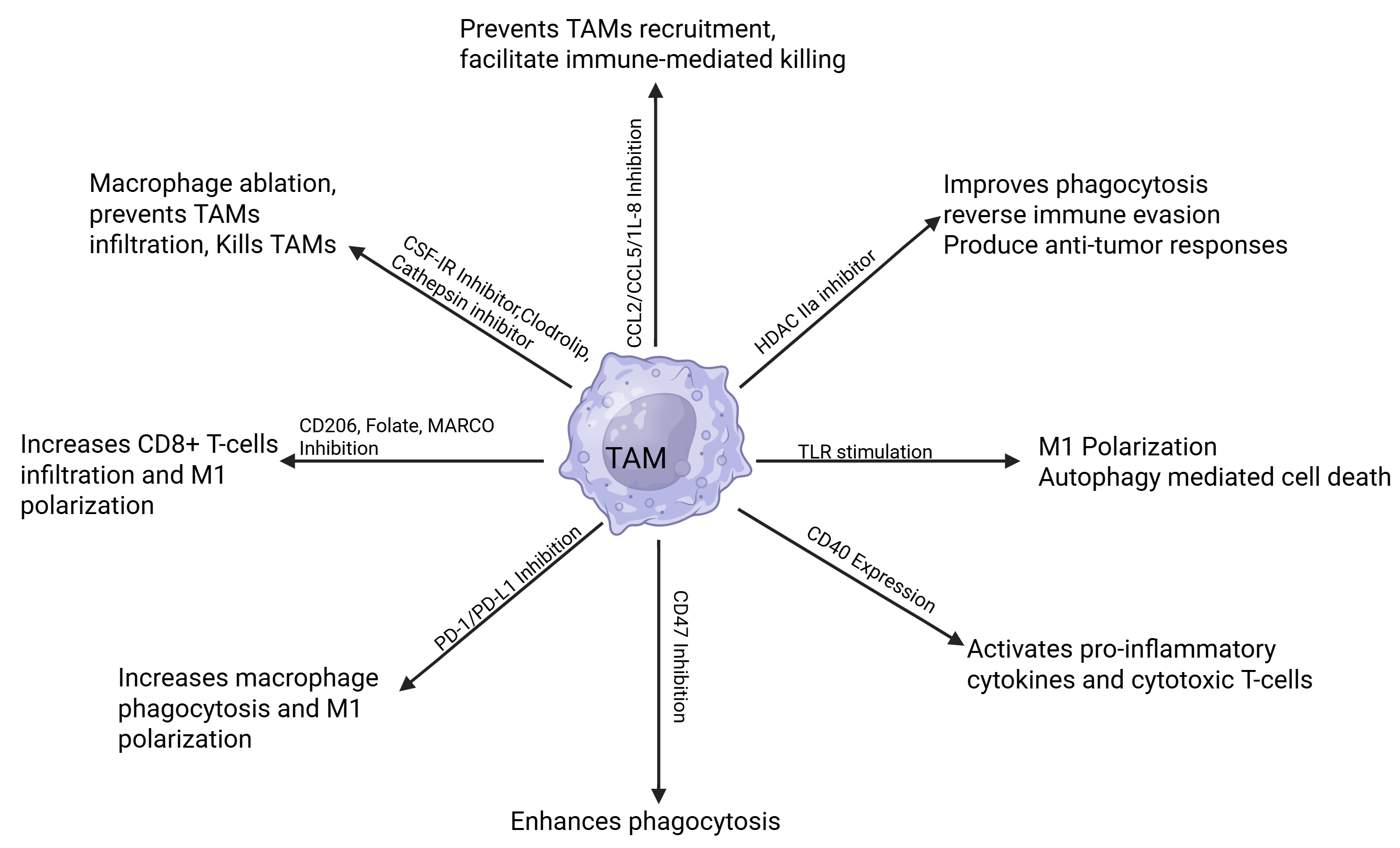
TAM-directed strategies fall into three complementary categories: depletion, recruitment blockade, and functional reprogramming. Depleting TAMs, particularly M2 macrophages, can be achieved with cathepsin inhibitors, which target lysosomal cathepsins that promote M2 polarization and tumor growth. Clodronate liposomes (Clodrolip) and CSF-1R inhibitors are also effective at depleting TAMs. Clodronate-loaded liposomes have demonstrated efficacy in ablating macrophages, preventing infiltration, and providing antitumor effects against metastasis, while CSF-1R inhibitors reduce TAM survival within the TME (294–299) (Fig. 2). To limit additional TAM recruitment to the tumor site, preclinical studies show that blocking chemokine axes effectively reduces monocyte migration. The CCL5/CCR5 axis is a key driver of monocyte recruitment into the TME, and its inhibition suppresses tumor growth and metastasis. Likewise, tumor-derived IL-8 recruits monocytes, and antibody-mediated neutralization of IL-8 can diminish recruitment and facilitate immune-mediated killing (300–303). Because CCL2/CCR2 and CXCL12/CXCR4 are major regulators of monocyte trafficking, their inhibition may further promote CD8⁺ T-cell infiltration and enhance antitumor responses (304–307) (Fig. 2).
Reprogramming TAMs toward an M1 phenotype is also a promising therapeutic avenue. Because M1 macrophages exhibit antitumor activity, enforcing this phenotype with interferon regulatory factor 5 (IRF5) can increase M1 macrophages in the TME (308–310). Epigenetic interventions, including histone acetylation and methylation, offer another therapeutic opportunity. The class IIa histone deacetylase inhibitor TMP195 reprograms monocytes/TAMs into highly phagocytic cells that recruit CD8⁺ T cells (311), and its combination with DNA methyltransferase inhibitors has been shown to reverse immune evasion by reprogramming TAMs. More broadly, HDAC inhibition enhances T-cell responses (312) (Fig. 2). In addition, because M-CSF and IL-34 promote M2 polarization via CSF1R, blocking this pathway can favor M1 polarization and strengthen antitumor immunity (313, 314). Finally, elevated IL-6 is associated with poor therapeutic response and M2 skewing; therefore, inhibiting IL-6-driven pathways may reduce M2 polarization and improve treatment outcomes (315).
As a complementary strategy to cytokine blockade, TLRs can be activated to enhance tumor recognition by upregulating M1-associated receptors, such as CD80/CD86, and downregulating M2 markers. In the presence of IFN-γ, TLR agonists preferentially drive M1 over M2 polarization. Agonists for TLR3, TLR4, TLR7, and TLR8 have been developed to reprogram M2 macrophages toward an M1 phenotype, thereby inducing autophagy-mediated tumor cell death (316–318) (Fig. 2). The cell-surface receptor CD40 has also been targeted therapeutically, as its activation induces pro-inflammatory cytokines and activates cytotoxic T cells to mount antitumor responses. Stimulating CD40 signaling in macrophages or other antigen-presenting cells (APCs) by binding to its natural ligand, CD40L, can reduce tumor growth (193, 319) (Fig. 2). CD47, often highly expressed on tumor cells, binds SIRPα on macrophages to deliver a “don’t-eat-me” signal that blocks phagocytosis of cancer cells and supports immune evasion. Antibodies targeting CD47 or SIRPα can enhance tumor-cell phagocytosis and reduce tumor growth and metastasis (320–324) (Fig. 2). Although CD47 has emerged as an attractive innate immune checkpoint, its clinical translation remains challenging. Most CD47-targeting agents have shown limited clinical success, largely because CD47 is ubiquitously expressed on normal cells, including red blood cells and platelets, leading to on-target hematologic toxicities. These limitations have prompted the development of next-generation strategies to improve tumor selectivity and minimize systemic toxicity (325–329). The mannose receptor (CD206), MARCO, and the folate receptor are additional targets because their expression is typically linked to M2-TAMs. Blocking these receptors can promote pro-inflammatory cytokine production, increase CD8⁺ T-cell infiltration, raise the M1 macrophage ratio, and decrease tumor growth (215–217) (Fig. 2).
Finally, stimulating macrophage phagocytic capacity through immune checkpoint blockade can further enhance their function within the TME. PD-L1, expressed on tumor and immune cells, binds to PD-1 on T cells, inducing immune tolerance. PD-L1, expressed on tumor and immune cells, binds PD-1 on both T cells and tumor-associated macrophages, thereby suppressing immune activation and macrophage phagocytosis. Blocking the PD-1/PD-L1 axis increases macrophage-mediated phagocytosis, both directly and indirectly, in part by enhancing IFN-γ secretion, which, in turn, promotes the expression of costimulatory molecules and pro-inflammatory mediators such as iNOS, CD40, and MHC II. These downstream signals enhance macrophage activation, survival, and functional remodeling in a T-cell-dependent manner, thereby contributing to improved control of tumor growth (328–331). Although PD-1/PD-L1 therapy shows promise, with monoclonal antibodies against ipilimumab (a CTLA-4 inhibitor) and nivolumab (a PD-1/PD-L1 inhibitor) now approved for antitumor activity, further improvements are needed, as only a small percentage of patients experience prolonged survival (332–336).
Despite numerous suggestions in the literature, few successes have emerged, and those that have are limited in scope. The CSF1R inhibitor pexidartinib and the anti-CSF1R antibody RG7155 (emactuzumab) are effective in conditions characterized by CSF1 overexpression and have shown a striking reduction in CSF1R⁺ CD163⁺ macrophages in tissues. A limitation of these therapies is that they often act as single agents, limiting their efficacy in solid tumors. Preclinical animal models suggest that even with a combination of anti-VEGF and taxane chemotherapy, there is a significant reduction in tumor burden in ovarian cancer models. Additionally, PD-1/PD-L1 blockade-based combination therapies have yielded better outcomes, including increased CD8⁺ T-cell infiltration and reduced tumor progression, compared to single-target therapies. A triple therapy of ipilimumab, nivolumab, and trabectedin in a phase II trial shows acceptable safety and preliminary clinical activity in patients with soft-tissue carcinoma. Lastly, the combination of the anti-CSF1R mAb emactuzumab and the PD-L1 mAb atezolizumab increased CD8⁺ T-cell infiltration, concomitant with a reduction in tumor-associated macrophages (337–342). The recruitment of myeloid cells, particularly immunosuppressive cells, into the TME is mediated by cytokines and chemokines. Therefore, strategies have been designed to inhibit chemokines, chemokine receptors, and signaling checkpoints, such as PI3K-γ, LILRB4, and JAK1/2, to block myeloid cell recruitment, and these strategies have shown efficacy in mouse models and early clinical studies. In a phase I clinical trial, the combination of the CCR2 inhibitor PF-04136309 with standard chemotherapy was shown to be safe and to inhibit TAM recruitment, thereby restricting tumor growth in pancreatic ductal adenocarcinoma. Similarly, Carlumab, a human anti-CCL2 IgG1κ monoclonal antibody, was well tolerated, with evidence of transient suppression of free CCL2 and of antitumor activity (343–348).
Recent clinical trials have also focused on TLR agonists to reprogram and restore M1 macrophages. A TLR9 agonist, MGN1703, has shown promising results in small-cell lung cancer. However, conclusions cannot be drawn due to the limited sample sizes and patient numbers. TLR4 agonists, such as GSK1795091, are under clinical investigation, although their therapeutic efficacy has not yet been fully established. Other reprogramming strategies involve inhibiting the PI3K/AKT signaling pathway, which is implicated in cancer pathogenesis, and have shown promise when PI3Kδ/γ is blocked with the PI3Kδ/γ inhibitor tenalisib (RP6530), either as a single agent or in combination with other immunotherapies, in patients with T-cell lymphoma. Additionally, a mouse model of breast and lung cancer demonstrates that loading mannosylated dual pH-responsive nanoparticles with siRNAs targeting placental growth factor and VEGF silences these genes, suppresses tumor proliferation, and inhibits metastasis (349–353).
5. Discussion
The TME contains immune cells that are often reprogrammed by local signals to promote tumor growth. The TME recruits a large proportion of monocytes/macrophages, which are reprogrammed toward M2 polarization. TAMs play a significant role in tumor progression; therefore, understanding how they are recruited, polarized, and respond to signals can guide the development of novel therapies for patients. A deeper understanding of the roles of receptors, cytokines, and chemokines in shaping TAMs will inform strategies to deplete or reprogram them to improve cancer treatment. Multiple studies show that monocytes/macrophages can defend the body against tumors. In the TME, monocytes are recruited as the first line of defense and express receptors and secrete cytokines/chemokines that trigger downstream cytotoxic CD8⁺ T-cell activity to control tumor progression and metastasis. However, the TME can reprogram monocytes and macrophages to shift their polarization toward suppression of antitumor activity and support of tumor growth (354, 355). Therefore, nanoplatform-based therapies that boost M1 macrophage activity while preventing M2 polarization are highly promising (Fig. 3).
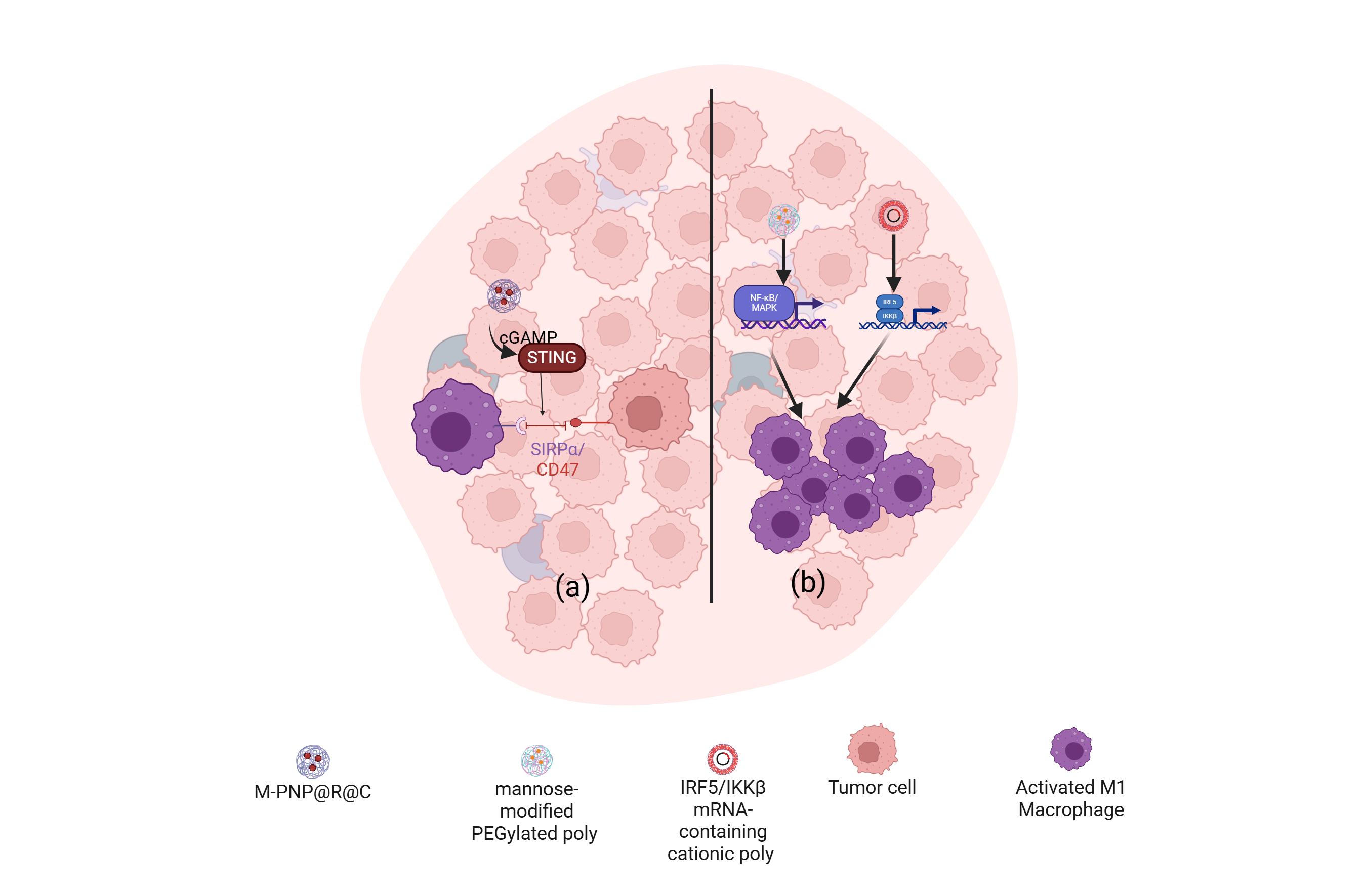
In this regard, a mannose-modified, pH-sensitive nanoplatform co-encapsulating resiquimod (R848), a TLR7/8 agonist, and 2’,3’-cyclic GMP-AMP (cGAMP), named M-PNP@R@C, repolarizes TAMs from an M2 to an M1 state. This intervention activates STING signaling, downregulates SIRPα, and blocks its interaction with CD47, thereby inhibiting the CD47-SIRPα axis. Because SIRPα suppresses phagocytosis, blocking this pathway reduces immune evasion (Fig. 3). Combination therapy with anti-CD47 and PD-L1 antibodies has shown higher survival rates and reduced metastasis in melanoma-grafted in vivo models (356). Additional nanotechnologies have been developed to reprogram M2 phenotypes, inhibit M2 recruitment, and deplete TAMs. For depletion, nanomaterials include a clodronate-cationic nanoliposome nanogel that depletes TAMs (357). Cell-derived vesicles from M1 macrophages carrying cGAMP can activate STING and disrupt the CD47-SIRPα axis (356, 358), and azide-modified exosomes from M1 macrophages have been designed to deliver IL-12, thereby increasing the M1/M2 ratio (359). For TAM reprogramming, proposed platforms include mannose-modified PEGylated PLGA nanoparticles, L-α-phosphatidylcholine/DSPE-PEG2000-amine formulations, and kinase-inhibiting amphiphiles that target NF-κB/MAPK or the CSF1-CSF1R axis. In addition, IRF5/IKKβ inhibitor of nuclear factor κ-B kinase subunit β (IKKβ) messenger RNA (mRNA)-loaded cationic poly (β-amino ester) nanoparticles reprogram M2 macrophages by promoting tumor-associated antigen release, activating MAPK signaling, and triggering M1 activation (360, 361).
Despite the promise, macrophage-targeted approaches face engineering challenges. Key issues include non-specific clearance or depletion of macrophages beyond TAMs, underscoring the need to spare TRMs (362, 363). Checkpoint modulation in TAMs can also provoke autoimmunity or therapeutic resistance. Thus, future nanomaterials should prioritize specificity for TAMs, controlled immune stimulation, and optimization to minimize off-target effects. Combining multiple mechanisms within a single nanoplatform may further enhance antitumor responses and limit progression and metastasis, while attention to non-invasive delivery and precise targeting remains essential.
Another promising angle is the microbiome. Gut bacteria influence macrophage numbers and the efficacy of PD-L1 immunotherapy. If we understand how microbes push macrophages toward M1 or M2 states, we could use them (delivered with nanoparticles) to reshape the TME and control TAMs (363–366). The microbiome is an important regulator of the tumor microenvironment, significantly influencing immune populations and immune regulation within the TME. The gut microbiome supports T-cell (including Treg) differentiation by releasing microbial metabolites and inflammatory mediators that contribute to immune suppression or activation. The microbiome and its metabolites also affect the efficacy of anticancer drugs. Both the gut and tumor-associated microbiomes can serve as biomarkers, and microbiome-informed strategies, including tumor-targeting bacteria and microbial-derived therapeutic agents, can be explored for tumor treatment. Conversely, the microbiome can contribute to cancer development and affect the response to therapy by releasing metabolites that can damage DNA. The microbiome can also regulate the TME by inducing inflammation that favors tumor progression (367–369). This bidirectional interplay between immune cells and the microbiome offers opportunities to design therapeutic strategies that modulate immune activation and enhance antitumor immunity within the TME, thereby controlling tumor growth.
In conclusion, macrophage-engineered therapies are highly promising for tumor targeting, but they require rigorous evaluation and rational combinations that act within the TME while minimizing effects on normal tissues. More broadly, monocyte-macrophage plasticity represents a central organizing principle of tumor immunology and an emerging therapeutic vulnerability for reshaping the tumor immune microenvironment.
References
1. Jakubzick C, Gautier EL, Gibbings SL, Sojka DK, Schlitzer A, Johnson TE, et al. Minimal differentiation of classical monocytes as they survey steady-state tissues and transport antigen to lymph nodes. Immunity. 2013;39(3):599–610. https://doi.org/10.1016/j.immuni.2013.08.007
2. Lavin Y, Kobayashi S, Leader A, Amir ED, Elefant N, Bigenwald C, et al. Innate immune landscape in early lung adenocarcinoma by paired single-cell analyses. Cell. 2017;169(4):750–765.e17. https://doi.org/10.1016/j.cell.2017.04.014
3. Zilionis R, Engblom C, Pfirschke C, Savova V, Zemmour D, Saatcioglu HD, et al. Single-cell transcriptomics of human and mouse lung cancers reveals conserved myeloid populations across individuals and species. Immunity. 2019;50(5):1317–1334.e10. https://doi.org/10.1016/j.immuni.2019.03.009
4. Kiss M, Caro AA, Raes G, Laoui D. Systemic reprogramming of monocytes in cancer. Front Oncol. 2020;10:1399. https://doi.org/10.3389/fonc.2020.01399
5. Ugel S, Cane S, De Sanctis F, Bronte V. Monocytes in the tumor microenvironment. Annu Rev Pathol. 2021;16:93–122. https://doi.org/10.1146/annurev-pathmechdis-012418-013058
6. Chen X, Li Y, Xia H, Chen YH. Monocytes in tumorigenesis and tumor immunotherapy. Cells. 2023;12(13):1673. https://doi.org/10.3390/cells12131673
7. Cassetta L, Pollard JW. A timeline of tumour-associated macrophage biology. Nat Rev Cancer. 2023;23(4):238–257. https://doi.org/10.1038/s41568-022-00547-1
8. Han J, Dong L, Wu M, Ma F. Dynamic polarization of tumor‑associated macrophages and their interaction with intratumoral T cells in an inflamed tumor microenvironment: from mechanistic insights to therapeutic opportunities. Front Immunol. 2023;14:1160340. https://doi.org/10.3389/fimmu.2023.1160340
9. Colak S, ten Dijke P. Targeting TGF-β signaling in cancer. Trends Cancer. 2017;3(1):56–71. https://doi.org/10.1016/j.trecan.2016.11.008
10. Kim J, Bae JS. Tumor‑associated macrophages and neutrophils in tumor microenvironment. Mediators Inflamm. 2016;2016:6058147. https://doi.org/10.1155/2016/6058147
11. Bejarano L, Jordão MJC, Joyce JA. Therapeutic targeting of the tumor microenvironment. Cancer Discov. 2021;11(4):933–959. https://doi.org/10.1158/2159-8290.cd-20-1808
12. De Visser KE, Joyce JA. The evolving tumor microenvironment: from cancer initiation to metastatic outgrowth. Cancer Cell. 2023;41(3):374–403. https://doi.org/10.1016/j.ccell.2023.02.016
13. Liu L, Li Y, Li B. Interactions between cancer cells and tumor-associated macrophages in tumor microenvironment. Biochim Biophys Acta Rev Cancer. 2025;1880(3):189344. https://doi.org/10.1016/j.bbcan.2025.189344
14. Bischoff P, Trinks A, Obermayer B, Pett JP, Wiederspahn J, Uhlitz F, et al. Single-cell RNA sequencing reveals distinct tumor microenvironmental patterns in lung adenocarcinoma. Oncogene. 2021;40(50):6748–6758. https://doi.org/10.1038/s41388-021-02054-3
15. De Zuani M, Xue H, Park JS, Dentro SC, Seferbekova Z, Tessier J, et al. Single-cell and spatial transcriptomics analysis of non-small cell lung cancer. Nat Commun. 2024;15:4388. https://doi.org/10.1038/s41467-024-48700-8
16. Cassetta L, Pollard JW. Targeting macrophages: therapeutic approaches in cancer. Nat Rev Drug Discov. 2018;17(12):887–904. https://doi.org/10.1038/nrd.2018.169
17. Li C, Xiao M, Geng S, Wang Y, Zeng L, Lai P, et al. Comprehensive analysis of human monocyte subsets using full-spectrum flow cytometry and hierarchical marker clustering. Front Immunol. 2024;15:1405249. https://doi.org/10.3389/fimmu.2024.1405249
18. Li M, Yang Y, Xiong L, Jiang P, Wang J, Li C. Metabolism, metabolites, and macrophages in cancer. J Hematol Oncol. 2023;16(1):80. https://doi.org/10.1186/s13045-023-01478-6
19. Mills CD, Kincaid K, Alt JM, Heilman MJ, Hill AM. M-1/M-2 macrophages and the Th1/Th2 paradigm. J Immunol. 2000;164(12):6166–6173. https://doi.org/10.4049/jimmunol.164.12.6166
20. Denardo DG, Barreto JB, Andreu P, Vasquez L, Tawfik D, Kolhatkar N, et al. CD4+ T cells regulate pulmonary metastasis of mammary carcinomas by enhancing protumor properties of macrophages. Cancer Cell. 2009;16(2):91–102. https://doi.org/10.1016/j.ccr.2009.06.018
21. Cassetta L, Fragkogianni S, Sims AH, Swierczak A, Forrester LM, Zhang H, et al. Human tumor-associated macrophage and monocyte transcriptional landscapes reveal cancer-specific reprogramming, biomarkers, and therapeutic targets. Cancer Cell. 2019;35(4):588–602.e10. https://doi.org/10.1016/j.ccell.2019.02.009
22. Curtaz CJ, Schmitt C, Herbert SL, Feldheim J, Schlegel N, Gosselet F, et al. Serum-derived factors of breast cancer patients with brain metastases alter permeability of a human blood-brain barrier model. Fluids Barriers CNS. 2020;17(1):31. https://doi.org/10.1186/s12987-020-00192-6
23. Dehqanzada ZA, Storrer CE, Hueman MT, Foley RJ, Harris KA, Jama YH, et al. Correlations between serum monocyte chemotactic protein-1 levels, clinical prognostic factors, and HER-2/neu vaccine-related immunity in breast cancer patients. Clin Cancer Res. 2006;12(2):478–486. https://doi.org/10.1158/1078-0432.ccr-05-1425
24. Sanford DE, Belt BA, Panni RZ, Mayer A, Deshpande AD, Carpenter D, et al. Inflammatory monocyte mobilization decreases patient survival in pancreatic cancer: a role for targeting the CCL2/CCR2 axis. Clin Cancer Res. 2013;19(13):3404–3415. https://doi.org/10.1158/1078-0432.ccr-13-0525
25. Rannikko JH, Hollmen M. Clinical landscape of macrophage-reprogramming cancer immunotherapies. Br J Cancer. 2024;131(4):627–640. https://doi.org/10.1038/s41416-024-02715-6
26. Henze AT, Mazzone M. The impact of hypoxia on tumor-associated macrophages. J Clin Invest. 2016;126(10):3672–3679. https://doi.org/10.1172/jci84427
27. Karimova AF, Khalitova AR, Suezov R, Markov N, Mukhamedshina Y, Rizvanov AA, et al. Immunometabolism of tumor-associated macrophages: A therapeutic perspective. Eur J Cancer. 2025;220:115332. https://doi.org/10.1016/j.ejca.2025.115332
28. Wong KL, Tai JJ, Wong WC, Han H, Sem X, Yeap WH, et al. Gene expression profiling reveals the defining features of the classical, intermediate, and nonclassical human monocyte subsets. Blood. 2011;118(5):e16–31. https://doi.org/10.1182/blood-2010-12-326355
29. Mulder K, Patel AA, Kong WT, Piot C, Halitzki E, Dunsmore G, et al. Cross-tissue single-cell landscape of human monocytes and macrophages in health and disease. Immunity. 2021;54(8):1883–1900.e5. https://doi.org/10.1016/j.immuni.2021.07.007
30. Petty AJ, Yang Y. Tumor-associated macrophages: implications in cancer immunotherapy. Immunotherapy. 2017;9(3):289–302. https://doi.org/10.2217/imt-2016-0135
31. Li Z, Li J, Bai X, Huang X, Wang Q. Tumor microenvironment as a complex milieu driving cancer progression: a mini review. Clin Transl Oncol. 2025;27(5):1943–1952. https://doi.org/10.1007/s12094-024-03697-w
32. Albonici L, Giganti MG, Modesti A, Manzari V, Bei R. Multifaceted role of the placental growth factor (PlGF) in the antitumor immune response and cancer progression. Int J Mol Sci. 2019;20(12):2970. https://doi.org/10.3390/ijms20122970
33. Balkwill FR, Capasso M, Hagemann T. The tumor microenvironment at a glance. J Cell Sci. 2012;125(Pt 23):5591–5596. https://doi.org/10.1242/jcs.116392
34. Kalbasi A, Komar C, Tooker GM, Liu M, Lee JW, Gladney WL, et al. Tumor-derived CCL2 mediates resistance to radiotherapy in pancreatic ductal adenocarcinoma. Clin Cancer Res. 2017;23(1):137–148. https://doi.org/10.1158/1078-0432.ccr-16-0870
35. Teicher BA, Fricker SP. CXCL12 (SDF-1)/CXCR4 pathway in cancer. Clin Cancer Res. 2010;16(11):2927–2931. https://doi.org/10.1158/1078-0432.ccr-09-2329
36. Pickup M, Novitskiy S, Moses HL. The roles of TGF-β in the tumour microenvironment. Nat Rev Cancer. 2013;13(11):788–799. https://doi.org/10.1038/nrc3603
37. Qian BZ, Pollard JW. Macrophage diversity enhances tumor progression and metastasis. Cell. 2010;141(1):39–51. https://doi.org/10.1016/j.cell.2010.03.014
38. Cassetta L, Pollard JW. Cancer immunosurveillance: role of patrolling monocytes. Cell Res. 2016;26(1):3–4. https://doi.org/10.1038/cr.2015.144
39. Kitamura T, Qian BZ, Pollard JW. Immune cell promotion of metastasis. Nat Rev Immunol. 2015;15(2):73–86. https://doi.org/10.1038/nri3789
40. Khalaf K, Hana D, Chou JT, Singh C, Mackiewicz A, Kaczmarek M. Aspects of the tumor microenvironment involved in immune resistance and drug resistance. Front Immunol. 2021;12:656364. https://doi.org/10.3389/fimmu.2021.656364
41. Veglia F, Perego M, Gabrilovich D. Myeloid-derived suppressor cells coming of age. Nat Immunol. 2018;19(2):108–119. https://doi.org/10.1038/s41590-017-0022-x
42. Jaillon S, Ponzetta A, Di Mitri D, Santoni A, Bonecchi R, Mantovani A. Neutrophil diversity and plasticity in tumour progression and therapy. Nat Rev Cancer. 2020;20(9):485–503. https://doi.org/10.1038/s41568-020-0281-y
43. Cox N, Pokrovskii M, Vicario R, Geissmann F. Origins, biology, and diseases of tissue macrophages. Annu Rev Immunol. 2021;39:313–344. https://doi.org/10.1146/annurev-immunol-093019-111748
44. Jahchan NS, Mujal AM, Pollack JL, Binnewies M, Sriram V, Reyno L, et al. Tuning the tumor myeloid microenvironment to fight cancer. Front Immunol. 2019;10:1611. https://doi.org/10.3389/fimmu.2019.01611
45. Woo SR, Corrales L, Gajewski TF. Innate immune recognition of cancer. Annu Rev Immunol. 2015;33:445–474. https://doi.org/10.1146/annurev-immunol-032414-112043
46. Esfahani K, Roudaia L, Buhlaiga N, Del Rincon SV, Papneja N, Miller WH. A review of cancer immunotherapy: from the past, to the present, to the future. Curr Oncol. 2020;27(Suppl 2):87–97. https://doi.org/10.3747/co.27.5223
47. Gren ST, Rasmussen TB, Janciauskiene S, Håkansson K, Gerwien JG, Grip O. A single-cell gene-expression profile reveals inter-cellular heterogeneity within human monocyte subsets. PLoS One. 2015;10(12):e0144351. https://doi.org/10.1371/journal.pone.0144351
48. Boyette LB, Macedo C, Hadi K, Elinoff BD, Walters JT, Ramaswami B, et al. Phenotype, function, and differentiation potential of human monocyte subsets. PLoS One. 2017;12(4):e0176460. https://doi.org/10.1371/journal.pone.0176460
49. Austermann J, Roth J, Barczyk-Kahlert K. The good and the bad: monocytes' and macrophages' diverse functions in inflammation. Cells. 2022;11(12):1979. https://doi.org/10.3390/cells11121979
50. Al-Khami AA, Zheng L, Del Valle L, Hossain F, Wyczechowska D, Zabaleta J, et al. Exogenous lipid uptake induces metabolic and functional reprogramming of tumor-associated myeloid-derived suppressor cells. Oncoimmunology. 2017;6(10):e1344804. https://doi.org/10.1080/2162402x.2017.1344804
51. Li J, Bolyard C, Xin G, Li Z. Targeting metabolic pathways of myeloid cells improves cancer immunotherapy. Front Cell Dev Biol. 2021;9:747863. https://doi.org/10.3389/fcell.2021.747863
52. Mantovani A, Marchesi F, Di Mitri D, Garlanda C. Macrophage diversity in cancer dissemination and metastasis. Cell Mol Immunol. 2024;21(11):1201–1214. https://doi.org/10.1038/s41423-024-01216-z
53. Wen S, Chen N, Hu Y, Huang L, Peng J, Yang M, et al. Elevated peripheral absolute monocyte count related to clinicopathological features and poor prognosis in solid tumors: systematic review, meta-analysis, and meta-regression. Cancer Med. 2021;10(5):1690–1714. https://doi.org/10.1002/cam4.3773
54. Kapoor G, Prakash S, Jaiswal V, Singh AK. Chronic inflammation and cancer: key pathways and targeted therapies. Cancer Invest. 2025;43(1):1–23. https://doi.org/10.1080/07357907.2024.2437614
55. Zhao H, Wu L, Yan G, Chen Y, Zhou M, Wu Y, et al. Inflammation and tumor progression: signaling pathways and targeted intervention. Signal Transduct Target Ther. 2021;6(1):263. https://doi.org/10.1038/s41392-021-00658-5
56. Oguma K, Oshima H, Oshima M. Inflammation, tumor necrosis factor and Wnt promotion in gastric cancer development. Future Oncol. 2010;6(4):515–526. https://doi.org/10.2217/fon.10.13
57. Yu L, Yuan J, Lou S, Huang A, Cai X, Ji J, et al. Factors affecting survival in patients with colorectal cancer: an umbrella review. J Transl Med. 2025;23:1185. https://doi.org/10.1186/s12967-025-06876-7
58. Wen J, Ye F, Huang X, Li S, Yang L, Xiao X, et al. Prognostic significance of preoperative circulating monocyte count in patients with breast cancer: based on a large cohort study. Medicine (Baltimore). 2015;94(49):e2266. https://doi.org/10.1097/md.0000000000002266
59. Shigeta K, Kosaka T, Kitano S, Yasumizu Y, Miyazaki Y, Mizuno R, et al. High absolute monocyte count predicts poor clinical outcome in patients with castration-resistant prostate cancer treated with docetaxel chemotherapy. Ann Surg Oncol. 2016;23(12):4115–4122. https://doi.org/10.1245/s10434-016-5354-5
60. Vajavaara H, Leivonen SK, Jørgensen J, Holte H, Leppä S. A low lymphocyte-to-monocyte ratio predicts poor outcome in high-risk aggressive large B-cell lymphoma. EJHaem. 2022;3(3):681–687. https://doi.org/10.1002/jha2.409
61. Mao Y, Chen D, Duan S, Zhao Y, Wu C, Zhu F, et al. Prognostic impact of pretreatment lymphocyte-to-monocyte ratio in advanced epithelial cancers: a meta-analysis. Cancer Cell Int. 2018;18:201. https://doi.org/10.1186/s12935-018-0698-5
62. Shibutani M, Maeda K, Nagahara H, Fukuoka T, Nakao S, Matsutani S, et al. The peripheral monocyte count is associated with the density of tumor-associated macrophages in the tumor microenvironment of colorectal cancer: a retrospective study. BMC Cancer. 2017;17(1):404. https://doi.org/10.1186/s12885-017-3395-1
63. Hayashi T, Fujita K, Nojima S, Hayashi Y, Nakano K, Ishizuya Y, et al. Peripheral blood monocyte count reflecting tumor-infiltrating macrophages is a predictive factor of adverse pathology in radical prostatectomy specimens. Prostate. 2017;77(14):1383–1388. https://doi.org/10.1002/pros.23398
64. de la Fuente J, Sharma A, Chari S, Majumder S. Peripheral blood monocyte counts are elevated in the pre-diagnostic phase of pancreatic cancer: A population based study. Pancreatology. 2019;19(8):1043–1048. https://doi.org/10.1016/j.pan.2019.10.002
65. Chen L, Qian J, Lin L, Lin J, Chen Q, Zhuang Z, et al. Prognostic value of preoperative lymphocyte-to-monocyte ratio in oral cancer patients and establishment of a dynamic nomogram. Oral Dis. 2021;27(5):1127–1136. https://doi.org/10.1111/odi.13629
66. Tham T, Olson C, Khaymovich J, Herman SW, Costantino PD. The lymphocyte-to-monocyte ratio as a prognostic indicator in head and neck cancer: a systematic review and meta-analysis. Eur Arch Otorhinolaryngol. 2018;275(7):1663–1670. https://doi.org/10.1007/s00405-018-4972-x
67. Barclay AN, Van den Berg TK. The interaction between signal regulatory protein alpha (SIRPalpha) and CD47: structure, function, and therapeutic target. Annu Rev Immunol. 2014;32:25–50. https://doi.org/10.1146/annurev-immunol-032713-120142
68. Chen YP, Kim HJ, Wu H, Price-Troska T, Villasboas JC, Jalali S, et al. SIRPalpha expression delineates subsets of intratumoral monocyte/macrophages with different functional and prognostic impact in follicular lymphoma. Blood Cancer J. 2019;9(10):84. https://doi.org/10.1038/s41408-019-0246-0
69. Patysheva M, Larionova I, Stakheyeva M, Grigoryeva E, Iamshchikov P, Tarabanovskaya N, et al. Effect of early-stage human breast carcinoma on monocyte programming. Front Oncol. 2021;11:800235. https://doi.org/10.3389/fonc.2021.800235
70. Kang SU, Cho SY, Jeong H, Han J, Chae HY, Yang H, et al. Matrix metalloproteinase 11 (MMP11) in macrophages promotes the migration of HER2-positive breast cancer cells and monocyte recruitment through CCL2-CCR2 signaling. Lab Invest. 2022;102(4):376–390. https://doi.org/10.1038/s41374-021-00699-y
71. Juusola M, Kuuliala K, Kuuliala A, Mustonen H, Vaha-Koskela M, Puolakkainen P, et al. Pancreatic cancer is associated with aberrant monocyte function and successive differentiation into macrophages with inferior anti-tumour characteristics. Pancreatology. 2021;21(2):397–405. https://doi.org/10.1016/j.pan.2020.12.025
72. Yin W, Lv J, Yao Y, Zhao Y, He Z, Wang Q, et al. Elevations of monocyte and neutrophils, and higher levels of granulocyte colony-stimulating factor in peripheral blood in lung cancer patients. Thorac Cancer. 2021;12(20):2680–2690. https://doi.org/10.1111/1759-7714.14103
73. Zhang B, Ye H, Ren X, Zheng S, Zhou Q, Chen C, et al. Macrophage-expressed CD51 promotes cancer stem cell properties via the TGF-β1/smad2/3 axis in pancreatic cancer. Cancer Lett. 2019;459:204–215. https://doi.org/10.1016/j.canlet.2019.06.005
74. Devalaraja S, To TKJ, Folkert IW, Natesan R, Alam MZ, Li M, et al. Tumor-derived retinoic acid regulates intratumoral monocyte differentiation to promote immune suppression. Cell. 2020;180(6):1098–1114.e16. https://doi.org/10.1016/j.cell.2020.02.042
75. Duluc D, Delneste Y, Tan F, Moles MP, Grimaud L, Lenoir J, et al. Tumor-associated leukemia inhibitory factor and IL-6 skew monocyte differentiation into tumor-associated macrophage-like cells. Blood. 2007;110(13):4319–4330. https://doi.org/10.1182/blood-2007-02-072587
76. Cheng P, Corzo CA, Luetteke N, Yu B, Nagaraj S, Bui MM, et al. Inhibition of dendritic cell differentiation and accumulation of myeloid-derived suppressor cells in cancer is regulated by S100A9 protein. J Exp Med. 2008;205(10):2235–2249. https://doi.org/10.1084/jem.20080132
77. Michielon E, Lopez Gonzalez M, Burm JLA, Waaijman T, Jordanova ES, de Gruijl TD, et al. Micro-environmental cross-talk in an organotypic human melanoma-in-skin model directs M2-like monocyte differentiation via IL-10. Cancer Immunol Immunother. 2020;69(11):2319–2331. https://doi.org/10.1007/s00262-020-02626-4
78. Veglia F, Sanseviero E, Gabrilovich DI. Myeloid-derived suppressor cells in the era of increasing myeloid cell diversity. Nat Rev Immunol. 2021;21(8):485–498. https://doi.org/10.1038/s41577-020-00490-y
79. Li T, Li X, Zamani A, Wang W, Lee CN, Li M, et al. c-Rel is a myeloid checkpoint for cancer immunotherapy. Nat Cancer. 2020;1(5):507–517. https://doi.org/10.1038/s43018-020-0061-3
80. Waight JD, Netherby C, Hensen ML, Miller A, Hu Q, Liu S, et al. Myeloid-derived suppressor cell development is regulated by a STAT/IRF-8 axis. J Clin Invest. 2013;123(10):4464–4478. https://doi.org/10.1172/jci68189
81. Sinha P, Okoro C, Foell D, Freeze HH, Ostrand-Rosenberg S, Srikrishna G. Proinflammatory S100 proteins regulate the accumulation of myeloid-derived suppressor cells. J Immunol. 2008;181(7):4666–4675. https://doi.org/10.4049/jimmunol.181.7.4666
82. Yan D, Wang J, Sun H, Zamani A, Zhang H, Chen W, et al. TIPE2 specifies the functional polarization of myeloid-derived suppressor cells during tumorigenesis. J Exp Med. 2020;217(2):e20182005. https://doi.org/10.1084/jem.20182005
83. Fultang N, Li X, Li T, Chen YH. Myeloid-derived suppressor cell differentiation in cancer: transcriptional regulators and enhanceosome-mediated mechanisms. Front Immunol. 2020;11:619253. https://doi.org/10.3389/fimmu.2020.619253
84. Li T, Bou-Dargham MJ, Fultang N, Li X, Pear WS, Sun H, et al. c-Rel-dependent monocytes are potent immune suppressor cells in cancer. J Leukoc Biol. 2022;112(4):845–859. https://doi.org/10.1002/jlb.1ma0422-518rr
85. Tumino N, Fiore PF, Pelosi A, Moretta L, Vacca P. Myeloid derived suppressor cells in tumor microenvironment: Interaction with innate lymphoid cells. Semin Immunol. 2022;61:101668. https://doi.org/10.1016/j.smim.2022.101668
86. Wu Y, Yi M, Niu M, Mei Q, Wu K. Myeloid-derived suppressor cells: an emerging target for anticancer immunotherapy. Mol Cancer. 2022;21(1):184. https://doi.org/10.1186/s12943-022-01657-y
87. Peterlin P, Debord C, Eveillard M, Garnier A, Le Bourgeois A, Guillaume T, et al. Peripheral levels of monocytic myeloid-derived suppressive cells before and after first induction predict relapse and survivals in AML patients. J Cell Mol Med. 2022;26(21):5486–5492. https://doi.org/10.1111/jcmm.17576
88. Kajihara N, Kobayashi T, Otsuka R, Nio-Kobayashi J, Oshino T, Takahashi M, et al. Tumor-derived interleukin-34 creates an immunosuppressive and chemoresistant tumor microenvironment by modulating myeloid-derived suppressor cells in triple-negative breast cancer. Cancer Immunol Immunother. 2023;72(4):851–864. https://doi.org/10.1007/s00262-022-03293-3
89. Jain MD, Zhao H, Wang X, Atkins R, Menges M, Reid K, et al. Tumor interferon signaling and suppressive myeloid cells are associated with CAR T-cell failure in large B-cell lymphoma. Blood. 2021;137(19):2621–2633. https://doi.org/10.1182/blood.2020007445
90. Bayik D, Bartels CF, Lovrenert K, Watson DC, Zhang D, Kay K, et al. Distinct cell adhesion signature defines glioblastoma myeloid-derived suppressor cell subsets. Cancer Res. 2022;82(22):4274–4287. https://doi.org/10.1158/0008-5472.can-21-3840
91. Singh L, Muise ES, Bhattacharya A, Grein J, Javaid S, Stivers P, et al. ILT3 (LILRB4) promotes the immunosuppressive function of tumor-educated human monocytic myeloid-derived suppressor cells. Mol Cancer Res. 2021;19(4):702–716. https://doi.org/10.1158/1541-7786.mcr-20-0622
92. Su MT, Kumata S, Endo S, Okada Y, Takai T. LILRB4 promotes tumor metastasis by regulating MDSCs and inhibiting miR-1 family miRNAs. Oncoimmunology. 2022;11(1):2060907. https://doi.org/10.1080/2162402x.2022.2060907
93. Arkhypov I, Ozbay Kurt FG, Bitsch R, Novak D, Petrova V, Lasser S, et al. HSP90alpha induces immunosuppressive myeloid cells in melanoma via TLR4 signaling. J Immunother Cancer. 2022;10:e005551. https://doi.org/10.1136/jitc-2022-005551
94. Alicea-Torres K, Sanseviero E, Gui J, Chen J, Veglia F, Yu Q, et al. Immune suppressive activity of myeloid-derived suppressor cells in cancer requires inactivation of the type I interferon pathway. Nat Commun. 2021;12(1):1717. https://doi.org/10.1038/s41467-021-22033-2
95. Zhan X, He Q, Sheng J, Jiang X, Lin L, Huang Y, et al. USP12 positively regulates M-MDSC function to inhibit antitumour immunity through deubiquitinating and stabilizing p65. Immunology. 2022;167(4):544–557. https://doi.org/10.1111/imm.13552
96. Alissafi T, Hatzioannou A, Mintzas K, Barouni RM, Banos A, Sormendi S, et al. Autophagy orchestrates the regulatory program of tumor-associated myeloid-derived suppressor cells. J Clin Invest. 2018;128(9):3840–3852. https://doi.org/10.1172/jci120888
97. De Gassart A, Camosseto V, Thibodeau J, Ceppi M, Catalan N, Pierre P, et al. MHC class II stabilization at the surface of human dendritic cells is the result of maturation-dependent MARCH I down-regulation. Proc Natl Acad Sci U S A. 2008;105(9):3491–3496. https://doi.org/10.1073/pnas.0708874105
98. Liu M, Zhou J, Liu X, Feng Y, Yang W, Wu F, et al. Targeting monocyte-intrinsic enhancer reprogramming improves immunotherapy efficacy in hepatocellular carcinoma. Gut. 2020;69(2):365–379. https://doi.org/10.1136/gutjnl-2018-317257
99. Arihara F, Mizukoshi E, Kitahara M, Takata Y, Arai K, Yamashita T, et al. Increase in CD14+HLA-DR-/low myeloid-derived suppressor cells in hepatocellular carcinoma patients and its impact on prognosis. Cancer Immunol Immunother. 2013;62(8):1421–1430. https://doi.org/10.1007/s00262-013-1447-1
100. Qian BZ, Li J, Zhang H, Kitamura T, Zhang J, Campion LR, et al. CCL2 recruits inflammatory monocytes to facilitate breast-tumour metastasis. Nature. 2011;475(7355):222–225. https://doi.org/10.1038/nature10138
101. Ikeda N, Asano K, Kikuchi K, Uchida Y, Ikegami H, Takagi R, et al. Emergence of immunoregulatory Ym1+Ly6Chi monocytes during recovery phase of tissue injury. Sci Immunol. 2018;3(28):eaat0207. https://doi.org/10.1126/sciimmunol.aat0207
102. Shibuya T, Kamiyama A, Sawada H, Kikuchi K, Maruyama M, Sawado R, et al. Immunoregulatory monocyte subset promotes metastasis associated with therapeutic intervention for primary tumor. Front Immunol. 2021;12:663115. https://doi.org/10.3389/fimmu.2021.663115
103. Sizemore S, Cicek M, Sizemore N, Ng KP, Casey G. Podocalyxin increases the aggressive phenotype of breast and prostate cancer cells in vitro through its interaction with ezrin. Cancer Res. 2007;67(13):6183–6191. https://doi.org/10.1158/0008-5472.can-06-3575
104. Zhang Y, Zhao Q, Du J. Regulatory role of ezrin in esophageal cancer progression via the PI3K-AKT signaling pathway. Hereditas. 2025;162:199. https://doi.org/10.1186/s41065-025-00554-w
105. Gomez KE, Wu F, Keysar SB, Morton JJ, Miller B, Chimed TS, et al. Cancer cell CD44 mediates macrophage/monocyte-driven regulation of head and neck cancer stem cells. Cancer Res. 2020;80(19):4185–4198. https://doi.org/10.1158/0008-5472.can-20-1079
106. Chen Q, Zhang XH, Massague J. Macrophage binding to receptor VCAM-1 transmits survival signals in breast cancer cells that invade the lungs. Cancer Cell. 2011;20(4):538–549. https://doi.org/10.1016/j.ccr.2011.08.025
107. Wang PC, Weng CC, Hou YS, Jian SF, Fang KT, Hou MF, et al. Activation of VCAM-1 and its associated molecule CD44 leads to increased malignant potential of breast cancer cells. Int J Mol Sci. 2014;15(3):3560–3579. https://doi.org/10.3390/ijms15033560
108. Arif AA, Huang YH, Freeman SA, Atif J, Dean P, Lai JCY, et al. Inflammation-induced metastatic colonization of the lung is facilitated by hepatocyte growth factor-secreting monocyte-derived macrophages. Mol Cancer Res. 2021;19(12):2096–2109. https://doi.org/10.1158/1541-7786.mcr-21-0009
109. Oh K, Lee OY, Shon SY, Nam O, Ryu PM, Seo MW, et al. A mutual activation loop between breast cancer cells and myeloid-derived suppressor cells facilitates spontaneous metastasis through IL-6 trans-signaling in a murine model. Breast Cancer Res. 2013;15(5):R79. https://doi.org/10.1186/bcr3473
110. Ouzounova M, Lee E, Piranlioglu R, El Andaloussi A, Kolhe R, Demirci MF, et al. Monocytic and granulocytic myeloid derived suppressor cells differentially regulate spatiotemporal tumour plasticity during metastatic cascade. Nat Commun. 2017;8:14979. https://doi.org/10.1038/ncomms14979
111. Roberts LM, Perez MJ, Balogh KN, Mingledorff G, Cross JV, Munson JM. Myeloid derived suppressor cells migrate in response to flow and lymphatic endothelial cell interaction in the breast tumor microenvironment. Cancers (Basel). 2022;14(12):3008. https://doi.org/10.3390/cancers14123008
112. Cha YJ, Koo JS. Role of tumor-associated myeloid cells in breast cancer. Cells. 2020;9(8):1785. https://doi.org/10.3390/cells9081785
113. Benzing C, Lam H, Tsang CM, Rimmer A, Arroyo-Berdugo Y, Calle Y, et al. TIMP-2 secreted by monocyte-like cells is a potent suppressor of invadopodia formation in pancreatic cancer cells. BMC Cancer. 2019;19(1):1214. https://doi.org/10.1186/s12885-019-6429-z
114. Hagerling C, Gonzalez H, Salari K, Wang CY, Lin C, Robles I, et al. Immune effector monocyte-neutrophil cooperation induced by the primary tumor prevents metastatic progression of breast cancer. Proc Natl Acad Sci U S A. 2019;116(43):21704–21714. https://doi.org/10.1073/pnas.1907660116
115. Wang R, Bao W, Pal M, Liu Y, Yazdanbakhsh K, Zhong H. Intermediate monocytes induced by IFN-γ inhibit cancer metastasis by promoting NK cell activation through FOXO1 and interleukin-27. J Immunother Cancer. 2022;10(1):e003539. https://doi.org/10.1136/jitc-2021-003539
116. Hanna RN, Cekic C, Sag D, Tacke R, Thomas GD, Nowyhed H, et al. Patrolling monocytes control tumor metastasis to the lung. Science. 2015;350(6263):985–990. https://doi.org/10.1126/science.aac9407
117. Guedez L, Jensen-Taubman S, Bourboulia D, Kwityn CJ, Wei B, Caterina J, et al. TIMP-2 targets tumor-associated myeloid suppressor cells with effects in cancer immune dysfunction and angiogenesis. J Immunother. 2012;35(6):502–512. https://doi.org/10.1097/cji.0b013e3182619c8e
118. Schulz C, Perdiguero EG, Chorro L, Szabo-Rogers H, Cagnard N, Kierdorf K, et al. A lineage of myeloid cells independent of Myb and hematopoietic stem cells. Science. 2012;336(6077):86–90. https://doi.org/10.1126/science.1219179
119. van Furth R, Cohn ZA. The origin and kinetics of mononuclear phagocytes. J Exp Med. 1968;128(3):415–435. https://doi.org/10.1084/jem.128.3.415
120. Boutilier AJ, Elsawa SF. Macrophage polarization states in the tumor microenvironment. Int J Mol Sci. 2021;22(13):6995. https://doi.org/10.3390/ijms22136995
121. Fu LQ, Du WL, Cai MH, Yao JY, Zhao YY, Mou XZ. The roles of tumor‑associated macrophages in tumor angiogenesis and metastasis. Cell Immunol. 2020;353:104119. https://doi.org/10.1016/j.cellimm.2020.104119
122. Nielsen SR, Schmid MC. Macrophages as key drivers of cancer progression and metastasis. Mediators Inflamm. 2017;2017:9624760. https://doi.org/10.1155/2017/9624760
123. Komohara Y, Fujiwara Y, Ohnishi K, Takeya M. Tumor‑associated macrophages: potential therapeutic targets for anti‑cancer therapy. Adv Drug Deliv Rev. 2016;99(Pt B):180–185. https://doi.org/10.1016/j.addr.2015.11.009
124. Sica A, Saccani A, Mantovani A. Tumor-associated macrophages: a molecular perspective. Int Immunopharmacol. 2002;2(8):1045–1054. https://doi.org/10.1016/s1567-5769(02)00064-4
125. Lahmar Q, Keirsse J, Laoui D, Movahedi K, Van Overmeire E, Van Ginderachter JA. Tissue-resident versus monocyte-derived macrophages in the tumor microenvironment. Biochim Biophys Acta Rev Cancer. 2016;1865(1):23–34. https://doi.org/10.1016/j.bbcan.2015.06.009
126. Ammarah U, Pereira-Nunes A, Delfini M, Mazzone M. From monocyte-derived macrophages to resident macrophages-how metabolism leads their way in cancer. Mol Oncol. 2024;18(7):1739–1758. https://doi.org/10.1002/1878-0261.13618
127. Davies LC, Taylor PR. Tissue-resident macrophages: then and now. Immunology. 2015;144(4):541–548. https://doi.org/10.1111/imm.12451
128. Ries CH, Cannarile MA, Hoves S, Benz J, Wartha K, Runza V, et al. Targeting tumor-associated macrophages with anti-CSF-1R antibody reveals a strategy for cancer therapy. Cancer Cell. 2014;25(6):846–859. https://doi.org/10.1016/j.ccr.2014.05.016
129. Scott CL, Zheng F, De Baetselier P, Martens L, Saeys Y, De Prijck S, et al. Bone marrow-derived monocytes give rise to self-renewing and fully differentiated Kupffer cells. Nat Commun. 2016;7:10321. https://doi.org/10.1038/ncomms10321
130. Murray PJ, Allen JE, Biswas SK, Fisher EA, Gilroy DW, Goerdt S, et al. Macrophage activation and polarization: nomenclature and experimental guidelines. Immunity. 2014;41(1):14–20. https://doi.org/10.1016/j.immuni.2014.06.008
131. Bill R, Wirapati P, Messemaker M, Roh W, Zitti B, Duval F, et al. CXCL9:SPP1 macrophage polarity identifies a network of cellular programs that control human cancers. Science. 2023;381(6657):515–524. https://doi.org/10.1126/science.ade2292
132. Kadomoto S, Izumi K, Mizokami A. Macrophage polarity and disease control. Int J Mol Sci. 2021;23(1):144. https://doi.org/10.3390/ijms23010144
133. Su X, Liang C, Chen R, Duan S. Deciphering tumor microenvironment: CXCL9 and SPP1 as crucial determinants of tumor-associated macrophage polarity and prognostic indicators. Mol Cancer. 2024;23:13. https://doi.org/10.1186/s12943-023-01931-7
134. Zhu S, Luo Z, Li X, Han X, Shi S, Zhang T. Tumor-associated macrophages: role in tumorigenesis and immunotherapy implications. J Cancer. 2021;12(1):54–64. https://doi.org/10.7150/jca.49692
135. Canli Ö, Nicolas AM, Gupta J, Finkelmeier F, Goncharova O, Pesic M, et al. Myeloid cell-derived reactive oxygen species induce epithelial mutagenesis. Cancer Cell. 2017;32(6):869–883.e5. https://doi.org/10.1016/j.ccell.2017.11.004
136. Moore RJ, Owens DM, Stamp G, Arnott C, Burke F, East N, et al. Mice deficient in tumor necrosis factor-alpha are resistant to skin carcinogenesis. Nat Med. 1999;5(7):828–831. https://doi.org/10.1038/10552
137. Noy R, Pollard JW. Tumor-associated macrophages: from mechanisms to therapy. Immunity. 2014;41(1):49–61. https://doi.org/10.1016/j.immuni.2014.06.010
138. Wang X, Lin Y. Tumor necrosis factor and cancer, buddies or foes? Acta Pharmacol Sin. 2008;29(11):1275–1288. https://doi.org/10.1111/j.1745-7254.2008.00889.x
139. Geeraerts X, Bolli E, Fendt SM, Van Ginderachter JA. Macrophage metabolism as therapeutic target for cancer, atherosclerosis, and obesity. Front Immunol. 2017;8:289. https://doi.org/10.3389/fimmu.2017.00289
140. Oates TC, Moura PL, Cross S, Roberts K, Baum HE, Haydn-Smith KL, et al. Defining the proteomic landscape of cultured macrophages and their polarization continuum. Immunol Cell Biol. 2023;101(10):947–963. https://doi.org/10.1111/imcb.12687
141. Palma A, Jarrah AS, Tieri P, Cesareni G, Castiglione F. Gene regulatory network modeling of macrophage differentiation corroborates the continuum hypothesis of polarization states. Front Physiol. 2018;9:1659. https://doi.org/10.3389/fphys.2018.01659
142. Liu X, Liu Y, Qi Y, Huang Y, Hu F, Dong F, et al. Signal pathways involved in the interaction between tumor-associated macrophages/TAMs and glioblastoma cells. Front Oncol. 2022;12:822085. https://doi.org/10.3389/fonc.2022.822085
143. Gabrilovich DI, Nagaraj S. Myeloid-derived suppressor cells as regulators of the immune system. Nat Rev Immunol. 2009;9(3):162–174. https://doi.org/10.1038/nri2506
144. Cortese N, Soldani C, Franceschini B, Barbagallo M, Marchesi F, Torzilli G, et al. Macrophages in colorectal cancer liver metastases. Cancers (Basel). 2019;11(5):633. https://doi.org/10.3390/cancers11050633
145. Condeelis J, Pollard JW. Macrophages: obligate partners for tumor cell migration, invasion, and metastasis. Cell. 2006;124(2):263–266. https://doi.org/10.1016/j.cell.2006.01.007
146. Murdoch C, Muthana M, Coffelt SB, Lewis CE. The role of myeloid cells in the promotion of tumour angiogenesis. Nat Rev Cancer. 2008;8(8):618–631. https://doi.org/10.1038/nrc2444
147. Pollard JW. Trophic macrophages in development and disease. Nat Rev Immunol. 2009;9(4):259–270. https://doi.org/10.1038/nri2528
148. Ruffell B, Affara NI, Coussens LM. Differential macrophage programming in the tumor microenvironment. Trends Immunol. 2012;33(3):119–126. https://doi.org/10.1016/j.it.2011.12.001
149. Cheng S, Li Z, Gao R, Xing B, Gao Y, Yang Y, et al. A pan-cancer single-cell transcriptional atlas of tumor infiltrating myeloid cells. Cell. 2021;184(3):792–809.e23. https://doi.org/10.1016/j.cell.2021.01.010
150. Coutant F, Pin JJ, Miossec P. Extensive phenotype of human inflammatory monocyte-derived dendritic cells. Cells. 2021;10(7):1663. https://doi.org/10.3390/cells10071663
151. van Vlerken-Ysla L, Tyurina YY, Kagan VE, Gabrilovich DI. Functional states of myeloid cells in cancer. Cancer Cell. 2023;41(3):490–504. https://doi.org/10.1016/j.ccell.2023.02.009
152. Villani AC, Satija R, Reynolds G, Sarkizova S, Shekhar K, Fletcher J, et al. Single-cell RNA-seq reveals new types of human blood dendritic cells, monocytes, and progenitors. Science. 2017;356(6335):eaah4573. https://doi.org/10.1126/science.aah4573
153. Barkal AA, Brewer RE, Markovic M, Kowarsky M, Barkal SA, Zaro BW, et al. CD24 signalling through macrophage Siglec-10 is a target for cancer immunotherapy. Nature. 2019;572(7769):392–396. https://doi.org/10.1038/s41586-019-1456-0
154. Becht E, Giraldo NA, Germain C, de Reyniès A, Laurent-Puig P, Zucman-Rossi J, et al. Immune contexture, immunoscore, and malignant cell molecular subgroups for prognostic and theranostic classifications of cancers. Adv Immunol. 2016;130:95–190. https://doi.org/10.1016/bs.ai.2015.12.002
155. Bruni D, Angell HK, Galon J. The immune contexture and Immunoscore in cancer prognosis and therapeutic efficacy. Nat Rev Cancer. 2020;20(11):662–680. https://doi.org/10.1038/s41568-020-0285-7
156. Jinushi M, Chiba S, Yoshiyama H, Masutomi K, Kinoshita I, Dosaka-Akita H, et al. Tumor-associated macrophages regulate tumorigenicity and anticancer drug responses of cancer stem/initiating cells. Proc Natl Acad Sci U S A. 2011;108(30):12425–12430. https://doi.org/10.1073/pnas.1106645108
157. Kuwada K, Kagawa S, Yoshida R, Sakamoto S, Ito A, Watanabe M, et al. The epithelial-to-mesenchymal transition induced by tumor-associated macrophages confers chemoresistance in peritoneally disseminated pancreatic cancer. J Exp Clin Cancer Res. 2018;37(1):307. https://doi.org/10.1186/s13046-018-0981-2
158. Ruffell B, Coussens LM. Macrophages and therapeutic resistance in cancer. Cancer Cell. 2015;27(4):462–472. https://doi.org/10.1016/j.ccell.2015.02.015
159. De Palma M, Mazzieri R, Politi LS, Pucci F, Zonari E, Sitia G, et al. Tumor-targeted interferon-alpha delivery by Tie2-expressing monocytes inhibits tumor growth and metastasis. Cancer Cell. 2008;14(4):299–311. https://doi.org/10.1016/j.ccr.2008.09.004
160. Gubin MM, Esaulova E, Ward JP, Malkova ON, Runci D, Wong P, et al. High-dimensional analysis delineates myeloid and lymphoid compartment remodeling during successful immune-checkpoint cancer therapy. Cell. 2018;175(4):1014–1030.e19. https://doi.org/10.1016/j.cell.2018.09.030
161. Xiao M, Li X. The impact of the tumor microenvironment on macrophages. Front Immunol. 2025;16:1572764. https://doi.org/10.3389/fimmu.2025.1572764
162. Movahedi K, Laoui D, Gysemans C, Baeten M, Stangé G, Van den Bossche J, et al. Different tumor microenvironments contain functionally distinct subsets of macrophages derived from Ly6C(high) monocytes. Cancer Res. 2010;70(14):5728–5739. https://doi.org/10.1158/0008-5472.can-09-4672
163. Lebegge E, Arnouk SM, Bardet PMR, Kiss M, Raes G, Van Ginderachter JA. Innate immune defense mechanisms by myeloid cells that hamper cancer immunotherapy. Front Immunol. 2020;11:1395. https://doi.org/10.3389/fimmu.2020.01395
164. Pollard JW. Tumour-educated macrophages promote tumour progression and metastasis. Nat Rev Cancer. 2004;4(1):71–78. https://doi.org/10.1038/nrc1256
165. Zhang QW, Liu L, Gong CY, Shi HS, Zeng YH, Wang XZ, et al. Prognostic significance of tumor-associated macrophages in solid tumor: a meta-analysis of the literature. PLoS One. 2012;7(12):e50946. https://doi.org/10.1371/journal.pone.0050946
166. Donkor MK, Lahue E, Hoke TA, Shafer LR, Coskun U, Solheim JC, et al. Mammary tumor heterogeneity in the expansion of myeloid-derived suppressor cells. Int Immunopharmacol. 2009;9(7):937–948. https://doi.org/10.1016/j.intimp.2009.03.021
167. Rodriguez PC, Hernandez CP, Quiceno D, Dubinett SM, Zabaleta J, Ochoa JB, et al. Arginase I in myeloid suppressor cells is induced by COX-2 in lung carcinoma. J Exp Med. 2005;202(7):931–939. https://doi.org/10.1084/jem.20050715
168. Colotta F, Allavena P, Sica A, Garlanda C, Mantovani A. Cancer-related inflammation, the seventh hallmark of cancer: links to genetic instability. Carcinogenesis. 2009;30(7):1073–1081. https://doi.org/10.1093/carcin/bgp127
169. Auffray C, Sieweke MH, Geissmann F. Blood monocytes: development, heterogeneity, and relationship with dendritic cells. Annu Rev Immunol. 2009;27:669–692. https://doi.org/10.1146/annurev.immunol.021908.132557
170. Zhang J, Cao J, Ma S, Dong R, Meng W, Ying M, et al. Tumor hypoxia enhances non-small cell lung cancer metastasis by selectively promoting macrophage M2 polarization through the activation of ERK signaling. Oncotarget. 2014;5(20):9664–9677. https://doi.org/10.18632/oncotarget.1856
171. Ohtaki Y, Ishii G, Nagai K, Ashimine S, Kuwata T, Hishida T, et al. Stromal macrophage expressing CD204 is associated with tumor aggressiveness in lung adenocarcinoma. J Thorac Oncol. 2010;5(10):1507–1515. https://doi.org/10.1097/jto.0b013e3181eba692
172. Hirayama S, Ishii G, Nagai K, Ono S, Kojima M, Yamauchi C, et al. Prognostic impact of CD204-positive macrophages in lung squamous cell carcinoma: possible contribution of Cd204-positive macrophages to the tumor-promoting microenvironment. J Thorac Oncol. 2012;7(12):1790–1797. https://doi.org/10.1097/jto.0b013e3182745968
173. Cai H, Zhang Y, Wang J, Gu J. Defects in macrophage reprogramming in cancer therapy: the negative impact of PD-L1/PD-1. Front Immunol. 2021;12:690869. https://doi.org/10.3389/fimmu.2021.690869
174. Xu C, Xia Y, Zhang BW, Drokow EK, Li HY, Xu S, et al. Macrophages facilitate tumor cell PD-L1 expression via an IL-1β-centered loop to attenuate immune checkpoint blockade. MedComm (2020). 2023;4(2):e242. https://doi.org/10.1002/mco2.242
175. Conte E. Targeting monocytes/macrophages in fibrosis and cancer diseases: Therapeutic approaches. Pharmacol Ther. 2022;234:108031. https://doi.org/10.1016/j.pharmthera.2021.108031
176. Tan SN, Hao J, Ge J, Yang Y, Liu L, Huang J, et al. Regulatory T cells converted from Th1 cells in tumors suppress cancer immunity via CD39. J Exp Med. 2025;222(4):e20240445. https://doi.org/10.1084/jem.20240445
177. Facciabene A, Peng X, Hagemann IS, Balint K, Barchetti A, Wang LP, et al. Tumour hypoxia promotes tolerance and angiogenesis via CCL28 and T(reg) cells. Nature. 2011;475(7355):226–230. https://doi.org/10.1038/nature10169
178. Curiel TJ, Coukos G, Zou L, Alvarez X, Cheng P, Mottram P, et al. Specific recruitment of regulatory T cells in ovarian carcinoma fosters immune privilege and predicts reduced survival. Nat Med. 2004;10(9):942–949. https://doi.org/10.1038/nm1093
179. Chen W, Jin W, Hardegen N, Lei KJ, Li L, Marinos N, et al. Conversion of peripheral CD4+CD25- naive T cells to CD4+CD25+ regulatory T cells by TGF-β induction of transcription factor Foxp3. J Exp Med. 2003;198(12):1875–1886. https://doi.org/10.1084/jem.20030152
180. Kuehnemuth B, Piseddu I, Wiedemann GM, Lauseker M, Kuhn C, Hofmann S, et al. CCL1 is a major regulatory T cell attracting factor in human breast cancer. BMC Cancer. 2018;18(1):1278. https://doi.org/10.1186/s12885-018-5117-8
181. Lv Q, Zhang Y, Gao W, Wang J, Hu Y, Yang H, et al. CSF1R inhibition reprograms tumor-associated macrophages to potentiate anti-PD-1 therapy efficacy against colorectal cancer. Pharmacol Res. 2024;202:107126. https://doi.org/10.1016/j.phrs.2024.107126
182. Wang J, Wu W, Yuan T, Wang L, Zang L, Liu Q, et al. Tumor-associated macrophages and PD-L1 in prostate cancer: a possible key to unlocking immunotherapy efficacy. Aging (Albany NY). 2024;16(1):445–465. https://doi.org/10.18632/aging.205378
183. Guo F, Kong W, Li D, Zhao G, Anwar M, Xia F, et al. M2-type tumor-associated macrophages upregulated PD-L1 expression in cervical cancer via the PI3K/AKT pathway. Eur J Med Res. 2024;29(1):357. https://doi.org/10.1186/s40001-024-01897-2
184. Guo F, Feng YC, Zhao G, Zhang R, Cheng ZZ, Kong WN, et al. Tumor-associated CD163+ M2 macrophage infiltration is highly associated with PD-L1 expression in cervical cancer. Cancer Manag Res. 2020;12:5831–5843. https://doi.org/10.2147/cmar.s257692
185. Deng R, Lu J, Liu X, Peng XH, Wang J, Li XP. PD-L1 Expression is highly associated with tumor-associated macrophage infiltration in nasopharyngeal carcinoma. Cancer Manag Res. 2020;12:11585–11596. https://doi.org/10.2147/cmar.s274913
186. Vonderheide RH, Burg JM, Mick R, Trosko JA, Li D, Shaik MN, et al. Phase I study of the CD40 agonist antibody CP-870,893 combined with carboplatin and paclitaxel in patients with advanced solid tumors. Oncoimmunology. 2013;2(1):e23033. https://doi.org/10.4161/onci.23033
187. Beatty GL, Chiorean EG, Fishman MP, Saboury B, Teitelbaum UR, Sun W, et al. CD40 agonists alter tumor stroma and show efficacy against pancreatic carcinoma in mice and humans. Science. 2011;331(6024):1612–1616. https://doi.org/10.1126/science.1198443
188. Li DK, Wang W. Characteristics and clinical trial results of agonistic anti-CD40 antibodies in the treatment of malignancies. Oncol Lett. 2020;20(5):176. https://doi.org/10.3892/ol.2020.12037
189. Vonderheide RH. Prospect of targeting the CD40 pathway for cancer therapy. Clin Cancer Res. 2007;13(4):1083–1088. https://doi.org/10.1158/1078-0432.ccr-06-1893
190. Hassan SB, Sørensen JF, Olsen BN, Pedersen AE. Anti-CD40-mediated cancer immunotherapy: an update of recent and ongoing clinical trials. Immunopharmacol Immunotoxicol. 2014;36(2):96–104. https://doi.org/10.3109/08923973.2014.890626
191. Djureinovic D, Wang M, Kluger HM. Agonistic CD40 antibodies in cancer treatment. Cancers (Basel). 2021;13(6):1302. https://doi.org/10.3390/cancers13061302
192. Labiano S, Roh V, Godfroid C, Hiou-Feige A, Romero J, Sum E, et al. CD40 agonist targeted to fibroblast activation protein α synergizes with radiotherapy in murine HPV-positive head and neck tumors. Clin Cancer Res. 2021;27(14):4054–4065. https://doi.org/10.1158/1078-0432.ccr-20-4717
193. Lum HD, Buhtoiarov IN, Schmidt BE, Berke G, Paulnock DM, Sondel PM, et al. In vivo CD40 ligation can induce T cell-independent antitumor effects that involve macrophages. J Leukoc Biol. 2006;79(6):1181–1192. https://doi.org/10.1189/jlb.0405191
194. Zhu GQ, Tang YL, Li L, Zheng M, Jiang J, Li XY, et al. Hypoxia inducible factor 1α and hypoxia inducible factor 2α play distinct and functionally overlapping roles in oral squamous cell carcinoma. Clin Cancer Res. 2010;16(19):4732–4741. https://doi.org/10.1158/1078-0432.ccr-10-1408
195. Tekin D, Dursun AD, Baştuğ M, Karaorman G, Fıçıcılar H. The effects of acute and intermittent hypoxia on the expressions of HIF-1α and VEGF in the left and right ventricles of the rabbit heart. Anadolu Kardiyol Derg. 2011;11(5):379–385. https://doi.org/10.5152/akd.2011.104
196. Davis L, Recktenwald M, Hutt E, Fuller S, Briggs M, Goel A, et al. Targeting HIF-2α in the tumor microenvironment: redefining the role of HIF-2α for solid cancer therapy. Cancers (Basel). 2022;14(5):1259. https://doi.org/10.3390/cancers14051259
197. Chouaib S, Messai Y, Couve S, Escudier B, Hasmim M, Noman MZ. Hypoxia promotes tumor growth in linking angiogenesis to immune escape. Front Immunol. 2012;3:21. https://doi.org/10.3389/fimmu.2012.00021
198. Xia W, Zhang X, Wang Y, Huang Z, Guo X, Fang L. Progress in targeting tumor-associated macrophages in cancer immunotherapy. Front Immunol. 2025;16:1658795. https://doi.org/10.3389/fimmu.2025.1658795
199. Liang D, Liu L, Zhao Y, Luo Z, He Y, Li Y, et al. Targeting extracellular matrix through phytochemicals: a promising approach of multi-step actions on the treatment and prevention of cancer. Front Pharmacol. 2023;14:1186712. https://doi.org/10.3389/fphar.2023.1186712
200. Lu P, Takai K, Weaver VM, Werb Z. Extracellular matrix degradation and remodeling in development and disease. Cold Spring Harb Perspect Biol. 2011;3(12):a005058. https://doi.org/10.1101/cshperspect.a005058
201. Chang Q, Bournazou E, Sansone P, Berishaj M, Gao SP, Daly L, et al. The IL-6/JAK/Stat3 feed-forward loop drives tumorigenesis and metastasis. Neoplasia. 2013;15(7):848–862. https://doi.org/10.1593/neo.13706
202. Huang SP, Wu MS, Shun CT, Wang HP, Lin MT, Kuo ML, et al. Interleukin-6 increases vascular endothelial growth factor and angiogenesis in gastric carcinoma. J Biomed Sci. 2004;11(4):517–527. https://doi.org/10.1007/bf02256101
203. Yang M, Wang L, Wang X, Wang X, Yang Z, Li J. IL-6 promotes FSH-induced VEGF expression through JAK/STAT3 signaling pathway in bovine granulosa cells. Cell Physiol Biochem. 2017;44(1):293–302. https://doi.org/10.1159/000484885
204. Cho JH, Lee HJ, Ko HJ, Yoon BI, Choe J, Kim KC, et al. The TLR7 agonist imiquimod induces anti-cancer effects via autophagic cell death and enhances anti-tumoral and systemic immunity during radiotherapy for melanoma. Oncotarget. 2017;8(15):24932–24948. https://doi.org/10.18632/oncotarget.15326
205. Figueiredo P, Lepland A, Scodeller P, Fontana F, Torrieri G, Tiboni M, et al. Peptide-guided resiquimod-loaded lignin nanoparticles convert tumor-associated macrophages from M2 to M1 phenotype for enhanced chemotherapy. Acta Biomater. 2021;133:231–243. https://doi.org/10.1016/j.actbio.2020.09.038
206. Liu CY, Xu JY, Shi XY, Huang W, Ruan TY, Xie P, et al. M2-polarized tumor-associated macrophages promoted epithelial-mesenchymal transition in pancreatic cancer cells, partially through TLR4/IL-10 signaling pathway. Lab Invest. 2013;93(7):844–854. https://doi.org/10.1038/labinvest.2013.69
207. Kwajah MM, Schwarz H. CD137 ligand signaling induces human monocyte to dendritic cell differentiation. Eur J Immunol. 2010;40(7):1938–1949. https://doi.org/10.1002/eji.200940105
208. Bartkowiak T, Curran MA. 4-1BB Agonists: multi-potent potentiators of tumor immunity. Front Oncol. 2015;5:117. https://doi.org/10.3389/fonc.2015.00117
209. Sanchez-Paulete AR, Labiano S, Rodriguez-Ruiz ME, Azpilikueta A, Etxeberria I, Bolaños E, et al. Deciphering CD137 (4-1BB) signaling in T-cell costimulation for translation into successful cancer immunotherapy. Eur J Immunol. 2016;46(3):513–522. https://doi.org/10.1002/eji.201445388
210. Kohrt HE, Houot R, Goldstein MJ, Weiskopf K, Alizadeh AA, Brody J, et al. CD137 stimulation enhances the antilymphoma activity of anti-CD20 antibodies. Blood. 2011;117(8):2423–2432. https://doi.org/10.1182/blood-2010-08-301945
211. Ito SE, Shirota H, Kasahara Y, Saijo K, Ishioka C. IL-4 blockade alters the tumor microenvironment and augments the response to cancer immunotherapy in a mouse model. Cancer Immunol Immunother. 2017;66(11):1485–1496. https://doi.org/10.1007/s00262-017-2043-6
212. Liu M, Kuo F, Capistrano KJ, Kang D, Nixon BG, Shi W, et al. TGF-β suppresses type 2 immunity to cancer. Nature. 2020;587(7832):115–120. https://doi.org/10.1038/s41586-020-2836-1
213. De Palma M, Lewis CE. Macrophage regulation of tumor responses to anticancer therapies. Cancer Cell. 2013;23(3):277–286. https://doi.org/10.1016/j.ccr.2013.02.013
214. Jaynes JM, Sable R, Ronzetti M, Bautista W, Knotts Z, Abisoye-Ogunniyan A, et al. Mannose receptor (CD206) activation in tumor-associated macrophages enhances adaptive and innate antitumor immune responses. Sci Transl Med. 2020;12(530):eaax6337. https://doi.org/10.1126/scitranslmed.aax6337
215. Georgoudaki AM, Prokopec KE, Boura VF, Hellqvist E, Sohn S, Östling J, et al. Reprogramming tumor‑associated macrophages by antibody targeting inhibits cancer progression and metastasis. Cell Rep. 2016;15(9):2000–2011. https://doi.org/10.1016/j.celrep.2016.04.084
216. Puig-Kröger A, Sierra-Filardi E, Domínguez-Soto A, Samaniego R, Corcuera MT, Gómez-Aguado F, et al. Folate receptor β is expressed by tumor-associated macrophages and constitutes a marker for M2 anti-inflammatory/regulatory macrophages. Cancer Res. 2009;69(24):9395–9403. https://doi.org/10.1158/0008-5472.can-09-2050
217. Cresswell GM, Wang B, Kischuk EM, Broman MM, Alfar RA, Vickman RE, et al. Folate receptor beta designates immunosuppressive tumor-associated myeloid cells that can be reprogrammed with folate-targeted drugs. Cancer Res. 2021;81(3):671–684. https://doi.org/10.1158/0008-5472.can-20-1414
218. Yin M, Li X, Tan S, Zhou HJ, Ji W, Bellone S, et al. Tumor-associated macrophages drive spheroid formation during early transcoelomic metastasis of ovarian cancer. J Clin Invest. 2016;126(11):4157–4173. https://doi.org/10.1172/jci87252
219. Pan Y, Yu Y, Wang X, Zhang T. Tumor-associated macrophages in tumor immunity. Front Immunol. 2020;11:583084. https://doi.org/10.3389/fimmu.2020.583084
220. Mantovani A, Allavena P, Sica A, Balkwill F. Cancer-related inflammation. Nature. 2008;454(7203):436–444. https://doi.org/10.1038/nature07205
221. Mantovani A, Schioppa T, Porta C, Allavena P, Sica A. Role of tumor-associated macrophages in tumor progression and invasion. Cancer Metastasis Rev. 2006;25(3):315–322. https://doi.org/10.1007/s10555-006-9001-7
222. Ugel S, De Sanctis F, Mandruzzato S, Bronte V. Tumor-induced myeloid deviation: when myeloid-derived suppressor cells meet tumor-associated macrophages. J Clin Invest. 2015;125(9):3365–3376. https://doi.org/10.1172/jci80006
223. Chen XJ, Wei WF, Wang ZC, Wang N, Guo CH, Zhou CF, et al. A novel lymphatic pattern promotes metastasis of cervical cancer in a hypoxic tumour-associated macrophage-dependent manner. Angiogenesis. 2021;24(3):549–565. https://doi.org/10.1007/s10456-020-09766-2
224. Fujikawa M, Koma YI, Hosono M, Urakawa N, Tanigawa K, Shimizu M, et al. Chemokine (C‑C motif) ligand 1 derived from tumor‑associated macrophages contributes to esophageal squamous cell carcinoma progression via CCR8‑mediated Akt/proline‑rich Akt substrate of 40 kDa/mammalian target of rapamycin pathway. Am J Pathol. 2021;191(4):686–703. https://doi.org/10.1016/j.ajpath.2021.01.004
225. Jiang Y, You Y, Xie Y, Zhou S, Ma M, Han B. Large extracellular vesicles derived from LPS-preconditioned cardiomyocytes alleviate myocarditis via mediating macrophage polarization and modulating p38 MAPK pathway. Front Immunol. 2025;16:1629676. https://doi.org/10.3389/fimmu.2025.1629676
226. Kumar A, Gupta P, Rana M, Chandra T, Dikshit M, Barthwal MK. Role of pyruvate kinase M2 in oxidized LDL-induced macrophage foam cell formation and inflammation. J Lipid Res. 2020;61(3):351–364. https://doi.org/10.1194/jlr.ra119000382
227. Tan Z, Xie N, Cui H, Moellering DR, Abraham E, Thannickal VJ, et al. Pyruvate dehydrogenase kinase 1 participates in macrophage polarization via regulating glucose metabolism. J Immunol. 2015;194(12):6082–6089. https://doi.org/10.4049/jimmunol.1402469
228. Yang H, Zhang Q, Xu M, Wang L, Chen X, Feng Y, et al. CCL2-CCR2 axis recruits tumor associated macrophages to induce immune evasion through PD-1 signaling in esophageal carcinogenesis. Mol Cancer. 2020;19(1):41. https://doi.org/10.1186/s12943-020-01165-x
229. Opdenakker G, Van Damme J. Cytokines and proteases in invasive processes: molecular similarities between inflammation and cancer. Cytokine. 1992;4(4):251–258. https://doi.org/10.1016/1043-4666(92)90064-x
230. Fein MR, He XY, Almeida AS, Bruzas E, Pommier A, Yan R, et al. Cancer cell CCR2 orchestrates suppression of the adaptive immune response. J Exp Med. 2020;217(10):e20181551. https://doi.org/10.1084/jem.20181551
231. Ma Y, Mattarollo SR, Adjemian S, Yang H, Aymeric L, Hannani D, et al. CCL2/CCR2-dependent recruitment of functional antigen-presenting cells into tumors upon chemotherapy. Cancer Res. 2014;74(2):436–445. https://doi.org/10.1158/0008-5472.can-13-1265
232. Fei L, Ren X, Yu H, Zhan Y. Targeting the CCL2/CCR2 axis in cancer immunotherapy: one stone, three birds? Front Immunol. 2021;12:771210. https://doi.org/10.3389/fimmu.2021.771210
233. Arwert EN, Harney AS, Entenberg D, Wang Y, Sahai E, Pollard JW, et al. A unidirectional transition from migratory to perivascular macrophage is required for tumor cell intravasation. Cell Rep. 2018;23(5):1239–1248. https://doi.org/10.1016/j.celrep.2018.04.007
234. Hu WM, Li M, Ning JZ, Tang YQ, Song TB, Li LZ, et al. FAM171B stabilizes vimentin and enhances CCL2-mediated TAM infiltration to promote bladder cancer progression. J Exp Clin Cancer Res. 2023;42:290. https://doi.org/10.1186/s13046-023-02860-5
235. Ren Z, Xu Z, Chang X, Liu J, Xiao W. STC1 competitively binding βPIX enhances melanoma progression via YAP nuclear translocation and M2 macrophage recruitment through the YAP/CCL2/VEGFA/AKT feedback loop. Pharmacol Res. 2024;204:107218. https://doi.org/10.1016/j.phrs.2024.107218
236. Zhang F, Li P, Liu S, Yang M, Zeng S, Deng J, et al. β-Catenin-CCL2 feedback loop mediates crosstalk between cancer cells and macrophages that regulates breast cancer stem cells. Oncogene. 2021;40(39):5854–5865. https://doi.org/10.1038/s41388-021-01986-0
237. Peranzoni E, Lemoine J, Vimeux L, Feuillet V, Barrin S, Kantari-Mimoun C, et al. Macrophages impede CD8 T cells from reaching tumor cells and limit the efficacy of anti-PD-1 treatment. Proc Natl Acad Sci U S A. 2018;115(17):E4041–4050. https://doi.org/10.1073/pnas.1720948115
238. Yu X, Wang D, Wang X, Sun S, Zhang Y, Wang S, et al. CXCL12/CXCR4 promotes inflammation-driven colorectal cancer progression through activation of RhoA signaling by sponging miR-133a-3p. J Exp Clin Cancer Res. 2019;38(1):32. https://doi.org/10.1186/s13046-018-1014-x
239. Tang C, Lei X, Xiong L, Hu Z, Tang B. HMGA1B/2 transcriptionally activated-POU1F1 facilitates gastric carcinoma metastasis via CXCL12/CXCR4 axis-mediated macrophage polarization. Cell Death Dis. 2021;12(5):422. https://doi.org/10.1038/s41419-021-03703-x
240. Roca H, Varsos ZS, Sud S, Craig MJ, Ying C, Pienta KJ. CCL2 and interleukin-6 promote survival of human CD11b+ peripheral blood mononuclear cells and induce M2-type macrophage polarization. J Biol Chem. 2009;284(49):34342–34354. https://doi.org/10.1074/jbc.m109.042671
241. Bonapace L, Coissieux M-M, Wyckoff J, Mertz KD, Varga Z, Junt T, et al. Cessation of CCL2 inhibition accelerates breast cancer metastasis by promoting angiogenesis. Nature. 2014;515(7525):130–133. https://doi.org/10.1038/nature13862
242. Campbell MJ, Tonlaar NY, Garwood ER, Huo D, Moore DH, Khramtsov AI, et al. Proliferating macrophages associated with high grade, hormone receptor negative breast cancer and poor clinical outcome. Breast Cancer Res Treat. 2011;128(3):703–711. https://doi.org/10.1007/s10549-010-1154-y
243. Yin JZ, Shi XQ, Wang MD, Du H, Zhao XW, Li B, et al. Arsenic trioxide elicits anti-tumor activity by inhibiting polarization of M2-like tumor-associated macrophages via Notch signaling pathway in lung adenocarcinoma. Int Immunopharmacol. 2023;117:109899. https://doi.org/10.1016/j.intimp.2023.109899
244. Azenshtein E, Luboshits G, Shina S, Neumark E, Shahbazian D, Weil M, et al. The CC chemokine RANTES in breast carcinoma progression: regulation of expression and potential mechanisms of promalignant activity. Cancer Res. 2002;62(4):1093–1102. PMID:11861388
245. Luboshits G, Shina S, Kaplan O, Engelberg S, Nass D, Lifshitz-Mercer B, et al. Elevated expression of the CC chemokine RANTES in advanced breast carcinoma. Cancer Res. 1999;59(18):4681–4687. PMID:10493525.
246. Mantovani A, Biswas SK, Galdiero MR, Sica A, Locati M. Macrophage plasticity and polarization in tissue repair and remodelling. J Pathol. 2013;229(2):176–185. https://doi.org/10.1002/path.4133
247. Nie Y, Huang H, Guo M, Chen J, Wu W, Li W, et al. Breast phyllodes tumors recruit and repolarize tumor‑associated macrophages via secreting CCL5 to promote malignant progression, which can be inhibited by CCR5 inhibition therapy. Clin Cancer Res. 2019;25(13):3873–3886. https://doi.org/10.1158/1078-0432.ccr-18-3421
248. Chen Z, Han F, Du Y, Shi H, Zhou W. Hypoxic microenvironment in cancer: molecular mechanisms and therapeutic interventions. Signal Transduct Target Ther. 2023;8(1):70. https://doi.org/10.1038/s41392-023-01332-8
249. Taylor MA, Lee YH, Schiemann WP. Role of TGF-β and the tumor microenvironment during mammary tumorigenesis. Gene Expr. 2011;15(3):117–132. https://doi.org/10.3727/105221611x13176664479322
250. David CJ, Massagué J. Contextual determinants of TGF-β action in development, immunity and cancer. Nat Rev Mol Cell Biol. 2018;19(7):419–435. https://doi.org/10.1038/s41580-018-0007-0
251. Xie F, Ling L, Van Dam H, Zhou F, Zhang L. TGF‑β signaling in cancer metastasis. Acta Biochim Biophys Sin (Shanghai). 2018;50(1):121–132. https://doi.org/10.1093/abbs/gmx123
252. Bai X, Yi M, Jiao Y, Chu Q, Wu K, et al. Blocking TGF‑β signaling to enhance the efficacy of immune checkpoint inhibitor. Onco Targets Ther. 2019;12:9527–9538. https://doi.org/10.2147/ott.s224013
253. Batlle E, Massagué J. Transforming growth factor-β signaling in immunity and cancer. Immunity. 2019;50(4):924–940. https://doi.org/10.1016/j.immuni.2019.03.024
254. Lonning S, Mannick J, McPherson JM. Antibody targeting of TGF‑β in cancer patients. Curr Pharm Biotechnol. 2011;12(12):2176–2189. https://doi.org/10.2174/138920111798808392
255. Gigante M, Gesualdo L, Ranieri E. TGF-β: a master switch in tumor immunity. Curr Pharm Des. 2012;18(27):4126–4134. https://doi.org/10.2174/138161212802430378
256. Zhang S, Che D, Yang F, Chi C, Meng H, Shen J, et al. Tumor-associated macrophages promote tumor metastasis via the TGF-β/SOX9 axis in non-small cell lung cancer. Oncotarget. 2017;8:99801–99815. https://doi.org/10.18632/oncotarget.21068
257. Gobert M, Treilleux I, Bendriss-Vermare N, Bachelot T, Goddard-Leon S, Arfi V, et al. Regulatory T cells recruited through CCL22/CCR4 are selectively activated in lymphoid infiltrates surrounding primary breast tumors and lead to an adverse clinical outcome. Cancer Res. 2009;69(5):2000–2009. https://doi.org/10.1158/0008-5472.can-08-2360
258. Schraufstatter IU, Zhao M, Khaldoyanidi SK, DiScipio RG. The chemokine CCL18 causes maturation of cultured monocytes to macrophages in the M2 spectrum. Immunology. 2012;135(4):287–298. https://doi.org/10.1111/j.1365-2567.2011.03541.x
259. Jafarzadeh A, Fooladseresht H, Minaee K, Bazrafshani MR, Khosravimashizi A, Nemati M, et al. Higher circulating levels of chemokine CCL22 in patients with breast cancer: evaluation of the influences of tumor stage and chemokine gene polymorphism. Tumour Biol. 2015;36(2):1163–1171. https://doi.org/10.1007/s13277-014-2739-6
260. Sánchez-Martín L, Estecha A, Samaniego R, Sánchez-Ramón S, Vega M, Sánchez-Mateos P. The chemokine CXCL12 regulates monocyte-macrophage differentiation and RUNX3 expression. Blood. 2011;117(1):88–97. https://doi.org/10.1182/blood-2009-12-258186
261. Reed JR, Stone MD, Beadnell TC, Ryu Y, Griffin TJ, Schwertfeger KL. Fibroblast growth factor receptor 1 activation in mammary tumor cells promotes macrophage recruitment in a CX3CL1-dependent manner. PLoS One. 2012;7(9):e45877. https://doi.org/10.1371/journal.pone.0045877
262. Li Z, Xi J, Li B, Liu Y, Wang G, Yu B, et al. SHP-2-induced M2 polarization of tumor associated macrophages via IL-4 regulate colorectal cancer progression. Front Oncol. 2023;13:1027575. https://doi.org/10.3389/fonc.2023.1027575
263. Azambuja JH, Ludwig N, Yerneni SS, Braganhol E, Whiteside TL. Arginase-1+ exosomes from reprogrammed macrophages promote glioblastoma progression. Int J Mol Sci. 2020;21(11):3990. https://doi.org/10.3390/ijms21113990
264. Li X, Chen L, Peng X, Zhan X. Progress of tumor-associated macrophages in the epithelial-mesenchymal transition of tumor. Front Oncol. 2022;12:911410. https://doi.org/10.3389/fonc.2022.911410
265. Guo Y, Deng X, Wang S, Yuan Y, Guo Z, Hao H, et al. SILAC proteomics based on 3D cell spheroids unveils the role of RAC2 in regulating the crosstalk between triple-negative breast cancer cells and tumor-associated macrophages. Int J Biol Macromol. 2024;254:127639. https://doi.org/10.1016/j.ijbiomac.2023.127639
266. Piao H, Fu L, Wang Y, Liu Y, Wang Y, Meng X, et al. A positive feedback loop between gastric cancer cells and tumor-associated macrophage induces malignancy progression. J Exp Clin Cancer Res. 2022;41:174. https://doi.org/10.1186/s13046-022-02366-6
267. Sun Y, Qian Y, Chen C, Wang H, Zhou X, Zhai W, et al. Extracellular vesicle IL-32 promotes the M2 macrophage polarization and metastasis of esophageal squamous cell carcinoma via FAK/STAT3 pathway. J Exp Clin Cancer Res. 2022;41:145. https://doi.org/10.1186/s13046-022-02348-8
268. Sica A, Schioppa T, Mantovani A, Allavena P. Tumour-associated macrophages are a distinct M2 polarised population promoting tumour progression: potential targets of anti-cancer therapy. Eur J Cancer. 2006;42(6):717–727. https://doi.org/10.1016/j.ejca.2006.01.003
269. Hu Q, Wu G, Wang R, Ma H, Zhang Z, Xue Q. Cutting edges and therapeutic opportunities on tumor-associated macrophages in lung cancer. Front Immunol. 2022;13:1007812. https://doi.org/10.3389/fimmu.2022.1007812
270. Biragyn A, Longo DL. Neoplastic "Black Ops": cancer's subversive tactics in overcoming host defenses. Semin Cancer Biol. 2012;22(1):50–59. https://doi.org/10.1016/j.semcancer.2012.01.005
271. Qi Y, Chang Y, Wang Z, Chen L, Kong Y, Zhang P, et al. Tumor-associated macrophages expressing galectin-9 identify immunoevasive subtype muscle-invasive bladder cancer with poor prognosis but favorable adjuvant chemotherapeutic response. Cancer Immunol Immunother. 2019;68(12):2067–2080. https://doi.org/10.1007/s00262-019-02429-2
272. Davuluri GVN, Chen CC, Chiu YC, Tsai HW, Chiu HC, Chen YL, et al. Autophagy drives Galectin-1 secretion from tumor-associated macrophages facilitating hepatocellular carcinoma progression. Front Cell Dev Biol. 2021;9:741820. https://doi.org/10.3389/fcell.2021.741820
273. Jang JH, Kim DH, Lim JM, Lee JW, Jeong SJ, Kim KP, et al. Breast cancer cell-derived soluble CD44 promotes tumor progression by triggering macrophage IL1β production. Cancer Res. 2020;80(6):1342–1356. https://doi.org/10.1158/0008-5472.can-19-2288
274. Jiang M, Liu L, Huang W, Qi Y, Li Y, Li B. HMGB1-activated tumor-associated macrophages promote migration and invasion via NF-κB/IL-6 signaling in oral squamous cell carcinoma. Int Immunopharmacol. 2024;126:111200. https://doi.org/10.1016/j.intimp.2023.111200
275. Liang M, Chen X, Wang L, Qin L, Wang H, Sun Z, et al. Cancer-derived exosomal TRIM59 regulates macrophage NLRP3 inflammasome activation to promote lung cancer progression. J Exp Clin Cancer Res. 2020;39:176. https://doi.org/10.1186/s13046-020-01688-7
276. Peng G, Wang C, Wang H, Qu M, Dong K, Yu Y, et al. Gankyrin-mediated interaction between cancer cells and tumor-associated macrophages facilitates prostate cancer progression and androgen deprivation therapy resistance. Oncoimmunology. 2023;12(1):2173422. https://doi.org/10.1080/2162402x.2023.2173422
277. Dai Y, Ji Z, Liang H, Jiang M, Wang L, Bao X, et al. CD44v5 domain regulates crosstalk between TNBC cells and tumor-associated macrophages by enhancing the IL-4R/STAT3 axis. Cancer Sci. 2024;115(7):2235–2253. https://doi.org/10.1111/cas.16200
278. Geng Y, Fan J, Chen L, Zhang C, Qu C, Qian L, et al. A Notch-dependent inflammatory feedback circuit between macrophages and cancer cells regulates pancreatic cancer metastasis. Cancer Res. 2021;81(1):64–76. https://doi.org/10.1158/0008-5472.can-20-0256
279. Wu C, Zhu Y, Jiang J, Zhao J, Zhang XG, Xu N. Immunohistochemical localization of programmed death-1 ligand-1 (PD-L1) in gastric carcinoma and its clinical significance. Acta Histochem. 2006;108(1):19–24. https://doi.org/10.1016/j.acthis.2006.01.003
280. Yao Y, Ye H, Qi Z, Mo L, Yue Q, Baral A, et al. B7-H4 (B7x)-mediated cross-talk between glioma-initiating cells and macrophages via the IL6/JAK/STAT3 pathway lead to poor prognosis in glioma patients. Clin Cancer Res. 2016;22(11):2778–2790. https://doi.org/10.1158/1078-0432.ccr-15-0858
281. Che F, Heng X, Zhang H, Su Q, Zhang B, Chen Y, et al. Novel B7-H4-mediated crosstalk between human non-Hodgkin lymphoma cells and tumor-associated macrophages leads to immune evasion via secretion of IL-6 and IL-10. Cancer Immunol Immunother. 2017;66(6):717–729. https://doi.org/10.1007/s00262-017-1961-7
282. Yuan L, Ye J, Fan D. The B7-H4 gene induces immune escape partly via upregulating the PD-1/Stat3 pathway in non-small cell lung cancer. Hum Immunol. 2020;81(5):254–261. https://doi.org/10.1016/j.humimm.2020.02.004
283. Wang Y, Ge WL, Wang SJ, Liu YY, Zhang ZH, Hua Y, et al. MiR-548t-5p regulates pancreatic ductal adenocarcinoma metastasis through an IL-33-dependent crosstalk between cancer cells and M2 macrophages. Cell Cycle. 2024;23(2):169–187. https://doi.org/10.1080/15384101.2024.2309026
284. Wang Q, Sun Z, Guo J, Li H, Zhang J, Zhang B, et al. Tumor-derived exosomal LINC01812 induces M2 macrophage polarization to promote perineural invasion in cholangiocarcinoma. Cancer Lett. 2025;617:217596. https://doi.org/10.1016/j.canlet.2025.217596
285. Pyonteck SM, Akkari L, Schuhmacher AJ, Bowman RL, Sevenich L, Quail DF, et al. CSF-1R inhibition alters macrophage polarization and blocks glioma progression. Nat Med. 2013;19(10):1264–1272. https://doi.org/10.1038/nm.3337
286. Sawa-Wejksza K, Kandefer-Szerszeń M. Tumor-associated macrophages as target for antitumor therapy. Arch Immunol Ther Exp (Warsz). 2018;66(2):97–111. https://doi.org/10.1007/s00005-017-0480-8
287. Allavena P, Sica A, Solinas G, Porta C, Mantovani A. The inflammatory micro-environment in tumor progression: the role of tumor-associated macrophages. Crit Rev Oncol Hematol. 2008;66(1):1–9. https://doi.org/10.1016/j.critrevonc.2007.07.004
288. Siegel RL, Miller KD, Wagle NS, Jemal A. Cancer statistics, 2023. CA Cancer J Clin. 2023;73(1):17–48. https://doi.org/10.3322/caac.21763
289. Shi D, Gao L, Wan XC, Li J, Tian T, Hu J, et al. Clinicopathologic features and abnormal signaling pathways in plasmablastic lymphoma: a multicenter study in China. BMC Med. 2022;20(1):483. https://doi.org/10.1186/s12916-022-02683-9
290. Chen DS, Mellman I. Elements of cancer immunity and the cancer-immune set point. Nature. 2017;541(7637):321–330. https://doi.org/10.1038/nature21349
291. Dempke WCM, Fenchel K, Uciechowski P, Dale SP. Second- and third-generation drugs for immuno-oncology treatment—The more the better? Eur J Cancer. 2017;74:55–72. https://doi.org/10.1016/j.ejca.2017.01.001
292. Guo S, Chen X, Guo C, Wang W. Tumour-associated macrophages heterogeneity drives resistance to clinical therapy. Expert Rev Mol Med. 2022;24:e17. https://doi.org/10.1017/erm.2022.8
293. Zhang W, Wang M, Ji C, Liu X, Gu B, Dong T. Macrophage polarization in the tumor microenvironment: Emerging roles and therapeutic potentials. Biomed Pharmacother. 2024;177:116930. https://doi.org/10.1016/j.biopha.2024.116930
294. Park JE, Dutta B, Tse SW, Gupta N, Tan CF, Low JK, et al. Hypoxia-induced tumor exosomes promote M2-like macrophage polarization of infiltrating myeloid cells and microRNA-mediated metabolic shift. Oncogene. 2019;38(26):5158–5173. https://doi.org/10.1038/s41388-019-0782-x
295. Salpeter SJ, Pozniak Y, Merquiol E, Ben-Nun Y, Geiger T, Blum G. A novel cysteine cathepsin inhibitor yields macrophage cell death and mammary tumor regression. Oncogene. 2015;34(50):6066–6078. https://doi.org/10.1038/onc.2015.51
296. Giraudo E, Inoue M, Hanahan D. An amino-bisphosphonate targets MMP-9-expressing macrophages and angiogenesis to impair cervical carcinogenesis. J Clin Invest. 2004;114(5):623–633. https://doi.org/10.1172/jci200422087
297. Zhang W, Zhu XD, Sun HC, Xiong YQ, Zhuang PY, Xu HX, et al. Depletion of tumor-associated macrophages enhances the effect of sorafenib in metastatic liver cancer models by antimetastatic and antiangiogenic effects. Clin Cancer Res. 2010;16(13):3420–3430. https://doi.org/10.1158/1078-0432.ccr-09-2904
298. Stanley ER, Chitu V. CSF-1 receptor signaling in myeloid cells. Cold Spring Harb Perspect Biol. 2014;6(6):a021857. https://doi.org/10.1101/cshperspect.a021857
299. Zeisberger SM, Odermatt B, Marty C, Zehnder-Fjallman AH, Ballmer-Hofer K, Schwendener RA. Clodronate-liposome-mediated depletion of tumour-associated macrophages: a new and highly effective antiangiogenic therapy approach. Br J Cancer. 2006;95(3):272–281. https://doi.org/10.1038/sj.bjc.6603240
300. Ban Y, Mai J, Li X, Mitchell-Flack M, Zhang T, Zhang L, et al. Targeting autocrine CCL5-CCR5 axis reprograms immunosuppressive myeloid cells and reinvigorates antitumor immunity. Cancer Res. 2017;77(11):2857–2868. https://doi.org/10.1158/0008-5472.can-16-2913
301. Cambien B, Richard-Fiardo P, Karimdjee BF, Martini V, Ferrua B, Pitard B, et al. CCL5 neutralization restricts cancer growth and potentiates the targeting of PDGFRβ in colorectal carcinoma. PLoS One. 2011;6(12):e28842. https://doi.org/10.1371/journal.pone.0028842
302. Alfaro C, Teijeira A, Oñate C, Pérez G, Sanmamed MF, Andueza MP, et al. Tumor-produced interleukin-8 attracts human myeloid-derived suppressor cells and elicits extrusion of neutrophil extracellular traps (NETs). Clin Cancer Res. 2016;22(15):3924–3936. https://doi.org/10.1158/1078-0432.ccr-15-2463
303. Dominguez C, McCampbell KK, David JM, Palena C. Neutralization of IL-8 decreases tumor PMN-MDSCs and reduces mesenchymalization of claudin-low triple-negative breast cancer. JCI Insight. 2017;2(21):e94296. https://doi.org/10.1172/jci.insight.94296
304. Li X, Yao W, Yuan Y, Chen P, Li B, Li J, et al. Targeting of tumour-infiltrating macrophages via CCL2/CCR2 signalling as a therapeutic strategy against hepatocellular carcinoma. Gut. 2017;66(1):157–167. https://doi.org/10.1136/gutjnl-2015-310514
305. Yang Y, Li J, Lei W, Wang H, Ni Y, Liu Y, et al. CXCL12-CXCR4/CXCR7 axis in cancer: from mechanisms to clinical applications. Int J Biol Sci. 2023;19(11):3341–3359. https://doi.org/10.7150/ijbs.82317
306. Shi Y, Riese DJ, Shen J. The role of the CXCL12/CXCR4/CXCR7 chemokine axis in cancer. Front Pharmacol. 2020;11:574667. https://doi.org/10.3389/fphar.2020.574667
307. Li X, Bu W, Meng L, Liu X, Wang S, Jiang L, et al. CXCL12/CXCR4 pathway orchestrates CSC-like properties by CAF recruited tumor associated macrophage in OSCC. Exp Cell Res. 2019;378(2):131–138. https://doi.org/10.1016/j.yexcr.2019.03.013
308. Malam Y, Loizidou M, Seifalian AM. Liposomes and nanoparticles: nanosized vehicles for drug delivery in cancer. Trends Pharmacol Sci. 2009;30(11):592–599. https://doi.org/10.1016/j.tips.2009.08.004
309. Zhang F, Parayath NN, Ene CI, Stephan SB, Koehne AL, Coon ME, et al. Genetic programming of macrophages to perform anti‑tumor functions using targeted mRNA nanocarriers. Nat Commun. 2019;10:3974. https://doi.org/10.1038/s41467-019-11911-5
310. Wang Y, Lin YX, Qiao SL, An HW, Ma Y, Qiao ZY, et al. Polymeric nanoparticles promote macrophage reversal from M2 to M1 phenotypes in the tumor microenvironment. Biomaterials. 2017;112:153–163. https://doi.org/10.1016/j.biomaterials.2016.09.034
311. Guerriero JL, Sotayo A, Ponichtera HE, Castrillon JA, Pourzia AL, Schad S, et al. Class IIa HDAC inhibition reduces breast tumours and metastases through anti-tumour macrophages. Nature. 2017;543(7645):428–432. https://doi.org/10.1038/nature21409
312. Quail DF, Joyce JA. Microenvironmental regulation of tumor progression and metastasis. Nat Med. 2013;19(11):1423–1437. https://doi.org/10.1038/nm.3394
313. Van Overmeire E, Stijlemans B, Heymann F, Keirsse J, Morias Y, Elkrim Y, et al. M-CSF and GM-CSF receptor signaling differentially regulate monocyte maturation and macrophage polarization in the tumor microenvironment. Cancer Res. 2016;76(1):35–42. https://doi.org/10.1158/0008-5472.can-15-0869
314. Mehta AK, Cheney EM, Hartl CA, Pantelidou C, Oliwa M, Castrillon JA, et al. Targeting immunosuppressive macrophages overcomes PARP inhibitor resistance in BRCA1-associated triple-negative breast cancer. Nature Cancer. 2021;2(1):66–82. https://doi.org/10.1038/s43018-020-00148-7
315. Dijkgraaf EM, Heusinkveld M, Tummers B, Vogelpoel LT, Goedemans R, Jha V, et al. Chemotherapy alters monocyte differentiation to favor generation of cancer-supporting M2 macrophages in the tumor microenvironment. Cancer Res. 2013;73(8):2480–2492. https://doi.org/10.1158/0008-5472.can-12-3542
316. Vidyarthi A, Khan N, Agnihotri T, Negi S, Das DK, Aqdas M, et al. TLR-3 stimulation skews M2 macrophages to M1 through IFN-αβ signaling and restricts tumor progression. Front Immunol. 2018;9:1650. https://doi.org/10.3389/fimmu.2018.01650
317. Müller E, Christopoulos PF, Halder S, Lunde A, Beraki K, Speth M, et al. Toll-like receptor ligands and interferon-γ synergize for induction of antitumor M1 macrophages. Front Immunol. 2017;8:1383. https://doi.org/10.3389/fimmu.2017.01383
318. Jarosz-Biej M, Smolarczyk R, Sprus-Lipka D, Szlag M, Urbaś Z, Czapla J, et al. TLR7 agonist imiquimod improves the therapeutic antitumor effect of high-dose-rate brachytherapy. Cancers (Basel). 2026;18(5):745. https://doi.org/10.3390/cancers18050745
319. Vonderheide RH. CD40 agonist antibodies in cancer immunotherapy. Annu Rev Med. 2020;71(1):47–58. https://doi.org/10.1146/annurev-med-062518-045435
320. Chao MP, Weissman IL, Majeti R. The CD47-SIRPα pathway in cancer immune evasion and potential therapeutic implications. Curr Opin Immunol. 2012;24(2):225–232. https://doi.org/10.1016/j.coi.2012.01.010
321. Jaiswal S, Jamieson CH, Pang WW, Park CY, Chao MP, Majeti R, et al. CD47 is upregulated on circulating hematopoietic stem cells and leukemia cells to avoid phagocytosis. Cell. 2009;138(2):271–285. https://doi.org/10.1016/j.cell.2009.05.046
322. Majeti R, Chao MP, Alizadeh AA, Pang WW, Jaiswal S, Gibbs KD Jr, et al. CD47 is an adverse prognostic factor and therapeutic antibody target on human acute myeloid leukemia stem cells. Cell. 2009;138(2):286–299. https://doi.org/10.1016/j.cell.2009.05.045
323. Chao MP, Tang C, Pachynski RK, Chin R, Majeti R, Weissman IL. Extranodal dissemination of non-Hodgkin lymphoma requires CD47 and is inhibited by anti-CD47 antibody therapy. Blood. 2011;118(18):4890–4901. https://doi.org/10.1182/blood-2011-02-338020
324. Edris B, Weiskopf K, Volkmer AK, Volkmer JP, Willingham SB, Contreras-Trujillo H, et al. Antibody therapy targeting the CD47 protein is effective in a model of aggressive metastatic leiomyosarcoma. Proc Natl Acad Sci U S A. 2012;109(17):6656–6661. https://doi.org/10.1073/pnas.1121629109
325. Kaur S, Cicalese KV, Banerjee R, Roberts DD. Preclinical and clinical development of therapeutic antibodies targeting functions of CD47 in the tumor microenvironment. Antib Ther. 2020;3(3):179–192. https://doi.org/10.1093/abt/tbaa017
326. Chen YC, Shi W, Shi JJ, Lu JJ. Progress of CD47 immune checkpoint blockade agents in anticancer therapy: a hematotoxic perspective. J Cancer Res Clin Oncol. 2022;148(1):1–14. https://doi.org/10.1007/s00432-021-03815-z
327. Bouwstra R, van Meerten T, Bremer E. CD47-SIRPα blocking-based immunotherapy: Current and prospective therapeutic strategies. Clin Transl Med. 2022;12(8):e943. https://doi.org/10.1002/ctm2.943
328. Gordon SR, Maute RL, Dulken BW, Hutter G, George BM, McCracken MN, et al. PD-1 expression by tumour-associated macrophages inhibits phagocytosis and tumour immunity. Nature. 2017;545(7655):495–499. https://doi.org/10.1038/nature22396
329. Riley JL. PD-1 signaling in primary T cells. Immunol Rev. 2009;229(1):114–125. https://doi.org/10.1111/j.1600-065x.2009.00767.x
330. Xiong H, Mittman S, Rodriguez R, Moskalenko M, Pacheco-Sanchez P, Yang Y, et al. Anti-PD-L1 treatment results in functional remodeling of the macrophage compartment. Cancer Res. 2019;79(7):1493–1506. https://doi.org/10.1158/0008-5472.can-18-3208
331. Hartley GP, Chow L, Ammons DT, Wheat WH, Dow SW. Programmed cell death ligand 1 (PD-L1) signaling regulates macrophage proliferation and activation. Cancer Immunol Res. 2018;6(10):1260–1273. https://doi.org/10.1158/2326-6066.cir-17-0537
332. Li JW, Deng C, Zhou X-Y, Deng R. The biology and treatment of Epstein-Barr virus-positive diffuse large B cell lymphoma, NOS. Heliyon. 2024;10(1):e23921. https://doi.org/10.1016/j.heliyon.2023.e23921
333. Toledo B, Zhu Chen L, Paniagua-Sancho M, Marchal JA, Perán M, Giovannetti E. Deciphering the performance of macrophages in tumour microenvironment: a call for precision immunotherapy. J Hematol Oncol. 2024;17(1):44. https://doi.org/10.1186/s13045-024-01559-0
334. Hodi FS, O'Day SJ, McDermott DF, Weber RW, Sosman JA, Haanen JB, et al. Improved survival with Ipilimumab in patients with metastatic melanoma. N Engl J Med. 2010;363(8):711–723. https://doi.org/10.1056/nejmoa1003466
335. Iwai Y, Hamanishi J, Chamoto K, Honjo T. Cancer immunotherapies targeting the PD-1 signaling pathway. J Biomed Sci. 2017;24(1):26. https://doi.org/10.1186/s12929-017-0329-9
336. Okazaki T, Chikuma S, Iwai Y, Fagarasan S, Honjo T. A rheostat for immune responses: the unique properties of PD-1 and their advantages for clinical application. Nat Immunol. 2013;14(12):1212–1218. https://doi.org/10.1038/ni.2762
337. Gomez-Roca CA, Italiano A, Le Tourneau C, Cassier PA, Toulmonde M, D'Angelo SP, et al. Phase I study of emactuzumab single agent or in combination with paclitaxel in patients with advanced/metastatic solid tumors reveals depletion of immunosuppressive M2-like macrophages. Ann Oncol. 2019;30(8):1381–1392. https://doi.org/10.1093/annonc/mdz163
338. Gordon EM, Chawla SP, Tellez WA, Younesi E, Thomas S, Chua-Alcala VS, et al. SAINT: A phase I/Expanded Phase II study using safe amounts of Ipilimumab, Nivolumab and Trabectedin as first-line treatment of advanced soft tissue sarcoma. Cancers (Basel). 2023;15(3):906. https://doi.org/10.3390/cancers15030906
339. Tap WD, Gelderblom H, Palmerini E, Desai J, Bauer S, Blay JY, et al. Pexidartinib versus placebo for advanced tenosynovial giant cell tumour (ENLIVEN): a randomised phase 3 trial. Lancet. 2019;394(10197):478–487. https://doi.org/10.1016/s0140-6736(19)30764-0
340. Lyons YA, Pradeep S, Wu SY, Haemmerle M, Hansen JM, Wagner MJ, et al. Macrophage depletion through colony stimulating factor 1 receptor pathway blockade overcomes adaptive resistance to anti-VEGF therapy. Oncotarget. 2017;8(57):96496–96505. https://doi.org/10.18632/oncotarget.20410
341. Moeller A, Kurzrock R, Botta GP, Adashek JJ, Patel H, Lee S, et al. Challenges and prospects of CSF1R targeting for advanced malignancies. Am J Cancer Res. 2023;13(7):3257–3265. PMID:37560003; PMCID: PMC10408490.
342. DeNardo DG, Ruffell B. Macrophages as regulators of tumour immunity and immunotherapy. Nat Rev Immunol. 2019;19(6):369–382. https://doi.org/10.1038/s41577-019-0127-6
343. Noel M, O'Reilly EM, Wolpin BM, Ryan DP, Bullock AJ, Britten CD, et al. Phase 1b study of a small molecule antagonist of human chemokine (C-C motif) receptor 2 (PF-04136309) in combination with nab-paclitaxel/gemcitabine in first-line treatment of metastatic pancreatic ductal adenocarcinoma. Invest New Drugs. 2020;38(3):800–811. https://doi.org/10.1007/s10637-019-00830-3
344. Sandhu SK, Papadopoulos K, Fong PC, Patnaik A, Messiou C, Olmos D, et al. A first-in-human, first-in-class, phase I study of carlumab (CNTO 888), a human monoclonal antibody against CC-chemokine ligand 2 in patients with solid tumors. Cancer Chemother Pharmacol. 2013;71(4):1041–1050. https://doi.org/10.1007/s00280-013-2099-8
345. Foubert P, Kaneda MM, Varner JA. PI3Kgamma activates integrin alpha(4) and promotes immune suppressive myeloid cell polarization during tumor progression. Cancer Immunol Res. 2017;5(11):957–968. https://doi.org/10.1158/2326-6066.cir-17-0143
346. Deng M, Gui X, Kim J, Xie L, Chen W, Li Z, et al. LILRB4 signalling in leukaemia cells mediates T cell suppression and tumour infiltration. Nature. 2018;562(7728):605–609. https://doi.org/10.1038/s41586-018-0615-z
347. Zak J, Pratumchai I, Marro BS, Marquardt KL, Zavareh RB, Lairson LL, et al. JAK inhibition enhances checkpoint blockade immunotherapy in patients with Hodgkin lymphoma. Science. 2024;384(6702):eade8520. https://doi.org/10.1126/science.ade8520
348. Nywening TM, Wang-Gillam A, Sanford DE, Belt BA, Panni RZ, Cusworth BM, et al. Targeting tumour-associated macrophages with CCR2 inhibition in combination with FOLFIRINOX in patients with borderline resectable and locally advanced pancreatic cancer: a single-centre, open-label, dose-finding, non-randomised, phase 1b trial. Lancet Oncol. 2016;17(5):651–662. https://doi.org/10.1016/s1470-2045(16)00078-4
349. Huen A, Haverkos BM, Zain J, Radhakrishnan R, Lechowicz MJ, Devata S, et al. Phase I/Ib study of Tenalisib (RP6530), a dual PI3K δ/γ inhibitor in patients with relapsed/refractory T-cell lymphoma. Cancers (Basel). 2020;12(8):2293. https://doi.org/10.3390/cancers12082293
350. Iyer SP, Huen A, Ai WZ, Jagadeesh D, Lechowicz MJ, Okada C, et al. Safety and efficacy of tenalisib in combination with romidepsin in patients with relapsed/refractory T-cell lymphoma: results from a phase I/II open-label multicenter study. Haematologica. 2024;109(1):209–219. https://doi.org/10.3324/haematol.2022.281875
351. Song Y, Tang C, Yin C. Combination antitumor immunotherapy with VEGF and PlGF siRNA via systemic delivery of multi-functionalized nanoparticles to tumor-associated macrophages and breast cancer cells. Biomaterials. 2018;185:117–132. https://doi.org/10.1016/j.biomaterials.2018.09.017
352. Thomas M, Ponce-Aix S, Navarro A, Riera-Knorrenschild J, Schmidt M, Wiegert E, et al. Immunotherapeutic maintenance treatment with toll-like receptor 9 agonist lefitolimod in patients with extensive-stage small-cell lung cancer: results from the exploratory, controlled, randomized, international phase II IMPULSE study. Ann Oncol. 2018;29(10):2076–2084. https://doi.org/10.1093/annonc/mdy326
353. Hug BA, Matheny CJ, Burns O, Struemper H, Wang X, Washburn ML. Safety, pharmacokinetics, and pharmacodynamics of the TLR4 agonist GSK1795091 in healthy individuals: results from a randomized, double-blind, placebo-controlled, ascending dose study. Clin Ther. 2020;42(8):1519–1534.e33. https://doi.org/10.1016/j.clinthera.2020.05.022
354. Binnewies M, Roberts EW, Kersten K, Chan V, Fearon DF, Merad M, et al. Understanding the tumor immune microenvironment (TIME) for effective therapy. Nat Med. 2018;24(5):541–550. https://doi.org/10.1038/s41591-018-0014-x
355. Bied M, Ho WW, Ginhoux F, Bleriot C. Roles of macrophages in tumor development: a spatiotemporal perspective. Cell Mol Immunol. 2023;20(9):983–992. https://doi.org/10.1038/s41423-023-01061-6
356. Li Y, Yi J, Ma R, Wang Y, Lou X, Dong Y, et al. A polymeric nanoplatform enhances the cGAS-STING pathway in macrophages to potentiate phagocytosis for cancer immunotherapy. J Control Release. 2024;373:447–462. https://doi.org/10.1016/j.jconrel.2024.07.039
357. Song C, Phuengkham H, Kim YS, Dinh VV, Lee I, Shin IW, et al. Syringeable immunotherapeutic nanogel reshapes tumor microenvironment and prevents tumor metastasis and recurrence. Nat Commun. 2019;10:3745. https://doi.org/10.1038/s41467-019-11730-8
358. Rao L, Wu L, Liu Z, Tian R, Yu G, Zhou Z, et al. Hybrid cellular membrane nanovesicles amplify macrophage immune responses against cancer recurrence and metastasis. Nat Commun. 2020;11:4909. https://doi.org/10.1038/s41467-020-18626-y
359. Qiu N, Wang G, Wang J, Zhou Q, Guo M, Wang Y, et al. Tumor-associated macrophage and tumor-cell dually transfecting polyplexes for efficient interleukin-12 cancer gene therapy. Adv Mater. 2021;33(2):2006189. https://doi.org/10.1002/adma.202006189
360. Ramesh A, Brouillard A, Kumar S, Nandi D, Kulkarni A. Dual inhibition of CSF1R and MAPK pathways using supramolecular nanoparticles enhances macrophage immunotherapy. Biomaterials. 2020;227:119559. https://doi.org/10.1016/j.biomaterials.2019.119559
361. Klichinsky M, Ruella M, Shestova O, Lu XM, Best A, Zeeman M, et al. Human chimeric antigen receptor macrophages for cancer immunotherapy. Nat Biotechnol 2020;38(8):947–953. https://doi.org/10.1038/s41587-020-0462-y
362. Daldrup-Link HE, Golovko D, Ruffell B, DeNardo DG, Castaneda R, Ansari C, et al. MRI of tumor-associated macrophages with clinically applicable iron oxide nanoparticles. Clin Cancer Res. 2011;17(17):5695–5704. https://doi.org/10.1158/1078-0432.ccr-10-3420
363. Etzerodt A, Tsalkitzi K, Maniecki M, Damsky W, Delfini M, Baudoin E, et al. Specific targeting of CD163+ TAMs mobilizes inflammatory monocytes and promotes T cell-mediated tumor regression. J Exp Med. 2019;216(10):2394–2411. https://doi.org/10.1084/jem.20182124
364. Routy B, Le Chatelier E, Derosa L, Duong CPM, Alou MT, Daillère R, et al. Gut microbiome influences efficacy of PD-1-based immunotherapy against epithelial tumors. Science. 2018;359(6371):91–97. https://doi.org/10.1126/science.aan3706
365. Killock D. Immunotherapy: Gut bacteria modulate responses to PD-1 blockade. Nat Rev Clin Oncol. 2018;15(1):6–7. https://doi.org/10.1038/nrclinonc.2017.182
366. Gopalakrishnan V, Spencer CN, Nezi L, Reuben A, Andrews MC, Karpinets TV, et al. Gut microbiome modulates response to anti-PD-1 immunotherapy in melanoma patients. Science. 2018;359(6371):97–103. https://doi.org/10.1126/science.aan4236
367. He Y, Huang J, Li Q, Xia W, Zhang C, Liu Z, et al. Gut microbiota and tumor immune escape: A new perspective for improving tumor immunotherapy. Cancers (Basel). 2022;14(21):5317. https://doi.org/10.3390/cancers14215317
368. González‐Sánchez P, DeNicola GM. The microbiome(s) and cancer: know thy neighbor(s). J Pathol. 2021;254(4):332–343. https://doi.org/10.1002/path.5661
369. Qiu Q, Lin Y, Ma Y, Li X, Liang J, Chen Z, et al. Exploring the emerging role of the gut microbiota and tumor microenvironment in cancer immunotherapy. Front Immunol. 2021;11:612202. https://doi.org/10.3389/fimmu.2020.612202
Declarations
Funding Statement
This work was supported by the National Institute of General Medical Sciences, National Institutes of Health (NIH) under award P20GM144041; the University of Mississippi Medical Center Intramural Research Support Program (IRSP); and the UMMC Cancer Center and Research Institute (CCRI) Intramural Research Grant.
Competing Interest
The authors declare no financial or personal relationships with other individuals or organizations that could inappropriately influence or bias the content of this work. All authors have read the final version of the manuscript and confirm that there are no competing interests.
Consent for Publication
Consent for publication: All authors have approved the final version of the manuscript.
Use of Artificial Intelligence Disclosure
This article was written by human contributors. Artificial intelligence-based tools were used to improve grammar, language, and readability without affecting the article's scientific content, data interpretation, or conclusions. The authors reviewed and verified all content to ensure its accuracy and integrity.
Data Availability Statement
“No datasets were generated or analyzed in the current study.”
Ethics approval and consent to participate
Not applicable, as this study did not involve the conduct of research.
Authors’ affiliations
1. Department of Cell and Molecular Biology, University of Mississippi Medical Center, Jackson, MS 39216, USA
2. Cancer Center and Research Institute, University of Mississippi Medical Center, Jackson, MS 39216, USA
3. Center for Immunology and Microbial Research, University of Mississippi Medical Center, Jackson, MS 39216, USA
CRediT authorship contribution statement
C.A.O. wrote the draft and created the figures. R.M. wrote the draft, revised and edited it. All authors have read and agreed to the published version of the manuscript.
ORCID ID
Christian A. Owusu: https://orcid.org/0009-0009-7190-115X
Ramona Moles: https://orcid.org/0000-0001-8576-2394